Abstract
Calorie restriction extends life span in organisms as diverse as yeast and mammals through incompletely understood mechanisms.The role of NAD+-dependent deacetylases known as Sirtuins in this process, particularly in the yeast Saccharomyces cerevisiae, is controversial. We measured chronological life span of wild-type and sir2Δ strains over a higher glucose range than typically used for studying yeast calorie restriction. sir2Δ extended life span in high glucose complete minimal medium and had little effect in low glucose medium, revealing a partial role for Sir2 in the calorie-restriction response under these conditions. Experiments performed on cells grown in rich medium with a newly developed genetic strategy revealed that sir2Δ shortened life span in low glucose while having little effect in high glucose, again revealing a partial role for Sir2. In complete minimal media, Sir2 shortened life span as glucose levels increased; whereas in rich media, Sir2 extended life span as glucose levels decreased. Using a genetic strategy to measure the strength of gene silencing at HML, we determined increasing glucose stabilized Sir2-based silencing during growth on complete minimal media. Conversely, increasing glucose destabilized Sir-based silencing during growth on rich media, specifically during late cell divisions. In rich medium, silencing was far less stable in high glucose than in low glucose during stationary phase. Therefore, Sir2 was involved in a response to nutrient cues including glucose that regulates chronological aging, possibly through Sir2-dependent modification of chromatin or deacetylation of a nonhistone protein.
Keywords: Sir2, gene silencing, heterochromatin, aging, nutrition
BIOLOGICAL aging is a complex process, occurring in most (Rose 1991), if not all (Gómez 2010), organisms, which involves changes in physiology over time (Rose et al. 2012). Late in life, especially once an organism has passed its peak in reproductive fitness, these physiological changes are often unfavorable for survival, eventually leading to death (Rose et al. 2012). The budding yeast Saccharomyces cerevisiae has been useful in the study of cellular aging, with many genes important in mammalian aging having been first identified in studies of yeast aging (Longo et al. 2012).
There have been two main approaches to the study of yeast life span (Longo et al. 2012). The first focuses on replicative life span, defined as the number of progeny an individual yeast mother cell can produce through mitosis before it can no longer divide, entering senescence (Mortimer and Johnston 1959). Replicative life span serves as a model of aging in actively dividing cell types, such as germ-line stem cells (Jazwinski 1990). The second measure of yeast life span, chronological life span, is defined as the length of time that cells in a stationary-phase culture remain viable and able to reenter the cell cycle upon introduction to fresh culture medium (Fabrizio and Longo 2007). Chronological life span serves as a model of aging in postmitotic cell types, such as terminally differentiated cells (Longo et al. 2012).
Calorie restriction, where calorie intake is reduced without a reduction in essential nutrients, extends life span and health span in organisms as diverse as yeast (Lin et al. 2002), invertebrates (Klass 1977), fish (Comfort 1963), and mammals (McCay et al. 1935) through incompletely understood mechanism(s). Both replicative and chronological yeast life span is increased with calorie restriction (Lin et al. 2000; Kaeberlein et al. 2004; Smith et al. 2007). Nutrient sensing and signaling pathways such as insulin/IGF, Tor, and the AMP kinase pathways have been implicated as effectors in calorie restriction-mediated longevity in various organisms (Anderson and Weindruch 2010), although exactly how they mediate the beneficial aging effects of calorie restriction requires further investigation. Changes in mitochondrial function (Anderson and Weindruch 2007; Zahn et al. 2007), fat usage and storage (Zhu et al. 2004, 2007), and insulin signaling (Chiba et al. 2007; Mair and Dillin 2008) are thought to play downstream roles in some organisms.
In addition to the nutrient-sensing pathways listed above, a class of NAD+-dependent protein deacetylases (Imai et al. 2000; Landry et al. 2000; Smith et al. 2000), known as Sirtuins, has been implicated in calorie restriction-mediated longevity (Guarente and Picard 2005). Sirtuins are named after Sir2, a protein found in the budding yeast S. cerevisiae whose primary role is the removal of acetyl groups from the N-terminal tails of histones H3 and H4 and some metabolic enzymes. The lysine at H4 position 16 is Sir2’s primary target for its role in gene silencing at HML and HMR, as well as at telomeres and ribosomal DNA (Rusche et al. 2003).
The work connecting Sirtuins to life extension via calorie restriction originally came from replicative aging studies of S. cerevisiae. Sir2 regulates life span, with deletion of SIR2 shortening life span, and overexpression of SIR2 extending it (Kaeberlein et al. 1999). S. cerevisiae grown on 0.5% glucose, considered by some as a calorie-restricted diet, have significantly longer replicative life spans than cells grown on 2% glucose, typically considered a calorically unrestricted diet. The longevity of 0.5% glucose-grown cells was initially shown to be dependent on Sir2: sir2Δ cells experienced no life span extension with calorie restriction (Lin et al. 2002). The authors argued that calorie content of the growth medium could influence NAD+ levels by affecting the redox balance of the cell. Since Sir2 depends on NAD+ for its enzymatic function, changing NAD+ levels could activate or inhibit Sir2, leading to downstream changes in aging and life span. In addition to being activated by NAD+, Sir2 is inhibited by nicotinamide (NAM), a compound produced when Sir2 consumes a molecule of NAD+ as part of the deacetylation reaction (Bitterman et al. 2002). A network of enzymes recycles NAM back to NAD+ to prevent NAM-induced inactivation of Sir2 (Sandmeier et al. 2002). Several of these enzymes are influenced by the levels of a variety of nutrients, including nitrogen (Medvedik et al. 2007), phosphorus (Lu et al. 2009), and carbon (Gasch et al. 2000), providing an alternate mechanism for nutrient sensing by Sir2.
Although observations linking Sir2 and calorie restriction were later supported by studies in other organisms (Tissenbaum and Guarente 2001; Rogina and Helfand 2004; Bordone et al. 2007), the original yeast conclusions (Kaeberlein et al. 2004) as well as related work in worms and flies (Burnett et al. 2011) have since been questioned. Additionally, studies of yeast chronological life span have revealed no role for Sir2 in the calorie-restriction aging response (Kaeberlein et al. 2006; Smith et al. 2007) despite Sir2’s ability to regulate chronological life span under some conditions (Fabrizio et al. 2005). The discrepancies in the literature, particularly with respect to replicative aging, have been attributed to differences in strain background and media composition (Couzin-Frankel 2011), leaving the role of Sir2 in calorie restriction, particularly in yeast, uncertain.
The mechanism(s) of calorie restriction-mediated longevity points to evolutionarily conserved nutrient sensing and signaling pathways like insulin/IGF1 (Gesing et al. 2014), AMPK (Greer et al. 2007), RAS/PKA (Wei et al. 2008), Tor/Sch9 (Kaeberlein et al. 2005), and possibly Sir2 (Guarente and Picard 2005; Kaeberlein and Powers 2007). The activities of these pathways are modified not just by sugar concentration, but also by the levels of many other nutrients (Santos et al. 2012). For instance, the levels of amino acids affect chronological aging (Maruyama et al. 2016) and could, in principle, alter the response to calorie restriction.
Yeast chronological aging is studied almost exclusively on synthetic complete minimal (SC) medium, which has high ammonium sulfate levels, and is weakly buffered against changes in pH compared to natural yeast substrates (Coghe et al. 2005; Sanchez 2008; Garde-Cerdán et al. 2011; Oliva et al. 2011) and standard yeast peptone (YP) medium (Weinberger et al. 2010). SC medium is used in chronological aging studies because yeast grown to saturation in this medium arrest efficiently in stationary phase, with most if not all cells remaining quiescent until plated to fresh medium (Longo and Fabrizio 2012). A molecular mechanism linked to pH in yeast has been proposed for chronological aging broadly and the calorie-restriction response specifically (Burtner et al. 2009), but it is unclear if it applies to cells grown in well-buffered environments.
It is important to note that there is no universally agreed upon calorie-restriction protocol. The full-calorie, ad libitum, diet used is somewhat arbitrary, and varies from organism to organism (Koubova and Guarente 2003; Piper and Bartke 2008; Taormina and Mirisola 2014). The arbitrary nature of calorie-restriction protocols is evident in replicative and chronological aging studies with S. cerevisiae. Both protocols use 2% glucose as the ad libitum, high calorie diet (Longo et al. 2012). But in nature, S. cerevisiae seems particularly well-adapted to high-sugar environments, with many yeast substrates containing well over 10% sugar by weight. For reference, brewing wort typically ranges from 8 to 12% w/v sugar (Coghe et al. 2005), tree saps contain as much as 16% sugar (Sanchez 2008), and grapes can contain >20% sugar (Garde-Cerdán et al. 2011; Oliva et al. 2011). All of these substrates are favored environments of S. cerevisiae.
A 2% glucose diet has been the standard sugar concentration in the majority of S. cerevisiae laboratory research papers over the last 100 years. The choice of 2% glucose is practical, as the growth rate of yeast is fairly constant between 2 and 20% or more glucose (Slator 1908; White 1955). Rather than 2% glucose being calorically relevant, it instead seems to be a relic of laboratory economic history. It is possible that the discrepancies in the literature over Sir2’s role in calorie restriction-mediated longevity is due to the “full-calorie” diet itself being on the edge of calorie restriction for S. cerevisiae. If true, using a higher concentration of sugar for the full-calorie diet could expand the dynamic range of phenotypic measurement, allowing previously missed effects of Sir2 on life span to be revealed.
To better understand Sir2’s role in life-span extension via calorie restriction, we determined the chronological life span of wild-type and sir2Δ cultures using a newly developed genetic strategy to allow for investigation of longevity in previously unexplored media conditions. By using 2% glucose as a calorie-restricted environment and 12% glucose as a full-calorie diet, we identified a role for Sir2 in calorie restriction-mediated chronological longevity that was highly dependent on the growth conditions. We also discovered that these same environments alter Sir-based silencing at HML in wild-type cells.
Materials and Methods
Yeast strains and media
Genotypes of strains from this study are given in Supplemental Material, Table S1. JRY10779 was derived from W303 (JRY4012) using standard yeast methodology. JRY10782 came from the laboratory strain collection. UCC8650 was provided by D. Gottschling. A PCR fragment containing pSCW11-cre-EBD and ∼500 bp of flanking sequence was generated using genomic DNA from UCC8650 as a template, which was transformed into JRY10782 to generate strain JRY10786. JRY10759, JRY10762, JRY10765, and JRY10768 were generated by mating JRY10776 to JRY4012, JRY527, JRY8822, or JRY9135, respectively, and selecting for diploids. In the case of liquid media, SC medium was produced according to the standard protocol (Fabrizio and Longo 2007), with minor changes as described. Media were prepared with either 2 or 12% w/v glucose, with 10% w/v sorbitol added only to 2% media to control for osmolarity differences between 2 and 12% glucose cultures. Additionally, SC media were supplemented with fourfold excesses of leucine, lysine, tryptophan, histidine, and uracil as needed to complement strain auxotrophies, and pH was adjusted to 5.0 with NaOH. YP medium was produced according to the standard protocol, also with minor changes as described. Media were prepared with either 2 or 12% w/v glucose, with 10% w/v sorbitol added only to 2% media as appropriate. Rather than autoclaving, which causes glucose to caramelize, media were filter sterilized using 0.2 μm polyethersulfone filters. Solid complete minimal media were prepared using complete drop-out supplement mixture lacking tryptophan (CSM-Trp) (Sunrise Science Products). Solid YP media was prepared according to the standard protocol. All solid media included 0.5, 2, 6, or 12% glucose, with or without sorbitol, as specified in figure legends.
Measurement of chronological life span
A total of 5 ml of seed cultures from individual colonies (in either 2%-glucose SC or 2%-glucose YP media) were grown overnight, shaking at 200 rpm at 30°. Flasks with 50 ml of appropriate media (both 2 and 12% glucose) were seeded to generate an initial cell concentration of 0.1 OD600 units and incubated at 200 rpm at 30° until growth ceased for a period of 24 hr as monitored by OD600 reading, usually on day 3 regardless of glucose concentration or type of medium. At this point, 100 μl aliquots of each culture were removed and the 50 ml cultures were left shaking at 200 rpm at 30°. A 10-fold serial dilution series was created from the removed 100 μl aliquot, generating 1:10, 1:100, 1:1000, and 1:10,000 dilutions. Then, 100 μl of the 1:10,000 dilution sample was spread evenly over a fresh 2%-glucose YP plate and incubated at 30° to determine the colony-forming units (CFU) score for each. At subsequent time points, additional 100 μl aliquots were removed from the still-shaking 50 ml cultures, diluted, plated, and counted as before. The CFU score for each culture decreased as a function of time, generating a life span curve for each. CFU scores generated were typically based on counting between 50 and 500 colonies. All experiments were performed in biological triplicate. Erlenmeyer flasks (Pyrex number 4980, stopper number 6) of 250 ml with 38-mm silicone sponge closures (Chemglass Life Sciences, CLS-1490-038) were used for all experiments. For the media-swap experiment (Figure S2), spent media were switched between wild type and sir2Δ on day 3 following inoculation of cultures.
Genetic strategy for measurement of chronological life span
JRY10786 expressed a gene encoding the cre recombinase, fused to the estradiol-binding domain of the murine estradiol receptor (cre-EBD), from daughter-cell-specific promoter pSCW11. Elsewhere in the genome, loxP sites flank a red fluorescent protein (RFP) upstream of a promoterless GFP. When cre-EBD is produced in a newly forming daughter cell in the presence of estradiol, cells switch from transcribing RFP to transcribing GFP. Seed cultures (5 ml) of individual colonies (in either 2%-glucose SC or 2%-glucose YP media) were grown overnight and diluted into fresh medium to an initial cell concentration of 0.1 OD600 units and cultured as above until day 3. At this point, 100 μl of each culture was removed. Then, 5 μl of 10 mM β-estradiol dissolved in DMSO was added to all 50 ml cultures to generate a final concentration of 1 μM, and the cultures were left shaking at 200 rpm at 30°. A 10-fold serial dilution series was created from the removed 100 μl aliquot, generating 1:10, 1:100, 1:1000, and 1:10,000 dilutions. Then, 100 μl of the 1:10,000 dilution sample was spread evenly over a fresh 2%-glucose YP plate and incubated at 30°. After 3 days, the plates were scanned face up using a Typhoon Trio (GE Healthcare Life Sciences, Buckinghamshire, England). The 488-nm laser and 520-nm emission filter were used to detect GFP fluorescence, and the 532-nm laser and 610-nm emission filter were used to detect RFP fluorescence. All colonies were counted, but only RFP-fluorescing colonies were used to generate a CFU score for each. At subsequent time points, additional 100 μl aliquots were removed, serially diluted, plated, and counted as before. The CFU score for each culture decreased as a function of time, generating a life span curve for each, as described above. YP experiments were performed in biological triplicate. SC, no-cre, and no-estradiol control experiments were performed in duplicate. Erlenmeyer flasks (Pyrex number 4980, stopper number 6) of 250 ml with 38-mm silicone sponge closures (Chemglass Life Sciences, CLS-1490-038) were used for all experiments.
Colony growth and imaging
Triplicate cultures were grown to log phase (0.4 OD600) in 2% glucose CSM-Trp (Sunrise Science Products) for data in Figure 7 or 2%-glucose YP for data in Figure 8. Tryptophan was excluded from the medium because its intrinsic fluorescence interferes with colony imaging. CSM medium is very similar to SC medium, especially with regard to pH buffering strength and total nitrogen, differing only in the specific proportions of amino acids, adenine, and uracil. Cultures were diluted to 1:20,000 and plated on appropriate solid medium (CSM-Trp or YP) containing 0.5, 2, 6, or 12% glucose, with or without sorbitol as specified. This resulted in 10–20 colonies per plate. CSM-Trp colonies were imaged on day 5 of growth, whereas YP colonies were imaged on day 4. All colonies were imaged using a Carl Zeiss (Thornwood, NY) Axio Zoom.V16 microscope and ZEN software, with a Carl Zeiss AxioCam MRm camera and PlanApo Z 0.5× objective.
Figure 7.
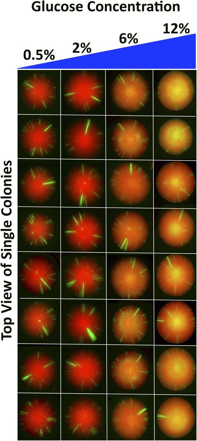
Effect of glucose on stability of Sir-based silencing during growth on complete minimal medium. The CRASH strain was plated on solid medium with 0.5, 2, 6, or 12% glucose and resulting colonies were imaged. The 0.5 and 12% colony images are shown side by side for greater contrast. Representative colonies from each media condition are shown above. Growth in lower glucose resulted in a higher loss-of-silencing rate, as evidenced by the higher rate of green sector formation. This trend holds with 2 and 6% glucose-grown colonies as well (data not shown).
Figure 8.
Effect of glucose on stability of Sir-based silencing during growth on YP medium. The CRASH strain was plated on solid medium with 0.5, 2, 6, or 12% glucose and resulting colonies were imaged. Representative colonies from each media condition are shown above. Growth in 6 and 12% glucose resulted in a high loss-of-silencing rate during late cell divisions, as evidenced by yellowing of the tops of colonies when red and green fluorescence channels are merged. Since silencing is being lost in cells that do not undergo many more cell divisions, large green sectors are not forming.
Stability of silencing in liquid YP culture
A 5 ml volume of seed cultures from individual colonies in 2%-glucose YP medium were grown overnight, shaking at 200 rpm at 30°. Then, 50 ml of appropriate media (both 2 and 12% glucose) was seeded to generate an initial cell concentration of 0.1 OD600 units. The 50 ml cultures shook at 200 rpm at 30°. Aliquots were removed at regular time intervals during log-phase growth and throughout stationary phase. Aliquots were diluted and plated to give ∼50–500 cells per plate. After 3 days, the plates were scanned face up using a Typhoon Trio as above. The number of all-green colonies was divided by the total number of colonies to determine silencing stability within a culture at that time point.
Data availability
Strains are available upon request. Table S1 contains descriptions of all strain genotypes.
Results
Calorie restriction extended yeast chronological life span over a broad range of sugar concentrations in complete minimal medium
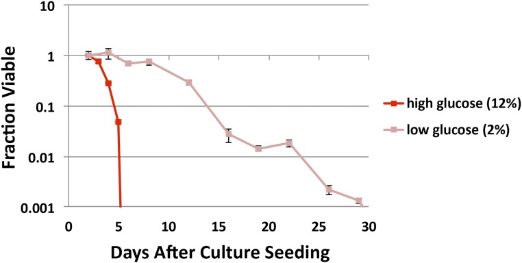
While calorie restriction extends yeast chronological life span when starting glucose concentration in complete minimal medium is <2%, chronological life span has only rarely been studied at higher glucose concentrations comparable to many natural niches of yeast (Smith et al. 2007; Longo et al. 2012). We measured the chronological life span of a wild-type strain (JRY4012) grown in either 2 or 12% glucose complete minimal medium. Sorbitol was added to the 2%-glucose medium to control for osmotic pressure differences between 2 and 12%-glucose media. Growth at 2% glucose resulted in a greater mean and maximal life span as compared to growth at 12% glucose (Figure 1). The higher osmotic pressure of 12%-glucose cultures did not cause the shortened life span of these cultures or influence the effect of sir2Δ on life span, as adding sorbitol to 2%-glucose cultures did not shorten life span and actually extended life span of 2% glucose-grown cells independent of Sir2 (Figure S1). Thus calorie restriction extended S. cerevisiae chronological life span across a wide range of glucose concentrations, not just from 2% and below. Therefore, these glucose concentrations defined a new and physiologically relevant context for studying calorie-level-mediated impacts on yeast chronological life span.
Figure 1.
Chronological life span in high- and low-glucose complete minimal medium. A wild-type strain was grown to stationary phase in either 2% (low) or 12% (high) glucose and aliquots were removed, diluted, and plated over time. Colonies that grew were counted and the daily total was normalized to the total number of colonies viable at the first time point to give the fraction viable. y-axis is cut off at 0.001 to zoom in on most relevant viability window.
sir2Δ dramatically extended chronological life span in 12%-glucose complete minimal cultures and had little effect in 2%-glucose cultures
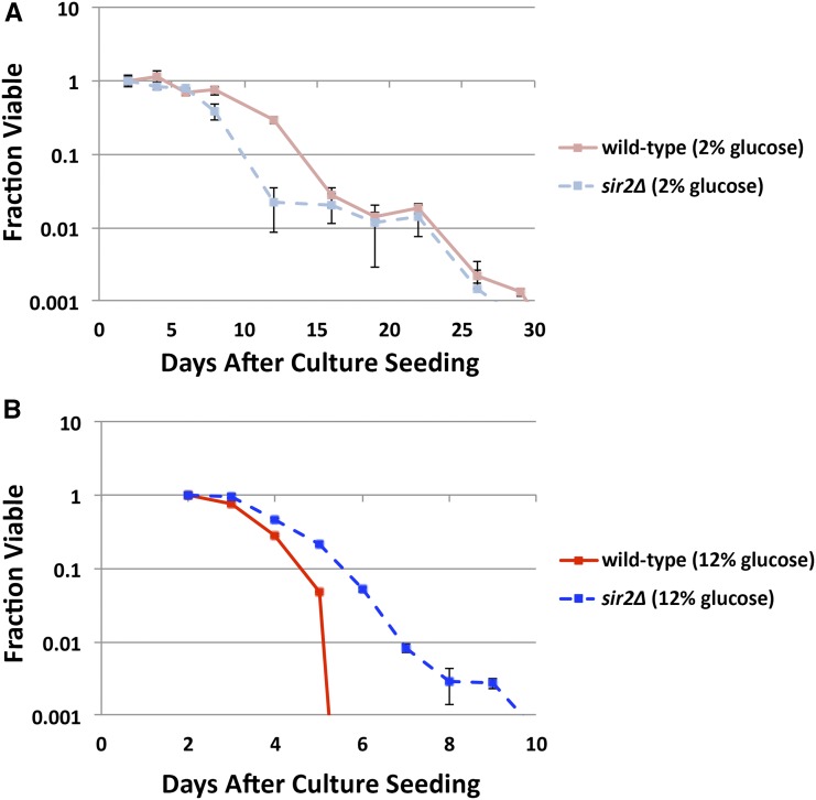
Previous studies have found that calorie restriction extends chronological life span independently of Sir2 in cultures grown in complete minimal 2%-glucose conditions (Smith et al. 2007), which we confirmed (Figure 2A). However, deletion of SIR2 dramatically increases maximum chronological life span of cultures when media is replaced with water, an extreme calorie-restriction state (Fabrizio et al. 2005). To determine whether Sir2 is involved in calorie restriction-mediated longevity under our new protocol, we measured chronological life span of wild-type and sir2Δ cultures in complete minimal medium with 2 or 12% glucose (Figure 2). Surprisingly, deletion of SIR2 dramatically extended both mean and maximum chronological life span in 12% glucose-grown cultures (Figure 2B). Therefore, Sir2’s impact on chronological life span varied dramatically depending on the initial calorie content of growth medium. In this experimental context, Sir2 acted to decrease life span as glucose was consumed from a starting level of 12%. Sir2’s effect on aging in 12% glucose seems to have both a cell-intrinsic and a cell-extrinsic component, as swapping of spent media between wild-type and sir2Δ cultures at the start of stationary phase resulted in a similar intermediate longevity phenotype for both genotypes (Figure S2). We discuss these two components more extensively below.
Figure 2.
Effect of sir2Δ on chronological life span in high- and low-glucose complete minimal medium. Wild-type and sir2Δ strains were grown to stationary phase in either (A) 2% or (B) 12% glucose and aliquots were removed, diluted, and plated over time. Colonies that grew were counted and the daily total was normalized to the total number of colonies viable at the first time point to give the fraction viable. (A) sir2Δ had no effect on life span in 2% glucose, (B) while it significantly extended life span in 12% glucose. y-axis is cut off at 0.001 to zoom in on most relevant viability window.
sir2Δ shortened chronological life span in 2%-glucose YP cultures while having little effect in 12%-glucose cultures
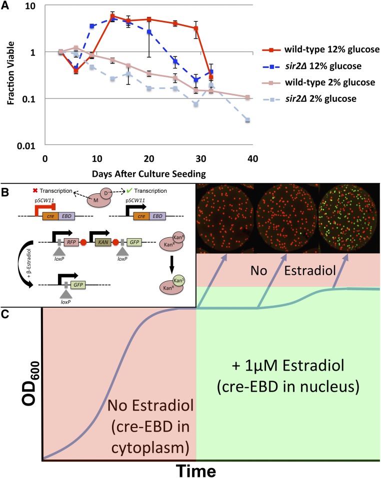
To determine how Sir2 regulates aging in cells grown in other media, we had to get around a previously documented phenomena in which a subpopulation of cells in a stationary-phase culture continue to divide, even as most cells in a culture remain quiescent (Allen et al. 2006) or die. Complex media like YP support larger nonquiescent populations in stationary-phase cultures than complete minimal medium does, which is one reason why complete minimal medium is used in chronological aging experiments (Fabrizio and Longo 2007). If a stationary-phase culture has a subpopulation of cells that continue to divide, that culture is now a mixture of both chronologically old cells and young cells. A culture that appears to retain viability for a longer period of time could reflect actual increased longevity of individual cells. Alternatively, it could result from higher rates of stationary-phase cell division producing new cells. Quantitatively rigorous studies of aging at the population level are nearly impossible under these conditions. Experiments using 12 and 2%-glucose YP revealed extensive cell division in ostensibly stationary-phase 12% glucose-grown cultures, complicating interpretation of aging dynamics (Figure 3A). This phenomenon, where stationary-phase cultures rapidly lose viability and a surviving subset reenters the cell cycle, appears to be an example of adaptive regrowth, which has been documented previously (Fabrizio et al. 2004), where nutrients released by the subset of dying cells fuel additional divisions of other cells in the same medium.
Figure 3.
Genetic strategy for studying chronological aging and regrowth in any medium. (A) High- and low-glucose YP media allow for significant levels of stationary-phase regrowth, complicating interpretation of aging results. (B) Diagram of genetic components. Cre protein is produced only in newly forming daughter cells, and is only present in the nucleus in the presence of estradiol. If cre is in the nucleus, it acts on loxP sites flanking RFP and upstream of GFP, changing cells from red to green. (C) Diagram of modified chronological life-span assay. Estradiol added to culture following log-phase growth period. Colonies formed from cells born after estradiol addition will fluoresce green and should be ignored when assaying chronological life span.
To circumvent this dilemma, we developed a genetic strategy, based in part on the mother enrichment program (Lindstrom and Gottschling 2009), to differentiate between old cells (those present at the very beginning of stationary phase) and young cells (those born during stationary phase) (Figure 3B). Expression of the cre recombinase (Abremski and Hoess 1984) is driven by the promoter for SCW11, a gene encoding a cell wall protein that is expressed only in newly forming daughter cells and never in old mother cells (Colman-Lerner et al. 2001; Doolin et al. 2001). Cre is also fused to the estradiol-binding domain of the murine estradiol receptor (cre-EBD). The cre-EBD hybrid protein remains in the cytoplasm until β-estradiol is added to the growth medium, when it binds to the estradiol-binding domain and allows cre-EBD to be shuttled into the nucleus (Lindstrom and Gottschling 2009). Elsewhere in the genome, an RFP gene, flanked by loxP sites, lies downstream of a constitutive promoter, and upstream of a promoterless GFP gene. In cultures of this strain lacking β-estradiol grown to stationary phase, cre-EBD protein is produced in all newly forming daughter cells, but remains in the cytoplasm, unable to act on loxP sites flanking RFP. Once cultures reach stationary phase, β-estradiol is spiked into the culture medium to a final concentration of 1 μM. Now, if a nonquiescent cell divides, the newly forming daughter cell will produce cre-EBD protein. Now in the presence of β-estradiol, cre-EBD is shuttled into the nucleus where it can act on the loxP sites, excising RFP and bringing GFP under the control of the constitutive promoter (Cheng et al. 2000). Rather than expressing RFP like their aging mothers, newly formed daughter cells express GFP. When diluted aliquots of the culture are plated to fresh medium over time, colonies forming on the plate will express either RFP or GFP, depending on whether they descended from an old mother cell or a young daughter cell, respectively (Figure 3C). By counting only RFP-expressing colonies, the resulting life-span curves represent the aging dynamics of old mother cells present at the start of stationary phase.
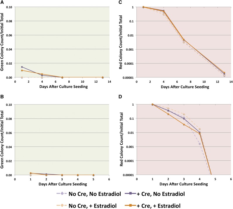
We first measured chronological life span of wild-type and sir2Δ isolates of this strain in 2 and 12%-glucose complete minimal media. Most, if not all, cells remained quiescent in stationary phase, with no increase in the proportion of GFP-expressing colonies beyond a low background level (Figure 4, A and B). Aging characteristics of wild-type and sir2Δ cultures, both 2 and 12% glucose, mirrored those observed without the use of this genetic strategy (Figure 4, C and D). There was also no significant change in aging dynamics in strains lacking cre-EBD or in strains grown in medium lacking β-estradiol, as expected.
Figure 4.
Chronological life span and stationary-phase regrowth in high- and low-glucose complete minimal medium. (A and B) Green or (C and D) red colonies were counted and normalized to the total number of colonies viable at the first time point. (A and C) represent 2%-glucose cultures, while (B and D) represent 12%-glucose cultures. Control experiments lacking cre and/or estradiol were included. Switching was never observed in experiments lacking cre, and no switching above background was observed without addition of estradiol. Complete minimal media did not support stationary-phase regrowth.
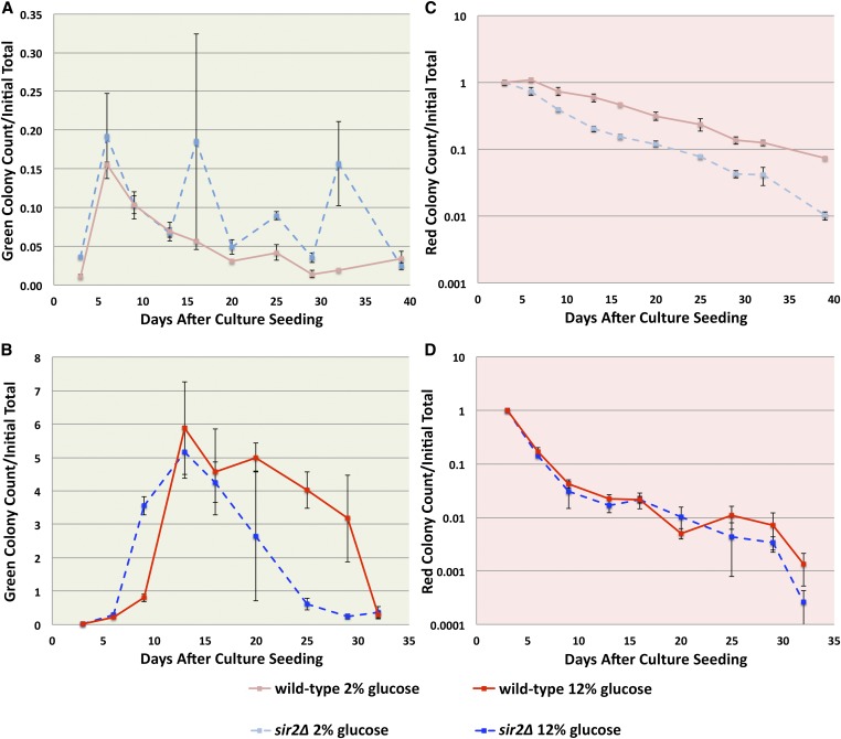
We then measured chronological life span of wild-type and sir2Δ isolates of this strain in 2 and 12%-glucose YP media. As previously reported, 2%-glucose YP cultures had a significant number of daughter cells born in stationary phase, with these cultures eventually being composed of ∼20% GFP-expressing cells (Figure 5A). The 12%-glucose cultures, on the other hand, were quickly heavily represented by newborn GFP-expressing cells (Figure 5B). By ignoring GFP-expressing colonies, life-span curves for 2 and 12%-glucose YP media represented only age-matched mother cells. Surprisingly, loss of Sir2 had no effect in 12%-glucose YP cultures (Figure 5D). In contrast, loss of Sir2 in 12%-glucose complete minimal cultures dramatically extended mean and maximal chronological life span under those conditions (Figure 2B). Also surprisingly, loss of Sir2 significantly shortened mean and maximal chronological life span in 2%-glucose YP cultures (Figure 5C). This again contrasted with loss of Sir2 on 2%-glucose complete minimal cultures, which had little if any effect on chronological life span under those conditions (Figure 2A).
Figure 5.
Effect of sir2Δ on chronological life span and stationary-phase regrowth in high- and low-glucose YP medium. (A and B) Green or (C and D) red colonies were counted and normalized to the total number of colonies viable at the first time point. (A and C) represent 2%-glucose cultures, while (B and D) represent 12%-glucose cultures. (A and B) Both glucose concentrations allowed for stationary-phase regrowth, (B) although regrowth was significantly increased in 12% glucose. Considering only red colonies, sir2Δ significantly decreased life span of 2%-glucose cultures (C), a phenotype that would have otherwise been obscured by stationary-phase regrowth of these sir2Δ cultures (A). In 12%-glucose cultures, however, sir2Δ had no significant effect on life span (D).
Sir2’s impact on chronological life span varied dramatically depending on aspects of the medium beyond sugar content. In YP, sir2Δ decreased life span in low glucose and had no effect in high glucose. Therefore, for wild-type cells growing in YP, Sir2 increased life span as starting calorie content decreased (Figure 5, C and D). In complete minimal medium, on the other hand, sir2Δ had no effect in low glucose and increased life span in high glucose. Therefore, for wild-type cells growing in minimal medium, Sir2 facilitated a decrease in life span as starting calorie content increased (Figure 2, A and B). These phenotypes provided an expanded view of the role for Sir2 in calorie restriction-mediated chronological longevity.
Increasing glucose concentration strengthened Sir-based silencing at HML in complete minimal medium
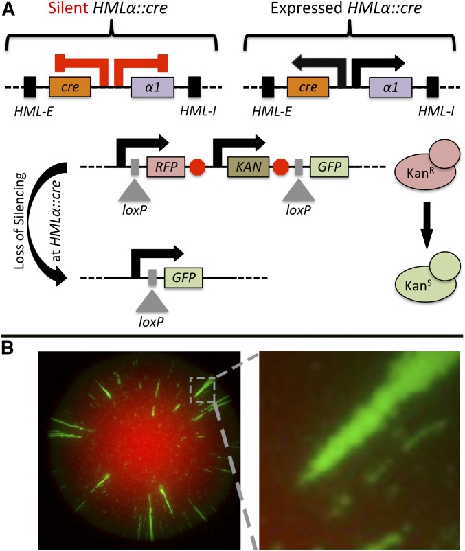
Sir2’s primary function is in transcriptional silencing through the deacetylation of histone tails, resulting in heterochromatin formation at discrete loci within the genome. Therefore, we explored whether this range of culture media also affected the strength of Sir2-based transcriptional silencing. The Sir2/3/4 complex is necessary for silencing HML, and loss of Sir function results in constitutive transcription of the α1 and α2 genes contained within (Rusche et al. 2003). We employed a recently developed method from our laboratory that marks cells that have experienced transient losses of silencing at the HML locus (Dodson and Rine 2015) (Figure 6). In this method, known as the cre-reported altered states of heterochromatin (CRASH) assay (Liu et al. 2016), the coding sequence of α2 at HML has been replaced with the coding sequence of cre, and cre is expressed only if Sir-based silencing at HML is disrupted. If cre protein is produced, it acts on loxP sites flanking RFP, upstream of GFP. Cells that have lost silencing, even transiently, switch from red to green fluorescence (Figure 6A).This switch is permanent and heritable. When cultures are diluted, plated, and grown into colonies; green sectors form in an otherwise red colony (Figure 6B). Each of these green sectors represents a loss-of-silencing event in the cell at the apex of the sector. The green sectoring rate of a strain can be assessed, either qualitatively or quantitatively (Dodson and Rine 2015; Liu et al. 2016), to determine the stability of Sir-protein based silencing at HML relative to wild type.
Figure 6.
CRASH assay design. (A) Diagram of genetic components of the CRASH assay. cre replaces the α2 gene at HML, and is only expressed if Sir-based heterochromatic silencing is lost at HML. If it is expressed, it acts on loxP sites flanking RFP and upstream of GFP, changing the cell from red to green. Descendants of these cells also express GFP rather than RFP. When colonies are grown, loss-of-silencing events result in visible green sectors (B), and the rate of green-sector formation represents the rate of loss of silencing for a particular genetic background or environmental condition.
We plated the wild-type CRASH strain on solid drop-out minimal medium on a range of glucose concentrations: 0.5, 2, 6, and 12%. Interestingly, increasing glucose concentration stabilized silencing at HML, with fewer green sectors seen in 12%-glucose colonies as compared to 0.5%-glucose colonies (Figure 7). Colonies grown on 2 and 6% glucose (data not shown) fit this general trend. Supplementing lower glucose media with sorbitol to achieve similar osmotic pressures in each case had no effect on silencing phenotypes (data not shown).
Increasing glucose concentration weakened Sir-based silencing at HML in YP medium
Because of the surprising differences in the effect of glucose concentrations on aging in the two media types, we explored the impact of increasing glucose concentration on silencing at HML in YP medium. Colonies from wild-type CRASH cells were imaged following growth on solid YP media containing 0.5, 2, 6, or 12% glucose. Quite surprisingly, increasing glucose levels caused a marked destabilization of silencing in a unique spatio-temporal pattern (Figure 8). All previously studied interventions that destabilized silencing in the CRASH assay resulted in loss-of-silencing events relatively early in colony growth and continued throughout the growth of the colony, forming visible green sectors of sizes proportional to the timing of the events (Dodson and Rine 2015; Liu et al. 2016). Here, however, loss-of-silencing events, particularly in 6 and 12% glucose, occurred late in colony development; preventing their outgrowth into a visible green sector. Instead, red cells and green cells were next to one another, giving a yellow appearance when the two fluorescence channels were merged. This appearance was not due to inappropriate expression of both fluorophores in a single cell, as streaking these colonies onto fresh solid medium to isolate single colonies revealed close to half of the cells in 6 and 12% glucose had switched from red to green (data not shown). Strikingly, 0.5 and 2% glucose-grown colonies completely lacked this phenotype, and appeared quite similar to one another. Hence, while glucose concentrations previously used to study calorie restriction showed no change in Sir-based silencing, higher glucose concentration had a dramatic effect on heterochromatin stability.
Stationary-phase cells in 12%-glucose liquid cultures lost silencing at high rates compared to 2% glucose-grown cultures
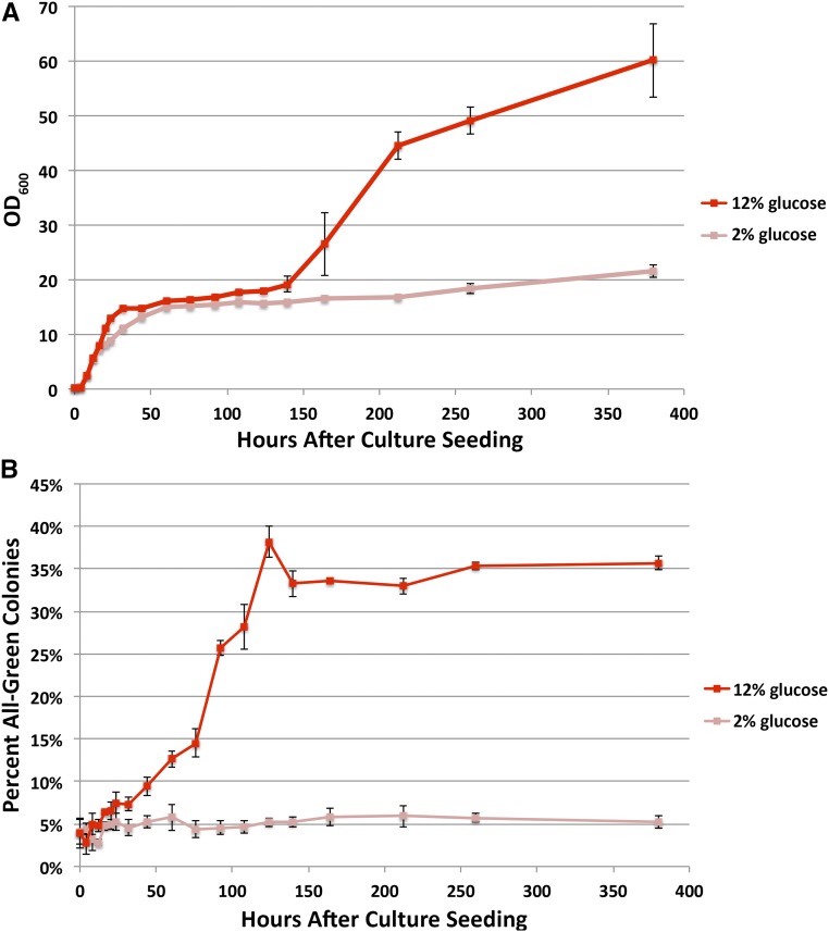
Since cells losing silencing in high glucose-grown colonies were not dividing much, if at all, at the time they were imaged, we wanted to see if stationary-phase cells in particular were more likely to lose silencing at HML, or whether the high glucose conditions caused elevated loss-of-silencing rates continuously during growth. We inoculated liquid YP medium containing either 2 or 12% glucose with the CRASH strain and grew cultures to stationary phase. At time points throughout log-phase growth and during stationary phase, aliquots were plated to fresh solid medium and the proportion of all-green colonies was quantified. For all-green colonies, the founding cell had to have lost silencing in liquid culture, before its plating. Cells grown in 12% glucose lost silencing at a rate similar to 2% glucose-grown cells during log-phase growth. In stationary phase, however, 12% glucose-grown cultures exhibited high rates of silencing loss, eventually plateauing with 35% of cells having lost silencing (Figure 9). Given that, under standard experimental conditions, wild-type cells lose silencing at HML only once every 1000 cell divisions (Dodson and Rine 2015), this rate represents a massive destabilization of Sir-based silencing based upon changes in physiology and not genotype. The period of silencing instability (Figure 9B) preceded the period of stationary-phase regrowth (Figure 9A).
Figure 9.
Loss of silencing in high- and low-glucose, stationary-phase liquid YP cultures. The CRASH strain was grown to stationary phase in 2 and 12%-glucose liquid YP media. (A) Cell density (OD600) was monitored throughout. Aliquots were also removed, diluted, and plated, and (B) all-green colonies that grew were quantified as a percentage of total colonies. The 12%-glucose cultures had high loss-of-silencing rates following log-phase growth, but preceding the period of culture regrowth [compare (A) to (B)].
Glucose control of heterochromatin was robust to genetic background
To determine whether the effect of glucose levels on heterochromatin stability was specific to the genetic background of our strains, we mated the haploid W303 CRASH strain to S. cerevisiae haploid strains of either W303 or s288c backgrounds. Additionally, we mated the CRASH strain to S. paradoxus and S. bayanus haploid strains. We plated all four of the resulting diploids on solid YP media with 0.5, 2, 6, or 12% glucose and imaged the resulting colonies as before. As with the W303 haploid strain, all of these diploid strains lost silencing at the tops of colonies grown on the higher glucose media (Figure 10).
Figure 10.
Glucose control of heterochromatin was robust to genetic background. The CRASH strain (w303) was mated to haploid isolates of S. cerevisiae (w303 and s288c), S. bayanus, and S. paradoxus genetic backgrounds. The resulting diploids were plated on solid YP media with 0.5, 2, 6, or 12% glucose and imaged after 4 days of growth. Representative colonies from each media condition and strain background are shown above. The previously observed loss-of-silencing phenotype (Figure 8) is apparent in all of these additional genetic backgrounds.
Discussion
This work demonstrated that calorie restriction extended chronological life span across a wider range of sugar concentrations, not just <2% (Figure 1). There is evidence that Sir2 does not mediate the extension of chronological life span in response to calorie restriction, at least in any previously used protocols (Smith et al. 2007). Even so, sir2Δ does extend the life span of cells experiencing extreme nutrient restriction in water, while having little effect in media with 2% glucose (Fabrizio et al. 2005). In the case of extreme nutrient restriction, Sir2 acts to oppose the beneficial effects on life span. However, because all nutrients are limiting in water, determining which nutrient(s) are involved is not possible. Under our new protocol, Sir2 has a clear role in reducing life span as glucose levels increase in complete minimal medium (Figure 2). Classically, calorie restriction-mediated longevity is thought of as a beneficial effect on life span of decreasing calorie content, rather than a detrimental effect on life span of increasing calorie content. However, these two views are logically equivalent ways of describing the same phenomenon. What is clear, however, is that Sir2 is not sufficient for calorie restriction-mediated longevity in this context, as 12%-glucose sir2Δ cultures did not phenocopy 2%-glucose sir2∆ cultures (Figure 2).
Sir2’s effect on aging in 12% glucose seems to have both a cell-intrinsic and a cell-extrinsic component. Swapping wild-type and sir2Δ 12% glucose-spent media at the start of stationary phase results in a similar, intermediate longevity phenotype for both genotypes (Figure S2). Therefore, the composition of stationary-phase media is not sufficient to determine life span. At least part of Sir2’s contribution came before stationary phase and irreversibly altered aging dynamics. Acidification of culture medium has been proposed as the primary mechanism for yeast chronological aging in unbuffered complete minimal media (Burtner et al. 2009). Sir2’s removal of acetyl groups from histones and other proteins could affect the acidity of the culture, possibly explaining Sir2’s negative role in aging in high glucose complete minimal cultures. Additionally, Sir2 modulates the activity of Pck1, an enzyme involved in gluconeogenesis (Lin et al. 2009). Sir2’s deacetylation of Pck1 may mediate the Sir-dependent effects on chronological life span (Orlandi et al. 2016). Our data are consistent with Sir2 altering the activity of Pck1 to affect culture acidification and ultimately life span. However, the intermediate longevity observed in the media-swap experiment implies that Sir2 must exert an additional effect, either through Pck1 or through another target, during log-phase growth. Addition of NAM in stationary phase phenocopied deletion of SIR2 with respect to chronological aging in 2% glucose and in extreme dietary restriction. Therefore, Sir2’s aging role in these contexts is restricted to its enzymatic function alone (Orlandi et al. 2016). If Pck1 mediates the Sir-dependent reduction in life span in 12%-glucose cultures, Pck1 function must be altered before stationary phase in this condition. Alternatively, a different Sir2 target could be responsible for the cell-intrinsic effects on life span seen following growth in 12% glucose.
Under some growth conditions, a fraction of stationary-phase cells regrow as others lyse (Fabrizio et al. 2004; Fabrizio and Longo 2007). This regrowth phenomenon was observed to such a high degree in 2 and 12%-glucose YP cultures that it initially prevented study of chronological aging in these conditions (Figure 3A). However, by deploying our newly developed genetic strategy to differentiate aging mother cells from newborn progeny (Figure 3B), it is clear that calorie restriction extends the life span of individual cells, even as the cultures themselves have lower total viability at many time points (comparing 12%-glucose data from Figure 3A to Figure 5D). In contrast to cultures grown in complete minimal medium, in YP medium sir2Δ had no effect on the life span of 12%-grown cells (Figure 5D), while it significantly shortened the mean and maximal life span of 2%-grown cells (Figure 5C). This phenotype was consistent with a partial role for Sir2 in calorie restriction-mediated longevity in YP medium in which Sir2 was necessary for the complete life span extension via calorie restriction. It is important to note that in YP medium sir2Δ 2% cultures did not phenocopy 12% cultures, demonstrating that Sir2 was not sufficient for calorie restriction-mediated longevity in this medium, just as it was not sufficient in complete minimal medium.
Sir2’s impact on calorie restriction-mediated longevity varied dramatically depending on type of growth medium. In YP, Sir2 extends life span as glucose is decreased. Conversely, in complete minimal medium, Sir2 shortens life span as glucose is increased. In both cases, Sir2 is responding to changes in glucose to alter aging dynamics in a way that makes 2%-glucose cultures live longer than 12%-glucose cultures. In both cases, the exact mechanism through which Sir2-mediated, glucose-dependent, life-span divergence occurs is unknown. It is also not clear whether a common downstream target, presumably a protein that is deacetylated by Sir2, mediates all of these effects, or whether different targets of Sir2 contribute to calorie restriction-mediated longevity in different growth media.
Interestingly, Sir-based silencing at HML was stabilized as glucose increases in minimal medium (CSM), while silencing is destabilized as glucose increases in YP medium (compare Figure 7 and Figure 8). This change in directionality is reminiscent of the change in directionality of sir2Δ-aging phenotypes when switching from complete minimal medium to YP medium. It will be interesting to see if the different effects of media act directly through Sir2, perhaps through NAD+ levels or phosphorylation (Kang et al. 2015) to alter Sir-based silencing at HML, or indirectly through other targets. Perhaps a common downstream target of glucose, and possibly Sir2, mediates the opposite effects on heterochromatic silencing seen in complete minimal and YP media as glucose levels increase.
The marked loss of silencing in 12%-glucose YP cultures during stationary phase points to the possibility that subtelomeric genes normally silenced by Sir2 (Aparicio et al. 1991) are expressed under these conditions, providing some advantage to cells. Interestingly, subtelomeric regions are enriched for genes with a role in metabolism (Ai et al. 2002), although few subtelomeric genes significantly increase in expression over wild-type levels in a sir2Δ strain during log-phase growth in 2% glucose (Ellahi et al. 2015). However, it is possible that expression levels of important metabolic genes do change in a Sir2-dependent manner at higher glucose concentrations and/or during stationary phase.
Previously, the study of S. cerevisiae chronological aging was mostly confined to complete minimal media in which stationary-phase cells almost exclusively enter a quiescent, nondividing state; ensuring that the population of cells being studied is as age-matched as possible. Complete minimal medium efficiently arrests stationary-phase cells because the pH of the medium drops significantly, below a level that allows for growth (Longo and Fabrizio 2012). Media like pH-buffered complete minimal, YP, and many natural yeast substrates do not reach such low pH’s, allowing for varying levels of regrowth. It is difficult, if not impossible, to interpret population-level aging data in a mixed-age population, especially when the relative distribution of individual ages is unknown. As a result, despite their potential physiological relevance, media allowing for stationary-phase growth have been avoided. With our new genetic strategy, however, yeast chronological aging can now be studied in any medium, regardless of its potential for stationary-phase growth. This is especially relevant to experiments in high calorie and pH-buffered media, which support high levels of stationary-phase regrowth (Figure 3; unpublished data). While data supports acidification of growth media via acetate production as the molecular mechanism driving yeast chronological aging (Burtner et al. 2009), it is unclear whether this mechanism is relevant in an environmental context where pH remains more stable. Given that the first gene studied using this strategy, SIR2, is shown to have dramatically different aging roles in different environmental contexts, it seems especially important to study aging across these contexts.
Taken together, a picture emerges of Sir2 as an important regulator of yeast aging that responds to nutrient cues including, but not limited to, glucose. It is especially intriguing that Sir2 can either extend or shorten chronological life span in response to changes in nutrients. Many genes have either a positive (e.g., SOD1; Longo et al. 1996) or a negative (e.g., SCH9; Longo 2003) impact on chronological life span, often independent of the limited number of environments tested. The variety of inputs that lead to a variety of outputs positions Sir2 as a central regulator, responding to nutritional cues to either extend or shorten life span. In times of nutrient scarcity, it makes sense for yeast to prioritize cell-cycle arrest and longevity over reproduction, as reproduction is energy expensive and their offspring would be born into an environment unlikely to support survival. By remaining viable and conserving scarce resources, the chances increase that a yeast cell might encounter an energy-rich substrate where it can begin growing again. In times of plenty, when yeast are dividing rapidly and often in competition with other microorganisms with even faster division rates, it makes sense to prioritize fast growth, even if accompanied by the high costs of genome instability and other forms of cellular damage that are thought to drive aging. Additionally, an apoptosis-like, cell-death program exists in yeast that is induced by chronological aging (Fabrizio and Longo 2008). Under certain conditions, this programmed cell death enhances the maximal chronological longevity of a culture by killing off most of the cells, releasing nutrients, and allowing for the regrowth of a robust, adapted subpopulation of cells (Fabrizio et al. 2004). It makes sense that, within a yeast colony or liquid culture that is essentially clonal, group selection (Leigh 2010; Frank 2013) could play a role in their evolution. Group selection could drive the development of an altruistic aging program as posited for S. cerevisiae (Fabrizio et al. 2004), one that benefits the culture as a whole at the expense of individual cells.
Supplementary Material
Supplemental material is available online at www.genetics.org/lookup/suppl/doi:10.1534/genetics.116.196485/-/DC1.
Acknowledgments
We thank Dan Gottschling for providing us with the mother enrichment program parent strain (UCC8650), from which we obtained the pSCW11-cre-EBD PCR fragment for use in our new genetic strategy. We also thank Anne Dodson, who provided JRY9628, JRY10776, and JRY10782. We also thank members of our laboratory, past and present, for helpful discussions that shaped this work. This work was supported by a National Institutes of Health predoctoral fellowship (5 F31 AG-047795-03) to D.F.M., as well as a grant from the National Institutes of Health to J.R. (GM-31105).
Footnotes
Communicating editor: A. Mitchell
Literature Cited
- Abremski K., Hoess R., 1984. Bacteriophage P1 site-specific recombination. Purification and properties of the Cre recombinase protein. J. Biol. Chem. 259: 1509–1514. [PubMed] [Google Scholar]
- Ai W., Bertram P. G., Tsang C. K., Chan T. F., Zheng X. F., 2002. Regulation of subtelomeric silencing during stress response. Mol. Cell 10: 1295–1305. [DOI] [PubMed] [Google Scholar]
- Allen C., Büttner S., Aragon A. D., Thomas J. A., Meirelles O., et al. , 2006. Isolation of quiescent and nonquiescent cells from yeast stationary-phase cultures. J. Cell Biol. 174: 89–100. [DOI] [PMC free article] [PubMed] [Google Scholar]
- Anderson R. M., Weindruch R., 2007. Metabolic reprogramming in dietary restriction. Interdiscip. Top. Gerontol. 35: 18–38. [DOI] [PMC free article] [PubMed] [Google Scholar]
- Anderson R. M., Weindruch R., 2010. Metabolic reprogramming, caloric restriction and aging. Trends Endocrinol. Metab. 21: 134–141. [DOI] [PMC free article] [PubMed] [Google Scholar]
- Aparicio O. M., Billington B. L., Gottschling D. E., 1991. Modifiers of position effect are shared between telomeric and silent mating-type loci in S. cerevisiae. Cell 66: 1279–1287. [DOI] [PubMed] [Google Scholar]
- Bitterman K. J., Anderson R. M., Cohen H. Y., Latorre-Esteves M., Sinclair D. A., 2002. Inhibition of silencing and accelerated aging by nicotinamide, a putative negative regulator of yeast Sir2 and human SIRT1. J. Biol. Chem. 277: 45099–45107. [DOI] [PubMed] [Google Scholar]
- Bordone L., Cohen D., Robinson A., Motta M. C., van Veen E., et al. , 2007. SIRT1 transgenic mice show phenotypes resembling calorie restriction. Aging Cell 6: 759–767. [DOI] [PubMed] [Google Scholar]
- Burnett C., Valentini S., Cabreiro F., Goss M., Somogyvári M., et al. , 2011. Absence of effects of Sir2 overexpression on lifespan in C. elegans and Drosophila. Nature 477: 482–485. [DOI] [PMC free article] [PubMed] [Google Scholar]
- Burtner C. R., Murakami C. J., Kennedy B. K., Kaeberlein M., 2009. A molecular mechanism of chronological aging in yeast. Cell Cycle 8: 1256–1270. [DOI] [PMC free article] [PubMed] [Google Scholar]
- Cheng T. H., Chang C. R., Joy P., Yablok S., Gartenberg M. R., 2000. Controlling gene expression in yeast by inducible site-specific recombination. Nucleic Acids Res. 28: E108. [DOI] [PMC free article] [PubMed] [Google Scholar]
- Chiba T., Yamaza H., Shimokawa I., 2007. Role of insulin and growth hormone/insulin-like growth factor-I signaling in lifespan extension: rodent longevity models for studying aging and calorie restriction. Curr. Genomics 8: 423–428. [DOI] [PMC free article] [PubMed] [Google Scholar]
- Coghe S., D’Hollander H., Verachtert H., Delvaux F. R., 2005. Impact of dark specialty malts on extract composition and wort fermentation. J. Inst. Brew. 111: 51–60. [Google Scholar]
- Colman-Lerner A., Chin T. E., Brent R., 2001. Yeast Cbk1 and Mob2 activate daughter-specific genetic programs to induce asymmetric cell fates. Cell 107: 739–750. [DOI] [PubMed] [Google Scholar]
- Comfort A., 1963. Effect of delayed and resumed growth on the longevity of a fish (Lebistes reticulatus, Peters) in captivity. Gerontologia 49: 150–155. [DOI] [PubMed] [Google Scholar]
- Couzin-Frankel J., 2011. Genetics. Aging genes: the sirtuin story unravels. Science 334: 1194–1198. [DOI] [PubMed] [Google Scholar]
- Dodson A. E., Rine J., 2015. Heritable capture of heterochromatin dynamics in Saccharomyces cerevisiae. eLife 4: e05007. [DOI] [PMC free article] [PubMed] [Google Scholar]
- Doolin M. T., Johnson A. L., Johnston L. H., Butler G., 2001. Overlapping and distinct roles of the duplicated yeast transcription factors Ace2p and Swi5p. Mol. Microbiol. 40: 422–432. [DOI] [PubMed] [Google Scholar]
- Ellahi A., Thurtle D. M., Rine J., 2015. The chromatin and transcriptional landscape of native Saccharomyces cerevisiae telomeres and subtelomeric domains. Genetics 200: 505–521. [DOI] [PMC free article] [PubMed] [Google Scholar]
- Fabrizio P., Longo V. D., 2007. The chronological life span of Saccharomyces cerevisiae. Methods Mol. Biol. 371: 89–95. [DOI] [PubMed] [Google Scholar]
- Fabrizio P., Longo V. D., 2008. Chronological aging-induced apoptosis in yeast. Biochim. Biophys. Acta 1783: 1280–1285. [DOI] [PMC free article] [PubMed] [Google Scholar]
- Fabrizio P., Battistella L., Vardavas R., Gattazzo C., Liou L. L., et al. , 2004. Superoxide is a mediator of an altruistic aging program in Saccharomyces cerevisiae. J. Cell Biol. 166: 1055–1067. [DOI] [PMC free article] [PubMed] [Google Scholar]
- Fabrizio P., Gattazzo C., Battistella L., Wei M., Cheng C., et al. , 2005. Sir2 blocks extreme life-span extension. Cell 123: 655–667. [DOI] [PubMed] [Google Scholar]
- Frank S. A., 2013. Natural selection. VII. History and interpretation of kin selection theory. J. Evol. Biol. 26: 1151–1184. [DOI] [PubMed] [Google Scholar]
- Garde-Cerdán T., Lorenzo C., Martínez-Gil A. M., Lara J. F., Pardo F., et al. , 2011. Evolution of nitrogen compounds during grape ripening from organic and non-organic monastrell – nitrogen consumption and volatile formation in alcoholic fermentation, pp. 123–138 in Research in Organic Farming, edited by Nokkoul R. InTech, Rijeka, Croatia. [Google Scholar]
- Gasch A. P., Spellman P. T., Kao C. M., Carmel-Harel O., Eisen M. B., et al. , 2000. Genomic expression programs in the response of yeast cells to environmental changes. Mol. Biol. Cell 11: 4241–4257. [DOI] [PMC free article] [PubMed] [Google Scholar]
- Gesing A., Al-Regaiey K. A., Bartke A., Masternak M. M., 2014. Growth hormone abolishes beneficial effects of calorie restriction in long-lived Ames dwarf mice. Exp. Gerontol. 58: 219–229. [DOI] [PMC free article] [PubMed] [Google Scholar]
- Gómez J. M., 2010. Aging in bacteria, immortality or not-a critical review. Curr. Aging Sci. 3: 198–218. [PubMed] [Google Scholar]
- Greer E. L., Dowlatshahi D., Banko M. R., Villen J., Hoang K., et al. , 2007. An AMPK-FOXO pathway mediates longevity induced by a novel method of dietary restriction in C. elegans. Curr. Biol. 17: 1646–1656. [DOI] [PMC free article] [PubMed] [Google Scholar]
- Guarente L., Picard F., 2005. Calorie restriction–the SIR2 connection. Cell 120: 473–482. [DOI] [PubMed] [Google Scholar]
- Imai S., Armstrong C. M., Kaeberlein M., Guarente L., 2000. Transcriptional silencing and longevity protein Sir2 is an NAD-dependent histone deacetylase. Nature 403: 795–800. [DOI] [PubMed] [Google Scholar]
- Jazwinski S. M., 1990. An experimental system for the molecular analysis of the aging process: the budding yeast Saccharomyces cerevisiae. J. Gerontol. 45: B68–B74. [DOI] [PubMed] [Google Scholar]
- Kaeberlein M., Powers R. W., 3rd, 2007. Sir2 and calorie restriction in yeast: a skeptical perspective. Ageing Res. Rev. 6: 128–140. [DOI] [PubMed] [Google Scholar]
- Kaeberlein M., McVey M., Guarente L., 1999. The SIR2/3/4 complex and SIR2 alone promote longevity in Saccharomyces cerevisiae by two different mechanisms. Genes Dev. 13: 2570–2580. [DOI] [PMC free article] [PubMed] [Google Scholar]
- Kaeberlein M., Kirkland K. T., Fields S., Kennedy B. K., 2004. Sir2-independent life span extension by calorie restriction in yeast. PLoS Biol. 2: E296. [DOI] [PMC free article] [PubMed] [Google Scholar]
- Kaeberlein M., Powers R. W., 3rd, Steffen K. K., Westman E. A., Hu D., et al. , 2005. Regulation of yeast replicative life span by TOR and Sch9 in response to nutrients. Science 310: 1193–1196. [DOI] [PubMed] [Google Scholar]
- Kaeberlein M., Steffen K. K., Hu D., Dang N., Kerr E. O., et al. , 2006. Comment on “HST2 mediates SIR2-independent life-span extension by calorie restriction”. Science 312: 1312. [DOI] [PubMed] [Google Scholar]
- Kang W. K., Kim Y. H., Kang H. A., Kwon K. S., Kim J. Y., 2015. Sir2 phosphorylation through cAMP-PKA and CK2 signaling inhibits the lifespan extension activity of Sir2 in yeast. eLife 4: e09709. [DOI] [PMC free article] [PubMed] [Google Scholar]
- Klass M. R., 1977. Aging in the nematode Caenorhabditis elegans: major biological and environmental factors influencing life span. Mech. Ageing Dev. 6: 413–429. [DOI] [PubMed] [Google Scholar]
- Koubova J., Guarente L., 2003. How does calorie restriction work? Genes Dev. 17: 313–321. [DOI] [PubMed] [Google Scholar]
- Landry J., Sutton A., Tafrov S. T., Heller R. C., Stebbins J., et al. , 2000. The silencing protein SIR2 and its homologs are NAD-dependent protein deacetylases. Proc. Natl. Acad. Sci. USA 97: 5807–5811. [DOI] [PMC free article] [PubMed] [Google Scholar]
- Leigh E. G., Jr, 2010. The group selection controversy. J. Evol. Biol. 23: 6–19. [DOI] [PubMed] [Google Scholar]
- Lin S. J., Defossez P. A., Guarente L., 2000. Requirement of NAD and SIR2 for life-span extension by calorie restriction in Saccharomyces cerevisiae. Science 289: 2126–2128. [DOI] [PubMed] [Google Scholar]
- Lin S. J., Kaeberlein M., Andalis A. A., Sturtz L. A., Defossez P. A., et al. , 2002. Calorie restriction extends Saccharomyces cerevisiae lifespan by increasing respiration. Nature 418: 344–348. [DOI] [PubMed] [Google Scholar]
- Lin Y. Y., Lu J. Y., Zhang J., Walter W., Dang W., et al. , 2009. Protein acetylation microarray reveals that NuA4 controls key metabolic target regulating gluconeogenesis. Cell 136: 1073–1084. [DOI] [PMC free article] [PubMed] [Google Scholar]
- Lindstrom D. L., Gottschling D. E., 2009. The mother enrichment program: a genetic system for facile replicative life span analysis in Saccharomyces cerevisiae. Genetics 183: 413–422. [DOI] [PMC free article] [PubMed] [Google Scholar]
- Liu T. Y., Dodson A. E., Terhorst J., Song Y. S., Rine J., 2016. Riches of phenotype computationally extracted from microbial colonies. Proc. Natl. Acad. Sci. USA 113: E2822–E2831. [DOI] [PMC free article] [PubMed] [Google Scholar]
- Longo V. D., 2003. The Ras and Sch9 pathways regulate stress resistance and longevity. Exp. Gerontol. 38: 807–811. [DOI] [PubMed] [Google Scholar]
- Longo V. D., Fabrizio P., 2012. Chronological aging in Saccharomyces cerevisiae. Subcell. Biochem. 57: 101–121. [DOI] [PMC free article] [PubMed] [Google Scholar]
- Longo V. D., Gralla E. B., Valentine J. S., 1996. Superoxide dismutase activity is essential for stationary phase survival in Saccharomyces cerevisiae. Mitochondrial production of toxic oxygen species in vivo. J. Biol. Chem. 271: 12275–12280. [DOI] [PubMed] [Google Scholar]
- Longo V. D., Shadel G. S., Kaeberlein M., Kennedy B., 2012. Replicative and chronological aging in Saccharomyces cerevisiae. Cell Metab. 16: 18–31. [DOI] [PMC free article] [PubMed] [Google Scholar]
- Lu S. P., Kato M., Lin S. J., 2009. Assimilation of endogenous nicotinamide riboside is essential for calorie restriction-mediated life span extension in Saccharomyces cerevisiae. J. Biol. Chem. 284: 17110–17119. [DOI] [PMC free article] [PubMed] [Google Scholar]
- Mair W., Dillin A., 2008. Aging and survival: the genetics of life span extension by dietary restriction. Annu. Rev. Biochem. 77: 727–754. [DOI] [PubMed] [Google Scholar]
- Maruyama Y., Ito T., Kodama H., Matsuura A., 2016. Availability of amino acids extends chronological lifespan by suppressing hyper-acidification of the environment in Saccharomyces cerevisiae. PLoS One 11: e0151894. [DOI] [PMC free article] [PubMed] [Google Scholar]
- McCay C. M., Crowell M. F., Maynard L. A., 1935. The effect of retarded growth upon the length of life span and upon the ultimate body size. Nutrition 5: 155–171. [PubMed] [Google Scholar]
- Medvedik O., Lamming D. W., Kim K. D., Sinclair D. A., 2007. MSN2 and MSN4 link calorie restriction and TOR to sirtuin-mediated lifespan extension in Saccharomyces cerevisiae. PLoS Biol. 5: e261. [DOI] [PMC free article] [PubMed] [Google Scholar]
- Mortimer R. K., Johnston J. R., 1959. Life span of individual yeast cells. Nature 183: 1751–1752. [DOI] [PubMed] [Google Scholar]
- Oliva J., Garde-Cerdán T., Martínez-Gil A. M., Salinas M., Barba A., 2011. Fungicide effects on ammonium and amino acids of monastrell grapes. Food Chem. 129: 1676–1680. [Google Scholar]
- Orlandi I., Pellegrino Coppola D., Strippoli M., Ronzulli R., Vai M., 2016. Nicotinamide supplementation phenocopies SIR2 inactivation by modulating carbon metabolism and respiration during yeast chronological aging. Mech. Ageing Dev. 6374: 30092–30096. [DOI] [PubMed] [Google Scholar]
- Piper M. D., Bartke A., 2008. Diet and aging. Cell Metab. 8: 99–104. [DOI] [PubMed] [Google Scholar]
- Rogina B., Helfand S. L., 2004. Sir2 mediates longevity in the fly through a pathway related to calorie restriction. Proc. Natl. Acad. Sci. USA 101: 15998–16003. [DOI] [PMC free article] [PubMed] [Google Scholar]
- Rose M. R., 1991. Evolutionary Biology of Aging. Oxford University Press, New York. [Google Scholar]
- Rose M. R., Flatt T., Graves J. L., Greer L. F., Martinez D. E., et al. , 2012. What is aging? Front. Genet. 3: 134. [DOI] [PMC free article] [PubMed] [Google Scholar]
- Rusche L. N., Kirchmaier A. L., Rine J., 2003. The establishment, inheritance, and function of silenced chromatin in Saccharomyces cerevisiae. Annu. Rev. Biochem. 72: 481–516. [DOI] [PubMed] [Google Scholar]
- Sanchez P. C., 2008. Philippine Fermented Foods: Principles and Technology. The University of The Philippines Press, Quezon City, Philippines. [Google Scholar]
- Sandmeier J. J., Celic I., Boeke J. D., Smith J. S., 2002. Telomeric and rDNA silencing in Saccharomyces cerevisiae are dependent on a nuclear NAD(+) salvage pathway. Genetics 160: 877–889. [DOI] [PMC free article] [PubMed] [Google Scholar]
- Santos J., Leão C., Sousa M. J., 2012. Growth culture conditions and nutrient signaling modulating yeast chronological longevity. Oxid. Med. Cell. Longev. 2012: 680304. [DOI] [PMC free article] [PubMed] [Google Scholar]
- Slator A., 1908. XXI.–Studies in fermentation. Part II. The mechanism of alcoholic fermentation. J. Chem. Soc. Trans. 93: 217–242. [Google Scholar]
- Smith D. L., Jr, McClure J. M., Matecic M., Smith J. S., 2007. Calorie restriction extends the chronological lifespan of Saccharomyces cerevisiae independently of the Sirtuins. Aging Cell 6: 649–662. [DOI] [PubMed] [Google Scholar]
- Smith J. S., Brachmann C. B., Celic I., Kenna M. A., Muhammad S., et al. , 2000. A phylogenetically conserved NAD+-dependent protein deacetylase activity in the Sir2 protein family. Proc. Natl. Acad. Sci. USA 97: 6658–6663. [DOI] [PMC free article] [PubMed] [Google Scholar]
- Taormina G., Mirisola M. G., 2014. Calorie restriction in mammals and simple model organisms. BioMed Res. Int. 2014: 308690. [DOI] [PMC free article] [PubMed] [Google Scholar]
- Tissenbaum H. A., Guarente L., 2001. Increased dosage of a sir-2 gene extends lifespan in Caenorhabditis elegans. Nature 410: 227–230. [DOI] [PubMed] [Google Scholar]
- Wei M., Fabrizio P., Hu J., Ge H., Cheng C., et al. , 2008. Life span extension by calorie restriction depends on Rim15 and transcription factors downstream of Ras/PKA, Tor, and Sch9. PLoS Genet. 4: e13. [DOI] [PMC free article] [PubMed] [Google Scholar]
- Weinberger M., Mesquita A., Caroll T., Marks L., Yang H., et al. , 2010. Growth signaling promotes chronological aging in budding yeast by inducing superoxide anions that inhibit quiescence. Aging (Albany NY) 2: 709–726. [DOI] [PMC free article] [PubMed] [Google Scholar]
- White J., 1955. Effect of seeding rate and sugar concentration on yeast growth and fermentation in aerated glucose worts. J. Inst. Brew. 61: 146–150. [Google Scholar]
- Zahn J. M., Poosala S., Owen A. B., Ingram D. K., Lustig A., et al. , 2007. AGEMAP: a gene expression database for aging in mice. PLoS Genet. 3: e201. [DOI] [PMC free article] [PubMed] [Google Scholar]
- Zhu M., Miura J., Lu L. X., Bernier M., DeCabo R., et al. , 2004. Circulating adiponectin levels increase in rats on caloric restriction: the potential for insulin sensitization. Exp. Gerontol. 39: 1049–1059. [DOI] [PubMed] [Google Scholar]
- Zhu M., Lee G. D., Ding L., Hu J., Qiu G., et al. , 2007. Adipogenic signaling in rat white adipose tissue: modulation by aging and calorie restriction. Exp. Gerontol. 42: 733–744. [DOI] [PMC free article] [PubMed] [Google Scholar]
Associated Data
This section collects any data citations, data availability statements, or supplementary materials included in this article.
Supplementary Materials
Data Availability Statement
Strains are available upon request. Table S1 contains descriptions of all strain genotypes.


