ABSTRACT
A wide range of signaling processes occurs at the cell surface through the reversible association of proteins from the cytosol to the plasma membrane. Some low abundant lipids are enriched at the membrane of specific compartments and thereby contribute to the identity of cell organelles by acting as biochemical landmarks. Lipids also influence membrane biophysical properties, which emerge as an important feature in specifying cellular territories. Such parameters are crucial for signal transduction and include lipid packing, membrane curvature and electrostatics. In particular, membrane electrostatics specifies the identity of the plasma membrane inner leaflet. Membrane surface charges are carried by anionic phospholipids, however the exact nature of the lipid(s) that powers the plasma membrane electrostatic field varies among eukaryotes and has been hotly debated during the last decade. Herein, we discuss the role of anionic lipids in setting up plasma membrane electrostatics and we compare similarities and differences that were found in different eukaryotic cells.
KEYWORDS: Anionic lipid, arabidopsis, biosensor, endocytosis, membrane surface charge, membrane territory, phosphatidylserine, phosphoinositide, phospholipid, plasma membrane identity
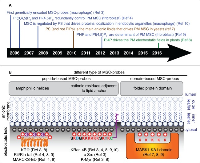
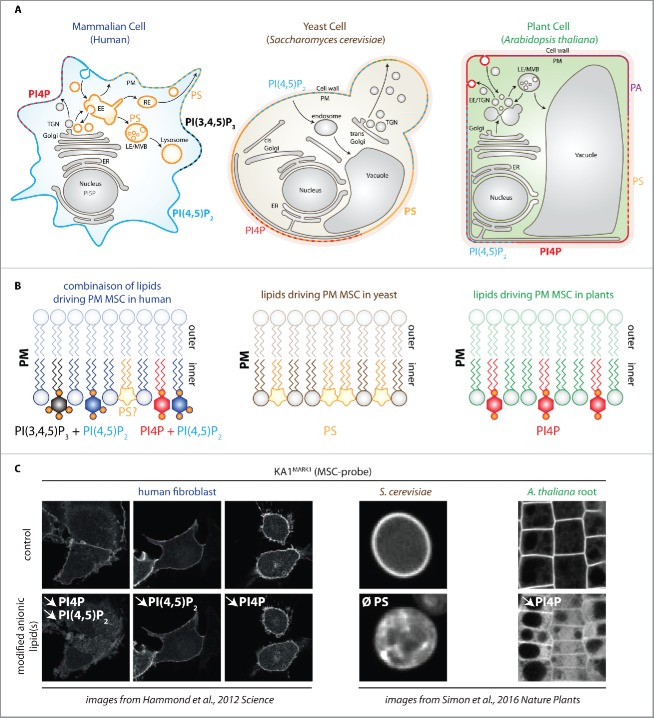
The inner leaflet of the plasma membrane (PM) of animal cells is composed of about 20% of anionic lipids that provide negative charges (electric field estimated at 5V/cm) giving the potential to permanently or transiently attract cytosolic cationic molecules, including peripheral membrane proteins.1 The concept of an electrostatic potential driven by membrane surface charges (MSC) was postulated long ago by biophysicists.2 However tools to sense this predicted feature were only developed during the last decade via the generation of genetically encoded biosensors (Fig. 1).3-7 These biosensors, which will be referred as MSC-probes thereafter, consist of cationic peptides or folded protein domains that transiently associate with anionic phospholipids based on their negative charges and irrespective of their head group (Fig. 1B).3,4,7 When fused to a fluorescent protein, these MSC-probes label strictly the cytosolic face of the plasma membrane in all eukaryotic cell type analyzed including yeast, plant and mammalian cells3,4,8-10 (Fig. 2C). This common feature highlights a unique signature of the plasma membrane as the most anionic membrane in cells. This particular plasma membrane property is paramount to localize signaling proteins, including for example small GTPases and kinases.3-10 However, in each eukaryotic kingdom, different anionic lipids are used to power this high plasma membrane electrostatic field (Fig. 2B).
Figure 1.
(A) Timeline showing landmark papers for the in vivo study of membrane surface charges (MSC) in various organisms. Color indicates the model system used in the study: blue, human cell lines; brown, Saccharomyces cerevisiae; Green, Arabidopsis thaliana and Nicotiana benthamiana. (B) Schematic representation of peptide-based MSC-probes (Left and middle panels) and domain-based MSC-probes (right panel). Black circles indicate negative membrane surface charges, red circles show cationic residues in MSC-probes that interact with MSC through electrostatic interactions, and purple circles indicate aromatic residues that provide hydrophobic interaction for membrane anchoring. The lipid anchor is represented in purple (for clarity only farnesylation is given as an example, but other lipid modifications have been used, such as the N-terminal myristoylation in c-Src or K-myr reporters, see ref 3). K-Ras4B MSC-probe corresponds to the C-terminal tail of K-Ras4B, c-Src probe corresponds to the N-terminal tail of c-Src, K-myr is a synthetic construct that has a N-terminal myristoylation adjacent to the K-Ras4B charged peptide. MSC, membrane surface charges, KA1 domain, Kinase Associated1 domain; MARK1, Microtubule Associated Regulated Kinase1; MARCKS-ED, Myristoylated Alanine-Rich C Kinase Substrate-Effector Domain.
Figure 2.
Contribution of different anionic phospholipids in plasma membrane surface charge. (A) schematic representation of human, yeast and plant cells. Anionic phospholipids that localize at the cell surface are indicated for each cell type. For clarity, PI3P, PI5P and PtdIns(3,4)P2 have been omitted, although they have been shown to localize at the plasma membrane in animal cells at very low quantity and/or upon specific stimuli.11 The localization of PS in plasma membrane-derived organelles is indicated by the orange color. Note that for practical purposes, dashes indicate the presence of several lipid species on the same membrane, however, this does not mean that they are necessarily organized in discrete domains. (B) schematic representation of the anionic lipids required for plasma membrane MSC in mammals (left), yeast (middle) and plants (right). Note that in human, PtdIns(4,5)P2 acts redundantly with either PtdIns(4)P or PtdIns(3,4,5)P3. (C) confocal pictures showing the localization of the KA1 domain of MARK1 in human fibroblast cells (left), S. cerevisiae (middle) and A. thaliana root epidermis (right). KA1 is a domain that interacts with all negatively charged lipids and therefore acts as a sensor of membrane electrostatics (so called MSC-probe). Top panels are control cells and bottom panels show conditions in which anionic phospholipids have been genetically or chemically perturbed. The targeted lipid(s) is indicated in white (downward pointing arrows indicate the reduction in the given lipid content and Ø total absence in the lipid in the Δcho1 yeast mutant). Note that KA1MARK1 localizes at the cell surface in mammals, yeasts and plants, but that this strict plasma membrane localization relies on different anionic phospholipid in these cells. EE, early endosome; LE, late endosome; RE, recycling endosomes; TGN, trans-golgi network; ER, endoplasmic reticulum; MSC, membrane surface charge. Pictures of fibroblasts are from Hammond et al.9 and pictures from yeast and plants are from Simon et al.8 The cartoon representing the cell from the top left cornel is inspired from Jean and Kiger 2012 and adapted by permission from Macmillan Publisher Ltd: [NATURE REVIEW MOLECULAR CELL BIOLOGY], ref. 12 copyright (2012).
Phosphoinositides cooperativity powers membrane electrostatics in mammals
In mammals, phosphatidylserine [PS], phosphatidylinositol-4-phosphate PtdIns(4)P, phosphatidylinositol- 4,5-biphosphate [PI(4,5)P2], and phosphatidylinositol-3,4,5-triphosphate [PtdIns(3,4,5)P3] are localized at the cell surface (Fig. 2A).11,12 These lipids are candidates to power the PM electrostatic field. Phosphoinositides are low abundant lipids but highly anionic, with PtdIns(4)P, PtdIns(4,5)P2 and PtdIns(3,4,5)P3 containing respectively 3, 5 and 7 net negative charges.13 Since PtdIns(4,5)P2 is a distinctive lipid of the plasma membrane and relatively abundant compared with other plasma membrane-localized phosphoinositides, it was a prime candidate to drive plasma membrane MSC. However, inducible PtdIns(4,5)P2 depletion at the plasma membrane has no effect on the localization of MSC-probes, suggesting that this lipid does not specify the plasma membrane electrostatic field on its own.4,9 Interestingly, inhibition of PtdIns(3,4,5)P3 synthesis by type-I PI3-Kinase inhibitors together with inducible depletion of PM PtdIns(4,5)P2 delocalized MSC-probes to intracellular compartments, showing that these lipids are redundantly required for PM MSC.4 Later on, concomitant inducible depletion of plasma membrane-associated PtdIns(4)P and PtdIns(4,5)P2 also demonstrated a role for PtdIns(4)P in plasma membrane surface charges together with PtdIns(4,5)P29 (Fig. 2C). Altogether, PtdIns(4,5)P2 seems to be critical in defining plasma membrane MSC in human cells but acts redundantly with PtdIns(3,4,5)P3 and/or PtdIns(4)P (Fig. 2B).
PtdInsPs are highly anionic but represent only 1–2% of total phospholipids in living cells.13 Other less anionic lipids might also contribute to MSC notably due to their higher abundance. In animals, PS represents about 10 to 20% of plasma membrane phospholipids but PS is less anionic than phosphoinositides (net charge −1).1,14 Inhibition of ATP synthesis prevents phosphorylation of PtdInsPs by kinases while lipid phosphatases are still active, triggering the rapid depletion of phosphoinositides from cellular membranes.10 However, this treatment does not affect the PS pool, since it is not constantly regulated by phosphorylation10 In this condition and therefore in the absence of PtdInsPs, MSC-probes lose their specific plasma membrane localization and relocalize to all PS-bearing organelles, including the PM but also all plasma membrane-derived organelles along the endocytic pathways (Fig. 2A).10 This result confirms the importance of phosphoinositides in driving the specific electrostatic signature of the cell surface.10 However, in the absence of phosphoinositides, MSC-probes partially retain their plasma membrane localization, suggesting a role for PS in plasma membrane MSC.10
In addition, because in the absence of phosphoinositide, MSC-probes localize to all PS-containing compartments,10 PS might be involved in driving the electrostatic properties of endocytic compartments. Bigay and Antonny proposed that PS defined an electrostatic territory in cells that corresponds to all PM-derived organelles.5,6 However, this hypothesis is mainly based on coincidence between the presence of negative charges, as visualized by MSC-probes, and the presence of PS on these membranes.10 To our knowledge, this theory has not been challenged by genetic and/or pharmacological perturbation(s) of the PS pool.
Overall, PtdInsPs are the main anionic lipids that regulate plasma membrane surface charge in mammals, while PS seems to have a broader role in controlling membrane electrostatics of all PM-derived organelles (Fig. 2B-C)3-6,9,10
Maintenance of plasma membrane electrostatics in yeast: It's all about PS
Based on findings in mammals, the potential involvement of PtdInsPs was analyzed in yeast. To address the relative role of PtdInsPs in plasma membrane electrostatic field, temperature-sensitive alleles that reduces both PtdIns(4)P and PtdIns(4,5)P2 or PtdIns(4,5)P2 alone were used. Surprisingly, at restrictive temperature, KINASE ASSOCIATED1 (KA1) domains, which are domains that bind to all anionic phospholipids and therefore act as MSC-probes, remain strictly localized at the PM in all these yeast mutant strains.7 These results suggest that unlike in animals, PtdInsPs do not play a major role in PM MSC.7
By contrast to mammals in which PS is spread all along the endocytic pathway,10,14,15 PS is highly enriched at the PM in yeast (Fig. 2A).7,10 Therefore, PS is a good candidate to specify plasma membrane electrostatics in yeast cells. Cho1p is the only PS synthase in yeast, and the cho1 mutant does not produce any PS.10,16 mislocalization of the KA1 MSC-probes in cho1 shows a prominent contribution of PS in plasma membrane surface charge (Fig. 2C).7,8 Altogether, these results suggest either no or minor roles of PtdInsPs in plasma membrane surface charge in yeast, while PS is the main anionic lipid regulating the plasma membrane electrostatic potential.7
PtdIns(4)P massively accumulates at the plasma membrane in plants and drives its electrostatic field
By contrast to yeast and animals, PtdIns(4)P massively accumulates at the plasma membrane in plants.8,17 In Arabidopsis root cells, short-term (up to 30 min) pharmacological inhibition of PI4-Kinase (PI4K) rapidly depletes the cellular PtdIns(4)P pool but has no effect on PtdIns(4,5)P2.8 This results is surprising since PtdIns(4)P is the precursor of PtdIns(4,5)P2. However, short-term depletion of PtdIns(4)P has also no effect on PtdIns(4,5)P2 in human fibroblast cells, suggesting that in both kingdoms the metabolism of these two lipids are largely independent within this short time frame.8,9 In addition, PtdIns(4)P is substantially more abundant than PtdIns(4,5)P2 in plant tissues,17,18 therefore the residual PtdIns(4)P molecules might be sufficient to sustain PtdIns(4,5)P2 synthesis. The relative abundance of PtdIns(4)P over PtdIns(4,5)P2 and its accumulation at the cell surface suggest that it might be involved in plasma membrane electrostatics. Indeed, inhibition of PI4K largely delocalized MSC-probes from the plasma membrane.8 In addition, genetic depletion of PtdIns(4)P specifically at the plasma membrane induced the ectopic localization of MSC-probes in less anionic endomembrane compartments.8 Together, these results indicate that PtdIns(4)P is important for plasma membrane electrostatics and that, by contrast to mammals, it does not act redundantly with PtdIns(4,5)P2.
However, it is worth noting that MSC-probes retain a certain degree of plasma membrane localization upon PtdIns(4)P depletion (Fig. 2C), and that therefore other anionic lipids might contribute to the plant plasma membrane electrostatic field.8 Candidate lipids include PtdIns(4,5)P2, PS and/or phosphatidic acid (PA) that all localized at the plasma membrane at least in some plant cell types (Fig. 2A).8,17,19 Pharmacological and/or genetic perturbation of PtdIns(4,5)P2 and PA indeed suggest that these lipids are involved in the plasma membrane localization of proteins with cationic stretches.20 Therefore, as seen for mammals, lipid cooperativity might also be important for membrane electrostatics in plants. Nevertheless, unlike in animals, depletion of PtdIns(4)P alone is sufficient to perturb PM electrostatics in plants (Fig. 2C), highlighting the unusual importance of PtdIns(4)P in specifying the identity of the plant plasma membrane.
Concluding remarks
To conclude, the plasma membrane is highly electronegative across eukaryotes, but differences exist concerning the lipids involved in the maintenance of plasma membrane electrostatics (Fig. 2B). The main difference comes from yeast where PS is the major anionic lipid that drives plasma membrane surface charge, while PtdInsPs are not required.7 This striking contrast brings the question of the role of PS in membrane electrostatics in multicellular eukaryotes such as plants or mammals. Indeed, while PS has been postulated to control electrostatic properties of plasma membrane-derived organelles,5,6 this has not been fully addressed experimentally. Future researches are therefore awaited to tackle this question. Similarly, it would be interesting to explore the contribution of PA in membrane electrostatics.
Disclosure of potential conflicts of interest
No potential conflicts of interest were disclosed.
Acknowledgment
We thank Laia Armengot, Isabelle Fobis-Loisy, Mehdi Bichir Doumane, Thierry Gaude, Vincent Bayle for critical comments on the manuscript, Julien Gronnier for the membrane template used in Fig. 1B and Fig. 2B, Gerald Hammond (University of Pittsburg, USA) for sharing the pictures of KA1(MARK1) in fibroblasts and Amy Kiger (UCSD, San Diego, USA) for the template of the animal cell in Fig. 2A. The orange spring in Fig. 1 was reproduced with permission from Vecteezy.com. Y.J. has received funding from the European Research Council—ERC Grant Agreement no. [3363360-APPL] under the European Union's Seventh Framework Program (FP/2007–2013).
References
- 1.Lemmon MA. Membrane recognition by phospholipid-binding domains. Nat Rev Mol Cell Biol 2008; 9:99-111; PMID:18216767; http://dx.doi.org/ 10.1038/nrm2328 [DOI] [PubMed] [Google Scholar]
- 2.McLaughlin S. The electrostatic properties of membranes. Annu Rev Biophys Biophys Chem 1989; 18:113-36; PMID:2660821; http://dx.doi.org/ 10.1146/annurev.bb.18.060189.000553 [DOI] [PubMed] [Google Scholar]
- 3.Yeung T, Terebiznik M, Yu L, Silvius J, Abidi WM, Philips M, Levine T, Kapus A, Grinstein S. Receptor activation alters inner surface potential during phagocytosis. Science 2006; 313:347-51; PMID:16857939; http://dx.doi.org/ 10.1126/science.1129551 [DOI] [PubMed] [Google Scholar]
- 4.Heo WD, Inoue T, Park WS, Kim ML, Park BO, Wandless TJ, Meyer T. PI(3,4,5)P3 and PtdIns(4,5)P2 lipids target proteins with polybasic clusters to the plasma membrane. Science 2006; 314:1458-61; PMID:17095657; http://dx.doi.org/ 10.1126/science.1134389 [DOI] [PMC free article] [PubMed] [Google Scholar]
- 5.Bigay J, Antonny B. Curvature, lipid packing, and electrostatics of membrane organelles: defining cellular territories in determining specificity. Dev Cell 2012; 23:886-95; PMID:23153485; http://dx.doi.org/ 10.1016/j.devcel.2012.10.009 [DOI] [PubMed] [Google Scholar]
- 6.Jackson CL, Walch L, Verbavatz JM. Lipids and their trafficking: an integral part of cellular organization. Dev Cell 2016; 39:139-53; PMID:27780039; http://dx.doi.org/ 10.1016/j.devcel.2016.09.030 [DOI] [PubMed] [Google Scholar]
- 7.Moravcevic K, Mendrola JM, Schmitz KR, Wang YH, Slochower D, Janmey PA, Lemmon MA. Kinase associated-1 domains drive MARK/PAR1 kinases to membrane targets by binding acidic phospholipids. Cell 2010; 143:966-77; PMID:21145462; http://dx.doi.org/ 10.1016/j.cell.2010.11.028 [DOI] [PMC free article] [PubMed] [Google Scholar]
- 8.Simon ML, Platre MP, Marquès-Bueno MM, Armengot L, Stanislas T, Bayle V, Caillaud MC, Jaillais Y. A PtdIns(4)P-driven electrostatic field controls cell membrane identity and signalling in plants. Nature Plants 2016; 2:16089; PMID:27322096; http://dx.doi.org/ 10.1038/nplants.2016.89 [DOI] [PMC free article] [PubMed] [Google Scholar]
- 9.Hammond GR, Fischer MJ, Anderson KE, Holdich J, Koteci A, Balla T, Irvine RF. PtdIns(4)P and PtdIns(4,5)P2 are essential but independent lipid determinants of membrane identity. Science 2012; 337:727-30; PMID:22722250; http://dx.doi.org/ 10.1126/science.1222483 [DOI] [PMC free article] [PubMed] [Google Scholar]
- 10.Yeung T, Gilbert GE, Shi J, Silvius J, Kapus A, Grinstein S. Membrane phosphatidylserine regulates surface charge and protein localization. Science 2008; 319:210-13; PMID:18187657; http://dx.doi.org/ 10.1126/science.1152066 [DOI] [PubMed] [Google Scholar]
- 11.Platre MP, Jaillais Y. Guidelines for the use of protein domains in acidic phospholipid imaging. Methods Mol Biol 2016; 1376:175-94; PMID:26552684; http://dx.doi.org/ 10.1007/978-1-4939-3170-5_15 [DOI] [PMC free article] [PubMed] [Google Scholar]
- 12.Jean S, Kiger AA. Coordination between RAB GTPase and phosphoinositide regulation and functions. Nat Rev Mol Cell Biol 2012; 13:463-70; PMID:22722608; http://dx.doi.org/ 10.1038/nrm3379 [DOI] [PubMed] [Google Scholar]
- 13.Balla T. Phosphoinositides: tiny lipids with giant impact on cell regulation. Physiol Rev 2013; 93:1019-37; PMID:23899561; http://dx.doi.org/ 10.1152/physrev.00028.2012 [DOI] [PMC free article] [PubMed] [Google Scholar]
- 14.Leventis PA, Grinstein S. The distribution and function of phosphatidylserine in cellular membranes. Annu Rev Biophys 2010; 39:407-27; PMID:20192774; http://dx.doi.org/ 10.1146/annurev.biophys.093008.131234 [DOI] [PubMed] [Google Scholar]
- 15.Yeung T, Heit B, Dubuisson JF, Fairn GD, Chiu B, Inman R, Kapus A, Swanson M, Grinstein S. Contribution of phosphatidylserine to membrane surface charge and protein targeting during phagosome maturation. J Cell Biol 2009; 185:917-28; PMID:19487458; http://dx.doi.org/ 10.1083/jcb.200903020 [DOI] [PMC free article] [PubMed] [Google Scholar]
- 16.Atkinson K, Fogel S, Henry SA. Yeast mutant defective in phosphatidylserine synthesis. J Biol Chem 1980; 255:6653-61; PMID:6771275 [PubMed] [Google Scholar]
- 17.Simon ML, Platre MP, Assil S, van Wijk R, Chen WY, Chory J, Dreux M, Munnik T, Jaillais Y. A multi-colour/multi-affinity marker set to visualize phosphoinositide dynamics in Arabidopsis. Plant J 2014; 77:322-37; PMID:24147788; http://dx.doi.org/ 10.1111/tpj.12358 [DOI] [PMC free article] [PubMed] [Google Scholar]
- 18.König S, Hoffmann M, Mosblech A, Heilmann I. Determination of content and fatty acid composition of unlabeled phosphoinositide species by thin-layer chromatography and gas chromatography. Anal Biochem 2008; 378:197-201; PMID:18466755; http://dx.doi.org/ 10.1016/j.ab.2008.03.052 [DOI] [PubMed] [Google Scholar]
- 19.Potocký M, Pleskot R, Pejchar P, Vitale N, Kost B, Zárský V. Live-cell imaging of phosphatidic acid dynamics in pollen tubes visualized by Spo20p-derived biosensor. New Phytol 2014; 203:483-94; PMID:24750036; http://dx.doi.org/ 10.1111/nph.12814 [DOI] [PubMed] [Google Scholar]
- 20.Barbosa ICR, Shikata H, Zourelidou M, Heilmann M, Heilmann I, Schwechheimer C. Phospholipid composition and a polybasic motif determine D6 PROTEIN KINASE polar association with the plasma membrane and tropic responses. Development 2016; 143(24):4687-700; PMID:27836964; http://dx.doi.org/ 10.1242/dev.137117 [DOI] [PubMed] [Google Scholar]


