Abstract
Candida albicans, along with some other non-albicans Candida species, is a group of yeast, which causes serious infections in humans that can be both systemic and superficial. Despite the fact that extensive efforts have been put into the discovery of novel antifungal agents, the frequency of these fungal infections has increased drastically worldwide. In our quest for the discovery of novel antifungal compounds, we had previously synthesized and screened quinoline containing 1,2,3-triazole (3a) as a potent Candida spp inhibitor. In the present study, two structural analogues of 3a (3b and 3c) have been synthesized to determine the role of quinoline and their anti-Candida activities have been evaluated. Preliminary results helped us to determine 3a and 3b as lead inhibitors. The IC50 values of compound 3a for C. albicans ATCC 90028 (standard) and C. albicans (fluconazole resistant) strains were 0.044 and 2.3 μg/ml, respectively while compound 3b gave 25.4 and 32.8 μg/ml values for the same strains. Disk diffusion, growth and time kill curve assays showed significant inhibition of C. albicans in the presence of compounds 3a and 3b. Moreover, 3a showed fungicidal nature while 3b was fungistatic. Both the test compounds significantly lower the secretion of proteinases and phospholipases. While, 3a inhibited proteinase secretion in C. albicans (resistant strain) by 45%, 3b reduced phospholipase secretion by 68% in C. albicans ATCC90028 at their respective MIC values. Proton extrusion and intracellular pH measurement studies suggested that both compounds potentially inhibit the activity of H+ ATPase, a membrane protein that is crucial for various cell functions. Similarly, 95–97% reduction in ergosterol content was measured in the presence of the test compounds at MIC and MIC/2. The study led to identification of two quinoline based potent inhibitors of C. albicans for further structural optimization and pharmacological investigation.
Introduction
Although sincere efforts are being continuously made for discovering new antifungal targets and drugs, the frequency of human fungal infections has increased drastically worldwide, [1–3]. Of particular concern are the ever-increasing incidences of hospital-acquired systemic mycoses caused by Candida species responsible for crude mortality rates of up to 50% in the United States alone [4]. Adding to this disease burden, superficial infections of skin and nails in humans are affecting ~25% of the general population worldwide [5]. Use of broad-spectrum antibiotics, suppression of immune response during organ transplantation, immune-suppressive agents during cancer treatment and HIV/AIDS cases have increased the chances of Candida spp infections, and hence further aggravating the condition [6]. Among different Candida spp, C. albicans is the major cause of candidiasis and accounts for 80% of the isolates from all forms of human candidiasis [7]. However, the number of infections caused by other non-albicans Candida species which includes C. glabrata, C. tropicalis, and C. parapsilosis has also increased significantly [8].
During both superficial and systemic infections, pathogenicity of Candida spp relies on a number of virulence factors including morphogenesis and capability to produce hydrolytic enzymes such as proteinases, phospholipases, and lipases. The ability of C. albicans to switch reversibly between yeast to filamentous or hyphal (pseudo or true, based on condition) form of growth has been well reported as an important virulence attribute [9]. Similarly, hydrolytic enzymes especially proteinases, phospholipases, and lipases help Candida spp with adhesion, invasion, host tissue damage and protection from host defense mechanism [10]. Various studies have explained the potential role of these hydrolytic enzymes in the pathogenicity of Candida spp [10–13]. In the modern age of drug discovery, the structure and function of potent targets play a very important role in designing better prototypic antimicrobial molecules. H+ ATPase, a member of P-type transport ATPase family, has been reported as a potential antifungal target [14–16]. This protein is essentially involved in the physiological functions of Candida spp such as maintenance of electrochemical gradient across cell membrane, nutrient uptake, regulation of intracellular pH and cell growth [17]. Plasma membrane H+ ATPase is unique to the fungus and is not available as a human protein. Hence this enzyme is crucial to the fungus and maybe explored as a potential antifungal target. Similarly, cytochrome P450-dependent enzyme lanosterol 14 α-demethylase (CYP51) is involved in the biosynthesis of ergosterol in fungi which is an important component of fungal membranes. Lanosterol 14 α-demethylase has already been explored as a potential drug target for azole based antifungal agents [18–19].
Moreover, the increased use of antifungal drugs has contributed to emerging resistance in Candida species which has prompted scientists worldwide to develop novel and more effective antifungal agents with a broad spectrum, better pharmacokinetic profile and low toxicity. In our previous study, we synthesized a series of novel 1,2,3-triazole derivatives from naturally bioactive alcohols and evaluated their anti-Candida activity as well as cytotoxicity [20]. The results showed that quinoline containing 1,2,3-triazole (3a) has great potential to inhibit the Candida spp in vitro. In the present work, we have extended our study further to determine the role of quinoline on anti-Candida activity; by replacing with its structural analogues (3b and 3c). We have explored the effect of selected lead inhibitors (3a and 3b) on the growth and secretion of hydrolytic enzymes in C. albicans. The plasma membrane H+ ATPase activity and ergosterol biosynthesis were also determined in the presence and absence of the test compounds.
Materials and methods
Chemistry
All the reagents and solvents purchased from Sigma-Aldrich, S.D. Fine, SRL and Hi Media, were used without further purification. Reaction was monitored by thin-layer chromatography (TLC) using pre-coated aluminum sheets (Silica gel 60 F254, Merck Germany) and spots were visualized under UV light. The IR spectra of the compounds were recorded on Agilent Cary 630 FT-IR spectrometer. 1H NMR and 13C NMR spectra were obtained in CDCl3 with reference to TMS as on internal standard using Bruker Spectrospin DPX-300 spectrometer at 300 MHz and 75 MHz respectively. Splitting patterns are designated as follows: s, singlet; d, doublet; t, triplet; m, multiplet. Chemical shift (δ) values are given in parts per million (ppm) and coupling constants (J) in Hertz (Hz). CHN analysis was recorded on Elemental Vario analyzer. Melting points were recorded on a digital Buchi melting point apparatus (M-560) and were reported uncorrected. Purification of the compounds was done by column chromatography using silica gel (230–400 mesh size) with petroleum ether/ethyl acetate (8:2) as eluent for alkynes and 10% methanol in DCM for triazole derivatives.
General procedure for the synthesis of alkynes (2a-c)
A round bottom flask with stir bar was charged with natural precursor (1.0 mmol) and anhydrous DMF (10 ml) and the reaction flask was cooled to 0°C. To this solution, potassium carbonate (1.0 mmol) was added, followed by slow addition of propargyl bromide (1.2 mmol). The reaction mixture was allowed to warm to room temperature and stirred overnight under argon. After completion of the reaction as confirmed by TLC, it was concentrated and water was added to the residue. The compound was extracted with ethyl acetate, dried over anhydrous sodium sulphate and concentrated under vacuo. The crude product was purified by column chromatography with petroleum ether: ethyl acetate (8:2) to give the desired alkynes (2a-c) in good to excellent yields. The synthesis of compound 2a [8-(Prop-2-ynyloxy)quinoline] has been previously reported in our lab [20].
5-Chloro-8-(prop-2-ynyloxy)quinoline (2b)
Light brown crystalline solid, yield:87%, Rf (pet. ether/ethyl acetate, 8:2) = 0.48, Anal (C12H8ClNO) calc. C 66.22, H 3.70, N 6.44, found: C 66.02, H 3.47, N 6.54%. IR (neat): ν (cm-1) 3144, 2121, 1615, 1592, 1499, 1469, 1456, 1367, 1311, 1242, 1169, 1095, 994, 967, 931, 829, 788, 754. 1H NMR (300 MHz, CDCl3) (δ, ppm): 8.35 (d, 1H, J = 7.8 Hz, Ar-H), 8.21 (d, 1H, J = 8.1 Hz, Ar-H), 7.93–7.85 (m, 2H, Ar-H), 7.45 (d, 1H, J = 7.63 Hz, Ar-H), 5.20 (s, 2H, OCH2), 2.54 (s, 1H, ≡CH).
1-(Prop-2-ynyloxy)naphthalene (2c)
Brown solid, yield: 78%, Rf (pet. ether/ethyl acetate, 8:2) = 0.45, Anal (C12H8ClNO) calc. C 85.69, H 5.53, found: C 85.32, H 5.21%. IR (neat): ν (cm-1) 3345, 2954, 2120, 1864, 1554, 1520, 1456, 1356, 1323, 1273, 1170, 1110, 1005, 987, 845, 831, 767, 745. 1H NMR (300 MHz, CDCl3) (δ, ppm): 8.20 (d, 1H, J = 7.6 Hz, Ar-H), 7.97 (d, 1H, J = 7.5 Hz, Ar-H), 7.56–7.39 (m, 4H, Ar-H), 7.10 (d, 1H, J = 6.9 Hz, Ar-H), 5.08 (s, 2H, OCH2), 2.50 (s, 1H, ≡CH).
Procedure for the synthesis of benzyl azide
A mixture of benzyl bromide (1.0 mmol) and sodium azide (3.0 mmol) in anhyd. DMF (10 ml) was stirred overnight at 70°C. After completion of the reaction as monitored by TLC, the reaction was quenched with water. The crude was extracted with ethyl acetate, washed with brine, dried over anhyd. sodium sulphate and concentrated under vacuo. The oily residue was used as such without any further purification.
General procedure for the synthesis of triazole derivatives (3a-c)
Equimolar amounts of alkyne (2a-c) and benzyl azide were dissolved in tert-butanol and water (1:2) mixture. To this reaction mixture, copper sulphate (0.05 eq) and sodium ascorbate (0.01 eq) were added and stirred at room temperature till the disappearance of starting materials as indicated by TLC. The reaction mixture was quenched with saturated brine and crude was extracted with ethyl acetate, dried over anhyd. Na2SO4 and concentrated under vacuo. The crude was purified by column chromatography using dichloromethane and methanol (9:1) as eluent to give 1,2,3-triazole derivatives in good to excellent yields. The compound 3a [1-Benzyl-4-(quinolin-1-yloxy)methyl)-1H-1,2,3-triazole] has been previously reported in our lab [20].
8-((1-Benzyl-1H-1,2,3-triazol-4-yl)methoxy)-5-chloroquinoline (3b)
Brown solid, M.pt. 148–149°C, yield: 65%, Rf (pet. ether/ethyl acetate, 5:5) = 0.15, Anal (C19H15ClN4O) calc. C 65.05 H 4.31 N 15.97, found: C 65.08 H 4.28 N 15.94%. IR (neat): ν (cm-1) 3065, 2924, 2851, 1721, 1605, 1587, 1512, 1460, 1423, 1380, 1343, 1296, 1262, 1225, 1162, 1140, 1061, 1035, 1018, 964, 855, 836, 737, 717, 678. 1H NMR (300 MHz, CDCl3) (δ, ppm): 8.95 (s, 1H, Ar-H), 8.52 (d, 1H, J = 8.4 Hz, Ar-H), 7.66 (s, 1H, triazole ring), 7.53–7.47 (m, 2H, Ar-H), 7.36–7.33 (m, 3H, Ar-H), 7.28–7.25 (m, 3H, Ar-H), 5.51 (s, 2H, OCH2), 5.50 (s, 2H, CH2). 13C NMR (75MHz, CDCl3) (δ, ppm): 153.05, 149.76, 144.02, 140.84, 134.26, 133.05, 129.12, 128.82, 128.17, 127.10, 126.46, 123.31, 122.84, 122.33, 110.10, 63.10, 54.28.
1-Benzyl-4-((naphthalen-1-yloxy)methyl)-1H-1,2,3-triazole (3c)
Dark brown solid, M.pt. 128–129°C, yield: 30%, Rf (DCM/ methanol, 9:1) = 0.735, Anal (C19H15ClN4O) calc. C 65.05 H 4.31 N 15.97, found: C 65.08 H 4.28 N 15.94%. IR (neat): ν (cm-1) 3065, 2924, 2851, 1721, 1605, 1587, 1512, 1460, 1423, 1380, 1343, 1296, 1262, 1225, 1162, 1140, 1061, 1035, 1018, 964, 855, 836, 737, 717, 678. 1H NMR (300 MHz, CDCl3) (δ, ppm): 8.92 (s, 1H, Ar-H), 8.43 (d, 1H, J = 7.8 Hz, Ar-H), 7.69 (s, 1H, triazole ring), 7.41–7.32 (m, 4H, Ar-H), 7.27–7.24 (m, 3H, Ar-H), 7.05–6.98 (m, 3H, Ar-H), 5.56 (s, 2H, OCH2), 5.48 (s, 2H, CH2).13C NMR (75MHz, CDCl3) (δ, ppm): 152.27, 148.56, 143.24, 139.76, 134.35, 133.25, 129.09, 128.76, 128.17, 127.26, 126.89, 123.24, 122.72, 122.13, 110.51, 62.97, 54.24.
In vitro anti-Candida activities
Determination of IC50 value
Broth dilution technique was employed to determine the IC50 values of all the synthesized 1,2,3 triazole derivatives (3a-c) against C. albicans ATCC 90028, C. glabrata ATCC 90030, C. tropicalis ATCC 750 and four clinical isolates of C. albicans namely D27, D31, D39 (FLC-sensitive) and D15.9 (FLC-resistant). The strains were maintained on yeast extract:peptone:dextrose (1:2:2:2.5) nutrient agar (2.5%) slants at 4°C. The test compounds were dissolved in DMSO with less than 4% concentration of it in the final test volume. Different concentrations (1000… 7.8 μg/mL) of test compounds in Sabouraud dextrose (SD) broth medium were dispensed into the wells, then inoculated with the test organism with approx. 2.5 × 10³ cells/mL and incubated at 37°C for 24 h. After incubation period, the optical density was measured at 600 nm by using Thermo Scientific Multiskan Go plate reader. The IC50 value was defined as the concentration of the test compound that causes 50% decrease in absorbance compared with that of the control (no test compound) [21]. All the experiments were done in triplicate at separate times.
Disk diffusion assay
The preliminary screening of anti-Candida activity showed that compound 3a still had better inhibition in comparison to its structural analogues, 3b and 3c. Moreover, compound 3b also showed significant inhibition including in FLC-resistant C. albicans strain, thus we selected compound 3a along with 3b as lead inhibitors. The present work is focussed on mainly C. albicans due to its pathological importance. Hence, besides one standard ATCC strain, a FLC-sensitive and FLC- resistant C. albicans clinical strains were used in the present study.
Disk diffusion assay was performed using C. albicans ATCC 90028 (standard), the two clinical isolates, C. albicans D27 (FLC-susceptible), and C. albicans D15.9 (FLC-resistant) for the lead inhibitors 3a and 3b. The C. albicans cells were inoculated into liquid SD medium and grown over night at 37°C. The cells were then centrifuged at 3000 rpm for 5 min, washed three times with PBS and then suspended in PBS again. Approximately, 105 cells/mL were inoculated into molten SD agar medium at 42°C and poured into 100-mm-diameter petriplates. Sterilized 4 mm disks of Whatman paper were placed on solid agar and 1, 2, and 4 μL of 3a and 2, 4, and 8 μL of 3b from the stock solution of 10 mg/mL were spotted on the disk. 2 μL of FLC from the stock of 10 mg/mL was also applied on the disks to serve as positive control. The diameter of zone of inhibition (ZOI) was recorded in millimetres after 48 h and was compared with that of control. This experiment was repeated thrice and values were shown in terms of mean ±standard error of mean.
Growth assay
The C. albicans cells were freshly revived by subculture on the SD agar plate. A loopful of inoculum was introduced into the SD broth and cells were grown for 16 h at 37°C before use. Approximately 2×10³ cells/mL were then inoculated into the freshly prepared 50 mL sterile SD medium. Different concentrations, equivalent to 2MIC, MIC, MIC/2, of test compounds were added separately into the conical flasks containing inoculated medium and incubated at 37°C and 160 rpm. Strain specific concentration of FLC was used as positive control viz 40 μg/mL for C. albicans ATCC 90028, 20 μg/mL for C. albicans D27 (FLC-susceptible) and 40 μg/mL for C. albicans D15.9 (FLC-resistant). At predetermined time periods (0, 2, 4, 6, 8, 10, 12, 14, 16, 18, 20 22, and 24 after incubation with agitation at 37°C), 1 mL aliquot of each sample was removed from the conical flask and growth was measured tubidometrically at 600 nm using Thermo Multiskan spectrophotometer. Optical density was recorded for each concentration against time (h).
Time kill curve study
Time-kill curves were used for testing the fungicidal or fungistatic nature of the lead inhibitors 3a and 3b. The experimental procedure was performed according to the method reported by Wang et al. [22]. C. albicans ATCC 90028, C. albicans D27 (FLC-susceptible) and C. albicans D15.9 (FLC-resistant) strains were used as test organisms. Fluconazole was used as a positive control. Freshly prepared sterile YEPD medium was inoculated by fresh culture (approx. 2×10³ cells) of test organisms in different conical flasks. The cells were exposed to the test compounds at 2MIC, MIC and MIC/2 values, separately. All the conical flasks having different sets of test compounds and concentrations were incubated at 37°C and 160 rpm. At predetermined time points (0, 4, 12, 24, and 48 h after incubation with agitation at 37°C), a 100-μL aliquot was removed from every solution and appropriately diluted in sterile water. A 100-μl aliquot from each dilution was spread over the YEPD agar plate. Colony counts were determined after incubation at 37°C for 24 h. All the time-kill curve experiments were conducted in duplicate and mean colony count data (log10 CFU/mL) were plotted as a function of time for each strain.
Effect on hydrolytic enzyme secretion in C. albicans
As described previously, secretion of hydrolytic enzymes (proteinases and phopholipases) play an important role in the pathogenicity of Candida spp. In the present study we have determined the effect of lead compounds 3a and 3b on the secretion of proteinases and phopholipases.
Proteinase assay
C. albicans ATCC 90028, C. albicans D27 (FLC-susceptible) and C. albicans D15.9 (FLC-resistant), previously identified as proteinase positive C. albicans [23] were transferred to flasks containing 5mL YEPD media and incubated at 37°C for 18 h. Following incubation, 1.5 mL of the yeast culture was taken into micro centrifuge tubes and centrifuged at 3000 rpm, for 5 min. The pellet obtained was washed by phosphate buffer saline (PBS) twice to remove the residual medium. After standardizing the suspensions (MacFarland standard), cells were exposed for 1 h to MIC and MIC/4 concentration of lead inhibitors 3a and 3b. Volumes of 1μL were placed at equidistant points on proteinase agar medium (0.1% yeast nitrogen base w/o amino acids; 0.145% ammonium sulfate; 2% glucose with 0.2% (w/v) BSA fraction V mixed into agar at ∼40°C) and incubated at 37°C for 3 days [24]. The experiment was performed in duplicate. Secreted proteinase enzyme will degrade BSA and form zone of clearance around the yeast colonies. An index (Pz value) was used to represent the extent of proteinase activity by different strains of C. albicans. A Pz value of 1.0 depicts no enzyme activity. It was calculated as follows:
Phospholipase assay
Phospholipase activity was performed using the method of Price et al. (1982) with minor modifications [25]. The cells were incubated at 37°C for 18 h and then centrifuged at 3000 rpm for 5 min. The pellet obtained was washed with PBS twice to remove the residual culture medium. After standardizing the suspension, cells were exposed for 1 h to the test compounds at MIC and MIC/4. Volumes of 1 μL were placed at equidistant points on phospholipase agar media plates (1% peptone; 3% glucose; 5.73% NaCl; 0.055% CaCl2 with 10% (v/v) egg yolk emulsion) and incubated at 37°C for 4 days. The secretion of phospholipases was determined by the formation of opaque zones due to precipitation around the yeast colonies. The assay was conducted in duplicate. Pz values were calculated as follows:
Proton extrusion measurement
The proton pumping activity of C. albicans was determined by monitoring acidification of external medium by measuring pH as describe earlier [17]. In brief, the mid-log phase cells were harvested from SD medium by centrifugation at 3000 rpm for 15 min which is followed by three washings with phosphate buffer saline (PBS). A solution containing 0.1 M KCl and 0.1 mM CaCl2 in distilled water was prepared. Compounds 3a and 3b at the concentrations of 50 and 100 μg/mL were separately added in this solution. Six sets of solution (10 mL) were prepared viz two having 50 and 100 μg/mL of 3a, two having 50 and 100 μg/mL of 3b, one having 10 μg/mL of FLC (as standard drug) and one is without test compound (control). Initial pH was adjusted to 7.0 using 0.01 M HCl/NaOH. Approximately 0.2 g of cells were suspended in each set of solution. Suspension was kept in a double-jacketed glass container with constant stirring. Again, the pH of suspension was maintained as 7.0 for 1 min by using 0.01 N NaOH. The rate of H+ extrusion was then monitored and calculated from the volume of 0.01 N NaOH consumed per min per mg weight of cells. Glucose has been reported as the stimulator of proton extrusion pump [17, 26]. Therefore above experiment was also repeated in the presence of 5 mM of glucose.
Measurement of intracellular pH (pHi)
In yeast cells, plasma membrane H+-ATPase helps to maintain internal pH between 6.0 and 7.5. Intracellular pH (pHi) was measured as reported earlier by Manzoor et al. [17]. Mid-log phase cells were harvested from SD medium and washed thrice with PBS. Subsequently, cells (0.1 g) were suspended in 5 mL solution of 0.1 M KCl, and 0.1 mM CaCl2 in distilled water. Compounds 3a and 3b were added to the suspension at a desired concentration of 100 μg/mL. 10 μg/mL of standard drug FLC was added to a separate suspension for comparison. Initial pH of the suspension was adjusted to 7.0 in each case and incubated for 30 min at 37°C with constant stirring. After incubation, the pH of suspension was again adjusted to 7.0 followed by the addition of nystatin (20 μM) and incubation at 37°C for 1 h. The change in pH of the suspension was monitored on a pH meter with constant stirring. The value of external pH at which nystatin permeabilization induced no further shift was taken as an estimate of pHi. Nystatin gives pHi values close to the cytoplasmic pH values, as it does not affect the mitochondria [27].
Transmission electron microscopic analysis
The morphology of C. albicans ATCC 90028 was analyzed using TEM following the standard protocol [28]. Mid-log phase cells were harvested, standardized (A600 ≈ 0.1) and exposed to MIC of lead inhibitors 3a and 3b for 1 h. Cells were then washed thrice with phosphate buffer solution to remove the residual medium and fixed overnight in 2.5% glutaraldehyde in phosphate/magnesium buffer (40 mM K2HPO4/KH2PO4, pH 6.5–0.5 mM MgCl2). Cells were washed twice for 15 min in 0.1 M sodium phosphate buffer (pH 6.0) and post-fixed for 2 h in 2% osmium tetroxide. Cells were again washed twice for 15 min in distilled water and then en bloc stained with 1% uranyl acetate (aqueous) for 30 min. After two further washes, cells were dehydrated in 95% and 100% ethanol. Cells were then exposed to propylene oxide for 2×10 min and infiltrated for 1 h in 1:1 propylene/epoxy embedding material (Epon) mixture and then overnight in fresh Epon. After polymerization for 48 h at 60°C, ultrathin sections were cut using a microtome (LeicaEM UC6) and transferred to a copper grid. Samples were stained with uranyl acetate (saturated solution of uranyl acetate in 50% alcohol) followed by lead citrate. Samples were washed three times in Milli-Q (MQ) water and dried by touching Whatman filter paper. Sections were examined with a FEI Tecnai G2 TF20 transmission electron microscope at 200 KV.
Ergosterol estimation assay
The total intracellular sterols were extracted as reported earlier with slight modifications [29]. As previous studies showed almost similar behavior of standard and FLC-sensitive strains of C. albicans, in the present study we used only standard C. albicans ATCC 90028 and compared with a FLC-resistant strain. Three separate conical flasks were used containing compounds 3a and 3b at MIC, MIC/2 and MIC/4 in SD broth inoculated with freshly cultured cells of C. albicans ATCC 90028 (standard) and FLC-resistant C. albicans. Fluconazole (40 μg/mL) was used as a positive control while untreated cells as negative control for comparison. All the conical flasks were incubated at 35°C for 16 h. After incubation, the cells were harvested at their stationary phase and the weight of pellet was determined. The pellet was treated with 25% alcoholic potassium hydroxide (KOH) solution followed by incubation at 85°C for 1 h. After incubation, sterol were extracted by the addition of n-heptane:distilled water (1:3). The heptane layers were transferred in fresh test tube, diluted five-fold in 100% ethanol and scanned spectrophotometrically between 230 and 300 nm. The presence of ergosterol and the late sterol intermediate 24(28) DHE in the extracted samples resulted in a characteristic four peak curve. The absence of detectable ergosterol in the extract was indicated as a flat line. Ergosterol content was calculated as percentage wet weight of the cells by the following equation:
here, % 24(28)DHE = [(A230/518)×F]/pellet weight, A281.5 and A230 absorbance at 281.5 nm and 230 nm, respectively and F is the dilution factor in alcohol.
Cytotoxicity
Cytotoxicity of compounds 3a and 3b was performed by hemolytic assay and cell proliferation assay.
Hemolytic assay
The hemolytic activities of the test compounds 3a and 3b along with standard drug FLC were determined on human red blood cells (hRBCs) [30]. Human blood from a healthy individual was collected in tubes containing EDTA as anti-coagulant. The erythrocytes were harvested by centrifugation for 10 min at 2000 rpm and 20°C, washed three times in PBS. To the pellet, PBS was added to yield a 10% (v/v) erythrocytes/PBS suspension. The 10% suspension was then diluted 1:10 in PBS. From each suspension, 100 μL was added in triplicate to 100 μL of a different dilution series of test compounds in the same buffer in micro centrifuge tubes. Total hemolysis was achieved with 1% Triton X-100. The tubes were incubated for 1 h at 37°C and then centrifuged for 10 min at 2000 rpm and 20°C. From the supernatant, 150 μL was transferred to a flat-bottomed microtiter plate (Tarson), and the absorbance was measured spectrophotometrically at 450 nm. The hemolysis percentage was calculated by following equation:
Cell proliferation assay
The in vitro cytotoxicity of compounds 3a and 3b to mammalian cells was also determined. The assay was performed in 96-well tissue culture-treated plates as described earlier [31]. Vero cells (monkey kidney fibroblasts) were seeded to the wells of 96-well plate at a density of 25,000 cells/well and incubated for 24 h. Samples at different concentrations were added and plates were again incubated for 48 h. The number of viable cells was determined by Neutral Red assay. IC50 values were obtained from dose response curves. Doxorubicin was used as a positive control for cytotoxicity.
Results
Chemistry
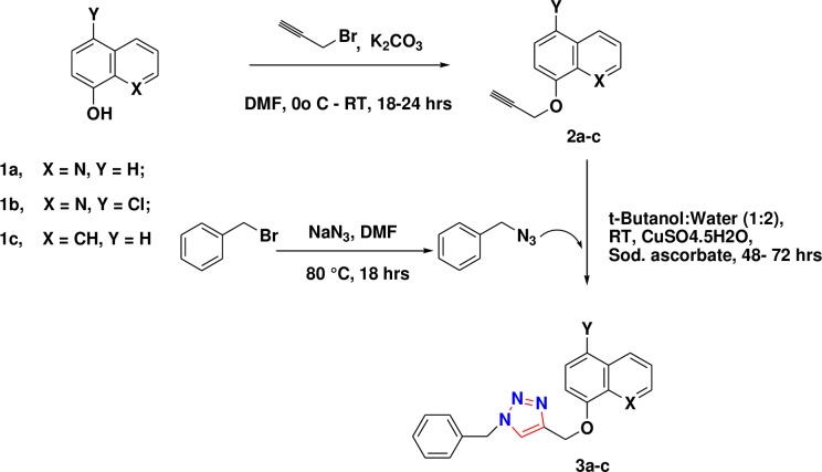
The two structural analogues (3b and 3c) of quinoline containing 1,2,3-triazole (3a) were synthesized as shown in Fig 1. The precursor alcohols were converted into 1,2,3-triazoles via copper catalyzed [3+2] azide-alkyne cycloaddition reaction (S1 and S2 Figs).
Fig 1.
Synthetic route of 1,2,3-triazole compounds (3a-c). 8-hydroxy quinoline (1a), 5-Cl- 8-hydroxy quinoline (1b) and 1-naphthol (1c) were used as precursor compounds which converted into 1,2,3 tiazoles.
Biological studies
Previously, we reported the synthesis and anti-Candida activities of eight novel 1,2,3 triazole derivatives (3a-h) and found that compound 3a displayed better activity than its natural bioactive compound 8-hydroxy quinoline. This compound showed an IC50 of 0.044, 12.02 and 3.60 μg/mL against C. albicans, C. glabrata and C. tropicalis, respectively. This enhanced anticandidal activity of compound 3a may be due to the presence of quinoline ring along with 1,2,3 triazole ring in its structure [20]. To determine the role of N-atom present in the quinoline ring and the effect of substitution on quinoline ring, we synthesized two new structural analogues (3b and 3c). Compound 3b with chlorine substitution on quinoline ring showed good anticandial activity with IC50 value of 25.4, 73.3 and 23.6 μg/mL against C. albicans, C. glabrata and C. tropicalis, respectively whereas compound 3c having naphthalene instead of quinoline ring showed slightly less activity with IC50 values of 91.2, 106.9 and 38.2 μg/mL against C. albicans, C. glabrata and C. tropicalis, respectively. The results hence indicate that N-atom plays an important role in the anticandidal activity and quinoline is a key moiety along with 1,2,3 triazole ring in the structure of these synthesized compounds. Hence compound 3a and 3b were selected for further evaluation of the biological activity of these compounds. Moreover, chlorine substitution in compound 3b might be the reason for its increased activity but compound 3a was still found to be the most potent anticandidal agent with lower IC50 values (Table 1).
Table 1. Anti-Candida activity of newly synthesized 1, 2, 3-triazoles.
| Compound | Anti-Candida Activity (IC50± S.D. in μg/mL) | ||||||
|---|---|---|---|---|---|---|---|
| C. albicans ATCC90028 | C. glabrata ATCC90030 | C. tropicalis ATCC750 | C. albicans D27 | C. albicans D31 | C. albicans D39 | C. albicans D15.9 | |
| 3a | 0.044 ± 0.11 | 12.02 ± 0.02 | 3.60 ± 0.07 | 1.9± 0.09 | 3.3±0.065 | 4.1±0.02 | 2.3±0.05 |
| 3b | 25.4± 0.76 | 73.3± 1.41 | 23.6± 0.83 | 33.7±1.51 | 41.5±1.24 | 47.1±0.39 | 32.8±1.25 |
| 3c | 91.2± 0.76 | 106.9 ± 1.41 | 38.2 ± 0.83 | 43.7±1.18 | 118.9±2.94 | 62.2±1.07 | 66.9±1.84 |
| FLC | 15.62 ± 0.65 | 7.81 ± 0.88 | 8.55 ± 1.59 | <7.5±0.00 | <7.5±0.00 | <7.5±0.00 | >1000±0.0 |
C. albicans D27, C. albicans D31, C. albicans D39 = clinical isolates of C. albicans (FLC sensitive); C. albicans D15.9 = clinical isolates of C. albicans (FLC resistant); FLC = Fluconazole.
The anticandial activity of compounds 3(a-c) was determined for clinical Candida isolates also besides the standard strains. Three FLC susceptible strains (C. albicans D27, C. albicans D31 and C. albicans D39) and one FLC resistant strain (C. albicans D15.9) were used in this study. Again, compound 3a showed best inhibition with IC50 values of 1.9, 3.3, 4.1 and 2.3 μg/mL against C. albicans D27, C. albicans D31, C. albicans D39 and C. albicans D15.9 strains, respectively. Among the structural analogues of 3a, 3b showed better anticandidal results than 3c with IC50 values of 33.7, 41.5, 47.1 and 32.8 μg/mL against C. albicans D27, C. albicans D31,C. albicans D39 and C. albicans D15.9, respectively as compared to 43.7, 118.9, 62.2 and 66.9 μg/mL in case of compound 3c, respectively (Table 1). Moreover, both the test compounds 3a (MIC 25 μg/mL) and 3b (MIC 250 μg/mL) showed many fold increased activity against FLC resistant strain in comparison to FLC (MIC >1 μg/mL). On the basis of these preliminary results, compounds 3a and 3b were selected for further biological evaluation. It is well known that 50–60% cases of candidiasis occur due infection with C. albicans [32]. Although, non-albicans species are also important, our study here is focused mainly on the C. albicans strains.
C. albicans ATCC 90028 (standard), clinical isolates C. albicans D27 (FLC-susceptible) and C. albicans D15.9 (FLC-resistant) were grown on solid agar plates and exposed to the various concentrations of lead inhibitory compounds (3a and 3b) as well as FLC. All the strains, including the FLC-resistant, showed higher susceptibility to both the test compounds, but 3a showed better results. Whatman filter disks, impregnated with 40 μg of 3a showed 39, 26 and 32 mm ZOI for C. albicans ATCC 90028 (standard), C. albicans D27 (FLC-susceptible) and C. albicans D15.9 (FLC-resistant), respectively. Similarly, the diameters of the ZOI around the disks impregnated with 80 μg of compound 3b were measured to be 29, 26, and 22 mm for C. albicans ATCC 90028 (standard), C. albicans D27 (FLC-susceptible) and C. albicans D15.9 (FLC-resistant), respectively. Compound 3a displayed much better inhibition in comparison to FLC against the standard and FLC-resistant strains. At the same concentration, compound 3a showed ZOI of 35 and 26 mm for C. albicans ATCC 90028 (standard) and C. albicans D15.9 (FLC-resistant) respectively, while FLC showed ZOI of 26 and 24 mm for the same strains (S3 Fig). The zone formed around the disks impregnated with compound 3a were clear which indicating towards its potential fungicidal nature while compound 3b and FLC showed turbid zones which indicated towards their fungistatic nature. The results are summarised in Table 2.
Table 2. Antifungal susceptibility using disk diffusion assay for the lead compounds 3a and 3b against C. albicans ATCC 90028, FLC-susceptible C. albicans D27 and FLC-resistant C. albicans D15.9.
| Compound (μg) | Zone of Inhibition (mm) | ||
|---|---|---|---|
| C. albicans ATCC 90028 (Standard) | C. albicans D27(FLC-susceptible) | C. albicans D15.9(FLC-resistant) | |
| 3a (40 μg) | 39± 1.12 | 26± 0.92 | 32± 1.28 |
| 3a (20 μg) | 35± 0.97 | 19± 0.95 | 26± 073 |
| 3a (10 μg) | 28± 0.72 | 14± 0.70 | 20± 0.21 |
| 3b (80 μg) | 29± 0.93 | 26± 1.17 | 22± 0.79 |
| 3b (40 μg) | 25± 0.90 | 18± 0.87 | 18± 0.09 |
| 3b (20 μg) | 18± 0.72 | 16± 0.54 | 15± 0.21 |
| FLC (20 μg) | 26± 1.02 | 27± 1.35 | 24± 0.74 |
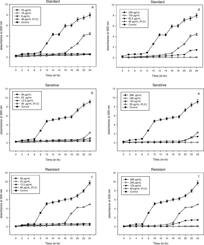
The life cycle of microbial cells is completed in four phases i.e. lag, log or exponential, stationary and decline phase. The growth of C. albicans ATCC 90028 (standard), clinical isolates C. albicans D27 (FLC-susceptible) and C. albicans D15.9 (FLC-resistant) was observed in the presence of compounds 3a and 3b at 2MIC, MIC and MIC/2. The untreated cells showed a lag phase of 6–8 h in all the three strains. The cells did not show any growth when exposed to compound 3a with a continuous lag phase of more than 24 h. However, at 40 μg/mL of FLC, lag phase was over by 16, 20 and 16 h in case of C. albicans standard, FLC-susceptible and FLC-resistant, respectively (Fig 2). Compound 3b also showed significant growth inhibition at 2MIC and MIC. At the sub-MIC values of compound 3b, growth was observed after 20, 18 and 14 h for C. albicans standard, FLC-susceptible and FLC-resistant, respectively. Thus, 3a showed high potential as an inhibitor of Candida cell growth even at sub-MIC concentrations. Moreover, compound 3b proved to be a good antifungal when compared to the standard drug FLC as an inhibitor of Candida cell growth.
Fig 2.
(a-f). Dose dependent growth curve of a) C. albicans ATCC 90028 b) FLC-susceptible C. albicans D27 and c) FLC-resistant C. albicans D15.9 in the presence compound 3a and d) C. albicans ATCC 90028 e) FLC-susceptible and f) FLC-resistant C. albicans in the presence of compound 3b. Growth was observed in the presence of test compounds at 2MIC, MIC and MIC/2 and was significantly inhibited at all concentrations. No growth was observed when cells were exposed to compound 3a with the lag phase extending beyond 24h. Error bars represent Mean±S.D. from three independent recordings.
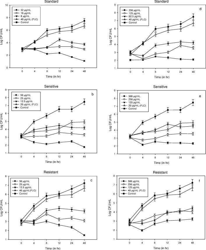
Fungistatic or fungicidal nature of lead inhibitory compounds (3a and 3b) was determined by time kill curve studies on C. albicans ATCC 90028 (standard) and the two clinical isolates of C. albicans (FLC-susceptible and resistant). At 2MIC, compound 3a was fungicidal whereas compound 3b was fungistatic as also observed in the growth curves. However, no complete fungicidal point was obtained for the compound 3a at 2MIC, but significant decrease in CFU/mL was observed after 48 h in all the three test strains. There was no significant difference in CFU/mL observed for the untreated and FLC (40 μg/mL) treated cells of C. albicans, both standard and FLC-resistant. As discussed earlier, compound 3b showed fungistatic behaviour as only slight increase in CFU/mL was recorded with time interval. However, this increase was dose dependent and significant inhibition was observed at higher concentrations. Moreover, both the test compounds showed better inhibition in comparison to the standard drug FLC (Fig 3).
Fig 3.
(a-f). Representative time kill curves of (a) C. albicans ATCC 90028 (b) FLC-susceptible C. albicans D27 and (c) FLC-resistant C. albicans D15,9 in the presence compound 3a and (d) C. albicans ATCC 90028 (e) FLC-susceptible and (f) FLC-resistant C. albicans in the presence of compound 3b. The results suggest that compound 3a has fungicidal nature while compound 3b is fungistatic. Error bars represent Mean±SD from three independent recordings.
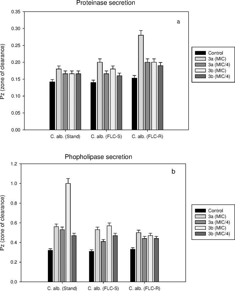
The test compounds (3a and 3b) were further explored to see their effects on the secretion of hydrolytic enzymes, proteinases and phospholipases. Although no significant inhibition of proteinase secretion was observed in the presence of either compound, yet compound 3a showed 45% inhibition of proteinase secretion in FLC-resistant strain of C. albicans at MIC. Results showed that proteinase secretion decreased by 21, 30 and 45% when treated with MIC of compound 3a and 14, 22 and 23.5% when treated with the MIC of compound 3b in C. albicans ATCC 90028 (standard), C. albicans D27 (FLC-susceptible) and C. albicans D15.9 (FLC-resistant), respectively. A slight decrease in % inhibition was observed when treated with lead compounds at MIC/4. Similarly, phospholipase secretion decreased by 43, 42 and 34% when treated with MIC of compound 3a and 68, 46 and 30% when treated with the MIC of compound 3b in C. albicans ATCC 90028 (standard), C. albicans D27 (FLC-susceptible) and C. albicans D15.9 (FLC-resistant), respectively. At lower concentrations (MIC/4), the decrease in phospholipase secretion was 40, 25 and 25% when treated with compound 3a and 32, 34 and 25% when treated with the compound 3b in standard, FLC-susceptible and FLC-resistant C. albicans, respectively. The results indicated that C. albicans D15.9 (FLC-resistant) is more sensitive to proteinase secretory activity of the fungus in comparison to phospholipase secretion when treated with compound 3a. The results are shown in (Fig 4).
Fig 4.
(a-b). Effect of compounds 3a and 3b on a) proteinase and b) phospholipase secretion by three strains of C. albicans. Pz value is the ratio of the diameter of colony to the diameter of colony plus zone of degradation/precipitation. High Pz value represents low enzyme activity. Compound 3a decreased proteinase secretion by two fold in FLC-resistant C. albicans D15.9. No phospholipase activity was observed in C. albicans ATCC 90028 after treatment with compound 3b. Error bars represents Mean±S.D. from three independent recordings.
The fungal plasma membrane H+ ATPase is a crucial enzyme that regulates intracellular pH and nutrient uptake into the cell. The process of proton extrusion is a phenomenon associated with enzyme activity which is stimulated in presence of glucose [33]. The inhibition of this protein causes the intracellular acidification which may result in cell death. The effect of the lead inhibitory compounds (3a and 3b) was investigated on the activity of H+ ATPase enzyme by determining the rate of H+ extrusion in the extracellular medium. The rate of H+ extrusion was calculated in μmoles min−1mg−1 Candida cells by titrating the cell suspension with 0.01N NaOH. H+ extrusion rate was also determined in presence of glucose which acts as a stimulator of H+ efflux pump. The results are summarized in Table 3. At 100 μg/mL, compound 3a showed 70, 45, and 40% inhibition of H+ extrusion in C. albicans ATCC 90028 (standard), clinical isolates C. albicans D27 (FLC-susceptible) and C. albicans D15.9 (FLC-resistant), respectively. Similarly, at the same concentration, 3b showed 47, 65, and 42% inhibition of H+ extrusion in C. albicans ATCC 90028 (standard), C. albicans D27 (FLC-susceptible) and C. albicans D15.9 (FLC-resistant), respectively. Both the compounds showed greater inhibition than standard drug FLC with 38, 49 and 25% inhibition of H+ extrusion in C. albicans standard, FLC-susceptible and FLC-resistant, respectively. In the presence of 5 mM of glucose, the rate of H+ extrusion increased by 33, 24 and 36% in the standard), FLC-susceptible and FLC-resistant C. albicans strains, respectively. Again, in the presence of glucose, 52, 52 and 54% inhibition by compound 3a and 64, 52 and 52% inhibition by compound 3b was calculated as H+ extrusion rate in the standard, FLC-susceptible and FLC-resistant strains, respectively. At low concentrations of the compounds, the inhibition of H+ extrusion rate decreased (S4 Fig).
Table 3. Effect of 3a and 3b on the rate of H+ efflux by C. albicans ATCC 90028, FLC-susceptible C. albicans D27 and FLC-resistant C. albicans D15.9 in the presence and absence of glucose (G).
Both the test compounds had a greater inhibitory effect on H+ efflux in comparison to FLC.
| Condition | Range of relative H+efflux rate (×10−6 mol min−1 mg cells−1) | ||
|---|---|---|---|
| C. albicans ATCC 90028 | C. albicans D27(FLC-susceptible) | C. albicans D15.9(FLC-resistant) | |
| Control | 36.65 ± 1.8 | 30.30 ± 1.5 | 27.80 ±1.3 |
| 3a (100 μg/mL) | 11.00 ± 0.5 | 16.60 ± 0.8 | 16.70 ± 0.8 |
| 3a (50 μg/mL) | 19.60 ± 0.9 | 20.00 ± 1.0 | 21.60 ± 1.0 |
| 3b (100 μg/mL) | 19.30 ± 0.9 | 16.00 ± 0.8 | 16.00 ± 0.8 |
| 3b (50 μg/mL) | 23.40 ± 1.1 | 17.60 ± 0.8 | 18.30 ± 0.9 |
| Glucose (G) (5mM) | 54.95 ± 2.7 | 40.00 ± 2.0 | 43.33 ± 2.1 |
| G + 3a (100 μg/mL) | 26.60 ± 1.3 | 19.30 ± 0.9 | 20.00 ± 1.0 |
| G + 3a (50 μg/mL) | 33.40 ± 1.6 | 21.60 ± 1.0 | 28.30 ± 1.4 |
| G + 3b (100 μg/mL) | 20.00 ± 1.0 | 19.30 ± 0.9 | 21.00 ± 1.0 |
| G + 3b (50 μg/mL) | 23.30 ± 1.1 | 22.30 ± 1.1 | 25.00 ± 1.2 |
| FLC(10 μg/mL) | 22.85 ± 1.1 | 15.35 ± 0.7 | 20.95 ± 1.0 |
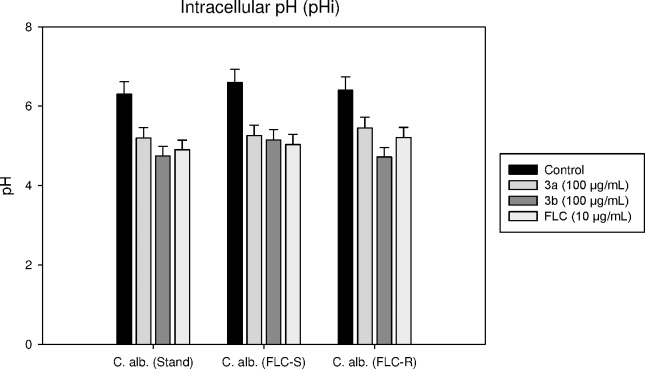
The enzyme activity of H+ ATPase can also be determined by evaluating intracellular pH of Candida cells. The intracellular pH of these cells is maintained within the range of 6.0–7.5 due to the activity of H+ ATPase. The inhibition of this protein blocks the permeability of H+ ions across plasma membrane which results in the alteration of pH inside the cell [17]. Here we studied the change in pHi in the presence of 3a and 3b which is, as mentioned earlier, directly related to H+ ATPase enzyme activity. In case of C. albicans ATCC 90028, the intracellular pH reduced from 6.3 to 5.2 and 4.7 in presence of compound 3a and 3b, respectively, while in C. albicans D27 (FLC-susceptible) it reduced from 6.6 to 5.2 and 5.1, respectively. Similarly, in C. albicans D15.9 (FLC-resistant), pH reduced from 6.4 to 5.4 and 4.7 in presence of compound 3a and 3b, respectively. The results are shown in Fig 5.
Fig 5. Intracellular pH of C. albicans ATCC 90028; FLC-susceptible C. albicans D27and FLC-resistant C. albicans D15.9 with and without the treatment of 100 μg/mL of test compounds 3a and 3b.
Fluconazole (10 μg/mL) was used as standard drug. The decrease in pH showed the accumulation of H+ ions due to reduced activity of the proton pump. The acidity error bars represent Mean±S.D. from three independent recordings.
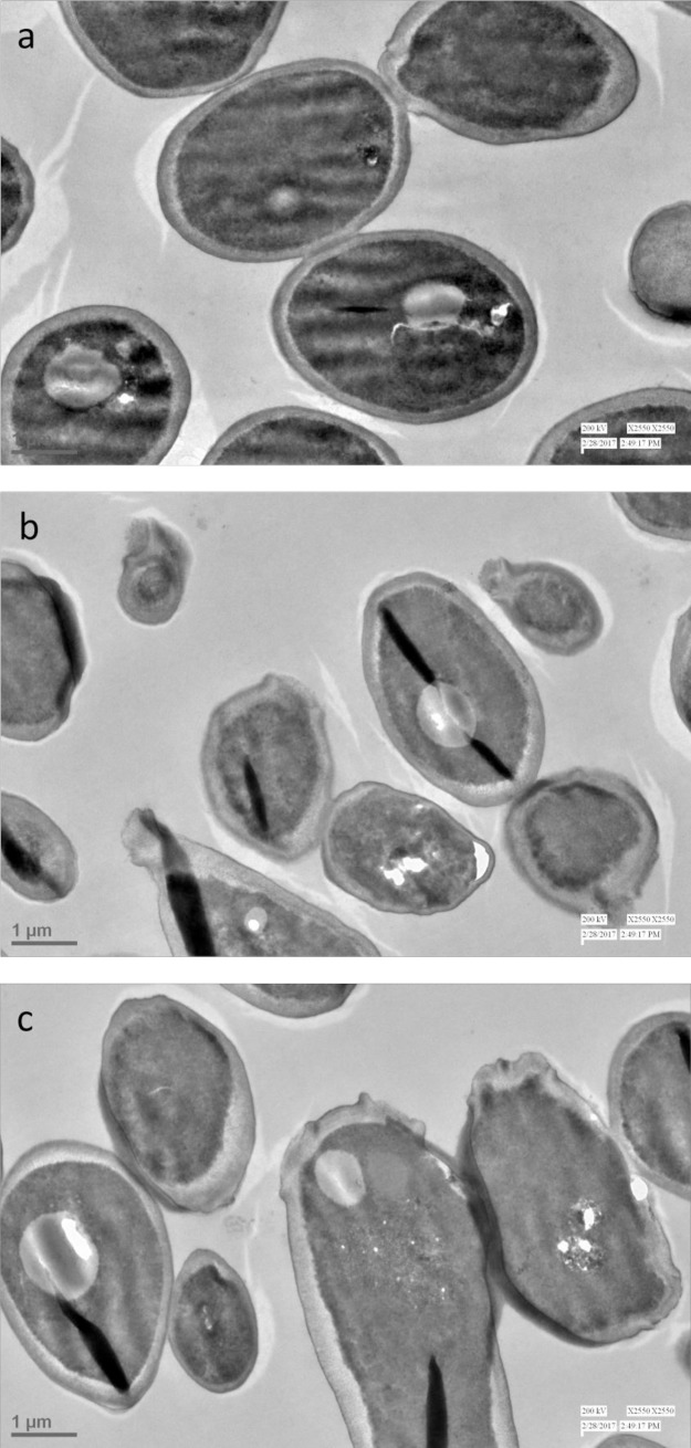
The effect of lead compounds (3a and 3b) on the morphology of C. albicans was monitored by transmission electron microscopy (TEM) to explore their possible mechanism of action. Candida cells treated with test compounds at their respective MICs and untreated control cells were used for TEM analysis. It was observed, as shown in Fig 6, that untreated cells are normal in shape with uniform cell density and intact cell membrane while treated cells showed several deformations. In case of exposed cells significant damage could be seen wherein the cell shape became abnormal and the cell membrane lost its integrity becoming irregular. It is well known that azole drugs like FLC inhibit ergosterol biosynthesis destroying both membrane integrity and function of some membrane-bound proteins and therefore affecting fungal cell growth and proliferation. Thus, we can conclude from the results of TEM analysis that the lead compounds (3a and 3b) may be interfering with biosynthesis of cell membrane. The result of phase contrast microscopy (phase plate 3, Nikon Eclipse 80i) also showed the clear morphological differences between treated and untreated cells (S5 Fig).
Fig 6.
(a-c). TEM images of C. albicans a) untreated cells; b) treated with compound 3a; and c) treated with compound 3b. The morphological characteristics of untreated cells are clearly distinct from cells treated with compound 3a and 3b. (scale bar = 1μm)
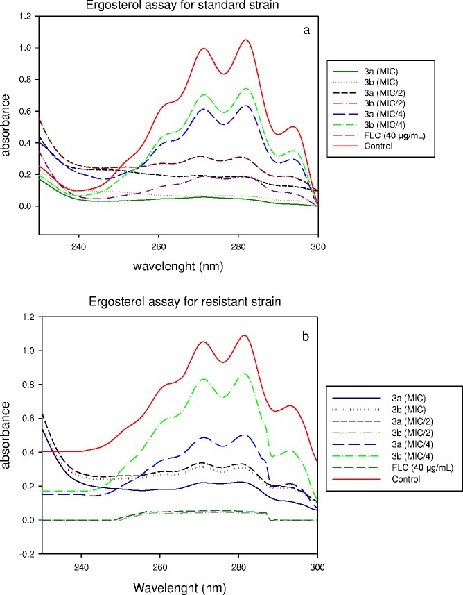
The effect of the lead inhibitors (3a and 3b) was also studied on ergosterol biosynthesis in Candida cells to support that azole drugs interfere with biosynthesis of cell membranes [34]. The ergosterol content of exposed cells was estimated in C. albicans ATCC 90028 and C. albicans D15.9 (FLC-resistant) strains by a previously reported method [29]. FLC was used as a positive control. The results showed a dose dependent decrease in ergosterol content when cells were grown in varying concentrations of test compounds. The mean decrease in total ergosterol content for C. albicans ATCC 90028 was found to be 48% at MIC/4, and 100% above MIC/2 of compound 3a. The mean decrease in total ergosterol content for C. albicans D15.9 (FLC-resistant) was found to be 30% at MIC/4, and 100% above MIC/2 of compound 3a. Similarly, the mean decrease in total ergosterol content for standard C. albicans ATCC 90028 was found to be 33% at MIC/4, and 100% above MIC/2 of 3b. However, the decrease in total ergosterol content for C. albicans D15.9 (FLC-resistant) was found to be 23% at MIC/4, 98% at MIC/2 and 100% at MIC of compound 3b. In the presence of 40 μg/mL of FLC, the decrease in ergosterol content was 100% and 96% respectively for C. albicans ATCC 90028 and C. albicans D15.9 (FLC-resistant). Our results suggest that the compounds 3a and 3b inhibit the total ergosterol content significantly for all the Candida strains tested here (Fig 7 and S6 Fig).
Fig 7.
UV spectrophotometric sterol profiles of (a) C. albicans ATCC 90028and (b) FLC-resistant C. albicans D15.9 after treatment with the test compounds 3a and 3b. Strains were grown for 16 h in SD broth containing various concentrations of test compounds (MIC/4, MIC/2, MIC). Sterols were extracted, and spectral profiles between 230 and 300 nm were determined.
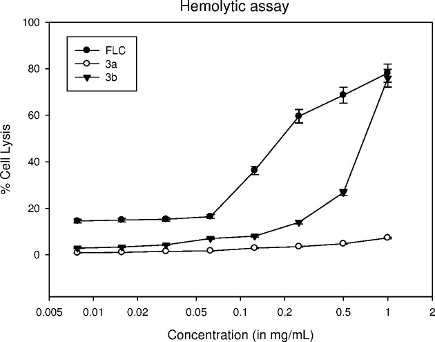
The cytotoxic effect of lead compounds (3a and 3b) was determined on human red blood cells (hRBCs) by hemolytic assay and compared with standard drug FLC for reference. At 500 μg/mL, compounds 3a and 3b showed 4.76 and 26.77% hemolysis of hRBCs while at the same concentration, standard drug FLC showed 68.63% hemolysis (Fig 8). Both the test compounds showed <5% hemolysis at their respective MIC values against standard Candida strain. Thus the results strongly suggest that these compounds have negligible toxic effect on hRBCs. To examine the effect of triazole derivatives (3a and 3b) on cell proliferation, they were screened for cytotoxicity against monkey kidney fibroblasts (VERO). A sub-confluent population of VERO cells was treated with increasing concentration of each compound and the number of viable cells was measured after 48 h by MTT cell viability assay. Doxorubicin was used as control which showed IC50 of >5 μg/mL. Neither compound was found cytotoxic upto 10 μg/mL.
Fig 8. Hemolytic activity of compounds 3a, 3b and FLC.
The hRBCs were collected, resuspended in PBS and treated with various concentrations of test compounds. Results show that the standard drug FLC exhibits greater cell lysis in comparison to compounds the test compounds
Discussion
C. albicans is an opportunistic fungus and is responsible for the most common fungal infection called candidiasis. These infections are continuously increasing all around the world with high mortality rate due to the limitations of current antifungal therapies. Thus, constant researches are required to generate new options for treatment of serious fungal infections with better armamentarium, low toxicity, and target specificity. Among the currently used antifungal agents, triazoles (e.g. fluconazole, itraconazole, posaconazole) are known for better therapeutic properties [35]. On the other hand, several reports including our lab have suggested towards the role of natural molecules as potential inhibitors of Candida spp [16, 28]. Recently we synthesized a series of 1,2,3-triazoles from naturally occurring bioactive molecules and found compound 3a as a promising inhibitor of Candida spp [20]. Compound 3a was synthesized from 8-hydroxy quinoline and we speculated that the quinoline, ring especially N-atom, is responsible for its enhanced anti-Candida activity. In this work, we synthesized and evaluated anti-Candida activities of two other structural analogues (3b and 3c) of compound 3a in order to determine the role of quinoline ring. We further performed various experiments to determine their possible mode of action in C. albicans. We obtained low IC50 values with compounds 3a and 3b against Candida spp from preliminary screening. Moreover, in vitro studies have shown that compounds 3a and 3b significantly inhibit FLC- susceptible as well as resistant clinical isolates of C. albicans. These results encouraged us to explore their possible effect on some important virulence attributes of C. albicans. We performed disk diffusion assay, growth curve studies and time kill assay to determine the fungicidal or fungistatic nature of compounds 3a and 3b. We demonstrated that at high concentrations compound 3a exhibits fungicidal activity whereas compound 3b showed fungistatic behaviour. It was noted that the performance of compound 3a was better than the standard drug FLC. Virulence attributes in pathogens can be used as alternative targets for drug development. Secretion of proteinases and phospholipases are important virulence factors in C. albicans which help in the invasion and damage of host tissues [36]. The significant inhibition in proteinase and phospholipase secretion by triazole-amino acid hybrids has been previously reported by our group [30], which motivated us to further demonstrate the effect of compounds 3a and 3b on these enzymes. Secretion of proteinases and phospholipases was inhibited in the presence of compounds 3a and 3b but it was not enough to conclude that these proteins maybe be used as potential antifungal targets. Compound 3a inhibited approximately two fold secretion of proteinases in FLC- resistant C. albicans and no phospholipase activity was observed in C. albicans ATCC 90028 after treatment with compound 3b. H+ ATPase, a member of P-type transport ATPase family, has been reported as a potential antifungal target [37, 38]. This protein is essentially involved in the physiological functions of Candida spp such as maintenance of electrochemical gradient across cell membrane, nutrient uptake, regulation of intracellular pH and cell growth. H+ ATPase of fungal origin is not similar to the human protein and might be a useful antifungal target [17]. Some previous studies have reported that FLC inhibits H+ ATPase activity in C. albicans [16, 39]. Therefore, we explored the action of the test compounds on the activity of plasma membrane H+ ATPase. We observed good to moderate inhibition in the H+ ATPase activity in absence and presence of glucose when treated with 50 and 100 μg/mL of the test compounds. Moreover, we also determined the internal pH of C. albicans in presence of compounds 3a and 3b and the results also supported the partial inhibition of H+ ATPase activity. The main focus of this study was to develop some new compounds with an azole moiety. It is well known that azoles have a common mode of action as they inhibit ergosterol biosynthesis by blocking cytochrome P450-dependent enzyme: lanosterol 14 α-demethylase. Ergosterol is the main sterol constituent of fungal membranes [40]. Thus, we examined the effect of test compounds on cell membranes and ergosterol biosynthesis by transmission electron microscopy and sterol profiling, respectively. We observed alterations in the morphology of treated C. albicans cells especially in cell membrane and cell organelles in comparison to the normal cells. This indicates that the test compounds are involved in the disintegration of the fungal cell membrane. The ergosterol content was negligible after treatment with the test compounds at MIC. Additionally, cell proliferation assay and haemolytic assay showed that both compounds 3a and 3b were significantly less cytotoxic. Thus, overall findings of this study indicate that compounds 3a and 3b have good anti-Candida activity and the mode of action is similar to FLC. The compounds do inhibit other virulence attributes in C. albicans but the main antifungal property may be due to the inhibition of ergosterol biosynthesis. Further, pharmacodynamic studies and in vivo efficacy of these compounds are required to establish these compounds as future antifungal agents.
Conclusion
The structural analogues of a previously determined anti-Candida compound 3a were synthesized. Based on the preliminary anti-Candida results, 3a and 3b were selected as lead compounds and further explored for their antifungal potential against C. albicans. Both compounds showed good anti-Candida activity for all the strains used. This study showed that both compounds 3a and 3b have significant inhibitory effect on the growth of FLC- sensitive as well as FLC- resistant C. albicans strains. Growth curve and time kill curve studies indicated that compound 3a has fungicidal nature while compound 3b was fungistatic. The secretion of extracellular hydrolytic enzymes (proteinases and phospholipases) was affected in the presence of both the test compounds. An altered cell membrane, reduced plasma membrane H+ ATPase activity and significant inhibition in ergosterol biosynthesis in the presence of these compounds suggest that plasma membrane of the pathogenic fungi is an important antifungal target for the newly synthesized triazoles. Hence, we conclude that compounds 3a and its analogue 3b are potential anti-Candida compounds with mode of action similar to FLC.
Supporting information
(JPG)
(JPG)
Disk diffusion assay of standard, FLC-susceptible and FLC-resistant C. albicans showing zone of inhibition in the presence of different concentration of test compounds 3a and 3b.
(TIF)
Inhibition of the rate of H+ efflux by C. albicans ATCC 90028; FLC-susceptible and FLC-resistant isolate of Candida in presence and absence of glucose in presence of compounds 3a and 3b, a) without glucose; b) with glucose.Error bars represents Mean±S.D. from three independent recordings.
(TIF)
Percentage inhibition of ergosterol in a) standard strain; and b) resistant strain showing by bar graph in presence of compounds 3a and 3b. Error bars represents mean±S.D. from three independent recordings.
(TIF)
(a-c). Phase contrast microscopy. Phase contrast microscopy was performed to determine the effect of lead inhibitor on the morphology of C. albicans. Mid-log phase cells were harvested, standardized (A600 ≈ 0.1) and treated with the MIC concentration of lead inhibitors 3a and 3b for 6 h. After treatment period, cells were washed thrice with phosphate buffer solution to remove residual medium. 10 μL of cell suspension was put over a clean glass slide and observed under phase contrast microscope (phase plate 3, Nikon Eclipse 80i). The morphological differences between a) un-treated cells of C. albicans and b-c) treated with compound 3a and 3b, respectively were observed.
(TIF)
Acknowledgments
Mohammad Abid gratefully acknowledges UGC, Govt. of India for RAMAN Postdoctoral Fellowship in USA (F. No. 5-123/2016 (IC)). MI acknowledges Non-NET fellowship support from UGC, India. The authors are thankful to Dr. Shabana I. Khan, Principal Scientist, National Center for Natural Products Research (NCNPR), School of Pharmacy, University of Mississippi, MS 38677, USA for doing cell-proliferation assay on Vero cells (monkey kidney fibroblasts). We are also thankful to Prof. Luqman A. Khan, JMI for providing the clinical strains of C. albicans and Ms. Babita Aneja for helping in spectral characterization.
Abbreviations
- FLC
Fluconazole
- ATCC
American Type Culture Collection
- MIC
Minimum Inhibitory Concentration
- IC50
Concentration required for 50% inhibition
- CDC
Center for Disease Control and Prevention
- CDCl3
Chloroform
- DCM
Dichloromethane
- DMF
Dimethylformamide
- TLC
Thin Layer Chromatography
- DMSO
Dimethyl sulfoxaide
- SD
Sabouraud Dextrose
- YEPD
Yeast Extract Peptone Dextrose
- h
hour/s
- TEM
Transmission Electron Microscopy
- DHE
Dehydroergosterol
- ZOI
Zone of inhibition
Data Availability
All relevant data are within the paper and its Supporting Information files.
Funding Statement
Mohammad Abid gratefully acknowledges UGC, Govt. of India for RAMAN Postdoctoral Fellowship in USA (F. No. 5-123/2016 (IC)). MI acknowledges Non-NET fellowship support from UGC, India.
References
- 1.Yao B, Haitao J, Yongbin C, Youjun Z, Jü Z, Jiaguo L, et al. Synthesis and antifungal activities of novel 2-aminotetralin derivatives. J. Med. Chem. 2007; 50(22): 5293–5300. doi: 10.1021/jm0701167 [DOI] [PubMed] [Google Scholar]
- 2.Tobudic S, Christina K, Elisabeth P. Azole‐resistant Candida spp.-emerging pathogens? Mycoses. 2012; 55(s1): 24–32. [Google Scholar]
- 3.Groll AH, Joanna L. New developments in invasive fungal disease. Future Microbiol. 2012; 7(2): 179–184. doi: 10.2217/fmb.11.154 [DOI] [PubMed] [Google Scholar]
- 4.CDC, U.S. Department of Health and Human Services, Atlanta, Georgia, 2013: 63–64.
- 5.Brown GD, Denning DW, Gow NA, Levitz SM, Netea MG, White TC. Hidden killers: human fungal infections. Sci. Transl. Med. 2012; 4(165): 165rv13–165rv13. doi: 10.1126/scitranslmed.3004404 [DOI] [PubMed] [Google Scholar]
- 6.Cao X, Zhaoshuan S, Yongbing C, Ruilian W, Tongkai C, Wenjing C, et al. Design, synthesis, and structure–activity relationship studies of novel fused heterocycles-linked triazoles with good activity and water solubility. J. Med. Chem. 2014: 57(9): 3687–3706. doi: 10.1021/jm4016284 [DOI] [PubMed] [Google Scholar]
- 7.Calderone RA. Introduction and historical perspectives. Candida and Candidiasis (Calderone R., ed). 2002; 15–25. ASM Press, Washington, DC. [Google Scholar]
- 8.Kauffman CA., José AV, Jack DS, Harry AG, David SM, Karchmer AM, et al. Prospective multicenter surveillance study of funguria in hospitalized patients. Clin. Infect. Dis. 2000: 30(1): 14–18. doi: 10.1086/313583 [DOI] [PubMed] [Google Scholar]
- 9.Jacobsen ID, Duncan W, Betty W, Sascha B, Julian RN, Bernhard H. Candida albicans dimorphism as a therapeutic target. Expert. Rev. Anti. Infect. Ther. 2012: 10(1): 85–93. doi: 10.1586/eri.11.152 [DOI] [PubMed] [Google Scholar]
- 10.Schaller M, Hans CK, Wilhelm S, Janine B, Wen CC, Bernhard H. Secreted aspartic proteinase (Sap) activity contributes to tissue damage in a model of human oral candidosis. Mol. Microbiol. 1999: 34(1): 169–180. [DOI] [PubMed] [Google Scholar]
- 11.Theiss S, Ganchimeg I, Audrey B, Marianne K, Chungn YL, Thomas N, et al. Inactivation of the phospholipase B gene PLB5 in wild-type Candida albicans reduces cell-associated phospholipase A 2 activity and attenuates virulence. Int. J. Med. Microbiol. 2006: 296(6): 405–420. doi: 10.1016/j.ijmm.2006.03.003 [DOI] [PMC free article] [PubMed] [Google Scholar]
- 12.Gácser A, Frank S, Cathrin K, László K, Wilhelm S, Joshua DN. Lipase 8 affects the pathogenesis of Candida albicans. Infect. Immun. 2007: 75(10): 4710–4718. doi: 10.1128/IAI.00372-07 [DOI] [PMC free article] [PubMed] [Google Scholar]
- 13.Wächtler B, Francesco C, Nadja J, Stephanie F, Frederic D, Martin S, et al. Candida albicans-epithelial interactions: dissecting the roles of active penetration, induced endocytosis and host factors on the infection process. PloS ONE. 2012; 7(5): e36952 doi: 10.1371/journal.pone.0036952 [DOI] [PMC free article] [PubMed] [Google Scholar]
- 14.Seto-Young D, Brian M, Mason AB, David SP. Exploring an antifungal target in the plasma membrane H+-ATPase of fungi. BBA Biomembranes. 1997; 1326(2): 249–256. [DOI] [PubMed] [Google Scholar]
- 15.Perlin DS, Seto YDONNA, Monk BC. The Plasma Membrane H+ ATPase of Fungi. Ann. N. Y. Acad. Sci. 1997; 834(1): 609–617. [DOI] [PubMed] [Google Scholar]
- 16.Ahmad A, Amber K, Snowber Y, Luqman AK, Nikhat M. Proton translocating ATPase mediated fungicidal activity of eugenol and thymol. Fitoterapia. 2010; 81(8): 1157–1162. doi: 10.1016/j.fitote.2010.07.020 [DOI] [PubMed] [Google Scholar]
- 17.Manzoor N, Amin M, Luqman AK. Effect of phosphocreatine on pHi and dimorphism in Candida albicans. Indian J. Exp. Biol. 2002; 40: 785–790. [PubMed] [Google Scholar]
- 18.Di Santo R, Andrea T, Roberta C, Maurizo B, Marino A, Federico C, et al. N-substituted derivatives of 1-[(aryl)(4-aryl-1 H-pyrrol-3-yl) methyl]-1 H-imidazole: synthesis, anti-Candida activity, and QSAR studies. J. Med. Chem. 2005; 48(16): 5140–5153. doi: 10.1021/jm048997u [DOI] [PubMed] [Google Scholar]
- 19.Sheng C, Wannian Z, Haitao J, Min Z, Yunlong S, Hui X, et al. Structure-based optimization of azole antifungal agents by CoMFA, CoMSIA, and molecular docking. J. Med. Chem. 2006; 49(8): 2512–2525. doi: 10.1021/jm051211n [DOI] [PubMed] [Google Scholar]
- 20.Irfan M, Babita A, Umesh Y, Shabana IK, Nikhat M, Constantin GD, et al. Synthesis, QSAR and anti-Candida evaluation of 1,2,3-triazoles derived from naturally bioactive scaffolds. Eur. J. Med. Chem. 2015; 93: 246–254. doi: 10.1016/j.ejmech.2015.02.007 [DOI] [PubMed] [Google Scholar]
- 21.Wayne PA. National committee for clinical laboratory standards. Performance standards for antimicrobial disc susceptibility testing. 2002; 12.
- 22.Wang S, Yan W, Wei L, Na L, Yongqiang Z, Guoqiang D, et al. Novel Carboline Derivatives as potent antifungal lead compounds: Design, synthesis, and biological evaluation. ACS Med. Chem. Lett. 2014; 5(5): 506–511. doi: 10.1021/ml400492t [DOI] [PMC free article] [PubMed] [Google Scholar]
- 23.Kumar DA, Sumathi M, Uma B, Seemi FB, Luqman AK. Antifungal resistance patterns, virulence attributes and spectrum of oral Candida species in patients with periodontal disease. Br. Microbiol. Res. J. 2015; 5(1): 68–75. [Google Scholar]
- 24.Rüchel R., Tegeler R, Trost M. A comparison of secretory proteinases from different strains of Candida albicans. Sabouraudia. 1982; 20(3): 233–244. [DOI] [PubMed] [Google Scholar]
- 25.Price MF, Ian DW, Layne OG. Plate method for detection of phospholipase activity in Candida albicans. Sabouraudia. 1982. 20(1): 7–14. [DOI] [PubMed] [Google Scholar]
- 26.Serrano R. In vivo glucose activation of the yeast plasma membrane ATPase. FEBS Lett. 1983; 156(1): 11–14. [DOI] [PubMed] [Google Scholar]
- 27.Kaur S, Mishra P, Prasad R. Dimorphism-associated changes in intracellular pH of Candida albicans. Biochim Biophys Acta. 1988; 972: 277–82. [DOI] [PubMed] [Google Scholar]
- 28.Khan A, Aijaz A, Luqman AK, Nikhat M. Ocimum sanctum (L.) essential oil and its lead molecules induce apoptosis in Candida albicans. Res. Microbiol. 2014; 165(6): 411–419. doi: 10.1016/j.resmic.2014.05.031 [DOI] [PubMed] [Google Scholar]
- 29.Arthington-Skaggs BA, David WW, Christine JM. Quantitation of Candida albicans ergosterol content improves the correlation between in vitro antifungal susceptibility test results and in vivo outcome after fluconazole treatment in a murine model of invasive candidiasis. Antimicrob. Agents Chemother. 2000; 44(8): 2081–2085. [DOI] [PMC free article] [PubMed] [Google Scholar]
- 30.Aneja B, Irfan M, Charu K, Jairajpuri MA, Maguire R, Kavanagh K, et al. Effect of novel triazole–amino acid hybrids on growth and virulence of Candida species: in vitro and in vivo studies. Org. Biomol. Chem. 2016; 14(45): 10599–10619. doi: 10.1039/c6ob01718e [DOI] [PubMed] [Google Scholar]
- 31.Manohar S, Rajesh UC, Shabana IK, Babu LT, Diwan SR. Novel 4-aminoquinoline-pyrimidine based hybrids with improved in vitro and in vivo antimalarial activity. ACS Med. Chem. Lett. 2012; 3(7): 555–559. [DOI] [PMC free article] [PubMed] [Google Scholar]
- 32.Martins N, Isabel CFRF, Lillian B, Sónia S, Mariana H. Candidiasis: predisposing factors, prevention, diagnosis and alternative treatment. Mycopathologia. 2014; 177(5–6): 223–240. doi: 10.1007/s11046-014-9749-1 [DOI] [PubMed] [Google Scholar]
- 33.Campetelli AN, Gabriela P, Carlos AA, Hector SB, Cesar HC. Activation of the plasma membrane H+‐ATPase of Saccharomyces cerevisiae by glucose is mediated by dissociation of the H+‐ATPase–acetylated tubulin complex. FEBS J. 2005; 272(22): 5742–5752. doi: 10.1111/j.1742-4658.2005.04959.x [DOI] [PubMed] [Google Scholar]
- 34.Schiaffella F, Macchiarulo A, Milanese L, Vecchiarelli A, Costantino G, Pietrella D, et al. Design, synthesis, and microbiological evaluation of new Candida albicans CYP51 inhibitors. J. Med. Chem. 2005; 48(24): 7658–7666. doi: 10.1021/jm050685j [DOI] [PubMed] [Google Scholar]
- 35.Maertens J, Raad I, Petrikkos G, Boogaerts M, Selleslag D, Petersen FB et al. Efficacy and safety of caspofungin for treatment of invasive aspergillosis in patients refractory to or intolerant of conventional antifungal therapy. Clin. Infect. Dis. 2004; 39(11): 1563–1571. doi: 10.1086/423381 [DOI] [PubMed] [Google Scholar]
- 36.Naglik JR, Challacombe SJ, Hube B. Candida albicans secreted aspartyl proteinases in virulence and pathogenesis. Microbiol. Mol. Biol. Rev. 2003; 67(3): 400–428. doi: 10.1128/MMBR.67.3.400-428.2003 [DOI] [PMC free article] [PubMed] [Google Scholar]
- 37.Seto-Young D, Monk BC, Mason AB, Perlin DS. Exploring an antifungal target in the plasma membrane H+-ATPase of fungi. Biochim. Biophys. Acta (BBA)-Biomembranes. 1997; 1326(2): 249–256. [DOI] [PubMed] [Google Scholar]
- 38.Perlin DS, Seto Young D, Monk BC. The Plasma Membrane H+ ATPase of Fungi. Annals of the New York Academy of Sciences. 1997; 834(1): 609–617. [DOI] [PubMed] [Google Scholar]
- 39.Khan N, Shreaz S, Rimple B, Muralidhar S, Manzoor N, Khan LA. Curcumin as a promising anticandidal of clinical interest. Can. J. Microbiol. 2011; 57(3): 204–210. doi: 10.1139/W10-117 [DOI] [PubMed] [Google Scholar]
- 40.de Montellano O, Paul R. Hydrocarbon hydroxylation by cytochrome P450 enzymes. Chem. Rev. 2009; 110(2): 932–948. [DOI] [PMC free article] [PubMed] [Google Scholar]
Associated Data
This section collects any data citations, data availability statements, or supplementary materials included in this article.
Supplementary Materials
(JPG)
(JPG)
Disk diffusion assay of standard, FLC-susceptible and FLC-resistant C. albicans showing zone of inhibition in the presence of different concentration of test compounds 3a and 3b.
(TIF)
Inhibition of the rate of H+ efflux by C. albicans ATCC 90028; FLC-susceptible and FLC-resistant isolate of Candida in presence and absence of glucose in presence of compounds 3a and 3b, a) without glucose; b) with glucose.Error bars represents Mean±S.D. from three independent recordings.
(TIF)
Percentage inhibition of ergosterol in a) standard strain; and b) resistant strain showing by bar graph in presence of compounds 3a and 3b. Error bars represents mean±S.D. from three independent recordings.
(TIF)
(a-c). Phase contrast microscopy. Phase contrast microscopy was performed to determine the effect of lead inhibitor on the morphology of C. albicans. Mid-log phase cells were harvested, standardized (A600 ≈ 0.1) and treated with the MIC concentration of lead inhibitors 3a and 3b for 6 h. After treatment period, cells were washed thrice with phosphate buffer solution to remove residual medium. 10 μL of cell suspension was put over a clean glass slide and observed under phase contrast microscope (phase plate 3, Nikon Eclipse 80i). The morphological differences between a) un-treated cells of C. albicans and b-c) treated with compound 3a and 3b, respectively were observed.
(TIF)
Data Availability Statement
All relevant data are within the paper and its Supporting Information files.


