Abstract
Recently, several new lipid-dependent species belonging to the genus Malassezia have been described. Some of them, such as Malassezia dermatis, Malassezia nana, and the tentatively named “Malassezia equi,” have similar phenotypes and are genetically close to Malassezia sympodialis Simmons et Guého 1990. DNA characterization by D1/D2 26S rRNA gene and internal transcribed spacer (ITS)-5.8S rRNA gene sequencing analysis of lipid-dependent strains from different animal species close to M. sympodialis is described and illustrated. Phylogenetic analysis of both the D1/D2 regions of 26S rRNA gene and ITS-5.8S rRNA gene sequences showed four distinct clusters. Cluster I included isolates from different animal species (horse, pig, and lamb) and the type culture of M. sympodialis. Cluster II included isolates from horses grouping close to the “M. equi” AJ305330 sequence. Cluster III comprised isolates mainly from goats. Cluster IV contained isolates mainly from cats grouping together with the M. nana AB075224 sequence. This last cluster included isolates from healthy and external otitic ears. All of these strains had identical 26S rRNA gene and ITS regions. It is not clear whether the value of these genetic differences is for the definition of species or whether they only demonstrate genetic variation among strains from different origins within M. sympodialis, which are in the course of differentiation and probably adaptation to specific animal hosts.
Malassezia species are lipophilic yeasts that are usually members of the normal mycobiota of the human skin and inhabit the skin of a variety of animal species. However, these yeasts are associated with a variety of dermatological disorders of the human skin, such as atopic dermatitis, dandruff, folliculitis, pityriasis versicolor or seborrheic dermatitis (13) and intravascular catheter-acquired infections (23). They have been implicated in different skin disorders in animals, mainly otitis externa and dermatitis (6, 15).
Since the genus Malassezia was created by Baillon in 1889, its taxonomy has been a matter of controversy. A few years ago, the genus was reclassified (14, 16) on the basis of studies of morphological, ultrastructural, physiological, and genetic characteristics, and seven species, which are now widely accepted, were proposed. They are the previously described Malassezia furfur, Malassezia pachydermatis, and Malassezia sympodialis and four new species, Malassezia obtusa, Malassezia globosa, Malassezia restricta, and Malassezia slooffiae.
Recently, four new lipid-dependent species belonging to the genus Malassezia have been described or proposed (19, 25, 32, 33). From these four species, Malassezia dermatis (32), Malassezia nana (19), and the tentatively named “Malassezia equi” (25) are close genetically to the type strain of M. sympodialis, and most of them also have some common morphological and physiological characteristics. M. sympodialis Simmons et Guého 1990 (30) was initially isolated from the auditory tract of a healthy human male and from the scalp of an AIDS patient suffering from tinea capitis. Although its pathogenic role is not yet elucidated, it is commonly isolated from healthy as well as diseased skin (1). M. dermatis was isolated from human patients with atopic dermatitis (32), M. nana was isolated from animals (cat and cows) (19), and “M. equi” was isolated from normal equine skin (25).
In our laboratory, Crespo et al. (3) described for the first time lipid-dependent yeasts associated with otitis externa in cats with similar morphological characteristics and some identical physiological characteristics to the M. sympodialis type species. Lipid-dependent yeasts were also isolated from the skin of horses and different domestic ruminants, as they are the major population of the lipophilic mycobiota in these animals. Difficulty in obtaining a high level of certainty in the identification of some of these lipid-dependent strains by means of these physiological tests was reported (7). The speciation of lipid-dependent isolates from animals by means of physiological tests presents some difficulties, and some of them cannot even be identified (3, 7, 8, 9, 19, 25).
Nucleic acid sequencing has already become an important tool that is useful for the identification of many microorganisms, including yeast species. Molecular systematics of yeasts has emphasized either coding and noncoding regions of the ribosomal DNA. Several studies determined that sequencing of the D1/D2 region could be used for the identification of most basidiomycetous yeast species and that closely related species or strains required sequencing of the internal transcribed spacer (ITS) region (10, 29).
The aim of this work was the DNA characterization of lipid-dependent strains from various domestic animal species close to the M. sympodialis type species by using D1/D2 26S and ITS-5.8S rRNA gene sequencing analysis to understand their phylogenetic relationships and to analyze their specific genetic variation. Phylogenetic relationships with the recently described or proposed M. sympodialis-related species are also analyzed and discussed.
MATERIALS AND METHODS
Strains used.
A total of 20 lipid-dependent isolates from domestic animals related to M. sympodialis were studied. The strains examined are listed in Table 1, and they were isolated from one animal each. Most of the strains from domestic animals used in the present study are from epidemiological studies carried out in our laboratory (3, 4, 6, 7). The following type strains were also included in this study: M. furfur CBS 1878NT, M. furfur CBS 7019NT, M. globosa CBS 7966T, M. obtusa CBS 7876T, M. pachydermatis CBS 1879 NT, M. restricta CBS 7877 T, M. slooffiae CBS 7956T, and M. sympodialis CBS 7222T. All of these strains were stored at −80°C (5).
TABLE 1.
Type strains and lipid-dependent isolates from animals used in this study
| Strain | Source | Genetic type | D1/D2 GenBank accession no. | ITS GenBank accession no. |
|---|---|---|---|---|
| MA73 CBS 9968 | Sheep, groin | I | AY743627 | AY743640 |
| MA80 CBS 9967 | Goat, ear | III | AY743618 | AY743647 |
| MA88 CBS 9986 | Cow, groin | II | AY743622 | AY743645 |
| MA125 | Horse, ear | III | AY743619 | AY743646 |
| MA146 CBS 9969 | Horse, anus | II | AY743621 | AY743641 |
| MA182 CBS 9970 | Horse, axilla | I | AY743620 | AY743638 |
| MA231 | Cat, ear | IV | AY743609 | AY743648 |
| MA237 | Cat, ear | IV | AY743612 | AY743649 |
| MA250 | Horse, anus | II | AY743623 | AY743642 |
| MA261 | Cat, ear | IV | AY743608 | AY743650 |
| MA262 CBS 9971 | Cat, otitis externa | IV | AY743610 | AY743651 |
| MA314 CBS 9972 | Cat, otitis externa | IV | AY743611 | AY743652 |
| MA333 | Goat, dorsum | III | AY743617 | AY743655 |
| MA383 | Goat, ear | III | AY743616 | AY743656 |
| MA400 CBS 9973 | Goat, ear | III | AY743615 | AY743657 |
| MA419 | Dog, otitis externa | IV | AY743613 | AY743653 |
| MA424 | Cat, ear | IV | AY743614 | AY743654 |
| MA461 | Horse, anus | II | AY743624 | AY743643 |
| MA470 | Horse, anus | II | AY743625 | AY743644 |
| MA477 | Pig | I | AY743628 | AY743639 |
| M. furfur CBS 1878NT | AY743602 | AY743634 | ||
| M. furfur CBS 7019NT | AY743603 | AY743635 | ||
| M. globosa CBS 7966T | AY743604 | AY743630 | ||
| M. obtusa CBS 7876T | AY743629 | AY743631 | ||
| M. pachydermatis CBS 1879NT | AY743605 | AY743637 | ||
| M. restricta CBS 7877T | AY743607 | AY743636 | ||
| M. slooffiae CBS 7956T | AY743606 | AY743633 | ||
| M. sympodialis CBS 7222T | AY743626 | AY743632 |
Isolation of DNA.
Cells were harvested from 4- to 5-day-old cultures in modified Dixon's medium (14). The DNA was prepared as described previously (12). The cells were incubated for 1 h at 65°C in 500 μl of extraction buffer (50 mM Tris-HCl, 50 mM EDTA, 3% sodium dodecyl sulfate, and 1% 2-mercaptoethanol). The lysate was extracted with phenol-chloroform-isoamyl alcohol (25:24:1, vol/vol/vol). Then 65 μl of 3 M sodium acetate and 75 μl of 1 M NaCl were added to 350 μl of the supernatant, and the resulting volume was incubated at 4°C for 30 min. DNA was recovered by isopropanol precipitation and washed with 70% (vol/vol) ethanol, dried under a vacuum, and resuspended in TE buffer (10 mM Tris-HCl, 1 mM EDTA [pH 8]). DNA was cleaned with the Geneclean kit II (BIO 101, Inc., La Jolla, Calif.) according to the manufacturer's instructions.
PCR and DNA sequencing of rRNA genes of the isolates.
The variable D1 and D2 regions of the 26S rRNA gene (at the 5′ end of the nuclear large subunit of the rRNA gene) were amplified by PCR by using the conserved fungal oligonucleotide primers NL1 and NL4 (26). The amplification process consisted of a predenaturation step at 94°C for 5 min, followed by 30 cycles of denaturation at 94°C for 45 s, annealing at 51°C for 1 min, and extension at 72°C for 3 min plus a final extension of 10 min at 72°C.
ITS and 5.8S rRNA genes were amplified by PCR with the conserved fungal primer pair ITS5 and ITS4 (35). The amplification process consisted of a predenaturation step at 94°C for 5 min, followed by 35 cycles of denaturation at 95°C for 30 s, annealing at 50°C for 1 min, and extension at 72°C for 1 min plus a final extension of 7 min at 72°C.
The amplified PCR products were purified by using a GFX PCR DNA and gel band purification kit (Amersham Pharmacia Biotech, Uppsala, Sweden) by following the supplier's protocol. The purified PCR products were sequenced directly by using a BigDye Terminator, version 3.1, cycle sequencing kit (Applied Biosystems, Gouda, The Netherlands) according to the manufacturer's recommendations. The 26S rRNA D1/D2 and ITS1-5.8S-ITS2 rRNA gene sequences were determined by using NL1 and NL4 and ITS5 and ITS4 as the sequencing primers, respectively. An Applied Biosystems 3100 sequencer was used to obtain the DNA sequences.
Sequence similarity and phylogenetic analyses.
The sequences were aligned by using Clustal X software (34). Phylogenetic and molecular evolutionary analyses were performed by using MEGA, version 2.1 (21). For neighbor-joining analysis (28), the distances between sequences were calculated by using Kimura's two-parameter model (20). A bootstrap analysis was conducted with 1,000 replications (11).
Nucleotide sequence accession number.
The nucleotide sequences of the D1/D2 26S rRNA gene and ITS-5.8S determined in this study have been deposited in GenBank, and their accession numbers are given in Table 1.
RESULTS
Molecular phylogenetic analysis.
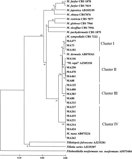
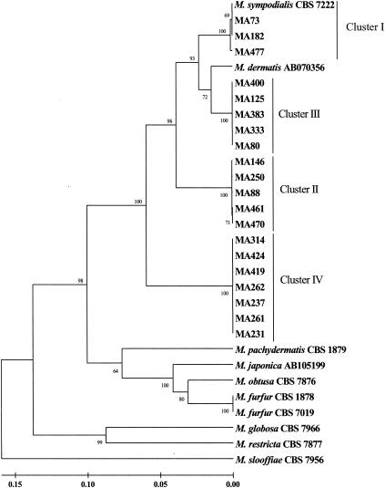
Fig. 1 shows the molecular phylogenetic tree based on the D1 and D2 regions of the 26S rRNA gene sequences constructed by the neighbor-joining method. Figure 2 shows the molecular phylogenetic tree based on the ITS-5.8S rRNA gene sequences. In both trees, the 20 lipid-dependent strains grouped into four phylogenetic clusters.
FIG. 1.
Molecular phylogenetic tree constructed by using the sequences of D1/D2 26S rRNA gene of members of the genus Malassezia and related Ustilaginomycetes species. The numbers at the branch points are the percentages of 1,000 bootstrapped data sets that supported the specific internal branches. Species with GenBank accession numbers represent sequences obtained from GenBank.
FIG. 2.
Molecular phylogenetic tree constructed by using the ITS1-5.8S rRNA gene-ITS2 sequences of members of the genus Malassezia. The numbers at the branch points are the percentages of 1,000 bootstrapped data sets that supported the specific internal branches. Species with GenBank accession numbers represent sequences obtained from GenBank.
Sequences of D1/D2 of 26S rRNA gene.
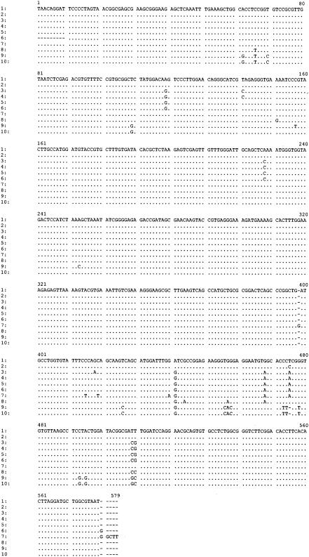
The aligned sequences of the D1/D2 region are shown in Fig. 3.
FIG. 3.
Alignment of D1/D2 sequences of M. sympodialis strains. M. sympodialis CBS 7222 is used as the model. Hyphens designate gaps added to permit alignment. Identical nucleotides are indicated by dots. Lanes: 1, M. sympodialis CBS 7222, MA73, and MA182; 2, MA477; 3, MA88, MA461, and MA470; 4, MA250; 5, MA146; 6, “M. equi” AJ305330; 7, M. dermatis AB070361; 8, MA80, MA125, MA333, MA383, and MA400; 9, M. nana AB075224; 10, MA231, MA237, MA261, MA262, MA314, MA419, and MA424.
Cluster I included isolates from different animal species (horse, pig, and lamb) and the type culture of M. sympodialis. The 26S rRNA gene (D1/D2 regions) sequenced from M. sympodialis CBS 7222 included 578 bp. The sequences of the strains from cluster I were completely identical to M. sympodialis CBS 7222, except for the strain MA477, which showed a single different nucleotide (dissimilarity, 0.17%).
The sequences of cluster I were close to the M. dermatis AB070361 sequence, which included 579 bp. These sequences differed from the sequence of M. dermatis at seven positions (dissimilarity, 1.21%).
Cluster II included isolates from horses grouping together with the “M. equi” AJ305330 sequence. The nucleotide analysis of the 26S rRNA gene of these strains showed two substitutions over 578 bp among them. The sequences of “M. equi” AJ305330 and MA146 were identical and differed from the sequence of M. sympodialis CBS 7222 at seven positions (dissimilarity, 1.21%). The strains MA88, MA461, and MA470 showed identical sequences and differed from the sequence of M. sympodialis CBS 7222 at nine positions (dissimilarity, 1.56%). The sequence of strain MA250 differed from the sequence of M. sympodialis CBS 7222 at eight positions (dissimilarity, 1.38%).
Cluster III comprised isolates mainly from goats. The section of DNA sequenced included 578 bp, and the sequences of all of the strains were identical, differing from the sequence of M. sympodialis CBS 7222 at eight positions (dissimilarity, 1.38%).
Cluster IV included isolates mainly from cats grouping together with the M. nana AB075224 sequence. The section of DNA sequenced included 577 bp. The sequences of all strains were identical and differed from the sequence of M. nana at 2 positions and from the sequence of M. sympodialis CBS 7222 at 16 positions (dissimilarity, 2.77%).
Sequences of ITS regions and 5.8S rRNA gene.
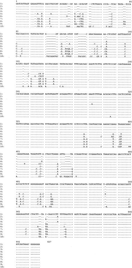
The aligned sequences of the 5.8S rRNA gene with the two intergenic spacers ITS1 and ITS2 are shown in Fig. 4.
FIG. 4.
Alignment of ITS1-5.8S rRNA gene-ITS2 sequences of M. sympodialis strains. M. sympodialis CBS 7222 is used as the model. Hyphens designate gaps added to permit alignment. Identical nucleotides are indicated by dots. Lanes: 1, M. sympodialis CBS 7222; 2, MA182; 3, MA477; 4, MA73; 5, M. dermatis AB070356; 6, MA80, MA125, MA333, MA383, and MA400; 7, MA88; 8, MA146 and MA250; 9, MA470; 10, MA231, MA237, MA261, MA262, MA314, MA419, and MA424.
The section of DNA sequenced from M. sympodialis CBS 7222 included 621 bp and contained the partial sequence of the 18S rRNA gene, the entire ITS1-5.8S-ITS2 region, and the partial sequence of the 26S rRNA gene. The ITS1 region occupied nucleotides 9 to 170, the 5.8S rRNA gene occupied nucleotides 171 to 326, and the ITS2 region occupied nucleotides 327 to 560.
The sequences of cluster I were similar. The length of the section of DNA sequenced ranged from 619 bp in MA477 to 623 bp in MA73. The sequence of MA182 included 621 bp and showed one base pair substitution from M. sympodialis CBS 7222. The nucleotide sequences analysis of these strains showed five substitutions among them and differences of less than four substitutions from those of M. sympodialis CBS 7222.
The sequences of cluster II showed identical ITS2 sequences and differed from the ITS2 sequence of M. sympodialis CBS 7222 at 31 positions. The sequences of strains MA470 and MA461 differed from the sequences of M. sympodialis CBS 7222 at 33 positions, MA146 and MA250 at 32 positions, and MA88 at 30 positions at the ITS1 region. Nucleotide sequence analysis of these isolates showed 3 substitutions among them at the ITS1 region and differences in more than 61 substitutions from those of M. sympodialis CBS 7222 (dissimilarity, 9.8 to 10.3%). Unfortunately, we were not able to analyze the “M. equi” ITS sequences because there is no sequence deposited in GenBank and we could not find any “M. equi” type strain in culture collections.
The sequences of cluster III were identical (620 bp) and differed from the sequence of M. sympodialis CBS 7222 at 39 positions: 18 in the ITS1 region and 21 in the ITS2 region (dissimilarity, 6.28%). The sequences of cluster III were close to the M. dermatis AB070356 sequence.
The sequences of cluster IV were identical (646 bp) and differed from the sequence of M. sympodialis CBS 7222 at 95 positions: 60 in the ITS1 region, 1 in the 5.8S region, and 34 in the ITS2 region (dissimilarity, 15.29%). The ITS1 sequence of M. nana AB075225 was identical to the ITS1 sequences of our isolates from cluster IV.
DISCUSSION
M. sympodialis Simmons et Guého 1990 was described by using two strains isolated from humans. These strains showed a strong lipophily and small daughter cells with occasional sympodial budding (30), different from M. furfur, which was the only previously described lipid-dependent species. The study of a large number of strains by, among other techniques, 26S rRNA sequence and nuclear DNA comparisons and some physiological tests resulted in a more precise characterization of the species M. sympodialis. These strains were represented by a unique rRNA sequence (16) and formed a physiological entity of lipid-dependent yeasts showing growth on glucose-peptone agar with 0.1 to 10% Tween 40, 60, or 80 as a sole source of lipid and no growth with 10% Tween 20 (14). However, all of the strains analyzed in these studies (14, 16) were exclusively recovered from humans, including isolates from normal skin, pityriasis versicolor, and folliculitis.
In our study, three isolates from different animal species (horse, lamb, and pig) clustered with the type species M. sympodialis CBS 7222. Only one strain had a single different nucleotide at the D1/D2 sequence. We observed ITS variation, but the strains demonstrated >99% sequence similarity. As previously reported, the gene sequences of ITS1 of Malassezia species are highly diverse (22).
At present, D1 and D2 26S rRNA gene sequences from almost all yeasts are used for species identification or phylogenetic analysis. Peterson and Kurtzman (27) correlated the biological species concept with the phylogenetic species concept by the extent of nucleotide substitutions in 26S rRNA gene sequences. Their study demonstrated that strains of a single biological species show less than 1% substitution in this region. It has been reported (2, 31) that conspecific strains generally demonstrated ≥99% sequence similarity in the ITS1 and ITS2 regions. According to this species concept, two new lipid-dependent species genetically close to M. sympodialis have been described: M. dermatis and M. nana. These species have similar phenotypes (some common physiological characteristics) to M. sympodialis (Table 2).
TABLE 2.
Some physiological characteristics of lipid-dependent Malassezia species
| Species | Result withc:
|
Reference(s) | |||||
|---|---|---|---|---|---|---|---|
| Catalase | Tween 20 (10%) (14) | Tween 40 or 60 (0.5%) (14) | Tween 80 (0.1%) (14) | Cremophor EL (24) | Esculin (24) | ||
| M. furfur | + | + | + | + | + | ± | 14 |
| M. globosa | + | − | − | − | − | − | 14 |
| M. japonica | + | − | + | − | NTb | NT | 33 |
| M. obtusa | + | − | − | − | − | + | 14 |
| M. restricta | − | − | − | − | − | − | 14 |
| M. slooffiae | + | ± | + | − | − | − | 14 |
| M. sympodialis | + | − | + | + | − | + | 14 |
| M. dermatis | + | + | + | + | + | − | 19, 32 |
| M. nana | + | ± | + | + | − | − | 19 |
| NUSV 1003a | + | − | + | + | NT | NT | 18 |
| MA262, MA314 | + | − | + | + | − | + | 3 |
M. nanaT
NT, not tested.
+, growth or positive reaction; −, no growth or negative reaction; ±, different reactions or variable growth results for different isolates or strains.
M. dermatis was isolated from skin lesions of patients with atopic dermatitis from Japan. The sequences of five isolates of this new species were completely identical in both 26S rRNA gene and ITS regions and clustered with M. sympodialis but had a high number of dissimilarities with it (32). However, the physiological characteristics of M. dermatis are identical to those of M. furfur (Table 2). It grows on glucose-peptone agar with 10% Tween 20 as a sole source of lipid, differing from M. sympodialis. Sympodial budding was not cited in these isolates. The isolates belonging to cluster I were close to the D1/D2 M. dermatis AB070361 sequence.
Cluster II included isolates from horses grouping close to the “M. equi” AJ305330 sequence, showing nearly identical D1/D2 and ITS sequences and being conspecific strains. Dissimilarities between these isolates and the M. sympodialis strain in their D1/D2 regions of 26S and ITS-5.8S were more than 1 and 9%, respectively. At the moment, there is not a valid description yet for “M. equi” and no physiological characteristics have been reported (25).
Cluster III comprised isolates mainly from goats. All strains were completely identical in both 26S rRNA gene and ITS regions. Dissimilarities between these isolates and the M. sympodialis strain in their D1/D2 regions of 26S and ITS-5.8S were 1.3 and 6.28%, respectively. They were conspecific and clearly distinct from M. sympodialis.
M. nana was isolated from animals in Japan and Brazil. It morphologically and physiologically resembles M. dermatis and M. sympodialis, but the phylogenetic trees based on the D1/D2 and ITS1 regions were clearly distinct from these two species (19). The type strain isolated from a cat in Japan was slightly different from the Brazilian strains isolated from cows. Our isolates from cluster IV, mainly from cats, showed the same ITS1 sequence as the M. nana type strain, and the D1/D2 sequences were nearly identical. All strains were completely identical in both 26S rRNA gene and ITS regions, and the dissimilarities between these isolates and the M. sympodialis strain in their D1/D2 regions of 26S and ITS-5.8S were 2.77 and 15.29%, respectively. This group includes two strains associated with otitis externa in cats (MA262 and MA314) (3) with identical physiological characteristics to M. sympodialis (Table 2). The type strain of M. nana (NUSV 1003) also showed physiological characteristics identical to those of M. sympodialis (18) (Table 2).
Special micromorphological characteristics, such as small size of the cells in comparison to other Malassezia spp. (3, 19), buds formed in a narrow base (3, 14, 19, 30), and occasional sympodial budding (3, 14, 30), have been cited for M. sympodialis-related species. However, the separation of Malassezia species based on morphological characteristics is unreliable (16), and their routine identification is currently based on the use of some physiological properties (14, 17), avoiding the use of morphological criteria, which are considered to be more subjective.
On the other hand, the difficulty in obtaining a high level of certainty in the identification of some lipid-dependent strains by means of physiological tests has been reported (3, 7, 8, 9, 18, 25). In addition, the isolation and identification of these strains continue to be difficult due to the low viability associated with some isolate types and the lack of suitable methods for its isolation and preservation (5).
Malassezia species are opportunistic yeasts and belong to the normal cutaneous microbiota. Some of them may act as pathogens when exposed to certain changes in the skin microclimate. In this study, we included lipid-dependent isolates from healthy skin and associated with diseased skin. The association of lipid-dependent isolates with human skin disease is well known. For animals, this association has been recently described (3, 4, 7, 19), and unfortunately, there are few lipid-dependent strains available in culture collections. Using D1/D2 and ITS sequencing analysis, strains associated with skin disease do not differ genetically from isolates from healthy skin. Genetic type IV included isolates from the ears of healthy cats and those from cats and a dog with otitis externa. All strains were completely identical in both the 26S rRNA gene and ITS regions. Usually, this technique does not allow differentiation between the two kinds of isolates. Other molecular techniques, such as randomly amplified polymorphic DNA or amplified fragment length polymorphism, could be more powerful tools to find clinical correlations.
Formal descriptions of possible new taxa will require a standard phenotypic study, which is not a part of this report. Some strains have enough variation to define possible new species or subtypes. Furthermore, some of the sequences included for comparison in the phylogenetic analysis belong to the recently described M. sympodialis-related species and were grouped close or even together with our strains in the proposed clusters. Scorzetti et al. (29), working with other kinds of basidiomycetous yeasts, considered that trees provided by ITS, D1/D2, and other genes, provide focal points for biological and systematic analyses of these yeasts. A sequence, per se, does not describe a species, and descriptions of new species should not rely solely on nucleotide data.
Finally, it is not clear whether the value of these genetic differences is to define species or whether they only demonstrate genetic variation among strains from different origins within M. sympodialis. Further studies are required to investigate whether the divergence between M. sympodialis and our strains is sufficient to resolve them as individual species or whether it only indicates that this species is in the course of differentiation and adaptation to specific animal hosts.
Acknowledgments
This work was supported by grant no. 2002SGR 00079 from DURSI, Generalitat de Catalunya, Spain.
REFERENCES
- 1.Boekhout, T., and E. Guého. 2002. Basidiomycetous yeasts, p. 551. In D. H. Howard (ed.), Pathogenic fungi in humans and animals. Marcel Dekker, Inc., New York, N.Y.
- 2.Chen, Y. C., J. D. Eisner, M. M. Kattar, S. L. Rassoulian-Barrett, K. LaFe, S. L. Yarfitz, A. P. Limaye, and B. T. Cookson. 2000. Identification of medically important yeasts using PCR-based detection of DNA sequence polymorphisms in the internal transcribed spacer 2 region of the rRNA genes. J. Clin. Microbiol. 38:2302-2310. [DOI] [PMC free article] [PubMed] [Google Scholar]
- 3.Crespo, M. J., M. L. Abarca, and F. J. Cabañes. 2000. Otitis externa associated with Malassezia sympodialis in two cats. J. Clin. Microbiol. 38:1263-1266. [DOI] [PMC free article] [PubMed] [Google Scholar]
- 4.Crespo, M. J., M. L. Abarca, and F. J. Cabañes. 2000. Atypical lipid-dependent Malassezia species isolated from dogs with otitis externa. J. Clin. Microbiol. 38:2383-2385. [DOI] [PMC free article] [PubMed] [Google Scholar]
- 5.Crespo, M. J., M. L. Abarca, and F. J. Cabañes. 2000. Evaluation of different preservation and storage methods for Malassezia spp. J. Clin. Microbiol. 38:3872-3875. [DOI] [PMC free article] [PubMed] [Google Scholar]
- 6.Crespo, M. J., M. L. Abarca, and F. J. Cabañes. 2002. Occurrence of Malassezia spp. in the external ear canals of dogs and cats with and without otitis externa. Med. Mycol. 40:115-121. [DOI] [PubMed] [Google Scholar]
- 7.Crespo, M. J., M. L. Abarca, and F. J. Cabañes. 2002. Occurrence of Malassezia spp. in horses and domestic ruminants. Mycoses 45:333-337. [DOI] [PubMed] [Google Scholar]
- 8.Duarte, E. R., M. M. Melo, R. C. Hahn, and J. S. Hamdan. 1999. Prevalence of Malassezia spp. in the ears of asymptomatic cattle and cattle with otitis in Brazil. Med. Mycol. 37:159-162. [PubMed] [Google Scholar]
- 9.Duarte, E. R., M. A. Lachance, and J. S. Hamdan. 2002. Identification of atypical strains of Malassezia spp. from cattle and dog. Can. J. Microbiol. 48:749-752. [DOI] [PubMed] [Google Scholar]
- 10.Fell, J. W., T. Boekhout, A. Fonseca, G. Scorzetti, and A. Statzell-Tallman. 2000. Biodiversity and systematics of basidiomycetous yeasts as determined by large-subunit rDNA D1/D2 domain sequence analysis. Int. J. Syst. Evol. Microbiol. 50:1351-1371. [DOI] [PubMed] [Google Scholar]
- 11.Felsenstein, J. 1985. Confidence limits on phylogenies: an approach using the bootstrap. Evolution 39:783-791. [DOI] [PubMed] [Google Scholar]
- 12.Ferrer, C., F. Colom, S. Frasés, E. Mulet, J. L. Abad, and J. L. Alló. 2001. Detection and identification of fungal pathogens by PCR and by ITS and 5.8S ribosomal DNA typing in ocular infection. J. Clin. Microbiol. 39:2873-2879. [DOI] [PMC free article] [PubMed] [Google Scholar]
- 13.Guého, E., T. Boekhout, H. R. Ashbee, J. Guillot, A. van Belkum, and J. Faergemann. 1998. The role of Malassezia species in the ecology of human skin and as pathogens. Med. Mycol. 36:220-229. [PubMed] [Google Scholar]
- 14.Guého, E., G. Midgley, and J. Guillot. 1996. The genus Malassezia with description of four new species. Antonie Leeuwenhoek 69:337-355. [DOI] [PubMed] [Google Scholar]
- 15.Guillot, J., and R. Bond. 1999. Malassezia pachydermatis: a review. Med. Mycol. 37:295-306. [DOI] [PubMed] [Google Scholar]
- 16.Guillot, J., and E. Guého. 1995. The diversity of Malassezia yeasts confirmed by rRNA sequence and nuclear DNA comparisons. Antonie Leeuwenhoek 67:297-314. [DOI] [PubMed] [Google Scholar]
- 17.Guillot, J., E. Guého, M. Lesourd, G. Midgley, G. Chévrier, and B. Dupont. 1996. Identification of Malassezia species. A practical approach. J. Mycol. Med. 6:103-110. [Google Scholar]
- 18.Hirai, A., R. Kano, K. Makimura, K. Yasuda, K. Konishi, H. Yamaguchi, and A. Hasegawa. 2002. A unique isolate of Malassezia from a cat. J. Vet. Med. Sci. 64:957-959. [DOI] [PubMed] [Google Scholar]
- 19.Hirai, A., R. Kano, K. Makimura, E. R. Duarte, J. S. Hamdan, M. A. Lachance, A. Yamaguchi, and A. Hasegawa. 2004. Malassezia nana sp. nov., a novel lipid-dependent yeast species isolated from animals. Int. J. Syst. Evol. Microbiol. 54:623-627. [DOI] [PubMed] [Google Scholar]
- 20.Kimura, M. 1980. A simple method for estimating evolutionary rates of base substitutions through comparative studies of nucleotide sequences. J. Mol. Evol. 16:111-120. [DOI] [PubMed] [Google Scholar]
- 21.Kumar, S., K. Tamura, I. B. Jakobsen, and M. Nei. 2001. Mega2: Molecular Evolutionary Genetics Analysis software. Bioinformatics 17:1244-1245. [DOI] [PubMed] [Google Scholar]
- 22.Makimura, K., Y. Tamura, M. Kudo, K. Uchida, H. Saito, and H. Yamaguchi. 2000. Species identification and strain typing of Malassezia species stock strains and clinical isolates based on the DNA sequences of nuclear ribosomal internal transcribed spacer 1 regions. J. Med. Microbiol. 49:29-35. [DOI] [PubMed] [Google Scholar]
- 23.Marcon, M. J., and D. A. Powell. 1992. Human infections due to Malassezia spp. J. Clin. Microbiol. 5:101-119. [DOI] [PMC free article] [PubMed] [Google Scholar]
- 24.Mayser, P., P. Haze, C. Papavassilis, M. Pickel, K. Gruender, and E. Guého. 1997. Differentiation of Malassezia species: selectivity of Cremophor EL, castor oil and ricinoleic acid for M. furfur. Br. J. Dermatol. 137:208-213. [DOI] [PubMed] [Google Scholar]
- 25.Nell, A., S. A. James, C. J. Bond, B. Hunt, and M. E. Herrtage. 2002. Identification and distribution of a novel Malassezia species yeast on normal equine skin. Vet. Rec. 150:395-398. [DOI] [PubMed] [Google Scholar]
- 26.O'Donnell, K. 1993. Fusarium and its near relatives, p. 225-233. In D. R. Reynolds and J. W. Taylor (ed.), Fungal holomorph: mitotic, meiotic and pleomorphic speciation in fungal systematics. CAB International, Wallingford, United Kingdom.
- 27.Peterson, S. W., and C. P. Kurtzman. 1991. Ribosomal RNA sequence divergence among sibling species of yeasts. Syst. Appl. Microbiol. 14:124-129. [Google Scholar]
- 28.Saitou, N., and M. Nei. 1987. The neighbor-joining method: a new method for reconstructing phylogenetic trees. Mol. Biol. Evol. 4:406-425. [DOI] [PubMed] [Google Scholar]
- 29.Scorzetti, G., J. W. Fell, A. Fonseca, and A. Statzell-Tallman. 2002. Systematics of basidiomycetous yeasts: a comparison of large subunit D1/D2 and internal transcribed spacer rDNA regions. FEMS Yeast Res. 2:495-517. [DOI] [PubMed] [Google Scholar]
- 30.Simmons, R. B., and E. Guého. 1990. A new species of Malassezia. Mycol. Res. 94:1146-1149. [Google Scholar]
- 31.Sugita, T., A. Nishikawa, R. Ikeda, and T. Shinoda. 1999. Identification of medically relevant Trichosporon species based on sequences of internal transcribed spacer regions and construction of a database for Trichosporon identification. J. Clin. Microbiol. 37:1985-1993. [DOI] [PMC free article] [PubMed] [Google Scholar]
- 32.Sugita, T., M. Takashima, T. Shinoda, H. Suto, T. Unno, R. Tsuboi, H. Ogawa, and A. Nishikawa. 2002. New yeast species Malassezia dermatis isolated from patients with atopic dermatitis. J. Clin. Microbiol. 40:1363-1367. [DOI] [PMC free article] [PubMed] [Google Scholar]
- 33.Sugita, T., M. Takashima, M. Kodama, R. Tsuboi, and A. Nishikawa. 2003. Description of a new yeast species, Malassezia japonica, and its detection in patients with atopic dermatitis and healthy subjects. J. Clin. Microbiol. 41:4695-4699. [DOI] [PMC free article] [PubMed] [Google Scholar]
- 34.Thompson, J. D., T. J. Gibson, F. Plewniak, F. Jeanmougin, and D. G. Higgins. 1997. The Clustal X windows interface: flexible strategies for multiple sequence alignment aided by quality analysis tools. Nucleic Acid Res. 24:4876-4882. [DOI] [PMC free article] [PubMed] [Google Scholar]
- 35.White, T. J., T. Bruns, S. Lee, and J. Taylor. 1990. Amplification and direct sequencing of fungi ribosomal RNA genes for phylogenetics, p. 315-322. In M. A. Innis, D. H. Gelfand, J. J. Sninsky, and T. J. White (ed.), PCR protocols. A guide to methods and applications. Academic Press, San Diego, Calif.


