Abstract
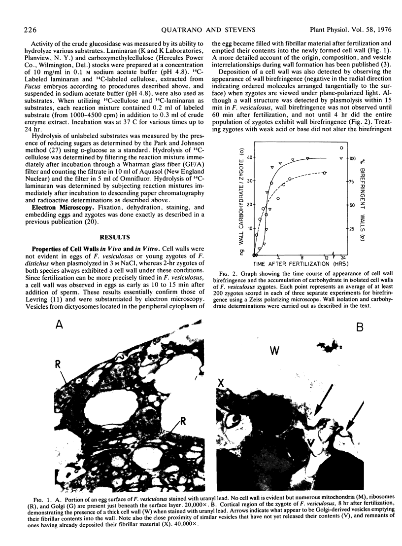
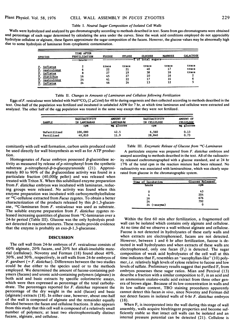
Fertilization triggers the assembly of a cell wall around the egg cell of three brown algae, Fucus vesiculosus, F. distichus, and F. inflatus. New polysaccharide polymers are continually being added to the cell wall during the first 24 hours of synchronous embryo development. This wall assembly involves the extracellular deposition of fibrillar material by cytoplasmic vesicles fusing with the plasma membrane. One hour after fertilization a fragmented wall can be isolated free of cytoplasm and contains equal amounts of cellulose and alginic acid with no fucose-containing polymers (fucans) present. Birefringence of the wall caused by oriented cellulose microfibrils is not detected in all zygotes until 4 hours, at which time intact cell walls can be isolated that retain the shape of the zygote. These walls have a relatively low ratio of fucose to xylose and little sulfate when compared to walls from older embryos. When extracts of walls from 4-hour zygotes are subjected to cellulose acetate electrophoresis at pH 7, a single fucan (F1) can be detected. By 12 hours, purified cell walls are composed of fucans containing a relatively high ratio of fucose to xylose and high levels of sulfate, and contain a second fucan (F2) which is electrophoretically distinct from F1. F2 appears to be deposited in only a localized region of the wall, that which elongates to form the rhizoid cell. Throughout wall assembly, the polyuronide block co-polymer alginic acid did not significantly vary its mannuronic (M) to guluronic (G) acid ratio (0.33-0.55) or its block distribution (MG, 54%; GG, 30%; MM, 16%). From 6 to 24 hours of embryo development, the proportion of the major polysaccharide components found in purified walls is stable. Alginic acid is the major polymer and comprises about 60% of the total wall, while cellulose and the fucans each make-up about 20% of the remainder. During the extracellular assembly of this wall, the intracellular levels of the storage glucan laminaran decreases. A membrane-bound β-1, 3-exoglucanase is found in young zygotes which degrades laminaran to glucose. It is postulated that hydrolysis of laminaran by this glucanase accounts, at least in part, for glucose availability for wall biosynthesis and the increase in respiration triggered by fertilization. The properties and function of alginic acid, the fucans, and cellulose are discussed in relation to changes in wall structure and function during development.
Full text
PDF


Images in this article
Selected References
These references are in PubMed. This may not be the complete list of references from this article.
- Boffey S. A., Northcote D. H. Pectin synthesis during the wall regeneration of plasmolysed tobacco leaf cells. Biochem J. 1975 Sep;150(3):433–440. doi: 10.1042/bj1500433. [DOI] [PMC free article] [PubMed] [Google Scholar]
- Brawley S. H., Wetherbee R., Quatrano R. S. Fine-structural studies of the gametes and embryo of Fucus vesiculosus L. (Phaeophyta). II. The cytoplasm of the egg and young zygote. J Cell Sci. 1976 Mar;20(2):255–271. doi: 10.1242/jcs.20.2.255. [DOI] [PubMed] [Google Scholar]
- Hanke D. E., Northcote D. H. Cell wall formation by soybean callus protoplasts. J Cell Sci. 1974 Jan;14(1):29–50. doi: 10.1242/jcs.14.1.29. [DOI] [PubMed] [Google Scholar]
- Hogsett W. E., Quatrano R. S. Isolation of Polysaccharides Sulfated during Early Embryogenesis in Fucus. Plant Physiol. 1975 Jan;55(1):25–29. doi: 10.1104/pp.55.1.25. [DOI] [PMC free article] [PubMed] [Google Scholar]
- Horine R. K., Ruesink A. W. Cell Wall Regeneration around Protoplasts Isolated from Convolvulus Tissue Culture. Plant Physiol. 1972 Oct;50(4):438–445. doi: 10.1104/pp.50.4.438. [DOI] [PMC free article] [PubMed] [Google Scholar]
- Larsen B., Haug A., Painter T. Sulphated polysaccharides in brown algae. 3. The native state of dfucoidan in Ascophyllum nodosum and Fucus vesiculosus. Acta Chem Scand. 1970;24(9):3339–3352. doi: 10.3891/acta.chem.scand.24-3339. [DOI] [PubMed] [Google Scholar]
- Novotny A. M., Forman M. The relationship between changes in cell wall composition and the establishment of polarity in Fucus embryos. Dev Biol. 1974 Sep;40(1):162–173. doi: 10.1016/0012-1606(74)90116-x. [DOI] [PubMed] [Google Scholar]
- Quatrano R. S. An ultrastructural study of the determined site of rhizoid formation in Fucus zygotes. Exp Cell Res. 1972 Jan;70(1):1–12. doi: 10.1016/0014-4827(72)90174-7. [DOI] [PubMed] [Google Scholar]
- Quatrano R. S., Crayton M. A. Sulfation of fucoidan in Fucus embryos. I. Possible role in localization. Dev Biol. 1973 Jan;30(1):29–41. doi: 10.1016/0012-1606(73)90045-6. [DOI] [PubMed] [Google Scholar]
- Seno N., Anno K., Kondo K., Nagase S., Saito S. Improved method for electrophoretic separation and rapid quantitation of isomeric chondroitin sulfates on cellulose acetate strips. Anal Biochem. 1970 Sep;37(1):197–202. doi: 10.1016/0003-2697(70)90280-0. [DOI] [PubMed] [Google Scholar]
- Tsai C. M., Hassid W. Z. Solubilization and Separation of Uridine Diphospho-d-glucose: beta-(1 --> 4) Glucan and Uridine Diphospho-d-glucose:beta-(1 --> 3) Glucan Glucosyltransferases from Coleoptiles of Avena sativa. Plant Physiol. 1971 Jun;47(6):740–744. doi: 10.1104/pp.47.6.740. [DOI] [PMC free article] [PubMed] [Google Scholar]
- Updegraff D. M. Semimicro determination of cellulose in biological materials. Anal Biochem. 1969 Dec;32(3):420–424. doi: 10.1016/s0003-2697(69)80009-6. [DOI] [PubMed] [Google Scholar]
- Woodland H. R., Gurdon J. B., Lingrel J. B. The translation of mammalian globin mRNA injected into fertilized eggs of Xenopus laevis. II. The distribution of globin synthesis in different tissues. Dev Biol. 1974 Jul;39(1):134–140. doi: 10.1016/s0012-1606(74)80015-1. [DOI] [PubMed] [Google Scholar]


