

Abstract
The rhesus macaque (Macaca Mulatta) is physiologically and phylogenetically similar to humans, and therefore represents an invaluable model for the pre-clinical assessment of the safety and feasibility of iPSC-derived cell therapies. We describe the use of an excisable polycistronic lentiviral STEMCCA vector to reprogram rhesus fibroblasts or bone marrow stromal cells (BMSCs) into RhiPSCs. After reprogramming, the pluripotency transgenes can be removed by transient expression of Cre, leaving a residual genetic tag that may be useful for identification of RhiPSC-derived tissues in vivo. Finally, we discuss steps to maintain pluripotency during passaging of RhiPSCs, required for successful utilization of RhiPSCs.
Keywords: induced pluripotent stem cells, reprogramming, non-human primate, rhesus macaque
Unit Title & Unit Introduction
Induced pluripotent stem cells represent an unlimited source of any cell type, and thus have myriad potential applications in basic and translational research as well as in the clinical regeneration of missing or damaged tissues. Safety and efficacy testing of iPSC-derived regenerative cellular therapies is most valuable utilizing an animal model sharing tissue and organ properties with humans. Rodent models are hindered by the significant differences between murine and human pluripotent cells, and xenografting of human iPSC-derived cells in immunodeficient mice cannot be used to study immunogeneticity, and is unlikely to result in normal integration of human tissues in a mouse environment. The overall approach to generation of RhiPSCs follows a similar progression as human iPSCs generation, but there are important modifications to the process and the maintenance of the generated RhiPSCs. This protocol outlines the steps required to generate RhiPSCs from either fibroblasts or BMSCs through the STEMCCA lentivirus reprogramming method (Sommer, et al. 2010). This method was selected because the transgene can be removed by transient expression of Cre-plasmid after reprogramming while leaving a non-expressed 303bp DNA tag which can be used to track iPSC-derived cells after autologous transplantation. Here, strategies are discussed for the generation and maintenance of RhiPSCs (see Basic Protocol 1 and Basic Protocol 2 respectively), and characterization of the generated lines (See Basic Protocol 3).
Note: Follow your institution's safety guidelines regarding handling of lentiviruses as well as non-human primate cells.
Rhesus induced pluripotent stem cell generation (Basic Protocol 1)
This protocol describes the generation of iPSCs from rhesus fibroblasts or BMSCs using the STEMCCA polycistronic lentiviral vector encoding the four human reprogramming factors, OCT4, SOX2, KLF4, and MYC, flanked by loxP sites (Sommer, et al. 2010). STEMCCA viruses (Sommer, et al. 2010) can be generated using 293T cells in the standard manner. Use of STEMCCA lentiviral vectors packaged with standard HIV components is sufficiently efficient to reprogram rhesus fibroblasts and BMSCs.
Note: For rhesus CD34+ cell reprogramming, the use of a chimeric modified HIV1-based packaging system for STEMCCA is recommended to improve efficiency (Uchida, et al. 2009).
Materials
DMEM, high glucose, pyruvate (Gibco cat. no. 11995)
DMEM (Lonza cat. no. 12-614F)
Alpha MEM (Gibco cat. no. 12561)
L-Glutamine (Gibco cat. no. 25030)
Fetal Bovine Serum (FBS) (Hyclone cat. no. SV30014.03)
KnockOut DMEM (KO/DMEM) (Gibco cat. no. 10829)
Knockout Serum Replacement (KSR) (Gibco cat. no. 10828)
Human Fibroblast like Growth Factor-basic (bFGF) (PeproTech cat. no. 100-18B)
MEM nonessential amino acids (NEAA) (Gibco cat. no. 11140)
Penicillin-Streptomycin-Glutamine (PSG) (Gibco cat. no. 10378)
2-mercaptoethanol (Sigma-Aldrich cat. no. M7522)
Dimethyl Sulfoxide (DMSO) (Sigma-Aldrich cat. no. D2650)
Matrigel™ (BD Bioscience cat. no. 354277)
Opti-MEM reduced serum medium (ThermoFisher, cat. no. 31985062)
EditPro Stem Transfection Reagent (MTI-GlobalStem, cat. no. 31985062)
Puromycin (Sigma cat. no. P8833)
Polybrene (Millipore cat. no. TR-1003-G)
ROCK inhibitor (Y-27632) (Stemgent cat. no. 04-0012)
MultiGrip™ 200μl Tips (Optional) (Denville cat. no. P3133-F)
Valproic acid (VPA) (Optional) (Stemgent cat. no. 04-0007)
Mouse embryonic fibroblasts (MEF), 2 million cells/vial (GlobalStem cat. no. GSC-6201G)
0.25% Trypsin-EDTA (1X) (Gibco cat. no. 25200-056)
Ultrapure Water with 0.1% Gelatin (Millipore cat. no. ES-006-B)
Hypoxia Chamber (BioSpherix cat. no.C-274)
O2 Controller (BioSpherix cat. no.P110)
CO2 Controller (BioSpherix cat. no. P120)
Cell Scraper (Corning cat. no. 3010)
Reprogramming
Day 0
- Plate 1×10⁵ cells/well in a 6-well plateFibroblasts or BMSCs between passages 4-6 tend to work best for reprogramming.
Day 1
- 2. Prepare transduction medium (parental media + polybrene 6 μg/ml).See Reagents and Solutions section.
3. Add concentrated STEMCCA virus to reach a final MOI of 7.5 to the transduction medium.
4. Add 1ml of transduction medium with viruses to each well of fibroblasts.
5. Incubate at 5% CO2 and 20% O2 overnight (no more than 12 h).
Day 2
6. Change media to parental media.
Day 4 (MEF plate preparation)
7. Add 1 mL of 0.1% gelatin to 12 wells of 6-well plates.
8. Transfer gelatin coated plates to an incubator for at least one hour.
- 9. Thaw 1 vial of MEFs in 9 mL of warm MEF culture media in a 15 mL conical.See Reagents and Solutions for MEF culture media recipe.
10. Centrifuge the conical at 250 × g for 5 minutes.
11. During centrifugation, aspirate gelatin from wells.
12. Aspirate supernatant off the cell pellet.
13. Resuspend pellet in 24 mL of MEF culture media (mix well).
- 14. Add 2 mL of cell suspension per well of a 6-well plate.Shake plate well to distribute MEFs evenly across the well.
- 15. Leave in incubator until ready to use.Discard plates that are not used within 3 days of preparation.
Day 5
16. Trypsinize transduced cells and plate 1×10⁵ cells per 10 cm dish or 2×10⁴ cells per each well of a 6-well plate coated with MEFs.
- 17. Culture in 5% CO2 and 5% O2 (hypoxic conditions) with 2 mL parental medium per well.Hypoxic culture has been shown to reduce spontaneous differentiation of rhesus embryonic stem cells (ESCs) (Mitalipov, et al. 2006). Similarly, our experience showed generating and maintaining RhiPSCs in normoxic conditions was extremely inefficient. Therefore it is highly recommended to set up hypoxic culture system for RhiPSC generation and culture. One can purchase multi-gas incubators or install a hypoxia chamber (e.g. BioSpherix) within a normal incubator (see Materials).
Day 6
18. Aliquot and warm necessary volume (2 mL per well) of RhiPSC medium.
- 19. Add 0.5 mM VPA to RhiPSC medium.See Reagents and Solutions section.
20. Change medium to RhiPSC medium + 0.5 mM VPA.
Day 7-13
21. Change media using fresh RhiPSC media + 0.5mM VPA every other day.
Day 14
22. Stop VPA treatment.
23. Add an additional aliquot of 2×10⁵ MEFs with fresh RhiPSC medium without VPA to each well of a 6-well plate.
Day 15- until colonies appear
- 24. Until ESC-like colonies appear, change media daily using RhiPSC medium.Between days 15-30, colonies with ESC-like morphology should appear. These should be mechanically removed individually, transferred onto new MEFs, and mechanically passaged every 4-5 days (see RhiPSC Passaging). RhiPSCs can be cryopreserved in ES-defined FBS supplemented with 10% DMSO or CryoStor® CS10 (StemCell Technologies) (see RhiPSC Cryopreservation).
Prepare MEF-conditioned medium (CM) to be used for feeder-free culture of iPSCs (Support protocol 1)
RhiPSCs often need to be cultured in feeder-free conditions for variety of reasons including transgene excision, genome editing, or directed in vitro differentiation. CM provides the essential secreted growth factors and cytokines provides by MEFs in standard RhiPSC culture. The following procedure can be scaled to stockpile CM.
Day 0
Add 15ml of 0.1% gelatin to each T165 flask and incubate for a minimum of 1 h at 37°C.
- Aspirate gelatin and plate 2 vials of MEFs in each T165 flask with 40 ml MEF medium.See Reagents and Solutions section.
Day 1
- 3. Prepare RhiPSC medium without bFGFSee Reagents and Solutions section.
4. After 24 h, wash the flask twice with 5 ml PBS.
5. Add 40 ml of RhiPSC medium without bFGF.
Day 2-11
6. After 24 h, collect CM from each flask and filter it with 0.22 μm filter.
7. Add RhiPSC medium without bFGF
8. Repeat 6 and 7 every day for 10 days.
- 9. Freeze CM at -20°CAdd bFGF after thawing medium, just before use.
Cre-mediated transgene excision
After generation of RhiPSC colonies, the pluripotency genes can be removed via cre-mediated excision. Theoretically this can be done at any time during iPSC culture, but excision is generally performed on early passage cells.
1-2 days before
-
Split RhiPSCs onto a Matrigel-coated 12-well plate using 1 ml CM with 10 µM ROCK inhibitor.
Generally, three wells are prepared for each cell line: two wells for excision and one well as a negative control. See UNIT 1C.2.7 for Matrigel plate preparation.
Day 1 (the following is based on transfection of one well)
2. Change medium with 1 ml CM.
-
3. Prepare transfection mixture.
For each well undergoing transfection:
62.5 µL warmed Opti-MEM medium
1 µg Cre plasmid (we use the Puro-T2A-Cre-GFP plasmid (Merling, et al. 2013))
6 µl EditPro Stem Transfection reagent
-
4. Incubate transfection mixture 10 min at room temperature.
This is optimized for the EditPro Stem kit, but lipofectamine based kits can all be used (Lipofectamine3000 (ThermoFisher, cat. no. L3000001)).
5. Mix transfection mixture well by pipetting up and down and add equal volumes of transfection mixture to each well. Gently shake plate.
6. Transfer plate(s) and incubate overnight in a 37°C incubator.
Day 2
7. Check GFP expression using a fluorescent microscope and replace medium with 1 ml CM containing 3 μg/ml puromycin.
Day 3-5
-
8. Change medium (using CM with puromycin) daily until all control cells (nontransfected well) die.
This process usually takes 2 to 3 days. It is recommended to use 3 μg/ml of puromycin for 1 day and then 2 μg/ml for the 2 additional days.
9. After successful selection has been completed, wash plate with PBS (add 0.5 ml per well and gently shake plate before aspirating PBS) and add 1.5 ml CM containing 10 μM ROCK inhibitor. Transfer cells to hypoxia chamber in incubator.
Day 6
10. Add 1 × 10⁵ feeder cells/well with RhiPSC medium.
Day 7 – until colonies appear
11. Change medium with RhiPSC medium daily until cells form colonies.
Week 3
12. Once colonies become large enough to pick, transfer them onto new 12-well feeder plates (one colony/well).
-
13. Expand colonies and extract DNA for confirmation of excision by transgene PCR and copy number qPCR.
See Basic Protocol 3
Rhesus iPSC Culture and Maintenance (Basic Protocol 2)
In general, RhiPSCs best maintain pluripotency long-term when passaged on MEFs. Feeder-free culture should be reserved for specific purposes. We have observed line-to-line variation and batch effects on growth rate and stability during both growth on MEFs and feeder free conditions. Therefore, it is important to identify the optimal culture conditions for each RhiPSC line, but in this protocol we provide an average procedure to be used as a starting point for culture and maintenance conditions applicable to most RhiPSCs.
Materials
KnockOut DMEM (KO/DMEM) (Gibco cat. no. 10829)
Knock Serum Replacement (KSR) (Gibco cat. no. 10828)
ES-defined FBS (Hyclone cat. no. SH30070.02E)
Human Fibroblast like Growth Factor-basic (bFGF) (PeproTech cat. no. 100-18B)
MEM nonessential amino acids (NEAA) (Gibco cat. no. 11140)
Penicillin-Streptomycin-Glutamine (PSG) (Gibco cat. no. 10378)
2-mercaptoethanol (Sigma-Aldrich cat. no. M7522)
Stereo Microscope (eg., Leica KL 200 LED)
ROCK inhibitor (Y-27632)@ (Stemgent cat. no. 04-0012)
RevitalCell™ Supplement (100×) (optional; ThermoFisher Scientific cat. no. A2644501)
Dispase (ThermoFisher Scientific cat. no. 17105-041)
CryoStor® CS10 (StemCell Technologies cat. no.07930)
RhiPSC Thawing
Stem cells are sensitive to the thawing process, and require careful attention by the operator, avoiding over-manipulation of the cells. RhiPSCs exhibit the highest viability in growth immediately following thawing when plated on MEFs as small aggregates of cells rather than as single cells. However, large clumps of cells tend to form differentiated colonies, which are not desirable. Typically 8-12 days after thawing, colonies are large enough to be manipulated for mechanical passaging.
Prepare MEF plates 1-2 days prior to thawing (6-well plate).
- Aliquot and warm necessary volume of thawing medium.Thawing medium can be RhiPSC media supplemented with either ROCK inhibitor (1000×), or RevitaCell™ Supplement (100×).
Transfer 9mL of thawing medium to a 15mL conical tube.
Thaw frozen vial of RhiPSCs in a 37°C water bath.
Transfer the cells gently into the 15mL conical tube using a 1 mL pipet.
Centrifuge the conical with balance at 200 × g for 4 minutes.
- Reconstitute pellet with 6 mL of thawing medium.While adding medium to the cell pellet, take care to pipet up and down a limited number of times to try and preserve some small cell aggregates and avoid making single cells.
- Add reconstituted pellet to desired number of wells of a 6-well MEF plate.Take care to appropriately remove media on MEF plates and wash with PBS at least twice before adding cells to the well. Media on MEFs should be changed to thawing media prior to thawing cells.
Place in a 37°C incubator at 5% CO2 and 5% O2.
- Change media daily with freshly warmed RhiPSC media and mechanically transfer appropriately sized colonies once they appear to new MEF plates.The number of colonies and rate at which they appear varies but typically between 8-12 days after thawing colonies should be present. In the later days of thawing, the MEF feeder layer begins to become sparse. Add fresh MEFs to wells that require more feeder cells.
RhiPSC Passaging
Rhesus iPSCs require mechanical passaging of colonies to new MEF plates or to feeder-free matrigel plates. It is important to passage RhiPSCs before colonies start to differentiate, since partially differentiated cells tend not to attach to new MEF plates, and do not yield quality cells for downstream experiments. We have experienced line-to-line variability in terms of colony size, rate at which colonies are ready for passaging, and overall maintenance quality on feeder or feeder-free conditions. With experience and a consistent approach to passaging RhiPSCs, we believe an operator can learn to discriminate which conditions are optimal for each line. A stereo microscope is absolutely necessary for passaging RhiPSCs. We suggest obtaining a small unit that can be placed within a biological safety cabinet to minimize the risk of contamination during passaging. We recommend using MultiGrip™ 200 μL tips (Denville) for passaging since they are longer and more flexible than standard 200 μL tips which in turn makes passaging easier and reduces the risk of contamination. Alternatively, an enzyme (Dispase) based approach can be used to passage RhiPSCs on MEFs. The enzymatic approach seems to work well with most lines on MEFs, but may have variable performance in some lines compared to mechanical processing.
Manual passaging
- Locate undifferentiated colonies under the stereo microscope and cut them into 6-10 pieces using a P200 tip.Figure 1 illustrates the differences between colonies that are of high quality (Figure 1A) that would be appropriate for passaging versus those that are already differentiated and therefore should be discarded (Figure 1B). The number of pieces that each colony can be broken into will vary based on the size of the colony. It is important to not make single cells or make pieces that are too small, as these have trouble forming new colonies. Repeat this process with other undifferentiated colonies present in the well.
- Using a P200 (or larger) tip, transfer the colony fragments to a new MEF or matrigel plate (6-well plate) with RhiPSC media or CM, respectively.The number of colony fragments being transferred to the new well can be varied based on whether you need to simply maintain a particular line versus expand the number of colonies for further use. We suggest that 8-12 pieces per well (6-well plate) be transferred for maintenance, versus more based on the operator's discretion for expansion. If transferring cells to feeder-free conditions (matrigel plate), supplement CM with ROCK inhibitor to enhance survival of passaged cells. Subsequent media change and passaging steps do not require the CM to be supplemented with ROCK inhibitor.
Replace with fresh RhiPS medium every day if cells are on MEF, or with CM if on matrigel plates.
Sub-culture wells every 3-5 days, based on the growth rate of the line.
Figure 1.
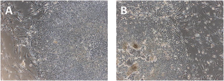
Identifying good quality RhiPSCs for passaging. (A) RhiPSC colony with ESC-like morphology that is ideal for passaging. (B) A representative RhiPSC colony of poor quality with obvious differentiation and cell death. Images were taken at 40×.
Enzymatic passaging
Visually inspect wells to confirm that the majority (>90%) of colonies are undifferentiated and are large enough for passaging.
- Prepare a working solution of dispase.See Reagents and Solutions. Dispase should be warmed to 37°C before use.
Aspirate cell culture media.
Wash cells gently with PBS and aspirate PBS.
- Add 1 mL of dispase to each well.The volume of dispase added to each well can be adjusted based on the size of the well. 1mL/well of dispase is suggested for a well in a 6 well plate.
- Transfer plate to incubator (37°C) for 8-15 minutes.Start checking on cells at 6 minutes of incubation. Look for edges of colonies to start lifting from plate. When 20-30% of colony has lifted the process is complete. MEFs will also come off plate during this process.
Aspirate dispase.
- Using a P1000 pipette, gently add 1 mL of CM to the corner of well to inactivate any residual dispase left in the well.Do NOT pipette up and down.
Aspirate media containing RhiPSCs.
Add 2-3 mL of CM containing RhiPSCs to well.
- Use P1000 pipette to break up large clumps.Make sure there are small clumps, since these will be the new islands that attach to new feeder plate. You will typically see a few large clumps which are usually the MEFs that came off during dispase incubation. Be careful to discard these clumps without removing too many RhiPSCs.
- Transfer the cell solution to new feeder plates with appropriate volumes of RhiPSC CM.A splitting ratio of 1:3 is typically used, but can be adjusted to meet needs. Cells that are maintained by dispase passaging tend to have better outcomes when CM is used for daily media change instead of standard RhiPSC media.
Change the media daily with RhiPSC CM.
RhiPSC Cryopreservation
Cryopreservation of RhiPSCs can be done with traditional “freezing media” formulations (90% ES-defined FBS, 10% DMSO) or with CryoStor® CS10 (StemCell Technologies). Importantly, RhiPSCs should be frozen as clumps rather than as single cells to enhance cryostability and viability after thawing. Below is the protocol for using CryoStor® CS10.
Remove culture medium from wells.
Add 2.2mL of cryopreservation medium to each.
Harvest RhiPSC colonies with MEF using a cell scraper.
- Transfer cells from each well to desired number of cryovials.We typically split one confluent well of RhiPSCs on MEF into two cryovials (1mL/vial).
Immediately place the cryovials into freezing containers and store in a -80°Cfreezer overnight.
Transfer the vials into liquid nitrogen the following day.
RhiPSC Characterization (Basic Protocol 3)
Careful characterization steps (immunostaining, RT-PCR, embryoid body differentiation, and teratoma formation) are useful tools to ensure complete reprogramming and assess the pluripotent state of the derived RhiPSCs. The protocols for these processes are not included since the methodologies are identical to standard approaches used for characterization of murine or human iPSCs (see Unit 1B.4, Unit 1C.16). However, we provide details regarding the rhesus-specific PCR primers and antibodies that can be employed for assessment of pluripotency.
Karyotyping of generated RhiPSC lines can be performed at either Oregon Health & Science University or Cell Line Genetics Inc.
Primer list
| Gene/Traget | Forwad sequence (5′-3′) | Reverse sequence (5′-3′) |
| OCT4 | CCTCACTTCACTGCACTGTA | CAGGTTTTCTTTCCCTAGCT |
| NANOG | TGAACCTCAGCTACAAACAG | TGGTGGTAGGAAGAGTAAAG |
| DPPA2 | CCCCTCCCTTGCCAACCATT | CACTGCCTTGCGTTTCCTCGA |
| AFP | AGCTTGGTGGTGGATGAAAC | CCCTCTTCAGCAAAGCAGAC |
| CD34 | GCGCTTTGCTTGCTGAGTTT | CCAAGGGTACTAGGTATTGC |
| NCAM | ATGGAAACTCTATTAAAGTGAACCTG | TAGACCTCATACTCAGCATTCCAGT |
| ACTIN | TGAAGTGTGACGTGGACATC | GGAGGAGCAATGATCTTGAT |
| Assay | Gene/Traget | Forwad sequence (5′-3′) | Reverse sequence (5′-3′) |
| Transgene PCR | MYC | GGAACTCTTGTGCGTAAGTCGATAG | GGAGGCGGCCCAAAGGGAGATCCG |
| Copy number qPCR | HIV-RRE | GGGAGCAGCAGGAAGCAC | TTGTCTGGCCTGTACCGTCA |
| Probe: ATGGGCGCAGCGTCAATGACG | |||
| STP | GGTGCCCTTCCTTGAGTTCA | CCCCTTGCACCCAGGAC | |
| Probe: CCCCAGGGATTCCCTCAGGTGTGT | |||
Antibody list
| Marker | Vendor | Source (dilution) | Secondary antibody (Vendor, dilution) |
| SSEA4 | Santa Cruz | 813-70 (1:50) | Alexa Fluor® 488 donkey anti-mouse IgG (Invitrogen, 1:500) |
| Nanog | R&D | Polyclonal (1:50) | Cy3-AffiniPure donkey anti-goat IgG (Jackson ImmunoResearch, 1:500) |
| OCT4 | Santa Cruz | C-10(1:50) | Alexa Fluor® 488 donkey anti-mouse IgG (Invitrogen, 1:500) |
| Tra-1-60 | Santa Cruz | TRA-1-60(1:50) | Alexa Fluor® 555 goat anti-mouse IgM (Invitrogen, 1:500) |
Reagents and Solutions
RhiPSC media
KnockOut DMEM (Gibco) supplemented with:
20% Knockout Serum Replacement
0.1mM NEAA
1% PSG
0.1 mM 2-mercaptoethanol
20 ng/ml bFGF
Store for up to 2 weeks at 4°C
Conditioned Media (RhiPSC media w/o bFGF)
Knockout DMEM (Gibco) supplemented with:
20% Knockout Serum Replacement
0.1mM NEAA
1% PSG
mM 2-mercaptoethanol
Store for up to 2 weeks at 4°C
MEF Culture Media
DMEM (Gibco cat. no. 11995) supplemented with:
10% FBS
1% NEAA
Fibroblast Culture Media
DMEM (Lonza cat no. 12-614F) supplemented with:
10% FBS
1% PSG
Store for up to 2 months at 4°C
BMSC Culture Media
Alpha MEM (Gibco cat no. 12561) supplemented with:
20% FBS
1% PSG
ROCK inhibitor
To make a 10 mM stock solution of Y27632, reconstitute 2 mg of the compound by adding 624.4 μl of DMSO. Make aliquots of 50 μl per tube and store at -20°C. To reach working concentration, stock can be used as 1000× – use 50 μl per 50 mL of medium to reach 10 nM. Store for up to 6 months at -20°C.
Polybrene
Stock Polybrene is 10 mg/mL, to achieve a final concentration of 6μg/ml, use 6 μl per 10 ml of medium.
Valporic Acid
Stock solution should be 100 mM, so dissolve 1 g VPA in 60.2 mL sterile water. Make sterile aliquots of 1 ml per tube and store them at -20°C for up to 6 months. To reach a working concentration (0.5 mM) of this stock dilute from200×.
Puromycin
Puromycin powder should be reconstituted with sterile water to reach a stock solution of 50 μg/ml. Store for up to 4 years at -20°C.
Dispase
Dissolve dispase powder with PBS to a working concentration of 1mg/mL. Filter sterilize through a 0.22 μm filter membrane. Store at 2-8°C for 24 months.
Commentary
Background Information
Since reports that differentiated adult murine and human cells could be reprogrammed to embryonic-like iPSCs via ectopic expression of a set of transcription factors, there has been exponential growth in the use of these cells for basic research (Takahashi and Yamanaka 2006; Yu, et al. 2007). In addition, the potential for differentiation of iPSCs to transplantable regenerative tissues useful clinically was realized immediately upon discovery, and has been pursued actively ever since. Immunocompetent large animal models to test the safety, efficacy and feasibility of iPSC-based regenerative therapies have been developed by several groups, including our own (Hong, et al. 2014). The basis for developing RhiPSCs stems from the same principles employed in derivation of human iPSCs – selective expression of Oct4, Sox2, Klf4, and Myc. Here we described an integrating lentiviral method for reprogramming rhesus fibroblasts or BMSCs to RhiPSCs. With the STEMCCA lentiviral reprogramming method followed by Cre-excision, a 303 bp DNA insert is left behind which can be used to track cells after autologous or allogeneic transplantation. However, integrating vectors have some risk of insertional mutagenesis, albeit low with the use of Cre-excised lentiviral enhancer-less designs.
Consequently, several non-integrating approaches, such as use of Sendai virus to deliver reprogramming factors, have been developed, and shown to be effective for iPSC generation from primary human adult cells (Ban, et al. 2011; Seki, et al. 2010). We have also used Sendai virus to reprogram rhesus macaque CD34+ hematopoietic progenitor cells to RhiPSCs; however, this method requires the cells be passaged upwards of ∼10 times post-reprogramming to dilute out the residual Sendai virus, until the Sendai genome is no longer detected by RT-PCR (Malik and Rao 2013).
Countless cell therapy protocols and strategies have been developed by leveraging iPSC technology; however, the use of rhesus or other non-human primate derived iPSCs is in its infancy. We have developed an autologous teratoma model, and demonstrated differentiation of RhiPSC towards mesodermal stromal cells that are able to produce bone in vivo following implantation (Hong, et al. 2014). Other groups have reported success in differentiating RhiPSCs towards neural lineages, and a number of groups are also working to develop NHP models for cardiac and hepatic differentiation and transplantation (Emborg, et al. 2013; Hallett, et al. 2015; Hong, et al. 2016). These and future studies should provide valuable information for moving iPSC-based cell therapies towards the clinic.
Critical Parameters and Troubleshooting
Successful RhiPSC generation and maintenance critically relies on the density of target BMSC or fibroblasts at the time of reprogramming, the oxygen concentration of incubator, and passaging technique. When the starting cell density of the original cells is higher than 1×10⁵ cells per 10 cm dish or 2×10⁴ cells per each well of a 6-well plate, the dish becomes confluent before the reprogrammed cells expand to make colonies, often leading to reprogramming failure. RhiPSCs also require hypoxic conditions (5% O2) to remain pluripotent, as has been reported for RhESC (Mitalipov et al., 2006). This degree of hypoxia can be achieved via multi-gas incubators or installation of hypoxic chambers within a traditional BL2 incubator. Under ambient O2, RhiPSCs tend to differentiate. It is recommended that these hypoxia chambers be calibrated periodically to ensure the desired oxygen concentration. Finally, the quality of colonies chosen for RhiPSC passaging is subjective, based on the knowledge, experience and skill of the individual scientist. When confluent colonies are passaged manually, the size of the pieces that are made from each colony will impact the overall attachment level and survival when transferred to a new MEF or matrigel plate. Overly large pieces can attach, but their edges tend to fold in on themselves leading spontaneous differentiation (UNIT 1C.10). Pieces that are too small have difficulty attaching and form colonies that are less compact on matrigel and tend not to form good quality colonies for maintenance or use in differentiation experiments. Beginners are recommended to practice making different size pieces and plating into different wells to better understand the effect colony fragment size has on attachment and colony quality. While more convenient than mechanical passaging, dispase-based passaging is not selective for differentiated versus undifferentiated colonies, so it is not suggested for continued maintenance of RhiPSC lines. However, this method is efficient for large-scale expansion prior to a differentiation experiment, or other specific purpose requiring large numbers of cells.
Anticipated Results
On average, the described protocol achieves a reprogramming efficiency of 0.05%. Distributing 120,000 BMSCs into the wells of a 6-well plate can be expected to produce roughly 60 colonies. RhiPSC should retain typical ESC morphology (Figure 1), normal karyotype, and express pluripotency proteins.
Time considerations
On average, 15-30 days are necessary to complete the reprogramming process, and select colonies for expansion and characterization. Freezing down early passage clones is desirable to assure a stable stock of RhiPSCs. Furthermore, it is advisable to perform Cre-excision of the reprogramming transgenes during early passages; however, with close attention to technique, it can performed on later passage cells.
Significance Statement.
Induced pluripotent stem cells (iPSCs) have great potential as a renewable and robust source of tissues for regenerative medicine; however, investigating the issues of safety and immunogenicity prior to clinical translation is paramount. Rhesus macaques are physiologically and phylogenetically similar to humans, and therefore are a relevant pre-clinical model organism for the development of iPSC-based cell therapies. Moreover, rhesus PSCs have been shown to be similar in terms of morphology, marker expression, and general culture requirements to their human counterpart, unlike murine PSC (Hong, et al. 2014; Liu, et al. 2008). Here we describe generation and maintenance of rhesus macaque iPSCs (RhiPSCs).
Acknowledgments
This research was supported by funding from the Division of Intramural Research at the National Heart, Lung and Blood Institute (NHLBI) at the National Institutes of Health.
Literature Cited
- Ban H, et al. Efficient generation of transgene-free human induced pluripotent stem cells (iPSCs) by temperature-sensitive Sendai virus vectors. Proc Natl Acad Sci U S A. 2011;108(34):14234–9. doi: 10.1073/pnas.1103509108. [DOI] [PMC free article] [PubMed] [Google Scholar]
- Emborg ME, et al. Induced Pluripotent Stem Cell-Derived Neural Cells Survive and Mature in the Nonhuman Primate Brain. Cell Rep. 2013 doi: 10.1016/j.celrep.2013.02.016. [DOI] [PMC free article] [PubMed] [Google Scholar]
- Hallett PJ, et al. Successful function of autologous iPSC-derived dopamine neurons following transplantation in a non-human primate model of Parkinson's disease. Cell Stem Cell. 2015;16(3):269–74. doi: 10.1016/j.stem.2015.01.018. [DOI] [PMC free article] [PubMed] [Google Scholar]
- Hong SG, et al. The Role of Nonhuman Primate Animal Models in the Clinical Development of Pluripotent Stem Cell Therapies. Mol Ther. 2016;24(7):1165–9. doi: 10.1038/mt.2016.131. [DOI] [PMC free article] [PubMed] [Google Scholar]
- Hong SG, et al. Path to the clinic: assessment of iPSC-based cell therapies in vivo in a nonhuman primate model. Cell Rep. 2014;7(4):1298–309. doi: 10.1016/j.celrep.2014.04.019. [DOI] [PMC free article] [PubMed] [Google Scholar]
- Liu H, et al. Generation of induced pluripotent stem cells from adult rhesus monkey fibroblasts. Cell Stem Cell. 2008;3(6):587–90. doi: 10.1016/j.stem.2008.10.014. [DOI] [PubMed] [Google Scholar]
- Malik N, Rao MS. A review of the methods for human iPSC derivation. Methods Mol Biol. 2013;997:23–33. doi: 10.1007/978-1-62703-348-0_3. [DOI] [PMC free article] [PubMed] [Google Scholar]
- Merling RK, et al. Transgene-free iPSCs generated from small volume peripheral blood nonmobilized CD34+ cells. Blood. 2013;121(14):e98–e107. doi: 10.1182/blood-2012-03-420273. [DOI] [PMC free article] [PubMed] [Google Scholar]
- Mitalipov S, et al. Isolation and characterization of novel rhesus monkey embryonic stem cell lines. Stem Cells. 2006;24(10):2177–86. doi: 10.1634/stemcells.2006-0125. [DOI] [PubMed] [Google Scholar]
- Seki T, et al. Generation of induced pluripotent stem cells from human terminally differentiated circulating T cells. Cell Stem Cell. 2010;7(1):11–4. doi: 10.1016/j.stem.2010.06.003. [DOI] [PubMed] [Google Scholar]
- Sommer CA, et al. Excision of reprogramming transgenes improves the differentiation potential of iPS cells generated with a single excisable vector. Stem Cells. 2010;28(1):64–74. doi: 10.1002/stem.255. [DOI] [PMC free article] [PubMed] [Google Scholar]
- Takahashi K, Yamanaka S. Induction of pluripotent stem cells from mouse embryonic and adult fibroblast cultures by defined factors. Cell. 2006;126(4):663–76. doi: 10.1016/j.cell.2006.07.024. [DOI] [PubMed] [Google Scholar]
- Uchida N, et al. Development of a human immunodeficiency virus type 1-based lentiviral vector that allows efficient transduction of both human and rhesus blood cells. J Virol. 2009;83(19):9854–62. doi: 10.1128/JVI.00357-09. [DOI] [PMC free article] [PubMed] [Google Scholar]
- Yu J, et al. Induced pluripotent stem cell lines derived from human somatic cells. Science. 2007;318(5858):1917–20. doi: 10.1126/science.1151526. [DOI] [PubMed] [Google Scholar]