Abstract
Background
Cerebral small vessel disease is a common cause of vascular cognitive impairment in older people, with no licensed treatment. Cerebral blood flow is reduced in small vessel disease. Tadalafil is a widely prescribed phosphodiesterase-5 inhibitor that increases blood flow in other vascular territories. The aim of this trial is to test the hypothesis that tadalafil increases cerebral blood flow in older people with small vessel disease.
Methods/design
Perfusion by Arterial Spin labelling following Single dose Tadalafil In Small vessel disease (PASTIS) is a phase II randomised double-blind crossover trial. In two visits, 7-30 days apart, participants undergo arterial spin labelling to measure cerebral blood flow and a battery of cognitive tests, pre- and post-dosing with oral tadalafil (20 mg) or placebo. Sample size: 54 participants are required to detect a 15% increase in cerebral blood flow in subcortical white matter (p < 0.05, 90% power). Primary outcomes are cerebral blood flow in subcortical white matter and deep grey nuclei. Secondary outcomes are cortical grey matter cerebral blood flow and performance on cognitive tests (reaction time, information processing speed, digit span forwards and backwards, semantic fluency).
Discussion
Recruitment started on 4th September 2015 and 36 participants have completed to date (19th April 2017). No serious adverse events have occurred. All participants have been recruited from one centre, St George’s University Hospitals NHS Foundation Trust.
Trial registration
European Union Clinical Trials Register: EudraCT number 2015-001235-20. Registered on 13 May 2015.
Electronic supplementary material
The online version of this article (doi:10.1186/s13063-017-1973-9) contains supplementary material, which is available to authorized users.
Keywords: Tadalafil, Cerebral blood flow, Vascular cognitive impairment, Vascular dementia, Phosphodiesterase, MRI, Arterial spin labelling
Background
Cerebral small vessel disease (SVD) is a frequent cause of vascular cognitive impairment (VCI) in older adults [1–4]. There is currently no licensed treatment for SVD or for VCI [1, 2]. There is evidence derived from some previous studies to suggest that cerebral blood flow (CBF) is reduced in SVD, particularly in subcortical white matter [5–10]. We hypothesised that increasing CBF has the potential to be both a symptomatic and a disease-modifying treatment for SVD and VCI.
Phosphodiesterase type 5 inhibitors (PDE5i) such as sildenafil and tadalafil are well-established pharmacological vasodilators that cause enhanced nitric oxide-cyclic guanosine monophosphate signalling in peripheral small arteries [11–13]. PDE5i are widely used in treatment of erectile dysfunction and pulmonary hypertension [13]. PDE5 messenger RNA and protein are also found in human brain tissue [12, 14, 15]. Side-effect profiles of PDE5i are well-known, and the drugs are well-tolerated in the target population [16–18]. In a meta-analysis of 28 placebo-controlled trials [18], the overall incidence of myocardial infarction, cardiovascular death or cerebrovascular death in tadalafil-treated patients did not differ from placebo. The incidence of these adverse events was independent of dosing regimen and duration of tadalafil therapy (up to 27 months) [18]. The choice of tadalafil (over other PDE5i) was based on long plasma half-life (17 h in healthy adults) [16, 17] and established brain penetration (brain-to-plasma ratio 1:10 in rodents and primates) [12, 19]. In this study, we will test whether single-dose tadalafil increases CBF in older people with neuroradiological and clinical evidence of SVD.
Methods/design
Objectives
The aim of this study is to test the hypothesis that tadalafil increases cerebral blood flow in subcortical areas in older people with symptomatic SVD.
Study design
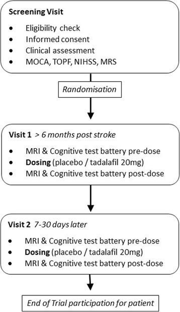
Perfusion by Arterial Spin labelling following Single dose Tadalafil In Small vessel disease (PASTIS) is a phase II double-blind crossover trial. Participants are randomised to order of treatment (tadalafil 20 mg, placebo; oral administration). Two visits are performed 7–30 days apart, with perfusion magnetic resonance imaging (MRI) and a battery of cognitive tests performed before and 3–5 h after dosing (see Fig. 1).
Fig. 1.

MOCA Montreal Cognitive Assessment, TOPF Test of Premorbid Functioning, NIHSS National Institutes of Health Stroke Scale, MRS Modified Rankin Scale, MRI Magnetic resonance imaging
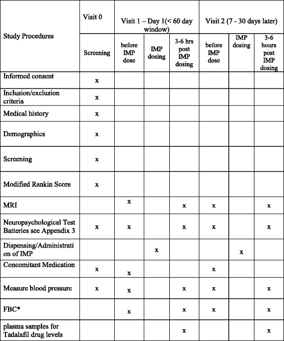
A Standard Protocol Items: Recommendations for Interventional Trials (SPIRIT) checklist is shown in Fig. 2 (see also Additional file 1).
Fig. 2.

Standard Protocol Items: Recommendations for Interventional Trials (SPIRIT) checklist: schedule of enrolment, interventions and assessments in the Perfusion by Arterial Spin labelling following Single dose Tadalafil In Small vessel disease (PASTIS) trial. From PASTIS protocol version 4, 27 Jan 2016
Trial endpoints
The primary endpoints are change in regional CBF in two sub-cortical brain areas (deep white matter and deep grey nuclei). The secondary endpoints are (1) change in regional CBF in cortical grey matter, (2) change in neuropsychological test performance and (3) plasma tadalafil concentration dependence of any changes observed.
Study setting
Participants are recruited from St George’s University Hospital NHS Foundation Trust and local Participant Identification Centre sites. All patient visits, data management and trial coordination are performed at St George’s. PASTIS has been adopted into the UK National Institute for Health Research Clinical Research Network Portfolio.
Participant characteristics
Participants are older people (men and women) without a diagnosis of dementia who have radiological and clinical evidence of symptomatic SVD. After informed consent is obtained, the following activities will occur at a screening visit (see Fig. 1):
Trial eligibility criteria check
Medical history
Concomitant medication checklist: medications, doses and frequencies
MRI suitability/contraindication checklist
Participant demographics, including ethnic origin
Next-of-kin and general practitioner contact details to be recorded if not already in medical notes, or check if still current and up-to-date
Affix Clinical Trials Alert sticker to front of the medical notes
Complete case report form screening page, ensuring the participant’s trial identifier is included
Test of Premorbid Functioning (TOPF) to establish estimated levels of cognitive functioning pre-illness
National Institutes of Health Stroke Scale (NIHSS)
Montreal Cognitive Assessment (MoCA) to establish estimated levels of cognitive functioning
Record the modified Rankin Scale score (mRS)
Inclusion criteria
Radiological evidence of cerebral SVD, defined as MRI evidence of lacunar infarcts (≤1.5 cm maximum diameter) and/or confluent deep white matter hyperintensities (WMH) (≥grade 2 on Fazekas scale)
- Clinical evidence of SVD, including the following:
- Lacunar stroke syndrome with symptoms lasting >24 h, occurring ≥6 months prior to visit 1; or
- Transient ischaemic attack (TIA) lasting <24 h with limb weakness, hemi-sensory loss or dysarthria ≥6 months previously and with MRI diffusion-weighted imaging performed acutely showing lacunar infarction, or, if MRI is not performed within 10 days of TIA, lacunar infarct in an anatomically appropriate area
Age ≥50 years
Imaging of the carotid arteries with Doppler ultrasound, computed tomographic angiography or magnetic resonance angiography in the previous 12 months demonstrating <70% stenosis in both internal carotid arteries or <50% stenosis in both internal carotids if measured in previous 12–60 months
Exclusion criteria
Known diagnosis of dementia
Cortical infarct (>1.5 cm maximum diameter)
Systolic blood pressure (BP) <90 mmHg and/or diastolic BP <50 mmHg
Creatinine clearance <30 ml/minute
Severe hepatic impairment
History of lactose intolerance
Concomitant use of PDE5i (e.g., sildenafil, tadalafil, vardenafil)
Receiving nicorandil or nitrates (e.g., isosorbide mononitrate, glyceryl trinitrate)
Weight >130 kg
Uncontrolled cardiac failure
Persistent or paroxysmal atrial fibrillation
History of gastric ulceration
History of ‘sick sinus syndrome’ or other supraventricular cardiac conduction conditions
Uncontrolled chronic obstructive pulmonary disease
Stroke or TIA within 6 months
MRI not tolerated or contraindicated
Known monogenic causes of stroke (e.g., cerebral autosomal dominant arteriopathy with subcortical infarcts and leukoencephalopathy)
Unable to provide informed consent
Randomisation
The randomisation list will be generated by Sharp Clinical Services (http://www.sharpservices.com/our-facilities/sharp-clinical-services-wales/; Crickhowell, UK) and will be done in blocks, as detailed in the client study information form kept in the sponsor site file. The participants will be acting as their own controls. Each participant will receive on two separate occasions a placebo dose and a tadalafil 20-mg immediate dose which appear identical in size, shape, weight and colour.
The patient pack numbers on the pharmacy shelf correlate directly with the next available pack number on the blinded randomisation list held in the pharmacy site file. Each patient pack contains two bottles, labelled as bottle A and bottle B. The randomisation list will be confidential to the trial statistician and will be summarised as treatment arm A and B, and not by tadalafil and placebo.
Measurement of regional cerebral blood flow
Whole-brain perfusion will be determined by pseudo-continuous arterial spin labelling (ASL) [20] in a 3-T MRI scanner (Achieva TX MRI scanner, Philips Medical Systems, Eindhoven, Netherlands). A total of 20-minute pseudo-continuous ASL acquisition time will be used to provide an adequate signal-to-noise ratio for CBF quantification in white matter. Other image data acquired will be as follows: an M0 image, to enable quantification of CBF; high-resolution 3D T1-weighted images for identification of grey and white matter regions of interest (including deep grey matter structures) for ASL analysis [20] and to map the ASL data to a standard brain atlas; fluid-attenuated inversion recovery (FLAIR) for delineation of WMH; and susceptibility-weighted imaging for detection of micro-haemorrhages. These will provide participant-specific WMH load and location of WMH. Total scanning time is under 60 minutes per MRI session.
Cognitive testing
Scores derived from the TOPF and MoCA instruments are recorded at the screening visit. These are included in the analyses as baseline data. They are not used as inclusion or exclusion criteria.
At the two dosing visits, the following neuropsychological tests are used: the Reaction Time subtest of the Cambridge Cognition Cambridge Neuropsychological Test Automated Battery, the Speed of Information Processing subtest of the Brain Injury Rehabilitation Trust Memory and Information Processing Battery, the Digit Span forwards and backwards subtest of the Repeatable Battery for the Assessment of Neuropsychological Status (RBANS), and the Semantic Fluency subtest of RBANS.
Biochemical analyses
A blood sample is taken at the end of visits 1 and 2 for haematocrit and full blood count analysis. Plasma samples are stored at −80 °C for subsequent analysis of plasma tadalafil concentration.
Details of the intervention
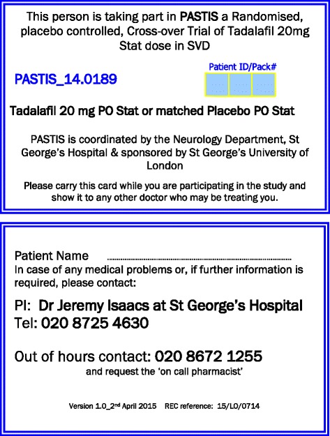
Each participant pack contains one bottle containing a single tadalafil 20-mg capsule and one identical bottle containing a single matched placebo capsule. At each visit, participants undergo cognitive tests and the first MRI scanning session of the day. Participants are then observed to swallow the appropriate investigational medicinal product (IMP) capsule and receive a standard light lunch (450–750 kcal and 500 ml of fluid). They undergo an equivalent, parallel version of the cognitive tests and the second MRI session of the day 3–5 h later. All participants are given a 24-h emergency contact card with the study title, details of the IMP, their participant trial number, investigator’s contact details, and out-of-hours contact details (see Fig. 3).
Fig. 3.

PASTIS trial emergency 24-hour contact card, supplied to each participant
All those involved in the study (researchers, radiologists, pharmacists and participants) are blinded to treatment allocation for the duration of the study. Emergency un-blinding will take place in circumstances such as serious adverse events (SAEs). Any SAEs and safety endpoints will be reported in line with clinical trial regulations (SI2004/1031) and the sponsor’s procedures. We do not anticipate any serious adverse reactions to the medication, because tadalafil is widely used clinically and is well-tolerated. The starting point for SAE monitoring is the first intervention visit, ending 5 days after the second visit (based on a drug elimination period of 6 half-lives for the study medication, using a 20-h half-life for tadalafil).
Power calculation
On the basis of previous ASL studies of regional CBF, we estimate baseline perfusion of 30 (±10) ml/100 g/minute (mean ± SD) in subcortical white matter and 70 (±15) ml/100 g/minute in deep grey nuclei [21, 22]. To detect a treatment effect of 15% (mean paired difference) with statistical power of 90%, a sample size of 24 is required in deep grey matter nuclei and 54 in subcortical white matter. We aim to recruit a target cohort of 54.
Statistical analysis
Baseline characteristics (age, sex, ethnic group, baseline BP, mRS score, NIHSS, TOPF, MoCA) will be summarised as mean (SD) or median (Q1, Q3) for continuous variables, depending on distribution, and as number (percent) for categorical variables. Changes in outcome variables will be calculated for each participant at each visit as post-dose value minus pre-dose value. Data will be analysed using a linear mixed effects regression model with fixed effects for treatment (drug vs. placebo), visit (visit 1, visit 2), treatment sequence and baseline response, as well as a random effect for participant nested within treatment sequence. Carry-over will be investigated by the treatment-by-visit interaction. If statistically significant, data from each visit will be analysed separately within linear regression models adjusted for treatment and pre-dose value. Clinical variables and other possible confounders (e.g., BP at the time of the scan) will be included in the linear mixed effects models as adjustment variables. These will be pre-specified in the statistical analysis plan.
All analyses will be done on an intention-to-treat basis, and no adjustment will be made for missing data. Statistical analyses will be performed using SAS® for Windows version 9.3 or later software (SAS Institute, Cary, NC, USA). A p value >0.05 indicates the absence of a statistically significant effect.
Data monitoring
Monitoring is performed by the sponsor clinical trials monitor in accordance with an agreed risk-based monitoring plan. Case report form entries are verified against the source documents and the participants’ medical notes. All data are entered directly from case report forms to the PASTIS Microsoft Access (Microsoft Corp., Redmond, WA, USA) database by the PASTIS research team. Data transfer from the case report form will be double-checked, and where corrections are required, these will carry a full audit trail and justification. Trial data storage conforms to St George’s institutional information governance policies. Trial data, evidence of monitoring and system audits will be made available for inspection by the sponsor and regulatory authorities as required.
Discussion
In this randomised, double-blind, crossover phase II study, we will test whether tadalafil (20 mg) increases CBF in older people with SVD. Tadalafil was chosen over other PDE5i (e.g., sildenafil or vardenafil) because of the documented brain penetration [12, 19] and longer plasma half-life of tadalafil [16, 17]. In the present trial, we are simply testing for acute changes in response to a single dose of tadalafil. For this purpose, a crossover design appeared optimal. In the event that a positive outcome is detected in the present study, it appears likely that a subsequent study testing tadalafil over a longer dosing period will be required. This will be needed to explore whether any tadalafil-mediated actions are maintained with chronic dosing and to test for any additional adverse reactions in participants who are likely to be taking concomitant stroke medications.
ASL was chosen to quantify regional CBF because it does not require injected radioisotopes or gadolinium compounds as tracers [20–22]. This MRI-based approach also enables acquisition of high-resolution 3D T1-weighted images, T2-weighted FLAIR images and susceptibility-weighted imaging. The neuropsychological tests that are used were chosen because each has four parallel versions of the test to be applied at each screening point (Fig. 1). The cognitive tests used measure processing speed, attention and executive function, which are affected in SVD, as well as working memory and semantic fluency. Nevertheless, it may be difficult to detect cognitive changes in such short-term follow-up as is employed in the present study. The cognitive data obtained from this trial may be of value in assessing sample size and feasibility for any subsequent trial of tadalafil in relation to cognitive function.
In addition to the European Union Clinical Trials Register (EudraCT number 2015-001235-20, date of registration 13 May 2015), the trial has been registered with ClinicalTrials.gov (NCT02450253, date of registration 18 May 2015). No SAEs have been observed so far. Inadvertent un-blinding due to the erectile effects of tadalafil has not occurred so far as we are aware. Spontaneous penile erection has been reported in a modest fraction (11%) of subjects taking 20 mg of tadalafil [16, 17]. PASTIS is the first phase II clinical trial of a selective PDE5i in older people with symptomatic SVD. Outcome data are expected in late 2017 and may inform a larger trial for re-purposing of tadalafil in SVD and VCI.
Trial status
The trial commenced on 4th September 2015. The PASTIS trial is ongoing. Patient recruitment has not been completed. As of 19 April 2017, 36 participants have completed the protocol.
Acknowledgements
We are very grateful to our colleagues at St George’s Hospital and St George’s University of London for their support of the PASTIS study.
Funding
The study is jointly funded by the Alzheimer’s Drug Discovery Foundation and the Alzheimer’s Society (grant reference 20140901). NC is funded by MRC doctoral training programme grant number MR/N013638/1. The funders have no input into trial design, data collection or data analysis.
Availability of data and materials
Not applicable.
Authors’ contributions
JDI, TRB, FAH, DR, SB, JBM, BM, UK, ACP, CK, ER and AHH contributed to study design. MMHP, ST and NC performed data collection. MMHP, TRB and CEH carried out data analysis. MMHP and NC drafted the manuscript. All authors contributed to revising the manuscript. AHH prepared the final manuscript. All authors read and approved the final manuscript.
Authors’ information
MMHP is a clinical research fellow at St George’s University Hospitals NHS Foundation Trust, London. NC is a neuroscience researcher at St George’s University of London. ST is a stroke research clinical studies coordinator at St George’s University Hospitals NHS Foundation Trust, London. SB is a consultant neuropsychologist at St George’s University Hospitals NHS Foundation Trust, London. FAH is a professor of medical physics at St George’s University of London. UK is a consultant neurologist at St George’s University Hospitals NHS Foundation Trust, London. CK is a consultant neurologist at Herlev Gentofte Hospital and associate professor of stroke medicine at University of Copenhagen, Denmark. JBM is a consultant neuroradiologist at St George’s University Hospitals NHS Foundation Trust, London. BM is a consultant is stroke medicine at Beaumont Hospital, Dublin. ACP is a consultant neurologist at St George’s University Hospitals NHS Foundation Trust, London. DR is a regulatory assurance manager, St George’s University of London. ER is a research consultant of nuclear medicine, Rigshospitalet Glostrup, Denmark. CEH is a biostatistician at the Robertson Centre for Biostatistics, University of Glasgow. TRB is a senior lecturer in image analysis at St George’s University of London. JDI is a Consultant neurologist and is clinical principal investigator on the PASTIS trial. AHH is a reader in cerebrovascular disease at St George’s University of London and is chief investigator on the PASTIS trial.
Competing interests
The authors declare that they have no competing interests.
Consent for publication
Not applicable.
Ethics approval and consent to participate
The PASTIS Study received NHS National Research Ethics Service approval on 6 May 2015 (London-Brent Research Ethics Committee reference 15/LO/0714). It received Medicines and Healthcare products Regulatory Agency authorisation (reference 16745/0222/001-0001) on 5 June 2015 and St George’s University of London research and development approval (protocol number 14.0189) on 4 September 2015. Informed consent to participate is obtained from all participants in the study. The trial opened for recruitment on 4 September 2015, and the first participant was enrolled on 14 September 2015. The PASTIS trial is sponsored by the Joint Research and Enterprise Office, St George’s University of London, Cranmer Terrace, London SW17 0RE, UK. A representative of the sponsor (DR) has had input into trial design and has contributed to this report.
Publisher’s Note
Springer Nature remains neutral with regard to jurisdictional claims in published maps and institutional affiliations.
Abbreviations
- ASL
Arterial spin labelling
- BP
Blood pressure
- CBF
Cerebral blood flow
- FLAIR
Fluid-attenuated inversion recovery
- IMP
Investigational medicinal product
- MoCA
Montreal Cognitive Assessment
- MRI
Magnetic resonance imaging
- mRS
Modified Rankin Scale
- NIHSS
National Institutes of Health Stroke Scale
- PASTIS
Perfusion by Arterial Spin labelling following Single dose Tadalafil In Small vessel disease
- PDE5i
Phosphodiesterase type 5 inhibitor
- RBANS
Repeatable Battery for the Assessment of Neuropsychological Status
- SAE
Serious adverse event
- SPIRIT
Standard Protocol Items: Recommendations for Interventional Trials
- SVD
Small vessel disease
- TIA
Transient ischaemic attack
- TOPF
Test of premorbid functioning
- VCI
Vascular cognitive impairment
- WMH
White matter hyperintensities
Additional file
SPIRIT 2013 checklist: recommended items to address in a clinical trial protocol and related documents. (DOC 120 kb)
Footnotes
Electronic supplementary material
The online version of this article (doi:10.1186/s13063-017-1973-9) contains supplementary material, which is available to authorized users.
Contributor Information
Mathilde M. H. Pauls, Email: mpauls@sgul.ac.uk
Natasha Clarke, Email: nclarke@sgul.ac.uk.
Sarah Trippier, Email: sarah.trippier@stgeorges.nhs.uk.
Shai Betteridge, Email: shai.betteridge@stgeorges.nhs.uk.
Franklyn A. Howe, Email: howefa@sgul.ac.uk
Usman Khan, Email: usman.khan4@nhs.net.
Christina Kruuse, Email: ckruuse@dadlnet.dk.
Jeremy B. Madigan, Email: jeremy.madigan@nhs.net
Barry Moynihan, Email: barrymoynihan@beaumont.ie.
Anthony C. Pereira, Email: anthony.pereira@stgeorges.nhs.uk
Debbie Rolfe, Email: drolfe@sgul.ac.uk.
Egill Rostrup, Email: egillr@dadlnet.dk.
Caroline E. Haig, Email: caroline.haig@glasgow.ac.uk
Thomas R. Barrick, Email: tbarrick@sgul.ac.uk
Jeremy D. Isaacs, Email: jeremy.isaacs@nhs.net
Atticus H. Hainsworth, Phone: +44 208 725 5586, Email: ahainsworth@sgul.ac.uk
References
- 1.O’Brien JT, Thomas A. Vascular dementia. Lancet. 2015;386:1698–706. doi: 10.1016/S0140-6736(15)00463-8. [DOI] [PubMed] [Google Scholar]
- 2.Pantoni L. Cerebral small vessel disease: from pathogenesis and clinical characteristics to therapeutic challenges. Lancet Neurol. 2010;9:689–701. doi: 10.1016/S1474-4422(10)70104-6. [DOI] [PubMed] [Google Scholar]
- 3.Esiri MM, Wilcock GK, Morris JH. Neuropathological assessment of the lesions of significance in vascular dementia. J Neurol Neurosurg Psychiatry. 1997;63:749–53. doi: 10.1136/jnnp.63.6.749. [DOI] [PMC free article] [PubMed] [Google Scholar]
- 4.Ighodaro ET, Abner EL, Fardo DW, Lin AL, Katsumata Y, Schmitt FA, et al. Risk factors and global cognitive status related to brain arteriolosclerosis in elderly individuals. J Cereb Blood Flow Metab. 2017;37:201–16. doi: 10.1177/0271678X15621574. [DOI] [PMC free article] [PubMed] [Google Scholar]
- 5.Markus HS, Lythgoe DJ, Ostegaard L, O’Sullivan M, Williams SC. Reduced cerebral blood flow in white matter in ischaemic leukoaraiosis demonstrated using quantitative exogenous contrast based perfusion MRI. J Neurol Neurosurg Psychiatry. 2000;69:48–53. doi: 10.1136/jnnp.69.1.48. [DOI] [PMC free article] [PubMed] [Google Scholar]
- 6.O’Sullivan M, Lythgoe DJ, Pereira AC, Summers PE, Jarosz JM, Williams SC, et al. Patterns of cerebral blood flow reduction in patients with ischemic leukoaraiosis. Neurology. 2002;59:321–6. doi: 10.1212/WNL.59.3.321. [DOI] [PubMed] [Google Scholar]
- 7.Schuff N, Matsumoto S, Kmiecik J, Studholme C, Du A, Ezekiel F, et al. Cerebral blood flow in ischemic vascular dementia and Alzheimer’s disease, measured by arterial spin-labeling magnetic resonance imaging. Alzheimers Dement. 2009;5:454–62. doi: 10.1016/j.jalz.2009.04.1233. [DOI] [PMC free article] [PubMed] [Google Scholar]
- 8.Yao H, Sadoshima S, Ibayashi S, Kuwabara Y, Ichiya Y, Fujishima M. Leukoaraiosis and dementia in hypertensive patients. Stroke. 1992;23:1673–7. doi: 10.1161/01.STR.23.11.1673. [DOI] [PubMed] [Google Scholar]
- 9.Bernbaum M, Menon BK, Fick G, Smith EE, Goyal M, Frayne R, et al. Reduced blood flow in normal white matter predicts development of leukoaraiosis. J Cereb Blood Flow Metab. 2015;35:1610–5. doi: 10.1038/jcbfm.2015.92. [DOI] [PMC free article] [PubMed] [Google Scholar]
- 10.Arba F, Mair G, Carpenter T, Sakka E, Sandercock PA, Lindley RI, et al. Cerebral white matter hypoperfusion increases with small-vessel disease burden: data from the Third International Stroke Trial. J Stroke Cerebrovasc Dis. doi: 10.1016/j.jstrokecerebrovasdis.2017.03.002. [DOI] [PubMed]
- 11.Conti M, Beavo J. Biochemistry and physiology of cyclic nucleotide phosphodiesterases: essential components in cyclic nucleotide signaling. Annu Rev Biochem. 2007;76:481–511. doi: 10.1146/annurev.biochem.76.060305.150444. [DOI] [PubMed] [Google Scholar]
- 12.Garcia-Osta A, Cuadrado-Tejedor M, Garcia-Barroso C, Oyarzabal J, Franco R. Phosphodiesterases as therapeutic targets for Alzheimer’s disease. ACS Chem Neurosci. 2012;3:832–44. doi: 10.1021/cn3000907. [DOI] [PMC free article] [PubMed] [Google Scholar]
- 13.Maurice DH, Ke H, Ahmad F, Wang Y, Chung J, Manganiello VC. Advances in targeting cyclic nucleotide phosphodiesterases. Nat Rev Drug Discov. 2014;13:290–314. doi: 10.1038/nrd4228. [DOI] [PMC free article] [PubMed] [Google Scholar]
- 14.Lakics V, Karran EH, Boess FG. Quantitative comparison of phosphodiesterase mRNA distribution in human brain and peripheral tissues. Neuropharmacology. 2010;59:367–74. doi: 10.1016/j.neuropharm.2010.05.004. [DOI] [PubMed] [Google Scholar]
- 15.Kruuse C, Khurana TS, Rybalkin SD, Birk S, Engel U, Edvinsson L, et al. Phosphodiesterase 5 and effects of sildenafil on cerebral arteries of man and guinea pig. Eur J Pharmacol. 2005;521:105–14. doi: 10.1016/j.ejphar.2005.07.017. [DOI] [PubMed] [Google Scholar]
- 16.Forgue ST, Patterson BE, Bedding AW, Payne CD, Phillips DL, Wrishko RE, et al. Tadalafil pharmacokinetics in healthy subjects. Br J Clin Pharmacol. 2006;61:280–8. doi: 10.1111/j.1365-2125.2005.02553.x. [DOI] [PMC free article] [PubMed] [Google Scholar]
- 17.Forgue ST, Phillips DL, Bedding AW, Payne CD, Jewell H, Patterson BE, et al. Effects of gender, age, diabetes mellitus and renal and hepatic impairment on tadalafil pharmacokinetics. Br J Clin Pharmacol. 2007;63:24–35. doi: 10.1111/j.1365-2125.2006.02726.x. [DOI] [PMC free article] [PubMed] [Google Scholar]
- 18.Kloner RA, Jackson G, Hutter AM, Mittleman MA, Chan M, Warner MR, et al. Cardiovascular safety update of tadalafil: retrospective analysis of data from placebo-controlled and open-label clinical trials of tadalafil with as needed, three times-per-week or once-a-day dosing. Am J Cardiol. 2006;97:1778–84. doi: 10.1016/j.amjcard.2005.12.073. [DOI] [PubMed] [Google Scholar]
- 19.Garcia-Barroso C, Ricobaraza A, Pascual-Lucas M, Unceta N, Rico AJ, Goicolea MA, et al. Tadalafil crosses the blood-brain barrier and reverses cognitive dysfunction in a mouse model of AD. Neuropharmacology. 2013;64:114–23. doi: 10.1016/j.neuropharm.2012.06.052. [DOI] [PubMed] [Google Scholar]
- 20.Alsop DC, Detre JA, Golay X, Gunther M, Hendrikse J, Hernandez-Garcia L, et al. Recommended implementation of arterial spin-labeled perfusion MRI for clinical applications: a consensus of the ISMRM perfusion study group and the European consortium for ASL in dementia. Magn Reson Med. 2015;73:102–16. doi: 10.1002/mrm.25197. [DOI] [PMC free article] [PubMed] [Google Scholar]
- 21.D’haeseleer M, Beelen R, Fierens Y, Cambron M, Vanbinst AM, Verborgh C, et al. Cerebral hypoperfusion in multiple sclerosis is reversible and mediated by endothelin-1. Proc Natl Acad Sci U S A. 2013;110:5654–8. doi: 10.1073/pnas.1222560110. [DOI] [PMC free article] [PubMed] [Google Scholar]
- 22.Colloby SJ, Firbank MJ, He J, Thomas AJ, Vasudev A, Parry SW, et al. Regional cerebral blood flow in late-life depression: arterial spin labelling magnetic resonance study. Br J Psychiatry. 2012;200:150–5. doi: 10.1192/bjp.bp.111.092387. [DOI] [PubMed] [Google Scholar]
Associated Data
This section collects any data citations, data availability statements, or supplementary materials included in this article.
Data Availability Statement
Not applicable.


