Abstract
Anaerobic thermophiles inhabit relic environments that resemble the early Earth. However, the lineage of these modern organisms co-evolved with our planet. Hence, these organisms carry both ancestral and acquired genes and serve as models to reconstruct early metabolism. Based on comparative genomic and proteomic analyses, we identified two distinct groups of genes in Thermovibrio ammonificans: the first codes for enzymes that do not require oxygen and use substrates of geothermal origin; the second appears to be a more recent acquisition, and may reflect adaptations to cope with the rise of oxygen on Earth. We propose that the ancestor of the Aquificae was originally a hydrogen oxidizing, sulfur reducing bacterium that used a hybrid pathway for CO2 fixation. With the gradual rise of oxygen in the atmosphere, more efficient terminal electron acceptors became available and this lineage acquired genes that increased its metabolic flexibility while retaining ancestral metabolic traits.
DOI: http://dx.doi.org/10.7554/eLife.18990.001
Research Organism: Other
eLife digest
Life may have arisen on our planet as far back as four billion years ago. Unlike today, the Earth’s atmosphere at the time had no oxygen and an abundance of volcanic emissions including hydrogen, carbon dioxide and sulfur gases. These dramatic differences have led scientists to wonder: how did the ancient microorganisms that inhabited our early planet make a living? And how has microbial life co-evolved with the Earth?
One way to answer these questions is to study bacteria that live today in environments that resemble the early Earth. Deep-sea hydrothermal vents are regions of the deep ocean where active volcanic processes recreate primordial conditions. These habitats support microorganisms that are highly adapted to live off hydrogen, carbon dioxide and sulfur gases, and studying these modern-day microorganisms could give insights into the earliest life on Earth.
Thermovibrio ammonificans is a bacterium that was obtained from an underwater volcanic system in the East Pacific. Giovannelli et al. have now asked if T. ammonificans might have inherited some of its genetic traits from a long-gone ancestor that also thrived off volcanic gases. The genetic makeup of this microorganism was examined for genes that would help it thrive at a deep-sea hydrothermal vent. Next, Giovannelli et al. compared these genes to related copies in other species of bacteria to reconstruct how the metabolism of T. ammonificans might have changed over time.
This approach identified a group of likely ancient genesthat allow a microorganism to use chemicals like hydrogen, carbon dioxide and sulfur to fuel its growth and metabolism. These findings support the hypothesis that an ancestor of T. ammonificans could live off volcanic gases and that the core set of genes involved in those activities had been passed on, through the generations, to this modern-day microorganism. Giovannelli et al. also identified a second group of genes in T. ammonificans that indicate that this bacterium also co-evolved with Earth’s changing conditions, in particular the rise in the concentration of oxygen.
The findings of Giovannelli et al. provide insight into how the metabolism of microbes has co-evolved with the Earth’s changing conditions, and will allow others to formulate new hypotheses that can be tested in laboratory experiments.
Introduction
Deep-branching, anaerobic, thermophilic Bacteria and Archaea inhabit relic environments that resemble the early Earth (Baross and Hoffman, 1985; Martin et al., 2008). Thermophily (Di Giulio, 2003, 2000), anaerobic metabolism (Baross and Hoffman, 1985; Martin et al., 2008; Schopf, 1983) and reliance on substrates of geothermal origin are among the proposed ancestral traits of these microorganisms (Baross and Hoffman, 1985; Di Giulio, 2003, 2000; Lane et al., 2010; Martin et al., 2008; Russell and Martin, 2004; Schopf, 1983). At the same time, their lineages have co-evolved with Earth and their genomes also carry more recently acquired traits. Therefore, these microorganisms can be used as models to reconstruct the evolution of metabolism.
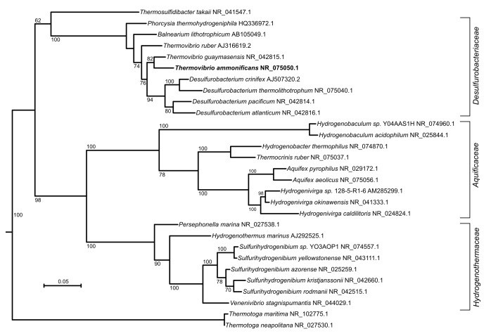
Thermovibrio ammonificans is part of the phylum Aquificae, a deep-branching group of thermophilic bacteria found in geothermal environments (Lebedinsky et al., 2007; Sievert and Vetriani, 2012). Based on phylogenetic analyses of the 16S rRNA gene as well as whole genomes, Aquificae are believed to be the earliest bacterial lineages having emerged on Earth along with the phyla Thermotogae and Thermodesulfobacteria (Di Giulio, 2003; Pitulle et al., 1994; Battistuzzi et al., 2004) (Figure 1, Figure 1—figure supplement 1). All the cultured members of this phylum are chemolithoautotrophs that use hydrogen as an energy source, have optimum growth temperatures between 65˚C and 95°C and rely on the reductive tricarboxylic acid cycle (rTCA) to convert carbon dioxide into biomass (Hügler et al., 2007; Hügler and Sievert, 2011; Sievert and Vetriani, 2012) (Table 1), making them ideal candidates to investigate the evolution of early metabolism.
Figure 1. Phylogenetic tree of 16S rRNA sequences computed from the current version of the aligned 16S rRNA database obtained from the arb-SILVA project (http://www.arb-silva.de/).
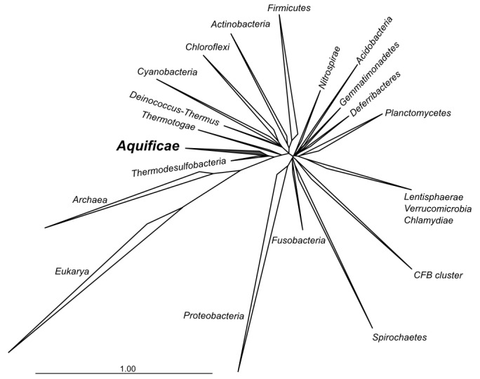
Figure 1—figure supplement 1. Maximum likelihood tree showing the 16S rRNA relationship of the Aquificae phylum.
Table 1.
Characteristic of representative members of Aquificae phylum.
| Family | Organism | Growth temp | Energy source | Carbon source | Terminal electron acceptor | Isolated from | Genome sequence accession number | References |
|---|---|---|---|---|---|---|---|---|
| Aquificaceae | Aquifex aeolicus VF5 | 95°C | H2 | CO2 | O2 | Underwater volcanic vents, Aeolic Islands Sicily, Italy | NC_000918.1 | (Deckert et al., 1998; Huber et al., 1992) |
| Hydrogenivirga sp.128–5 R1-11 | 75°C | S2O32-, S0 | CO2 | NO3-, O2 | Lau Basin hydrothermal vent area, Pacific Ocean | NZ_ABHJ00000000 | (Nakagawa et al., 2004; Reysenbach et al., 2009) | |
| Hydrogenobacter thermophilus TK-6 | 75°C | H2 | CO2 | O2 | Hot springs in Izu and Kyushu, Japan | NC_017161.1 | (Arai et al., 2010; Kawasumi et al., 1984) | |
| Hydrogenobaculum sp. Y04AAS1* | - | - | - | - | Marine hydrothermal area, Vulcano Island, Italy | NC_011126.1 | (Reysenbach et al., 2009; Stohr et al., 2001) | |
| Thermocrinis ruber DSM 12173 | 85°C | H2, S2O32-, S0 | CO2 | O2 | Octopus Spring, Yellowstone National Park, Wyoming, USA | PRJNA75073 | (Huber et al., 1998) | |
| Desulfurobacteriaceae | Balnearium lithotrophicum 17S | 70–75˚C | H2 | CO2 | S0 | Deep-sea hydrothermal vent chimney, Suiyo Seamount, Japan | Not sequenced | (Takai et al., 2003b) |
| Desulfurobacterium thermolithotrophum DSM 11699 | 70°C | H2 | CO2 | S0, S2O32-, SO2- | Deep-sea hydrothermal chimney, mid-Atlantic ridge, Atlantic Ocean | NC_015185.1 | (Göker et al., 2011; L'Haridon et al., 1998) | |
| Phorcysia thermohydrogeniphila HB-8 | 75°C | H2 | CO2 | S0, NO3- | Tube of Alvinella pompejana tubeworms, deep-sea hydrothermal vents 13 ˚N, East Pacific Rise, Pacific Ocean | Not sequenced | (Pérez-Rodríguez et al., 2012) | |
| Thermovibrio ammonificans HB-1 | 75°C | H2 | CO2 | S0, NO3- | Deep sea hydrothermal vents 9˚N, East Pacific Rise, Pacific Ocean | NC_014926.1 | (Giovannelli et al., 2012; Vetriani et al., 2004) | |
| Hydrogenothermaceae | Hydrogenothermus marinus VM1 | 65°C | H2 | CO2 | O2(1–2%) | Deep sea hydrothermal vents 9˚N, East Pacific Rise, Pacific Ocean | Not sequenced | (Stohr et al., 2001) |
| Persephonella marina EX-H1 | 73°C | S0 | CO2 | O2, NO3- | Deep sea hydrothermal vents 9˚N, East Pacific Rise, Pacific Ocean | NC_012440.1 | (Götz et al., 2002; Reysenbach et al., 2009) | |
| Sulfurihydrogenibium sp. YO3AOP11 | 70°C | S2O32-, S0 | CO2 | O2 | Calcite Hot Springs, Yellowstone National Park, USA | NC_010730.1 | (Reysenbach et al., 2009; Takai et al., 2003a) | |
| Venenivibrio stagnispumantis | 70°C | H2 | CO2 | O2 | Terrestrial hot spring Champagne Pool, Waiotapu, New Zealand | Not sequenced | (Hetzer et al., 2008) | |
| Incertae sedis | Thermosulfidibacter takai ABI70S6T | 70°C | H2 | CO2 | S0 | deep-sea hydrothermal field at Southern Okinawa Trough, Japan | Not sequenced | (Nunoura et al., 2008) |
*– Strain not formally described whose genome sequence is available. For these strains, the physiological information reported in Table S1 have been collected from MIG associated with the sequencing or from the closest validly published species.
The ability to synthesize new biomass from inorganic precursors, i.e. autotrophic carbon fixation, is a critical step in the global carbon cycle and is considered a key invention during the early evolution of metabolism (Braakman and Smith, 2012; Fuchs, 2011; Hügler and Sievert, 2011). Among the six known pathways of carbon fixation (reviewed in Fuchs, 2011; Hügler and Sievert, 2011), the rTCA cycle (present in the Aquificae, among others) and the reductive acetyl-CoA pathway (present in acetogenic bacteria and methanogens), represent good candidates for the ancestral carbon fixation pathway (Martin and Russell, 2003; Russell and Hall, 2006). The emergence of a reductive acetyl-CoA pathway has been associated with the FeS-rich minerals at alkaline hydrothermal vents (Martin and Russell, 2007, 2003; Wächtershäuser, 1988a) and the presence of homologous core enzymes in both Bacteria and Archaea potentially support its ancestral nature (Fuchs, 2011; Russell and Martin, 2004). The rTCA cycle is considered the modern version of a proposed prebiotic autocatalytic cycle fueled by the formation of the highly insoluble mineral pyrite in sulfur-rich hydrothermal environments (Wächtershäuser, 1988b, 1990). The rTCA cycle is widespread among anaerobic and microaerophilic bacteria including all Aquificae, chemolithoautotrophic Epsilonproteobacteria, Chlorobi, and Nitrospirae (Berg, 2011; Fuchs, 2011; Hügler and Sievert, 2011) in addition to few other bacterial strains. A recent phylometabolic reconstruction hypothesized that all extant pathways can be derived from an ancestral carbon fixation network consisting of a hybrid rTCA cycle / reductive acetyl-CoA pathway (Braakman and Smith, 2012).
Here, we used a comparative genomic approach coupled to proteomic analyses to reconstruct the central metabolism of Thermovibrio ammonificans (Giovannelli et al., 2012; Vetriani et al., 2004), and provide evidence of ancestral and acquired metabolic traits in the genome of this bacterium. We suggest that the last common ancestor within the Aquificae possessed the reductive acetyl-CoA pathway in addition to the previously described rTCA cycle. Furthermore, we show that these two pathways may still coexist within the Desulfurobacteraceae lineage. The simultaneous presence of both pathways of carbon fixation may represent a modern analog of the early carbon fixation phenotype, and suggests that the redundancy of central metabolic pathways could have been common in ancestral microorganisms.
Results
General genome structure and central metabolism of T. ammonificans.
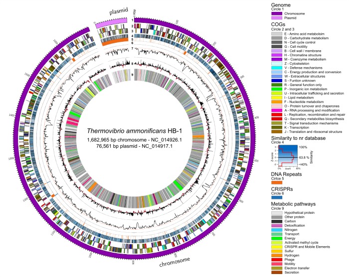
The genome of T. ammonificans HB-1 consists of one chromosome and one plasmid of 1,682,965 bp and 76,561 bp, respectively (Figure 2). The genome encodes for 1890 genes, 1831 of which are protein coding genes and 75.16% of the protein coding genes could be assigned a putative function (Giovannelli et al., 2012). The chromosome contains three ribosomal RNA operons, the first two with a 5 S-23S-16S alignment (coordinates 149,693–152,859 bp and 1,168,776–1,172,141 bp antisense strand, respectively) and the other with a 16 S-23S-5S (coordinates 1,604,527–1,609,585 bp). The second copy of the operon is flanked by the largest CRISPR found in the genome (Figure 2, circle 6). Several other repeats were identified in the chromosome (Figure 2, circle 5). The plasmid contained mainly ORFs with unknown hypothetical functions (Figure 2, circle 9) with the exception of a putative RNA polymerase sigma factor of probable archaeal origin, a DNA Topoisomerase I (43.2% similarity to Hydrogenivirga sp. 128–5 R1-6), a type II secretion protein E (identified also in the preliminary proteome, Figure 3, Figure 3—source data 1) and an ArsR-like regulatory protein, both of which had homologs among the Firmicutes. All other plasmid genes were ORFans with no significant similarities in the database and contained highly repeated DNA (Figure 2, circle 5).
Figure 2. Thermovibrio ammonificans HB-1 genomic map highlighting main genomic features.
The circular chromosome and plasmid are drawn together. Several other repeats were identified in the chromosome (circle 5). From the outer circle: 1 – Dimension of the genome in base pairs (chromosome in violet, plasmid in pink); 2 and 3 – sense and antisense coding sequences colored according to their COGs classification (see legend in the figure); 4 – similarity of each coding sequence with sequences in the non-redundant database; 5 – position of tandem repeat in the genome. A total of 46 tRNA were identified, comprising all of the basic 20 amino acid plus selenocysteine; 6 – position of CRISPRs; 7 – GC mol% content, the red line is the GC mean of the genome (52.1 mol%); 8 – GC skew calculated as G+C/A+T; 9 – localization of coding genes arbitrarily colored according to the metabolic pathways reconstructed (see Figure 1). Colors are consistent trough the entire paper. Total dimension and accession number for the chromosome and plasmid are given.
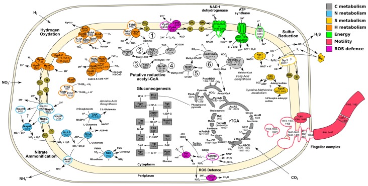
Figure 3. Central metabolism of T. ammonificans HB-1.
Enzyme names are reported together with the gene locus number (Theam_number). Primary compounds involved in reactions were also reported, however visualized reactions are not complete. Pathways were arbitrarily color coded according to their reconstructed function and are consistent throughout the paper. Solid shapes represent genes for which the enzyme was found in the proteome, while outlined shapes were only identified in the genome. Circled numbers 1 to 6 represent reaction numbers of the putative reductive acetyl-CoA pathway as described in the text. Abbreviations: NITRATE AMMONIFICATION: NapCMADGH – Periplasmic nitrate reductase complex; NirA – Putative nitrite reductase; AmtB – ammonia transporter; GlnA – L-Glutamine synthetase; GltA – Glutamate synthetase. HYDROGEN OXIDATION: HynABC – Ni-Fe Membrane bound hydrogenase; FrhACB – Cytosolic Ni-Fe hydrogenase/putative coenzyme F420 hydrogenase; HupD – Cyrosolic Ni-Fe hydrogenase maturation protease; HypA-F – Hydrogenases expression/synthesis accessory proteins; EchABCEF – Ech membrane bound hydrogenase complex; HydAB – Cytosolic Ni-Fe hydrogenases potentially involved in ferredoxin reduction; Hdr? – Missing heterodisulfide reductase CoB-CoM; FdhABC – Formate dehydrogenase. ENERGY PRODUCTION: NADH dehydrogenase and ATP synthetase are reported without the names of the single units; Nhe – Sodium/hydrogen symporter. SULFUR REDUCTION: Sqr – Putative sulfate quinone reductase involved in sulfur respiration; Nsr? – FAD/NAD nucleotide-disulphide oxidoreductase; Aps – Sulfate adenylyl transferase and kinase involved in assimilation of sulfur. FLAGELLAR COMPLEX: for simplicity single unit names are not reported. REDUCTIVE ACETYL-CoA: Fhs – Proposed reverse formyl-THF deformylase; PurHT – Formyltransferases (phosphoribosylaminoimidazolecarboxamide and phosphoribosylglycinamide) FolD – Methenyl-THF cyclohydrolase and dehydrogenase; MetVF – Methylene-THF reductase; MetR – Putative methyl-transferase; CodH – Carbon monoxyde dehydrogenase; AcsA – Acetyl-CoA ligase/synthase; AccABCD – Acetyl-CoA carboxylase. REDUCTIVE CITRIC ACID CYCLE: AclAB – ATP-citrate lyase; Mdh – Malate dehydrogenase; FumAB – Fumarate hydratase; FrdAB – Fumarate reductase; SucCD – Succinyl-CoA synthetase; OorABCD – 2-Oxoglutarate synthase; Idh2 – Isocitrate dehydrogenase/2-oxoglutarate carboxylase; AcnB – Aconitate hydratase; PorABDG – Pyruvate synthase; PycAB - Pyruvate carboxylase; PpsA – Phosphoenolpyruvate synthase water dikinase; PyK – Pyruvate:water dikinase. GLUCONEOGENESIS: Eno – Enolase; Pgm – Phosphoglycerate mutase; Pgk – Phosphoglycerate kinase; Gapor – Glyceraldehyde-3-phosphate dehydrogenase; Gapdh – Glyceraldehyde 3-phosphate dehydrogenase; Fba – Predicted fructose-bisphosphate aldolase; Tim – Triosephosphate isomerase; Fbp – Fructose-1,6-bisphosphatase I; Pgi – Phosphoglucose isomerase; Pgm – Phosphoglucomutase.
DOI: http://dx.doi.org/10.7554/eLife.18990.007
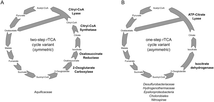
Figure 3—figure supplement 1. Reductive TCA cycle variants found in extant bacterial lineages.
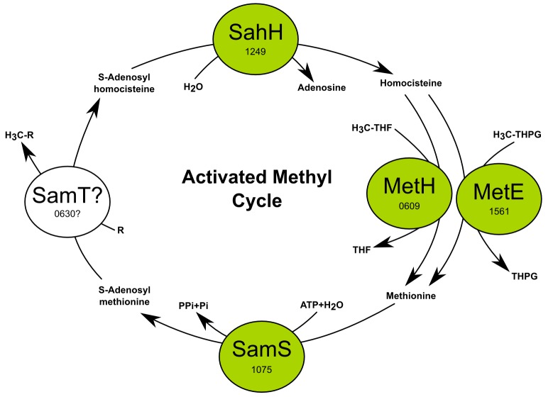
Figure 3—figure supplement 2. The activated methyl cycle of T. ammonificans reconstructed from the genome.
By combining genome-scale analyses with known physiological traits, we were able to reconstruct the metabolic network of T. ammonificans (Figure 3). As part of the central metabolism of T. ammonificans, we identified the major pathways for carbon fixation, hydrogen oxidation and nitrate reduction to ammonia, and confirmed them by identifying the corresponding expressed enzymes in the proteome (Figure 3, Figure 3—source data 1). Further, we compared the central metabolism of T. ammonificans with closely related species within the Aquificae, and with ecologically similar Epsilonproteobacteria. Comparative genomic analyses with closely related strains provide information about the evolutionary history of the group, while comparison with phylogenetically distant, but ecologically similar species may reveal common adaptive strategies to similar habitats.
The proteome of T. ammonificans
780 proteins, comprising approximately 43% of all predicted ORFs in the database, were identified in cell samples of T. ammonificans grown under nitrate reducing conditions (Figure 3, Figure 3—source data 1), indicating a very high congruence between database entries and sample peptides. Among the most abundant proteins were – besides those involved in general housekeeping functions like the translation elongation factor Tu, ribosomal proteins and chaperones – many rTCA cycle enzymes. The four key enzymes of the rTCA cycle (see next paragraph for details) together yielded a relative abundance of 6% of all identified proteins in the samples. Most of the enzymes putatively involved in the reductive acetyl-CoA pathway (Table 2), including the carbon-monoxide dehydrogenase (CodH), were also detected, although at a lower abundance of 1.25% of all proteins.
Table 2.
Enzymes involved in the putative reductive acetyl-CoA pathway in T. ammonificans HB-1, closest relative homolog and homologs within the Aquificae phylum.
|
Desulfurobac
teriaceae |
Hydrogenothe
rmaceae |
Aquificaceae | |||||||||||
|---|---|---|---|---|---|---|---|---|---|---|---|---|---|
| Reaction |
Putative
enzyme |
Putative gene locus | Closest relative |
Putative
origin‡ |
D. thermolitho
trophum |
Desulfuro
bacterium sp. TC5-1 |
P. marina |
Sulfurihydro
genibium sp. YO3AOP1 |
H. themophilus |
Hydrogeno
baculum sp. Y04AAS1H |
Hydrogeni
virga sp. 128–5 R1-6 |
T. ruber | A. aeolicus |
| 1 | Formate dehydrogenase |
Theam_1020 |
Nitratiruptor
sp. SB155-2 sim. 55% YP_001357016 |
Methanogens | f.e.§ - 30% YP_00428203# |
-** | 56% YP_002730364 | f.e.§ - 32% YP_001930236 | f.e.§ - 33% YP_003433330 | - | - | f.e.§ - 27% YP_003474076 | - |
| 2* | 5-formyltetra hydrofolate cyclo-ligase |
Theam_1206 |
Hydrogenivirga
sp. 128–5 R1-1 sim. 41% WP_008285842 |
Deltaproteo
bacteria / Gram + |
41% YP_004281339 | 44% WP_022846876.1 | f.e.§ - 36% YP_002729868 | f.e.§ - 38% YP_001931834 | f.e.§ - 38% YP_003431710 | f.e.§ - 30% YP_002121539 | 41% WP_008285842 | 40% YP_003472981 | 43% 1SOU_A |
| formyltetra hydrofolate deformylase / hydrolase |
Theam_0826 |
Hydrogenivirga
sp. 128–5 R1-1 sim. 70% WP_008287030 |
Bacteria | 82% YP_004281077 | 77% WP_022846479 | - | - | - | - | 70% WP_008287030 | 65% NP_214247 | - | |
| phosphori bosylglycina mide formyl transferase 2 |
Theam_1211 |
P. marina
sim. 76% YP_002731257 |
Methanogens
/ Deltaproteo bacteria |
78% YP_004281335 | 64% WP_022847512 | 76% YP_002731257 | 73% YP_001931730 | f.e.§ - 28% YP_003433072 | 67% YP_007499479 | 74% WP_008288476 | f.e.§ - 25% YP_003473050 | f.e.§ - 30% NP_213168 | |
| phosphori bosylaminoi midazole carboxamide formyltransferase /IMP cyclohydrolase |
Theam_0328 |
P. marina
sim. 59% YP_002731268 |
Aquificae /
Bacteria |
86% YP_004281681 | 78% WP_022847359 | 59% YP_002731268 | 56% YP_001931239 | 53% YP_003433270 | 50% YP_007499473 | 57% WP_008288360 | 52% YP_003474337 | 56% NP_214344 | |
| 3 | methylenetetra hydrofolate dehydrogenase / methenyltetra hydrofolate cyclohydrolase |
Theam_1261 |
A. aeolicus sim. 60% NP_214304 |
Aquificae / Gram + |
87% YP_004281147 | 74% WP_022846217 | 55% YP_002731476 | 58% YP_001931240 | 59% YP_005511318 | 58% YP_001931240 | - | 58% YP_003473104 | 60% NP_214304 |
| 4 | methylenetetra hydrofolate reductase |
Theam_0919 |
Methanosrcina barkeri sim. 48% YP_305816 |
Methanogens | 87% YP_004281147 | 74% WP_022846217 | 55% YP_002731476 | 58% YP_001931240 | 59% YP_005511318 | 58% YP_002121558 | f.e.§ - 24% WP_008286783 | 58% YP_003473104 | 60% NP_214304 |
| 5 | 5-methyltetra hydrofolate corrinoid/iron sulfurprotein methyltransferase |
Theam_1184 |
Clostridium glycolicum
sim. 42% WP_018589788 |
Gram + | 82% YP_004281484 | 72% WP_022846603 | f.e.§ - 31% YP_002729885 | f.e.§ - 38% YP_001930381 | f.e.§ - 37% YP_003432344 | f.e.§ - 34% YP_002122031 | f.e.§ - 38% WP_008287326 | f.e.§ - 36% YP_003473438 | f.e.§ - 34% NP_213375 |
| 6† | Carbon-monoxide dehydrogenase |
Theam_1337 |
Candidatus Methano peredens sp. BLZ1 sim. 56% KPQ43483 |
Archaea / Gram + | 87% WP_013638427 38% WP_013638030 |
76% WP_022847143 | 41% YP_002729904 | - | - | - | - | - | - |
* – Reactions are numbered according to Figure 1. For reaction two we reported the possible enzymes that could substitute the missing 10-fomyl-THF synthetase (Fhs). The enzymes are listed in order of decreasing likelihood of their involvement in the reaction based on putative substrate affinity.
† – Reaction six is catalyzed by the Acs/CodH complex, reported here separately.
‡ – Putative origin of the gene was calculated as consensus taxonomic assignment of the first 100 bastp hits against the nr database. Double assignment implies equal number of assignments to the two taxonomic groups.
§ – f.e. = functional equivalent annotated in the genome with similarity below 40% with T. ammonificans equivalent gene. Not considered a true homolog in the present study.
# – Pairwise similarity to T. ammonificans translated gene and accession number of the homologs.
** – Missing homolog or functional annotated equivalent in the genome.
Carbon fixation
The genome and proteome of T. ammonificans (Figure 3) supports previous evidence, based on detection of key genes and measurements of enzyme activities, that carbon fixation occurs via the rTCA cycle (Hügler et al., 2007). The rTCA is widespread among anaerobic and microaerophilic bacteria including all Aquificae, chemolithoautotrophic Epsilonproteobacteria, Chlorobi, Nitrospina aaaand Nitrospirae, in addition to few other bacterial strains (e.g. Desulfobacter hydrogenophilus, Magnetococcus marinus MC-1 and the endosymbiont of the vent tubeworm, Riftia pachyptila; Hügler and Sievert, 2011). We identified all the genes responsible for the functioning of the rTCA in the genome and proteome of T. ammonificans (Figure 3), including the key enzymes fumarate reductase (frdAB, gene loci Theam_1270–1275), ATP-citrate (pro-S)-lyase (aclAB, 1021–1022), 2-oxoglutarate synthase (oorABCD, 1410–1413) and isocitrate dehydrogenase (idh2, 1023). T. ammonificans synthesizes 2-oxoglutarate from succinyl-CoA rather than via citrate, a feature in common with methanogenic Archaea and Desulfurococcales, and present in numerous strict anaerobes that use the rTCA cycle (Fuchs, 2011). Comparative analyses showed that several genes associated with the rTCA cycle and the gluconeogenesis pathway were highly conserved in all Aquificae, with the exception of the genes coding for the ATP-citrate lyase and the isocitrate-dehydrogenase, which are absent in the Aquificaceae (see details below) (Hügler et al., 2007). In general, T. ammonificans genes had a high percent identity to the genes of D. thermolithotrophum, its closest relative whose genome sequence is available.
Two separate variants of the rTCA cycle are known (Braakman and Smith, 2012; Hügler et al., 2007). In the two-step variant – also known as symmetric variant, with respect to the enzymatic reaction catalyzed in the two arcs of the rTCA cycle – citrate cleavage is accomplished by the combined action of the enzymes citryl-CoA synthetase (CCS) and citryl-CoA lyase (CCL) and the carboxylation of 2-oxoglutarate is catalyzed by 2-oxoglutarate carboxylase and oxalosuccinate reductase (Aoshima, 2007; Aoshima and Igarashi, 2008; Hügler and Sievert, 2011). This variant of the rTCA cycle was previously proposed as ancestral (Braakman and Smith, 2012; Hügler et al., 2007). By contrast, in the one-step variant of the rTCA cycle, also known as asymmetric variant and more widely distributed, the cleavage of citrate and the carboxylation of 2-oxoglutarate are performed in a one-step fashion (Braakman and Smith, 2012; Hügler et al., 2007)(Figure 3, Figure 3—figure supplement 1). This variant is found in T. ammonificans.
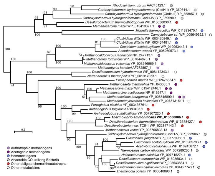
The genome of T. ammonificans also codes for an incomplete reductive acetyl-CoA pathway (Figure 3 and Table 2). The key enzyme of this carbon fixation pathway, which is found in acetogens, methanogens, sulfate-reducers and anaerobic ammonium oxidizers (anammox; Berg et al., 2010), is the bifunctional heterotetramer enzyme carbon monoxide dehydrogenase/acetyl-CoA synthase (CODH/ACS). The reductive acetyl-CoA pathway is believed to be the most ancient carbon fixation pathway on Earth (Fuchs, 2011). In T. ammonificans, the genes coding for the CO-dehydrogenase (codh catalytic subunit Theam_1337; Table 2 and Figure 3) is present and also expressed. However, rather than coding for the classical bifunctional type II CodH, T. ammonificans codes for the type V, which is present in numerous deep-branching organisms (Figure 4) and whose function is unknown (Techtmann et al., 2012). Sequence analysis of the T. ammonificans CodH protein sequence revealed the presence of conserved residues necessary for the interaction with Acs and for the coordination of the NiFeS cluster. We hypothesize that the type V CodH, which, based on structural and size considerations may represent the most ancestral version of this class of enzymes (Frank Robb, personal communication), may catalyze the reduction of CO2 to CO. The codh gene of T. ammonificans shared high similarity with that of D. thermolithotrophum (87 similarity), while the similarity with the next closest homologous genes dropped below 54%. The genome of D. thermolithotrophum also codes for a classical bifunctional Type II CodH (Dester_0417 and Dester_0418; Figure 4) in addition to the type V CodH. The two genomes also share a CodH iron-sulfur accessory protein (Theam_0999 and Dester_0418, 81% similarity). Additional laboratory analyses will be required to confirm the role of the type V CodH in carbon fixation.
Figure 4. Neighbor-joining tree showing the position of the carbon monoxide dehydrogenase, CodH, of T. ammonificans (in bold).
Bootstrap values based on 1000 replications are shown at branch nodes. Only bootstrap values above 50% are reported. Bar, 10% estimated substitutions. The metabolism of each organism is reported on the side.
Gluconeogenesis
T. ammonificans uses the Embden-Meyerhof-Parnas pathway to synthesize pentose and hexose monosaccharides. We identified in the genome the key genes of this pathway, fructose-1,6-biphosphatase (fbp, Theam_1323; identified also in the proteome, Figure 3) and all other genes necessary for the functioning of the pathway (Figure 3). While the fbp gene is present in Desulfurobacterium thermolithotrophum, and in the genome of Sulfurohydrogenibium sp. YO3AOP1, Persephonella marina and Hydrogenivirga sp. 128–5 R1-6, it is absent from the genome of A. aeolicus (Deckert et al., 1998) and H. thermophilus, suggesting that in those species an alternative unidentified pathway may be active.
Hydrogen oxidation
Hydrogen is the only electron donor used by T. ammonificans and its importance in the metabolism of this bacterium is reflected by the diversity of hydrogenases found in the genome, whose encoding genes are organized in two large clusters (Figure 2 circle 9). The first cluster is composed of eight genes encoding for the complete Group 4 Ech hydrogenases (echABCDEFG, Theam_0476–0482) and the alpha, beta, gamma and delta subunits of the Group 3b multimeric cytoplasmic Hyd sulfhydrogenase (hydABCD, Theam_0484–0487). The second hydrogenase cluster in the genome codes for the maturation protein HypF (hypF, Theam_1116), the three subunit of the Group 3a cytoplasmic F420-reducing hydrogenase Frh (frhACB, Theam_1117–1119) and the Group 1 hydrogenases Hyn (hynABC, Theam_1121–1123) with associated maturation factors/chaperones Hyp (Theam_1124–1128). We also found other hydrogenase maturation factors (Theam_0922–0924) and the formate dehydrogenase Fdh (fdhABC, Theam_1020 and Theam_0966–0967 respectively) scattered in the genome.
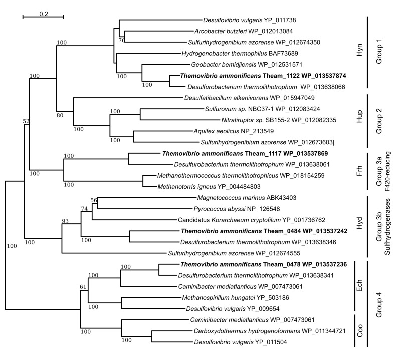
We reconstructed the phylogeny of T. ammonificans [NiFe]-hydrogenases (Figure 5), and we assigned them to known classes of hydrogenases (Groups 1 through 4) according to their phylogenetic position in the tree and the presence of known conserved amino acid motifs typical of each group (Vignais and Billoud, 2007). Group 1 [NiFe]-hydrogenases are normally coupled to anaerobic respiration through electron transfer to the membrane quinone and menaquinone pool. Comparative analyses of the hydrogenases revealed that the Group 1 cluster in T. ammonificans (Hyn) is more similar to the hydrogenases found in Deltaproteobacteria than to homologs in other Aquificae (Figure 5). This finding raises interesting questions about the origin of the hydrogen oxidation Group 1 enzymes in T. ammonificans and suggests that an event of lateral gene transfer occurred between T. ammonificans and the Deltaproteobacteria. Further, the periplasmic nitrate reductase (napA) and the nap operon structure in T. ammonificans also appears to be closely related to that of Deltaproteobacteria, suggesting that a horizontal gene transfer event involved the entire hydrogen oxidation (Hyn)/nitrate reduction respiratory pathway (see discussion).
Figure 5. Neighbor-Joining tree showing the position and classification of the [NiFe]-hydrogenases found in the genome of T.ammonificans.
Bootstrap values based on 1000 replication are shown at branch node. Bar, 20% estimated substitution.
Contrarily to members of the Aquificaceae and Hydrogenothermaceae, T. ammonificans and D. thermolithotrophum genomes code for both Group 3 cytoplasmic hydrogenases, Group 3a F420-reducing hydrogenases (frhACB) and Group 3b multimeric cytoplasmic sulfhydrogenases (hydABCD; Figure 5; Jeon et al., 2015). Comparative analyses of these two clusters revealed that other members of the Desulfurobacteriaceae family were the closest relatives, while the next closest enzymes were those found in hyperthermophilic Euryarchaeota (i.e. Pyrococcus abyssi) and in methanogens (Figure 5). Overall, these results imply a polyphyletic origin for the group 3 cytoplasmic hydrogenases. Comparative analyses revealed similarities with hyperthermophilic Euryarchaeota and methanogens (Figure 5), suggesting a possible common ancestry for the group 3 cytoplasmic hydrogenases of Archaea and Desulfurobacteriaceae. Together with Group 3b sulfhydrogenases and Group 4 Ech, F420-reducing hydrogenases are in fact typically found in methanogens in which the F420-cofactor is used as an electron carrier in the reduction of CO2 or other C1 compounds to methane (Vignais and Billoud, 2007). Cultures of T. ammonificans grown with nitrate as the terminal electron acceptor did not present the typical autofluorescence of methanogens due to F420-cofactor fluorescence when observed under UV in epifluorescence microscopy and the genome lacks the gene involved in the biosynthetic pathway of the F420-coenzyme. Due to these observations, we suggest that these hydrogenases may work in conjunction with ferredoxin in T. ammonificans. Group 3b Hyd sulfhydrogenases appear to be of mixed origins despite being present in a single operon. Three of the subunits share similarities with the Thermococcales (Euryarchaeota; hydBCD) while the catalytic subunit (HydA) appears to be unique to the Desulfurobacteriacea, not having any other significant homolog in the database (Figure 6). Comparative analyses indicated that Ech shared similarity with Epsilonproteobacteria Group 4 hydrogenases (Figure 5). Interestingly, Group 2 hydrogenases are found in microaerobic members of the Aquificaceae and Hydrogenothermaceae and in microaerobic Epsiloproteobacteria but are not found in anaerobes, suggesting a possible link with oxygen adaptation. In contrast, Group 3a is found in strict anaerobes, including T. ammonificans and methanogens (Figure 5).
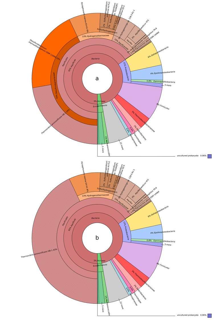
Figure 6. Blastp best hit (cut off at 40% similarity) for the T. ammonificans CDS: (a) Desulfurobacterium thermolithotrophum is included in the database; (b) D. thermolithotrophum and Desulfurobacterium sp.
TC5-1 are excluded from the database (best hits are outside of the Desulfurobacteriaceae family). The interactive versions of the Krona plots are available for download at DOI: 10.6084/m9.figshare.3178528.
Finally, Ech hydrogenases are the only Group 4 hydrogenases found in T. ammonificans. Both the reductive TCA cycle and the reductive acetyl-CoA pathway involve reaction steps that are driven by reduced ferredoxin (Fuchs, 2011). In the case of T. ammonificans, the enzymes formate dehydrogenase, CO-dehydrogenase, pyruvate synthase and 2-oxoglutarate synthase require reduced ferredoxin. We hypothesize that T. ammonificans can accomplish ferredoxin reduction in two different ways: the first involves cytosolic hydrogenases (Group 3, Frh and Hyd; Figure 3) commonly found in methanogens, where they reduce ferredoxin via electron bifurcation (Fuchs, 2011); the second involves membrane bound hydrogenases (Group 4, Ech; Figure 3). With the exception of the F420-reducing (Frh) hydrogenases, representatives of the remaining three groups were identified in the proteome of T. ammonificans (Figure 3).
Nitrate ammonification
T. ammonificans HB-1 can use nitrate as a terminal electron acceptor and reduces it to ammonium (Vetriani et al., 2004). Periplasmic nitrate reductases (Nap) have been studied extensively in some Proteobacteria, and are comprised of the catalytic enzymes – typically a heterodimer (NapAB) – associated with various other subunits involved in channeling electrons to the NapA reactive center (Moreno-Vivián et al., 1999; Vetriani et al., 2014). In T. ammonificans, the genes encoding for the Nap complex are organized in a single operon, napCMADGH. Comparative genomic analyses with genomes from Aquificae and Proteobacteria indicate that the nap operon structure of T. ammonificans is unique to this organism, and shares similarity with Desulfovibrio desulfuricans and other Deltaproteobacteria. All other Aquificae whose genomes are available have a Nap operon similar to that of Epsilon- and Gammaproteobacteria (Vetriani et al., 2014). Moreover, unlike other members of the Aquificae, the nitrate reductase of T. ammonificans (encoded by gene Theam_0423, expressed in the proteome; Figure 3) appears to be of the monomeric type (sensu; Jepson et al., 2006), and the NapB subunit is missing (Figure 3). We also identified a putative nitrite reductase-encoding gene possibly involved in the reduction of nitrite to ammonium (NirA; Theam_1000, Figure 3), which was also expressed in the proteome (Figure 3). However, the exact mechanism through which T. ammonificans reduces nitrite to ammonium remains to be experimentally elucidated.
Sulfur reduction
While T. ammonificans is able to reduce elemental sulfur to hydrogen sulfide (Vetriani et al., 2004), the sulfur reduction pathway remains unclear. We identified in the genome a putative polysulfide reductase of the sulfide-quinone reductase family (sqr, Theam_0841, Figure 3). We propose that this membrane-bound complex can utilize polysulfide formed from the reaction of S0 with sulfide (naturally enriched in hydrothermal systems) via quinone oxidation (Figure 3). The putative sqr gene of T. ammonificans shares similarities with other Aquificae and Epsilonproteobacteria (average 56% similarity to both groups). In particular, the sqr gene appears to be conserved also in Caminibacter mediatlanticus (Figure 7; Giovannelli et al., 2011), whose sulfur reduction pathway is yet to be elucidated. Another possibility is that T. ammonificans uses a NAD or FAD-dependent reductase (nsr, Theam_1321) to reduce sulfur, a mechanism previously described in the sulfur-reducing archaeon Pyrococcus furiosus (Schut et al., 2013, 2007). However, the putative polysulfide reductase of T. ammonificans has only about 33% identity to homologous enzymes of sulfur reducing bacteria and archaea identified in the database.
Figure 7. Hives plot presenting the comparative genomic analysis of the T. ammonificans (Tam) genome with the closest relative available genomes of D. thermolithotrophum (Des) and the ecologically similar Epsilonproteobacterium C. mediatlanticus (Cam).
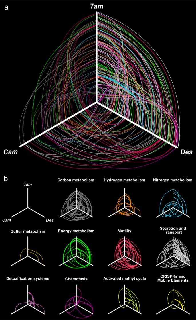
The three axes represent the organism’s linearized genomes with the origin of replication at the center of the figure. Genome size was normalized for visualization purpose. (A) Localization of homologous genes and syntenic regions among the three genomes. Lines colored according to the general pathway code adopted in Figure 3 connect homologous coding sequences. Lines opacity is proportional to similarity between sequences (darker = higher similarity), line width is proportional to the extent of the syntenic area. (B) Hives panel of the homologous genes divided by pathways. Collinear lastZ alignments and interactive dot-plot replicating the analysis are accessible at the GenomeEvolution website with the following permanent addresses: T. ammonificans and D. thermolithotrophum (http://genomevolution.org/r/9vb3); T. ammonificans and C. mediatlanticus (http://genomevolution.org/r/9vb4); D. thermolithotrophum and C. mediatlanticus (http://genomevolution.org/r/9vb8).
Resistance to oxidative stress
T. ammonificans is a strict anaerobe. However, its genome codes for genes involved in the detoxification of oxygen radicals, including a catalase/peroxidase (Theam_0186), whose activity was previously detected (Vetriani et al., 2004), a putative superoxide reductase (Theam_0447), a cytochrome-C peroxidase (Theam_1156) and a cytochrome bd complex (Theam_0494–0496, Figure 3). The latter three enzymes were identified in the proteome (Figure 3). The cythochrome bd complex has been shown to contribute to oxygen tolerance in other anaerobic bacteria (Das et al., 2005), and to contribute to the detoxification of nitrous oxide radicals (Mason et al., 2009).
Energy conversion
The genome of T. ammonificans contains two gene clusters coding for the NADH dehydrogenase (Theam_0731–0745 and Theam_1488–1498). The genes appear to have undergone an inversion during the duplication process and the second copy is missing four genes in the middle of the operon (nuoEFG and a transcriptional regulator). The two clusters share on average 60% gene similarity. Only one copy of the gene for the synthesis of ATP synthetase complex was present (Theam_1605–1606 and Theam_1656–1662). Three different hydrogen-sodium symporters (nhe, Theam_0443, 0413 and 0647) were present with low pairwise similarity. NADH dehydrogenase second cluster and ATP synthetase complex appear to be conserved in the close relative D. thermolithotrophum and in the Epsilonproteobacterium, C. mediatlanticus TB-2 (Figure 3).
Motility, cell sensing and biofilm formation
T. ammonificans posses one to two terminal flagella (Vetriani et al., 2004). The genes involved in flagella formation and assembly are prevalently organized in a single large cluster in the genome (from Theam_1440 to Theam_1468, Figure 7), putatively organized in three distinct operons. The hook-filament junction proteins genes flgK and flgL (Theam_1384 and Theam_1385), the filament and filament cap proteins fliC and fliD (Theam_1160 and Theam_1162) together with few flagellar maturation factors and motor switch genes (motA and motB, Theam_1087 and Theam_1085–1086, respectively) are instead scattered in the genome. The entire cluster of genes shares similarities with D. thermolitotrophum, although the similarity for some of the proteins is as low as 50%. Despite this, comparative analyses revealed a similar organization in D. thermolitotrophum where the cluster appears inverted in the same relative genomic position and constituted one of the main region of synteny (Figure 7b). By contrast, the flagellar genes in the Epsilonproteobacterium, Caminibacter mediatlanticus TB-2, are scattered throughout the genome (Figure 7b). Numerous genes for adhesion and pili were present, generally organized in small clusters (Figure 7b).
Four different putative methyl-accepting chemotaxis sensory transducer proteins were found in the genome (mcp, Theam_0157, 0165, 0845 and 1027). One of those, Theam_0845, is surrounded by receptors cheW and cheA, and by the response regulator cheY. The entire group is organized in a single operon, cheYVBWAZ conserved in D. thermolithotrophum and other Aquificales with the exception of cheW, which shares higher similarities with the Epsilonproteobacterium, C. mediatlanticus TB-2 (Figure 6). We failed to find in the genome of T. ammonificans a homolog of the primary receptor encoding gene cheR (Wadhams and Armitage, 2004). We identified in the genome numerous membrane transporters for molybdenum (modCBA, Theam_0787–0789), iron (fhuDBC, Theam_0192–0194), zinc (znuAB, Theam_0238 and 0689) and cobalt/nickel (cbiOQMN, Theam_1522, 1523, 1525 and 1526) all involved in the uptake of important micro-elements for enzyme and cofactor synthesis. We also identified the complete type II secretion pathway, which is conserved in gram-negative bacteria and responsible for protein and toxin translocation to the extracellular milieu. Proteins secreted by the type II pathway include proteases, cellulases, pectinases, phospholipases, lipases, toxins and in some cases type III and IV pili (Douzi et al., 2012). In some bacteria, the expression of secretion type II pathway genes are regulated either by quorum-sensing or by the environmental factors at the site of colonization (Douzi et al., 2012; Sandkvist, 2001). Biofilm formation in bacteria is often linked to quorum-sensing mechanisms, regulating the settlement of planktonic cells in relationship to environmental sensing, substrate suitability and cell densities. It is likely that T. ammonificans maintains a mostly attached lifestyle in hydrothermal vent environments due to the high turbulence associated with fluid flux and mixing with seawater. We speculate that the type II pathway genes, together with chemotaxis and flagellar/adhesion genes may play a key role in the formation of biofilm. Extracellular secreted proteins may in fact be used to ‘condition’ the colonized substrate and to interact/remodel existing and newly formed biofilms.
Activated methyl cycle
The activated methyl cycle is a conserved pathway in which S-methyl groups from L-methionine are converted into a biochemically reactive form through insertion into an adenosyl group (Vendeville et al., 2005). S-adenosyl-L-methionine provides activated methyl groups for use in the methylation of proteins, RNA, DNA and certain metabolites central to the core cell machinery. The activated methyl cycle exists in two recognized forms, involving respectively luxS or sahH genes (Vendeville et al., 2005). Genome-based reconstruction of the activated methyl cycle indicated that, in T. ammonificans, involves the sahH gene, while a luxS homolog is absent (Figure 3—figure supplement 2). Methionine methyl groups are adenosylated by a methionine adenosyltransferase (samS, Theam_1075). The methyl group is thus activated and readily available for the S-adenosylmethionine-dependent methyltransferase (samT) for methylation of substrate. In T. ammonificans, samT is a pseudogene, as it contains two stop codons (Theam_0630). Homologs of samT are absent in the genome of the other Aquificae and the pathway appears to be incomplete. Only in P. marina and D. themolithotrophum the functionally equivalent DNA-cytosine methyltransferase (dcm) is present. The presence of a gap in such a central pathway in the Aquificae and the retrieval of a samT-like pseudogene in T. ammonificans raises interesting question on the functioning of the activated methyl cycle in those organisms. An interesting working hypothesis is that enzyme thermal stability at the physiological temperature of these bacteria might have driven the loss of samT and the replacement by a not yet identified alternative enzyme in most Aquificae.
Discussion
Evidence for a reductive acetyl-CoA pathway in the Desulfurobacteraceae lineage
The reductive acetyl-CoA pathway is present in acetogenic bacteria, methanogens, sulfate-reducers and anammox bacteria (Berg, 2011; Fuchs, 2011; Hügler and Sievert, 2011). It is believed to be the oldest carbon fixation pathway on Earth, due to the centrality of acetyl-CoA in metabolism, the low energy requirement (~1 ATP required vs 2–3 ATP for the rTCA cycle) and the limited necessity of de novo protein assembly (Berg et al., 2010; Fuchs, 2011). We hypothesize that the reductive acetyl-CoA pathway is used as an additional or alternative pathway to fix CO2 in T. ammonificans.
The finding of a potentially active reductive acetyl-CoA pathway (reactions 1 to 6; Figure 3) in T. ammonificans and other members of the Desulfurobacteraceae is intriguing. The pathway is missing reaction 2, the formyl-THF synthesis for which no obvious 10-formyl-THF synthetase was found (Figure 3). However, the 10-formyl-THF synthetase of the reductive acetyl-CoA pathway may have been replaced by alternative enzymes, such as 5-formyl-THF cycloligase (Theam_1206) or 10-formyl-THF deformylase (Theam_0826) working in reverse (Table 2, Figure 3). Promiscuous enzymes that function in more than one pathway have been long hypothesized and recently described in Escherichia coli (Jensen, 1976; Kim et al., 2010). Assuming low substrate specificity, other candidate enzymes with similar substrate requirements are the phosphoribosylglycinamide formyltransferase (Theam_1211) or phosphoribosylaminoimidazolecarboxamide formyltransferase/IMP cyclohydrolase (Theam_0328), which could catalyze the synthesis of formyl-THF, albeit with sub-optimal kinetics. While all four enzymes were detected in the proteomic analysis (Figure 3), we think that the possible involvement of a 5-formyl-THF cycloligase in the synthesis of formyl-THF in T. ammonificans is particularly appealing, as this would provide an evolutionary link between the N5-formate uptake of methanogens and N10-formate uptake in acetogens (Braakman and Smith, 2012). Despite the absence of the 10-formyl-THF synthetase, the genome of T. ammonificans encodes and expresses a type V CodH (Figure 3).
We investigated the available genomes of other members of the Aquificae for the presence of genes homologous to the putative reductive acetyl-CoA pathway of T. ammonificans (Table 2). The similarity of the reductive acetyl-CoA pathway-related genes of T. ammonificans to those from organisms outside the Desulfurobacteriaceae is low. The type V codH gene identified in T. ammonificans is found exclusively in members of the family Desulfurobacteriaceae, with exception of a low similarity homolog present in Persephonella marina (41% amino acid identity) (Table 2). Furthermore, most of the genes involved in reaction 3, 4 and 5 (Figure 3) have homologs in other Aquificae or are substituted by functional equivalents with low amino acid identity (<40%). To understand the relationship of the T. ammonificans CodH with the homologous enzymes of methanogens, homoacetogens and sulfate-reducers, we reconstructed the phylogenetic history of these enzymes with a particular focus on type V CodH (Figure 4). Sequences from the Desulfurobacteriaceae appear to be related to those present in the Archaea, Ferroglobus placidus and Archaeoglobus spp., and similar to Methanosarcina spp. and homoacetogenic bacteria. The CodH found in CO-utilizing bacteria is only distantly related to the one found in T. ammonificans (Techtmann et al., 2012), while the CodH sequence of Persephonella marina appears to be one of a kind, with the enzyme of Ferroglobus placidus as its closest relative (44% similarity). Taken together, these findings suggest that, within the Aquificae, the type V CodH is exclusive to the Desulfurobacteriaceae.
The retrieval of the CodH enzyme from the proteome of T. ammonificans (Figure 3, Figure 3—source data 1), along with the catalytic subunit of the formate dehydrogenase (FdhA), suggests that the reductive acetyl-CoA pathway could be operational in T. ammonificans in addition to the rTCA cycle. The simultaneous presence of two distinct carbon fixation pathways has been confirmed so far only in the endosymbiont of the deep-sea vent tubeworm Riftia pachyptila (Markert et al., 2007). Recent findings suggest that the Riftia symbiont can switch from the Calvin-Benson-Bassham (CBB) to the rTCA cycles in response to low-energy supply (e.g., low concentrations of hydrogen sulfide in the vent fluids, being the rTCA cycle more energetically favorable than the CBB cycle; Markert et al., 2007). Genomic evidence of the simultaneous presence of different carbon fixation pathways came also from genome analysis of Ammonifex degensii and Ferroglobus placidus (Berg, 2011; Hügler and Sievert, 2011), where the reductive acetyl-CoA pathway seems to be coupled to an incomplete CBB cycle. The use of different carbon fixation strategies could be advantageous under energy limiting conditions (e.g., deep biosphere), could optimize overall carbon fixation or provide different precursors for biosynthetic pathways, and could be more widespread than originally thought.
Further laboratory analyses will identify the exact role of the type V CodH in T. ammonificans and the potential contribution of the reductive acetyl-CoA to overall carbon fixation.
Comparative genomic of T. ammonificans: ancestral and acquired metabolic traits
The genome of T. ammonificans HB-1 displays a large degree of mosaicism (Figure 6). When comparing the coding sequences (CDS) of T. ammonificans with available genomes, 34% of the CDS shared higher similarity to genes outside the Aquificae, suggesting that these genes were acquired from more distantly related taxa.
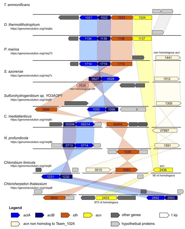
We carried out comparative genomic analyses among T. ammonificans and all available Aquificae genomes (Table 3). Desulfurobacterium thermolithotrophum DSM 11699 (its closest relative whose genome was sequenced) and Caminibacter mediatlanticus TB-2 (an Epsilonproteobacterium which, albeit phylogenetically distant, shares the same physiology and occupies a similar ecological niche as T. ammonificans, only at lower temperature; Figure 7) were further used as a direct comparison. We identified areas of synteny between T. ammonificans and the closely related D. thermolithotrophum in the region surrounding the genes encoding for key enzymes responsible for the citrate cleavage in the rTCA cycle (Figure 7b and Figure 8). This region is conserved also within the Hydrogenothermaceae (in particular P. marina) and members of the Epsilonproteobacteria.
Table 3.
List of the genomes belonging to the Aquificae phylum used for comparative genomic analyses.
| Organism | Genome acc. number | Genome length | GC content | Num. genes |
|---|---|---|---|---|
| Aquifex aeolicus VF5 | NC_000918 | 1,590,791 | 43% | 1782 |
| Desulfurobacterium sp. TC5-1 | NZ_ATXC01000001 | 1,653,625 | 40% | 1680 |
| Desulfurobacterium thermolithotrophum DSM 11699 | NC_015185 | 1,541,968 | 35% | 1561 |
| Hydrogenivirga sp. 128–5 R1-1 | NZ_ABHJ01000551 | 3,038,240 | 44% | 3756 |
| Hydrogenobacter thermophilus TK-6 | NC_013799 | 1,743,135 | 44% | 1897 |
| Hydrogenobaculum sp. Y04AAS1 | NC_011126 | 1,559,514 | 35% | 1631 |
| Persephonella marina EX-H1 | NC_012440 | 1,983,966 | 37% | 2067 |
| Persephonella sp. IF05-L8 | NZ_JNLJ01000001 | 1,828,858 | 35% | 1920 |
| Sulfurihydrogenibium azorense Az-Fu1 | NC_012438 | 1,640,877 | 33% | 1722 |
| Sulfurihydrogenibium sp. YO3AOP1 | NC_010730 | 1,838,442 | 32% | 1832 |
| Sulfurihydrogenibium subterraneum DSM 15120 | NZ_JHUV01000001 | 1,610,181 | 32% | 1701 |
| Sulfurihydrogenibium yellowstonense SS-5 | NZ_ABZS01000228 | 1,534,471 | 33% | 1637 |
| Thermocrinis albus DSM 14484 | NC_013894 | 1,500,577 | 47% | 1631 |
| Thermocrinis ruber DSM 23557 | NZ_CP007028 | 1,521,037 | 45% | 1625 |
| Thermocrinis sp. GBS | NZ_JNIE01000001 | 1,315,625 | 41% | 1417 |
| Thermosulfidibacter takaii ABI70S6 | NZ_AP013035 | 1,816,670 | 43% | 1844 |
| Thermovibrio ammonificans HB-1 | NC_014926 | 1,759,526 | 52% | 1820 |
Figure 8. Syntheny diagram presenting the genome organization around the rTCA key enzyme ATP citrate lyase.
In T. ammonificans the two subunits of the ATP citrate lyase enzyme are organized in a single operon together with the isocitrate dehydrogenase and the aconitate dehydratase. The numbers inside each gene represent the locus number for the organism. Shaded color connects synthenic regions. The website address below each organism name is a permanent link to the pairwise analysis performed on the Genome Evolution server (http://genomevolution.org/) against T. ammonificans.
The conserved regions between the genomes of T. ammonificans and D. thermolithotrophum included the two hydrogenase clusters, the flagellum and the NADP dehydrogenase complex. The order and position of the hydrogenase and NADP dehydrogenase clusters were highly conserved also in C. mediatlanticus, while flagellar genes were scattered throughout the genome (Figure 7). These results suggest that the genes encoding hydrogen utilization and functions related to energy conversion are among the oldest genomic regions present in those organisms, and overall are conserved across phyla. When we analyzed the central metabolism of T. ammonificans using comparative genomic approaches, the presence of two distinct groups of genes became evident: the first group was related to early-branching bacterial or archaeal lineages and coded for enzymes involved in several central metabolic pathways, while the second group of genes appeared to have been acquired later. Among the first group of genes, we identified: (I) the cytoplasmic [Ni-Fe]-hydrogenases Group 3 that were related to the enzymes found in methanogens and thermophilic Euryarchaeota, in which they are involved in ferredoxin reduction (Figure 3 and Figure 8); (II) sulfur-reduction related genes (Figure 3); and (III) the genes coding for the enzymes of the reductive acetyl-CoA pathway (Figure 3). These pathways are either present in early branching Archaea, or are directly involved in metabolic reactions that do not require oxygen (or oxygen by-products) and use substrates of geothermal origin that are likely to have been abundant on Early Earth. Moreover, most key reactions in these pathways are catalyzed by enzymes that are extremely sensitive to oxygen. Our findings are consistent with a recent reconstruction of the ancestral genome of the last universal common ancestor (LUCA), which suggest that LUCA was a hydrogen-dependent autotroph capable of S utilization that could fix CO2 via the reductive acetyl-CoA pathway (Weiss et al., 2016). Taken together, these observations suggest that part of the central metabolism of T. ammonificans is ancestral and emerged prior to oxygenic photosynthesis.
By contrast, some genes and associated metabolic pathways appear to have been acquired by the T. ammonificans lineage at a later time. One example is the ability to conserve energy by nitrate reduction. The hypothesis that nitrate respiration is an acquired trait in T. ammonificans is supported by the structure of the nap operon. The monomeric napA gene of T. ammonificans is homologous to that of Desulfovibrio desulfuricans and other Deltaproteobacteria (Figure 6; online interactive version) and apparently has been acquired laterally. Furthermore, it shares high similarity with assimilatory nitrate reductases (Nas) and it could be an evolutionary intermediate form between assimilatory Nas and the dimeric respiratory Nap. In line with this observation, phylogenetic analyses suggest that the membrane bound [Ni-Fe]-hydrogenases of Group 1 and 4 are of delta/epsilonproteobacterial origin (Figure 5). Based on the finding that Group 1 Hyn hydrogenases were expressed when T. ammonificans was grown under nitrate-reducing conditions (Figure 3), we suggest that these enzymes are linked through the membrane quinone pool to nitrate reduction and may have been acquired simultaneously with the nap genes.
A second example is represented by the oxygen radical detoxification enzymes encoded by the genome of T. ammonificans. We propose that such genes are not part of the core, or ancestral genome of T. ammonificans, but that they evolved as a response to exposure to toxic oxygen radicals. In the modern ocean, catalase and peroxidase may provide protection to oxygen-sensitive enzymes during transient exposure to oxygen associated with entrainment of deep seawater in hydrothermal fluids, or when the organism is displaced into the water column. This second scenario may have important implication for the dispersal of vent microorganisms to other vent sites. We hypothesize that T. ammonificans is able to deal with oxygen exposures using two different mechanisms: the first response mechanism would be activated following exposure to oxygen at physiological temperatures (60–80°C). In that case, the oxygen-stress related genes are induced, protecting the cell machinery against damages. The second mechanism would be a response to prolonged exposure to oxygen at temperatures below the physiological threshold. Such prolonged exposure would trigger a metabolic shut-down, enabling survival of the organism in cold seawater. The latter hypothesis is supported by experiments where batch cultures of T. ammonificans were exposed for a prolonged time at 4°C, which revealed the survival of the bacterium despite being exposed to oxygen (data not shown).
In conclusion, the nitrate reduction pathway, as well as oxygen stress-related genes, may represent adaptations acquired by the T. ammonificans lineage to cope with the rise of oxygen on Earth.
Insight into the evolution of carbon fixation
Six different pathways of carbon fixation are known to date (Fuchs, 2011). In the modern biosphere, the Calvin-Benson-Bassham cycle is the dominant mechanism of CO2 fixation. Yet, other anaerobic pathways have been investigated and are proposed to represent the ancestral autotrophic carbon fixation pathway. In particular, the reductive acetyl-CoA pathway is thought to be among the earliest carbon fixation pathways that have emerged on Earth (Fuchs, 2011). This hypothesis is supported by numerous observations: (I) the presence of this pathway in the early branching methanogenic archaea; (II) its low energy requirements and low need of de novo protein synthesis; (III) its capacity to incorporate different one-carbon compounds and carbon monoxide of geothermal origin, and (IV) the extreme oxygen sensitivity of its key enzymes which have common roots in Bacteria and Archaea (reviewed in Berg et al., 2010; Fuchs, 2011; Hügler and Sievert, 2011). Despite differences in their structure, all extant carbon fixation pathways can be theoretically derived from a putative rTCA cycle/reductive acetyl-CoA hybrid pathway, as proposed by a recent phylometabolic reconstruction of the evolution of carbon fixation (Braakman and Smith, 2012). According to this study, both Aquificae and acetogenic bacteria represent the closest living example of the archetypal network, and both diverged from the pre-LUCA pathway under the selective pressure of energy-efficiency (acetogens) and oxygen sensitivity (Aquificae). Further, a recent reconstruction of the genome of LUCA based on gene phyletic pattern reconstruction is consistent with some of these findings and suggests that LUCA’s genomic makeup point to autotrophic acetogenic and methanogenic roots and to the ancestry of the reductive acetyl-CoA pathway (Weiss et al., 2016).
The presence of a CodH type V enzyme in the genome of T. ammonificans and other members of the Desulfurobacteriaceae, and the presence of a type II CodH in Desulfurobacterium thermolithotrophum suggest that the reductive acetyl-CoA pathway could be operational in extant members of the Desulfurobacteriaceae. Furthermore, this finding supports the scenario proposed by Braakman and Smith that a complete and operational reductive acetyl-CoA pathway was present in the ancestor of the phylum Aquificae (Braakman and Smith, 2012). Comparative analyses revealed that the CodH enzyme is conserved only within the Desulfurobacteriaceae, consistent with the strict anaerobic nature of the members of this family and the extreme oxygen sensitivity of CodH. The rise of oxygen has been interpreted as one of the factors responsible for the diversification of carbon fixation from the ancestral pathway, and could explain the subsequent loss of the CodH in the generally facultative microaerobic Hydrogenothermaceae and Aquificaceae within the Aquificae (Table 2).
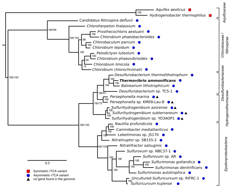
Comparative genomic analyses of rTCA cycle genes allow the description of a possible evolutionary scenario for the rTCA cycle in Aquificae. Members of the Aquificaceae (e.g., Aquifex aeolicus; Figure 9 and Figure 3—figure supplement 1) have the two-step version of the rTCA cycle (Figure 3—figure supplement 1A), where citrate cleavage is accomplished by the combined action of the enzymes citryl-CoA synthetase and citryl-CoA lyase (encoded by the ccl gene), and the carboxylation of 2-oxoglutarate is catalyzed by the two enzymes 2-oxoglutarate carboxylase and oxalosuccinate reductase (Aoshima et al., 2004; Braakman and Smith, 2012). Since citryl-CoA synthetase and 2-oxoglutarate carboxylase likely evolved by duplication of the genes for the succinyl-CoA synthetase and pyruvate carboxylase (Aoshima, 2007; Braakman and Smith, 2012), a complete rTCA cycle could have evolved in the Aquificaceae from an ancestral, incomplete version of the cycle. In contrast, the two other groups of Aquificae, the Hydrogenothermaceae and the Desulfurobacteriaceae (Figure 1—figure supplement 1), use the one-step – and more recent – version of the rTCA cycle (Figure 10, Figure 9 and Figure 3—figure supplement 1B), involving ATP citrate lyase (ACL, encoded by the acl gene) and isocitrate dehydrogenase, that carry out citrate cleavage and 2-oxoglutarate carboxylation in two single enzyme reactions, respectively (Braakman and Smith, 2012).
Figure 9. Phylogenetic tree of ATP citrate lyase amino acid sequences.
The tree was constructed with Bayesian inference and maximum likelihood methods and presents the phylogenetic relationship among the concatenated subunit of the ATP citrate lyase in different organisms known to use the rTCA cycle. The numbers near the nodes represent the bayesian posterior probability (left number) and the maximum-likelihood confidence values based on 1000 bootstrap replications (right number). Bar, 30% estimated substitutions. Accession numbers are reported in Figure 9—source data 1. Squares – symmetric rTCA cycle variant characterized by the presence of two-step enzyme reactions for the cleavage of citrate and the carboxylation of 2-oxoglutarate. Circles – asymmetric rTCA cycle variant characterized by one-step reaction enzymes catalyzing the above reactions. Triangles – presence of a citryl-CoA lyase in the genome. See Supplementary Materials for details.
DOI: http://dx.doi.org/10.7554/eLife.18990.018
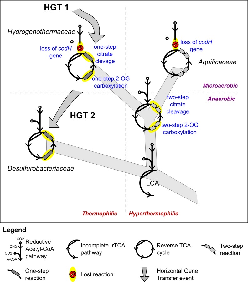
Figure 10. Proposed evolution of the carbon fixation pathway in the Aquificae phylum.
Proposed evolution of the carbon fixation pathway in the Aquificae phylum and reconstruction of the ancestral carbon fixation for the last common ancestor (LCA) based on the results of integrated phylogenetic, comparative genomic and phylometabolic analyses.
The enzyme responsible for citrate cleavage, the ATP citrate lyase, likely evolved through gene fusion of the genes of CCS and CCL (Aoshima et al., 2004). Phylogenetic analyses of ACL suggests that this gene fusion event did not happen within the Aquificae, as Nitrospira and Chlorobia have an evolutionary older version of ACL than Hydrogenothermaceae and Desulfurobacteriaceae (Figure 9; Hügler et al., 2007). Hence, it is likely that these two groups acquired ACL through HGT (Figure 9 and Figure 8; Hügler et al., 2007). Furthermore, our comparative analysis showed that: (I) in the Hydrogenothermaceae and in the Desulfurobacteriaceae, the enzymes of the first half of the rTCA cycle, as well as aconitase (acnA), share a common ancestor with the Aquificaceae and can be considered part of the core genome of the phylum Aquificae, while the remaining enzymes are either the result of gene duplication or have been acquired by horizontal gene transfer (Hügler et al., 2007); (II) the two-step citrate cleavage is exclusive to the Aquificacea (Figure 9); (III) the ccl gene is still present in the Hydrogenothermacea (in addition to acl), and one sequenced strain of the Hydrogenothermaceae (S. azorense) still uses the two-step carboxylation of 2-oxoglutarate, suggesting that the two-step version of the rTCA cycle was present in the ancestor of both the Aquificaceae and the Hydrogenothermaceae (Figure 9 and Figure 10). However, neither of the genes for the two-step citrate cleavage or two-step 2-oxoglutrate carboxylation is present in the Desulfurobacteriaceae (Figure 9). Finally, synteny analyses show that the four enzymes necessary to complement the one-step rTCA cycle variant in the Desulfurobacteriaceae from a theoretical ancestral linear rTCA are organized in a single operon (Figure 8).
Taking into consideration evidence from comparative genomics and phylogenetic analyses, the most parsimonious interpretation of the data suggests that the last common ancestor of the Aquificae had an incomplete form of the rTCA that did not proceed past the synthesis of 2-oxoglutarate, and that later on the cycle was closed following two independent evolutionary trajectories (Figure 10): (I) Gene duplication in the lineage that lead to the Aquificaceae and Hydrogenothermaceae; and (II) gene acquisition by horizontal gene transfer in the Desulfurobacteriaceae and Hydrogenothermaceae (the latter replaced the two-step version of the rTCA cycle with the one-step version) (Figure 9 and Figure 10). The reasons behind the presence of two distinct rTCA cycle variants within the extant Aquificae are not known. Braackman and Smith hypothesized that the one-step reactions might have evolved as a way to improve the thermodynamic efficiency of the rTCA cycle (Braakman and Smith, 2012). We hypothesize that temperature may also have played a role in preserving the ancient, more symmetric, two-step citrate cleavage rTCA cycle variant in Aquificaceae (Figure 10). Members of this group have optimum growth temperatures (75–95°C) higher than those of the two other groups (60–75°C; Table 1), and the ‘ancient’ enzymes might be more stable at these high temperatures. In contrast, the ‘newer’ enzymes that catalyze the one-step citrate cleavage might have evolved to function optimally at lower temperatures.
Different members of the Aquificae use either one of the two versions of the rTCA cycle, and all the extant members of this phylum possess the genes encoding for the enzymes of the reductive acetyl-CoA pathway, with the exception of codH (encoding for the CO-dehydrogenase), which is only found in the Desulfurobacteriaceae. Therefore, we hypothesize that the last common ancestor of the Aquificae possessed the complete reductive acetyl-CoA pathway (Figure 10). While members of the Desulfurobacteriaceae kept the CodH due to their obligate anaerobic lifestyle, microaerophilic Aquificae (Hydrogenothermaceae and Aquificaceae) lost this extremely oxygen-sensitive enzyme (Figures 4 and 10). The presence of the gene encoding CodH in P. marina (Hydrogenothermaceae) is the only exception, and suggests that codH was lost independently in the two lineages (Figure 10).
Alltogether, our results suggest that the last common ancestor of the Aquificae combined the reductive acetyl-CoA pathway with an incomplete form of the rTCA that did not proceed past the synthesis of 2-oxoglutarate (Figure 10). A similar incomplete version of the rTCA pathway, consisting only of the reactions from acetyl-CoA to 2-oxoglutarate, is present in extant methanogens (Berg, 2011). Thus, our phylometabolic reconstruction of the ancestral state of carbon fixation in the Aquificae (Figure 10) is conceptually consistent with chemoautotrophic processes of extant Bacteria and Archaea, and may represent the earliest carbon fixation pathway.
Conclusions
We propose that the ancestor of Thermovibrio ammonificans was originally a hydrogen oxidizing, sulfur reducing bacterium that used a hybrid carbon fixation pathway for CO2 fixation. The simultaneous presence of the rTCA cycle and of the reductive acetyl-CoA pathway of carbon fixation in T. ammonificans may represent a modern analog of the early carbon fixation phenotype, and suggests that the redundancy of central metabolic pathways was common in ancestral microorganisms. With the gradual rise of oxygen in the atmosphere, more efficient terminal electron acceptors became available and this lineage acquired genes that increased its metabolic flexibility - e.g., the capacity to respire nitrate - along with enzymes involved in the detoxification of oxygen radicals. However, this lineage also retained its core, or ancestral, metabolic traits. Given the early branching nature of the phylum Aquificae and the ability of T. ammonificans and the Desulfurobacteriaceae to thrive in hydrothermal environments relying on energy sources of geothermal origins (namely carbon dioxide, hydrogen and elemental sulfur), we argue that these microorganisms represent excellent models to investigate how metabolism co-evolved with Earth’s changing environmental conditions.
Materials and methods
Strain isolation was described in (Vetriani et al., 2004). Growth condition, DNA extraction, sequencing strategy and automatic annotation were published in (Giovannelli et al., 2012).
Manual curation of the genome
Manual curation of the genome was performed using blastn and blastp (McGinnis and Madden, 2004) against the non-redundant database (Pruitt et al., 2007), the conserved domain database (Marchler-Bauer et al., 2005), the Kyoto Encyclopedia of Genes and Genomes (Kanehisa and Goto, 2000) and the PFAM database (Sonnhammer et al., 1998). Coding sequence similarities were compared using translated protein sequence. Metabolic pathways were reconstructed on the basis of available genomic, physiologic and biochemical information and using Kyoto Encyclopedia of Genes and Genomes (Kanehisa and Goto, 2000) and SEED (Overbeek et al., 2014) pathways as template.
Comparative genomics
Genome maps were drawn using Circos (Krzywinski et al., 2009), parsing blast results with ad hoc bash scripting. Comparative analyses between the genomes of T. ammonificans, those of representative members of the Aquificae (reported in Table 3), Desulfurobacterium thermolithotrophum (Göker et al., 2011) and Caminibacter mediatlanticus (Giovannelli et al., 2011) were performed using the GenomeEvolution pipeline CoGe (Lyons et al., 2008). Aquificae genomes were selected among all available genome within this phylum to maximize diversity while minimizing redundancy. All the available genomes of validly published Aquificae species were selected for analysis. To this set we added the reference genomes for the genera Hydrogenivirga and Hydrogenobaculum, as no genome sequence is available for validly published species of these genera. We also selected two additional genomes belonging to the genera Desulfurobacterium (the closest relative to the genus Thermovibrio) and Persephonella, respectively. Excluded genomes include either alternative assemblies of selected genomes or closely related genomes with a gapped genome similarity above 90%. LastZ pairwise alignments of selected genomes were used to draw three-way plots using the Hive Plot software (Krzywinski et al., 2012). Gene context and operonic structures were reconstructed using BioCyc (Karp et al., 2005) and FgenesB. Operons were manually screened and their structure selected based on gene context and available literature on the specific gene. When it was not possible to discriminate between the two alternative operonic predictions, both were reported in the text. Similarities between T. ammonificans genes and other prokaryotic genomes were found performing blastp analyses against the nr database. The top three blast results were retained and further analyzed, ranking the genes for their best hits. The procedure was repeated removing from the database the genome of D. thermolithotrophum and Desulfurobacterium sp. TC5-1, thus searching for the best hit outside of the Desulfurobacteriaceae family. The results were analyzed and interactive Krona plots (Ondov et al., 2011) drawn linking the T. ammonificans genes with its closest match in the database. The interactive plots are accessible at DOI: 10.6084/m9.figshare.3178528. Images were drawn or edited using the open source vector drawing program Inkscape (http://inkscape.org/).
Phylogenetic analyses
Phylogenetic analyses were performed using the approaches defined below for each tree. The phylogenetic tree presented in Figure 1 was computed from the current 16S rRNA database alignment available from the ARB-SILVA project (http://www.arb-silva.de). The maximum likelihood three was computed from the ARB-SILVA alignment using PHYLML (Guindon and Gascuel, 2003) and the GTR model. The phylogenetic tree in Figure 1—figure supplement 1 was constructed by aligning complete or near complete 16S rRNA sequences obtained from NCBI and representing the phylum Aquificae. The 16S rRNA sequences were aligned with ClustalO (Thompson et al., 1997) and Gblocks (Castresana, 2000) and the alignment was manually refined using SEAVIEW (Galtier et al., 1996). The maximum likelihood phylogeny was inferred from the alignment of 1455 sites using PHYML (Guindon and Gascuel, 2003), the GRT model and 1000 bootstrap replications. The CODH tree presented in Figure 4 was computed using a selected set of amino acid sequences and the neighbor-joining method. Alignments were obtained using Muscle (Edgar, 2004) and Gblocks (Castresana, 2000), manually refined using SEAVIEW, and phylogenetic distances calculated using 255 sites and the Observed Divergence matrix. The neighbor-joining method was used to evaluate tree topologies using Phylo_win (Galtier et al., 1996) and their robustness was tested by bootstrap analysis with 1000 resamplings. The tree for the catalytic subunit of the [NiFe]-hydrogenases presented in Figure 5 was computed using a selected set of amino acid sequences and the same approach described above for the CODH tree and was based on 539 sites. The accession number for the sequences used in trees presented in Figure 1—figure supplement 1, Figures 4 and 5 are reported within each tree. The ATP citrate lyase phylogenetic tree was reconstructed with Bayesian inference and maximum likelihood methods. Both subunit of the ATP citrate lyase (AclB and AclA) were aligned individually to retrieved homologs using Muscle (Edgar, 2004) and SEAVIEW (Galtier et al., 1996). The alignments were concatenated and refined using Gblock (Castresana, 2000). A hypothetical ancestral ATP citrate lyase enzyme was manually constructed by concatenating the citryl-CoA synthetase and citryl-CoA lyase of H. thermophilus and A. aeolicus, respectively, and used as the outgroup (Hügler et al., 2007). The maximum likelihood phylogeny was inferred from the alignment using PHYML with the Wag substitution model (Whelan and Goldman, 2001) and 1000 bootstrap resamplings. The substitution model was selected based on AIC values using ProTest3 (Darriba et al., 2011). Bayesian phylogeny was inferred using MrBayes (Ronquist and Huelsenbeck, 2003) performing 500,000 generations with two parallel searches with the Wag amino acid matrix model (selected using forward selection among all possible substitution models) and a burn-in of 125,000 generations. Both tree were computed on 1035 identified sites. Accession numbers for the tree presented are reported in Figure 9—source data 1. A combination of phylogenetic analysis and comparative genomic analyses were used to identify lateral gene transfer events.
Phylometabolic reconstruction of the carbon fixation pathway within the aquificae
Phylometabolic analysis (Braakman and Smith, 2012) was used to investigate the carbon fixation pathway in T. ammonificans and its evolutionary relationship to the carbon fixation pathways present in other members of the Aquificae phylum. In phylometabolic analyses, the metabolic pathways of the organism under investigation are compared to those found in related organisms both within and across neighboring clades. By focusing on the pathways, the comparison may reveal variations in multi-enzyme functional units, providing context for the completion of the pathway within the networks of individual organism, while also allowing for the identification of ancestral states and horizontal gene transfer events. The resulting phylometabolic tree includes multiple complete pathways to common essential metabolites, and suggests which evolutionary substitutions are allowed (at either organism or ecosystem levels) among these pathways (see [Braakman and Smith, 2012] and reference therein for a more extensive description of the principles underlying this approach). We reconstructed the carbon fixation pathways in representative genomes of the Aquificae, and compared them. Information regarding the carbon fixation metabolic network was implemented using phylogenetic and comparative genomic information to help reconstruct possible ancestral states of the carbon fixation network based on maximum parsimony principles.
Protein extraction, digestion and identification
T. ammonificans cell pellets were washed in TE buffer (10 mM Tris-HCl pH 7.5, 10 mM EDTA pH 8.0, containing Roche cOmplete protease inhibitor) and soluble proteins were extracted as described by (Heinz et al., 2012). Briefly, cells were disrupted by sonication (2 × 25 s), cell debris was pelleted and protein concentrations in the supernatant were determined according to (Bradford, 1976). 20 µg of protein extract were loaded onto a precast 10% polyacrylamide mini gel in technical triplicates for 1D PAGE (150 V, 45 min). After staining with Coomassie Brilliant Blue, protein-containing gel lanes were excised and cut into 10 equal subsamples each, which were destained (200 mM NH4HCO3, 30% acetonitrile) and digested with trypsin solution (1 µg/ml, Promega, Madison WI, USA) at 37°C over night, before peptides were eluted from the gel pieces in an ultrasonic bath (15 min). As described by (Xing et al., 2015), peptide mixes were subjected to reversed phase C18 column chromatography on a nano-ACQUITY-UPLC (Waters Corporation, Milford, MA, USA). Mass spectrometry (MS) and MS/MS data were recorded with an online-coupled LTQ-Orbitrap mass spectrometer (Thermo Fisher Scientific Inc., Waltham, MA, USA). MS data were searched against the forward-decoy T. ammonificans protein database using Sequest (Thermo Fisher Scientific, San Jose, CA, USA; version 27, revision 11) and identifications were filtered and validated in Scaffold (http://www.proteomesoftware.com), as described previously (Heinz et al., 2012). Data for all three technical replicates were merged and exclusive unique peptide count values of all proteins were exported for calculation of normalized spectral abundance factors (NSAF), which are given in Figure 3—source data 1 in %, i.e., as relative abundance of each protein in % of all identified proteins.
Acknowledgements
The authors gratefully acknowledge the support of the Deep Carbon Observatory. Thanks to Sandra Beyer for assistance in the lab and to Sebastian Grund for MS measurements. The genome of Thermovibrio ammonificans was sequenced under the auspices of the US Department of Energy. Work on T. ammonificans was supported, in part, by NSF grants MCB 04–56676, OCE 03–27353, MCB 08–43678, OCE 09–37371, OCE 11–24141, OCE11-36451, and NASA grant NNX15AM18G to CV, NSF grant MCB 15–17567 to CV and DG, and by the New Jersey Agricultural Experiment Station. DG was supported by a Postdoctoral Fellowship from the Institute of Marine and Coastal Sciences and a Postdoctoral Fellowship from the Center for Dark Energy Biosphere Investigations (C-DEBI). Funding to SMS was provided by NSF grant OCE-1136727 and a senior fellowship by the Alfried Krupp Wissenschaftskolleg Greifswald, Germany. This publication was in part supported by the ELSI Origins Network (EON), which is supported by a grant from the John Templeton Foundation. The opinions expressed in this publication are those of the authors and do not necessarily reflect the views of the John Templeton Foundation. This paper is C-DEBI contribution 368.
Funding Statement
The funders had no role in study design, data collection and interpretation, or the decision to submit the work for publication.
Funding Information
This paper was supported by the following grants:
National Science Foundation MCB 04-56676 to Costantino Vetriani.
National Aeronautics and Space Administration NNX15AM18G to Costantino Vetriani.
National Science Foundation OCE 03-27353 to Costantino Vetriani.
National Science Foundation MCB 08-43678 to Costantino Vetriani.
National Science Foundation OCE 09-37371 to Costantino Vetriani.
National Science Foundation OCE 11-24141 to Costantino Vetriani.
National Science Foundation MCB 15-17567 to Donato Giovannelli, Costantino Vetriani.
National Science Foundation OCE-1136727 to Stefan M Sievert.
Additional information
Competing interests
The authors declare that no competing interests exist.
Author contributions
DG, Conceptualization, Data curation, Formal analysis, Investigation, Methodology, Writing—original draft, Writing—review and editing.
SMS, Conceptualization, Formal analysis, Funding acquisition, Methodology, Writing—review and editing.
MH, Conceptualization, Formal analysis, Investigation, Writing—review and editing.
SM, Data curation, Formal analysis, Methodology, Writing—review and editing.
DB, Formal analysis, Methodology, Writing—review and editing.
TS, Conceptualization, Data curation, Methodology, Writing—review and editing.
CV, Conceptualization, Formal analysis, Supervision, Funding acquisition, Writing—original draft, Project administration, Writing—review and editing.
References
- Aoshima M, Igarashi Y. Nondecarboxylating and decarboxylating isocitrate dehydrogenases: oxalosuccinate reductase as an ancestral form of isocitrate dehydrogenase. Journal of Bacteriology. 2008;190:2050–2055. doi: 10.1128/JB.01799-07. [DOI] [PMC free article] [PubMed] [Google Scholar]
- Aoshima M, Ishii M, Igarashi Y. A novel enzyme, citryl-CoA lyase, catalysing the second step of the citrate cleavage reaction in Hydrogenobacter thermophilus TK-6. Molecular Microbiology. 2004;52:763–770. doi: 10.1111/j.1365-2958.2004.04010.x. [DOI] [PubMed] [Google Scholar]
- Aoshima M. Novel enzyme reactions related to the tricarboxylic acid cycle: phylogenetic/functional implications and biotechnological applications. Applied Microbiology and Biotechnology. 2007;75:249–255. doi: 10.1007/s00253-007-0893-0. [DOI] [PubMed] [Google Scholar]
- Arai H, Kanbe H, Ishii M, Igarashi Y. Complete genome sequence of the thermophilic, obligately chemolithoautotrophic hydrogen-oxidizing bacterium Hydrogenobacter thermophilus TK-6. Journal of Bacteriology. 2010;192:2651–2652. doi: 10.1128/JB.00158-10. [DOI] [PMC free article] [PubMed] [Google Scholar]
- Baross JA, Hoffman SE. Submarine hydrothermal vents and associated gradient environments as sites for the origin and evolution of life. Origins of Life and Evolution of the Biosphere. 1985;15:327–345. doi: 10.1007/BF01808177. [DOI] [Google Scholar]
- Battistuzzi FU, Feijao A, Hedges SB, Balir Hedger S. A genomic timescale of prokaryote evolution: insights into the origin of methanogenesis, phototrophy, and the colonization of land. BMC Evolutionary Biology. 2004;4:44. doi: 10.1186/1471-2148-4-44. [DOI] [PMC free article] [PubMed] [Google Scholar]
- Berg IA, Kockelkorn D, Ramos-Vera WH, Say RF, Zarzycki J, Hügler M, Alber BE, Fuchs G. Autotrophic carbon fixation in archaea. Nature Reviews Microbiology. 2010;8:447–460. doi: 10.1038/nrmicro2365. [DOI] [PubMed] [Google Scholar]
- Berg IA. Ecological aspects of the distribution of different autotrophic CO2 fixation pathways. Applied and Environmental Microbiology. 2011;77:1925–1936. doi: 10.1128/AEM.02473-10. [DOI] [PMC free article] [PubMed] [Google Scholar]
- Braakman R, Smith E. The emergence and early evolution of biological carbon-fixation. PLoS Computational Biology. 2012;8:e1002455. doi: 10.1371/journal.pcbi.1002455. [DOI] [PMC free article] [PubMed] [Google Scholar]
- Bradford MM. A rapid and sensitive method for the quantitation of microgram quantities of protein utilizing the principle of protein-dye binding. Analytical Biochemistry. 1976;72:248–254. doi: 10.1016/0003-2697(76)90527-3. [DOI] [PubMed] [Google Scholar]
- Castresana J. Selection of conserved blocks from multiple alignments for their use in phylogenetic analysis. Molecular Biology and Evolution. 2000;17:540–552. doi: 10.1093/oxfordjournals.molbev.a026334. [DOI] [PubMed] [Google Scholar]
- Darriba D, Taboada GL, Doallo R, Posada D. ProtTest 3: fast selection of best-fit models of protein evolution. Bioinformatics. 2011;27:1164–1165. doi: 10.1093/bioinformatics/btr088. [DOI] [PMC free article] [PubMed] [Google Scholar]
- Das A, Silaghi-Dumitrescu R, Ljungdahl LG, Kurtz DM. Cytochrome bd oxidase, oxidative stress, and dioxygen tolerance of the strictly anaerobic bacterium Moorella thermoacetica. Journal of Bacteriology. 2005;187:2020–2029. doi: 10.1128/JB.187.6.2020-2029.2005. [DOI] [PMC free article] [PubMed] [Google Scholar]
- Deckert G, Warren PV, Gaasterland T, Young WG, Lenox AL, Graham DE, Overbeek R, Snead MA, Keller M, Aujay M, Huber R, Feldman RA, Short JM, Olsen GJ, Swanson RV. The complete genome of the hyperthermophilic bacterium Aquifex aeolicus. Nature. 1998;392:353–358. doi: 10.1038/32831. [DOI] [PubMed] [Google Scholar]
- Di Giulio M. The universal ancestor lived in a thermophilic or hyperthermophilic environment. Journal of Theoretical Biology. 2000;203:203–213. doi: 10.1006/jtbi.2000.1086. [DOI] [PubMed] [Google Scholar]
- Di Giulio M. The ancestor of the bacteria domain was a hyperthermophile. Journal of Theoretical Biology. 2003;224:277–283. doi: 10.1016/S0022-5193(03)00164-4. [DOI] [PubMed] [Google Scholar]
- Douzi B, Filloux A, Voulhoux R. On the path to uncover the bacterial type II secretion system. Philosophical Transactions of the Royal Society B: Biological Sciences. 2012;367:1059–1072. doi: 10.1098/rstb.2011.0204. [DOI] [PMC free article] [PubMed] [Google Scholar]
- Edgar RC. MUSCLE: multiple sequence alignment with high accuracy and high throughput. Nucleic Acids Research. 2004;32:1792–1797. doi: 10.1093/nar/gkh340. [DOI] [PMC free article] [PubMed] [Google Scholar]
- Fuchs G. Alternative pathways of carbon dioxide fixation: insights into the early evolution of life? Annual Review of Microbiology. 2011;65:631–658. doi: 10.1146/annurev-micro-090110-102801. [DOI] [PubMed] [Google Scholar]
- Galtier N, Gouy M, Gautier C. SEAVIEW and PHYLO_WIN: two graphic tools for sequence alignment and molecular phylogeny. Bioinformatics. 1996;12:543–548. doi: 10.1093/bioinformatics/12.6.543. [DOI] [PubMed] [Google Scholar]
- Giovannelli D, Ferriera S, Johnson J, Kravitz S, Pérez-Rodríguez I, Ricci J, O'Brien C, Voordeckers JW, Bini E, Vetriani C. Draft genome sequence of caminibacter mediatlanticus strain TB-2, an epsilonproteobacterium isolated from a deep-sea hydrothermal vent. Standards in Genomic Sciences. 2011;5:135–143. doi: 10.4056/sigs.2094859. [DOI] [PMC free article] [PubMed] [Google Scholar]
- Giovannelli D, Ricci J, Pérez-Rodríguez I, Hügler M, O'Brien C, Keddis R, Grosche A, Goodwin L, Bruce D, Davenport KW, Detter C, Han J, Han S, Ivanova N, Land ML, Mikhailova N, Nolan M, Pitluck S, Tapia R, Woyke T, Vetriani C. Complete genome sequence of Thermovibrio ammonificans HB-1(T), a thermophilic, chemolithoautotrophic bacterium isolated from a deep-sea hydrothermal vent. Standards in Genomic Sciences. 2012;7:82–90. doi: 10.4056/sigs.2856770. [DOI] [PMC free article] [PubMed] [Google Scholar]
- Guindon S, Gascuel O. A simple, fast, and accurate algorithm to estimate large phylogenies by maximum likelihood. Systematic Biology. 2003;52:696–704. doi: 10.1080/10635150390235520. [DOI] [PubMed] [Google Scholar]
- Göker M, Daligault H, Mwirichia R, Lapidus A, Lucas S, Deshpande S, Pagani I, Tapia R, Cheng JF, Goodwin L, Pitluck S, Liolios K, Ivanova N, Mavromatis K, Mikhailova N, Pati A, Chen A, Palaniappan K, Han C, Land M, Hauser L, Pan C, Brambilla EM, Rohde M, Spring S, Sikorski J, Wirth R, Detter JC, Woyke T, Bristow J, Eisen JA, Markowitz V, Hugenholtz P, Kyrpides NC, Klenk HP. Complete genome sequence of the thermophilic sulfur-reducer Desulfurobacterium thermolithotrophum type strain (BSA(T)) from a deep-sea hydrothermal vent. Standards in Genomic Sciences. 2011;5:407–415. doi: 10.4056/sigs.2465574. [DOI] [PMC free article] [PubMed] [Google Scholar]
- Götz D, Banta A, Beveridge TJ, Rushdi AI, Simoneit BR, Reysenbach AL. Persephonella marina gen. nov., sp. nov. and Persephonella guaymasensis sp. nov., two novel, thermophilic, hydrogen-oxidizing microaerophiles from deep-sea hydrothermal vents. International Journal of Systematic and Evolutionary Microbiology. 2002;52:1349–1359. doi: 10.1099/00207713-52-4-1349. [DOI] [PubMed] [Google Scholar]
- Heinz E, Williams TA, Nakjang S, Noël CJ, Swan DC, Goldberg AV, Harris SR, Weinmaier T, Markert S, Becher D, Bernhardt J, Dagan T, Hacker C, Lucocq JM, Schweder T, Rattei T, Hall N, Hirt RP, Embley TM. The genome of the obligate intracellular parasite trachipleistophora hominis: new insights into microsporidian genome dynamics and reductive evolution. PLoS Pathogens. 2012;8:e1002979. doi: 10.1371/journal.ppat.1002979. [DOI] [PMC free article] [PubMed] [Google Scholar]
- Hetzer A, McDonald IR, Morgan HW. Venenivibrio stagnispumantis gen. nov., sp. nov., a thermophilic hydrogen-oxidizing bacterium isolated from champagne pool, waiotapu, New zealand. International Journal of Systematic and Evolutionary Microbiology. 2008;58:398–403. doi: 10.1099/ijs.0.64842-0. [DOI] [PubMed] [Google Scholar]
- Huber R, Eder W, Heldwein S, Wanner G, Huber H, Rachel R, Stetter KO. Thermocrinis ruber gen. nov., sp. nov., A pink-filament-forming hyperthermophilic bacterium isolated from yellowstone national park. Applied and Environmental Microbiology. 1998;64:3576–3583. doi: 10.1128/aem.64.10.3576-3583.1998. [DOI] [PMC free article] [PubMed] [Google Scholar]
- Huber R, Wilharm T, Huber D, Trincone A, Burggraf S, König H, Reinhard R, Rockinger I, Fricke H, Stetter KO. Aquifex pyrophilus gen. nov. sp. nov., represents a novel group of Marine hyperthermophilic hydrogen-oxidizing bacteria. Systematic and Applied Microbiology. 1992;15:340–351. doi: 10.1016/S0723-2020(11)80206-7. [DOI] [Google Scholar]
- Hügler M, Huber H, Molyneaux SJ, Vetriani C, Sievert SM. Autotrophic CO2 fixation via the reductive tricarboxylic acid cycle in different lineages within the phylum Aquificae: evidence for two ways of citrate cleavage. Environmental Microbiology. 2007;9:81–92. doi: 10.1111/j.1462-2920.2006.01118.x. [DOI] [PubMed] [Google Scholar]
- Hügler M, Sievert SM. Beyond the Calvin cycle: autotrophic carbon fixation in the ocean. Annual Review of Marine Science. 2011;3:261–289. doi: 10.1146/annurev-marine-120709-142712. [DOI] [PubMed] [Google Scholar]
- Jensen RA. Enzyme recruitment in evolution of new function. Annual Review of Microbiology. 1976;30:409–425. doi: 10.1146/annurev.mi.30.100176.002205. [DOI] [PubMed] [Google Scholar]
- Jeon JH, Lim JK, Kim MS, Yang TJ, Lee SH, Bae SS, Kim YJ, Lee SH, Lee JH, Kang SG, Lee HS. Characterization of the frhAGB-encoding hydrogenase from a non-methanogenic hyperthermophilic archaeon. Extremophiles. 2015;19:109–118. doi: 10.1007/s00792-014-0689-y. [DOI] [PubMed] [Google Scholar]
- Jepson BJ, Marietou A, Mohan S, Cole JA, Butler CS, Richardson DJ. Evolution of the soluble nitrate reductase: defining the monomeric periplasmic nitrate reductase subgroup. Biochemical Society Transactions. 2006;34:122–126. doi: 10.1042/BST0340122. [DOI] [PubMed] [Google Scholar]
- Kanehisa M, Goto S. KEGG: kyoto encyclopedia of genes and genomes. Nucleic Acids Research. 2000;28:27–30. doi: 10.1093/nar/28.1.27. [DOI] [PMC free article] [PubMed] [Google Scholar]
- Karp PD, Ouzounis CA, Moore-Kochlacs C, Goldovsky L, Kaipa P, Ahrén D, Tsoka S, Darzentas N, Kunin V, López-Bigas N. Expansion of the BioCyc collection of pathway/genome databases to 160 genomes. Nucleic Acids Research. 2005;33:6083–6089. doi: 10.1093/nar/gki892. [DOI] [PMC free article] [PubMed] [Google Scholar]
- Kawasumi T, Igarashi Y, Kodama T, Minoda Y. Hydrogenobacter thermophilus gen. nov., sp. nov., an extremely thermophilic, aerobic, hydrogen-oxidizing bacterium. International Journal of Systematic Bacteriology. 1984;34:5–10. doi: 10.1099/00207713-34-1-5. [DOI] [Google Scholar]
- Kim J, Kershner JP, Novikov Y, Shoemaker RK, Copley SD. Three serendipitous pathways in E. coli can bypass a block in pyridoxal-5'-phosphate synthesis. Molecular Systems Biology. 2010;6:436. doi: 10.1038/msb.2010.88. [DOI] [PMC free article] [PubMed] [Google Scholar]
- Krzywinski M, Birol I, Jones SJ, Marra MA. Hive plots--rational approach to visualizing networks. Briefings in Bioinformatics. 2012;13:627–644. doi: 10.1093/bib/bbr069. [DOI] [PubMed] [Google Scholar]
- Krzywinski M, Schein J, Birol I, Connors J, Gascoyne R, Horsman D, Jones SJ, Marra MA. Circos: an information aesthetic for comparative genomics. Genome Research. 2009;19:1639–1645. doi: 10.1101/gr.092759.109. [DOI] [PMC free article] [PubMed] [Google Scholar]
- L'Haridon S, Cilia V, Messner P, Ragúenès G, Gambacorta A, Sleytr UB, Prieur D, Jeanthon C. Desulfurobacterium thermolithotrophum gen. nov., sp. nov., a novel autotrophic, sulphur-reducing bacterium isolated from a deep-sea hydrothermal vent. International Journal of Systematic Bacteriology. 1998;48 Pt 3:701–711. doi: 10.1099/00207713-48-3-701. [DOI] [PubMed] [Google Scholar]
- Lane N, Allen JF, Martin W. How did LUCA make a living? Chemiosmosis in the origin of life. BioEssays. 2010;32:271–280. doi: 10.1002/bies.200900131. [DOI] [PubMed] [Google Scholar]
- Lebedinsky AV, Chernyh NA, Bonch-Osmolovskaya EA. Phylogenetic systematics of microorganisms inhabiting thermal environments. Biochemistry. 2007;72:1299–1312. doi: 10.1134/S0006297907120048. [DOI] [PubMed] [Google Scholar]
- Lyons E, Pedersen B, Kane J, Alam M, Ming R, Tang H, Wang X, Bowers J, Paterson A, Lisch D, Freeling M. Finding and comparing syntenic regions among Arabidopsis and the outgroups papaya, poplar, and grape: coge with rosids. Plant Physiology. 2008;148:1772–1781. doi: 10.1104/pp.108.124867. [DOI] [PMC free article] [PubMed] [Google Scholar]
- Marchler-Bauer A, Anderson JB, Cherukuri PF, DeWeese-Scott C, Geer LY, Gwadz M, He S, Hurwitz DI, Jackson JD, Ke Z, Lanczycki CJ, Liebert CA, Liu C, Lu F, Marchler GH, Mullokandov M, Shoemaker BA, Simonyan V, Song JS, Thiessen PA, Yamashita RA, Yin JJ, Zhang D, Bryant SH. CDD: a conserved domain database for protein classification. Nucleic Acids Research. 2005;33:D192–D196. doi: 10.1093/nar/gki069. [DOI] [PMC free article] [PubMed] [Google Scholar]
- Markert S, Arndt C, Felbeck H, Becher D, Sievert SM, Hügler M, Albrecht D, Robidart J, Bench S, Feldman RA, Hecker M, Schweder T. Physiological proteomics of the uncultured endosymbiont of Riftia pachyptila. Science. 2007;315:247–250. doi: 10.1126/science.1132913. [DOI] [PubMed] [Google Scholar]
- Martin W, Baross J, Kelley D, Russell MJ. Hydrothermal vents and the origin of life. Nature Reviews Microbiology. 2008;6:805–814. doi: 10.1038/nrmicro1991. [DOI] [PubMed] [Google Scholar]
- Martin W, Russell MJ. On the origins of cells: a hypothesis for the evolutionary transitions from abiotic geochemistry to chemoautotrophic prokaryotes, and from prokaryotes to nucleated cells. Philosophical Transactions of the Royal Society B: Biological Sciences. 2003;358:59–85. doi: 10.1098/rstb.2002.1183. [DOI] [PMC free article] [PubMed] [Google Scholar]
- Martin W, Russell MJ. On the origin of biochemistry at an alkaline hydrothermal vent. Philosophical Transactions of the Royal Society B: Biological Sciences. 2007;362:1887–1926. doi: 10.1098/rstb.2006.1881. [DOI] [PMC free article] [PubMed] [Google Scholar]
- Mason MG, Shepherd M, Nicholls P, Dobbin PS, Dodsworth KS, Poole RK, Cooper CE. Cytochrome bd confers nitric oxide resistance to Escherichia coli. Nature Chemical Biology. 2009;5:94–96. doi: 10.1038/nchembio.135. [DOI] [PubMed] [Google Scholar]
- McGinnis S, Madden TL. BLAST: at the core of a powerful and diverse set of sequence analysis tools. Nucleic Acids Research. 2004;32:W20–W25. doi: 10.1093/nar/gkh435. [DOI] [PMC free article] [PubMed] [Google Scholar]
- Moreno-Vivián C, Cabello P, Martínez-Luque M, Blasco R, Castillo F. Prokaryotic nitrate reduction: molecular properties and functional distinction among bacterial nitrate reductases. Journal of Bacteriology. 1999;181:6573–6584. doi: 10.1128/jb.181.21.6573-6584.1999. [DOI] [PMC free article] [PubMed] [Google Scholar]
- Nakagawa S, Nakamura S, Inagaki F, Takai K, Shirai N, Sako Y. Hydrogenivirga caldilitoris gen. nov., sp. nov., a novel extremely thermophilic, hydrogen- and sulfur-oxidizing bacterium from a coastal hydrothermal field. International Journal of Systematic and Evolutionary Microbiology. 2004;54:2079–2084. doi: 10.1099/ijs.0.03031-0. [DOI] [PubMed] [Google Scholar]
- Nunoura T, Oida H, Miyazaki M, Suzuki Y. Thermosulfidibacter takaii gen. nov., sp. nov., a thermophilic, hydrogen-oxidizing, sulfur-reducing chemolithoautotroph isolated from a deep-sea hydrothermal field in the southern okinawa trough. International Journal of Systematic and Evolutionary Microbiology. 2008;58:659–665. doi: 10.1099/ijs.0.65349-0. [DOI] [PubMed] [Google Scholar]
- Ondov BD, Bergman NH, Phillippy AM. Interactive metagenomic visualization in a web browser. BMC Bioinformatics. 2011;12:385. doi: 10.1186/1471-2105-12-385. [DOI] [PMC free article] [PubMed] [Google Scholar]
- Overbeek R, Olson R, Pusch GD, Olsen GJ, Davis JJ, Disz T, Edwards RA, Gerdes S, Parrello B, Shukla M, Vonstein V, Wattam AR, Xia F, Stevens R. The SEED and the rapid annotation of microbial genomes using subsystems technology (RAST) Nucleic Acids Research. 2014;42:D206–D214. doi: 10.1093/nar/gkt1226. [DOI] [PMC free article] [PubMed] [Google Scholar]
- Pitulle C, Yang Y, Marchiani M, Moore ER, Siefert JL, Aragno M, Jurtshuk P, Fox GE. Phylogenetic position of the genus Hydrogenobacter. International Journal of Systematic Bacteriology. 1994;44:620–626. doi: 10.1099/00207713-44-4-620. [DOI] [PubMed] [Google Scholar]
- Pruitt KD, Tatusova T, Maglott DR. NCBI reference sequences (RefSeq): a curated non-redundant sequence database of genomes, transcripts and proteins. Nucleic Acids Research. 2007;35:D61–D65. doi: 10.1093/nar/gkl842. [DOI] [PMC free article] [PubMed] [Google Scholar]
- Pérez-Rodríguez I, Grosche A, Massenburg L, Starovoytov V, Lutz RA, Vetriani C. Phorcysia thermohydrogeniphila gen. nov., sp. nov., a thermophilic, chemolithoautotrophic, nitrate-ammonifying bacterium from a deep-sea hydrothermal vent. International Journal of Systematic and Evolutionary Microbiology. 2012;62:2388–2394. doi: 10.1099/ijs.0.035642-0. [DOI] [PubMed] [Google Scholar]
- Reysenbach AL, Hamamura N, Podar M, Griffiths E, Ferreira S, Hochstein R, Heidelberg J, Johnson J, Mead D, Pohorille A, Sarmiento M, Schweighofer K, Seshadri R, Voytek MA. Complete and draft genome sequences of six members of the Aquificales. Journal of Bacteriology. 2009;191:1992–1993. doi: 10.1128/JB.01645-08. [DOI] [PMC free article] [PubMed] [Google Scholar]
- Ronquist F, Huelsenbeck JP. MrBayes 3: bayesian phylogenetic inference under mixed models. Bioinformatics. 2003;19:1572–1574. doi: 10.1093/bioinformatics/btg180. [DOI] [PubMed] [Google Scholar]
- Russell MJ, Hall AJ. The onset and early evolution of life. Geol. Soc. Am. Mem. 2006;198:1–32. doi: 10.1130/2006.1198(01). [DOI] [Google Scholar]
- Russell MJ, Martin W. The rocky roots of the acetyl-CoA pathway. Trends in Biochemical Sciences. 2004;29:358–363. doi: 10.1016/j.tibs.2004.05.007. [DOI] [PubMed] [Google Scholar]
- Sandkvist M. Type II secretion and pathogenesis. Infection and Immunity. 2001;69:3523–3535. doi: 10.1128/IAI.69.6.3523-3535.2001. [DOI] [PMC free article] [PubMed] [Google Scholar]
- Schopf JW. Earth’s Earliest Biosphere: Its Origin and Evolution. 1983. [Google Scholar]
- Schut GJ, Boyd ES, Peters JW, Adams MW. The modular respiratory complexes involved in hydrogen and sulfur metabolism by heterotrophic hyperthermophilic archaea and their evolutionary implications. FEMS Microbiology Reviews. 2013;37:182–203. doi: 10.1111/j.1574-6976.2012.00346.x. [DOI] [PubMed] [Google Scholar]
- Schut GJ, Bridger SL, Adams MW. Insights into the metabolism of elemental sulfur by the hyperthermophilic archaeon Pyrococcus furiosus: characterization of a coenzyme A- dependent NAD(P)H sulfur oxidoreductase. Journal of Bacteriology. 2007;189:4431–4441. doi: 10.1128/JB.00031-07. [DOI] [PMC free article] [PubMed] [Google Scholar]
- Sievert S, Vetriani C. Chemoautotrophy at Deep-Sea Vents: past, present, and future. Oceanography. 2012;25:218–233. doi: 10.5670/oceanog.2012.21. [DOI] [Google Scholar]
- Sonnhammer EL, Eddy SR, Birney E, Bateman A, Durbin R. Pfam: multiple sequence alignments and HMM-profiles of protein domains. Nucleic Acids Research. 1998;26:320–322. doi: 10.1093/nar/26.1.320. [DOI] [PMC free article] [PubMed] [Google Scholar]
- Stohr R, Waberski A, Völker H, Tindall BJ, Thomm M. Hydrogenothermus marinus gen. nov., sp. nov., a novel thermophilic hydrogen-oxidizing bacterium, recognition of Calderobacterium hydrogenophilum as a member of the genus Hydrogenobacter and proposal of the reclassification of Hydrogenobacter acidophilus as Hydrogenobaculum acidophilum gen. nov., comb. nov., in the phylum 'Hydrogenobacter/Aquifex'. International Journal of Systematic and Evolutionary Microbiology. 2001;51:1853–1862. doi: 10.1099/00207713-51-5-1853. [DOI] [PubMed] [Google Scholar]
- Takai K, Kobayashi H, Nealson KH, Horikoshi K. Sulfurihydrogenibium subterraneum gen. nov., sp. nov., from a subsurface hot aquifer. International Journal of Systematic and Evolutionary Microbiology. 2003a;53:823–827. doi: 10.1099/ijs.0.02506-0. [DOI] [PubMed] [Google Scholar]
- Takai K, Nakagawa S, Sako Y, Horikoshi K. Balnearium lithotrophicum gen. nov., sp. nov., a novel thermophilic, strictly anaerobic, hydrogen-oxidizing chemolithoautotroph isolated from a black smoker chimney in the Suiyo Seamount hydrothermal system. International Journal of Systematic and Evolutionary Microbiology. 2003b;53:1947–1954. doi: 10.1099/ijs.0.02773-0. [DOI] [PubMed] [Google Scholar]
- Techtmann SM, Lebedinsky AV, Colman AS, Sokolova TG, Woyke T, Goodwin L, Robb FT. Evidence for horizontal gene transfer of anaerobic carbon monoxide dehydrogenases. Frontiers in Microbiology. 2012;3:132. doi: 10.3389/fmicb.2012.00132. [DOI] [PMC free article] [PubMed] [Google Scholar]
- Thompson JD, Gibson TJ, Plewniak F, Jeanmougin F, Higgins DG. The CLUSTAL_X windows interface: flexible strategies for multiple sequence alignment aided by quality analysis tools. Nucleic Acids Research. 1997;25:4876–4882. doi: 10.1093/nar/25.24.4876. [DOI] [PMC free article] [PubMed] [Google Scholar]
- Vendeville A, Winzer K, Heurlier K, Tang CM, Hardie KR. Making 'sense' of metabolism: autoinducer-2, LuxS and pathogenic bacteria. Nature Reviews Microbiology. 2005;3:383–396. doi: 10.1038/nrmicro1146. [DOI] [PubMed] [Google Scholar]
- Vetriani C, Speck MD, Ellor SV, Lutz RA, Starovoytov V. Thermovibrio ammonificans sp. nov., a thermophilic, chemolithotrophic, nitrate-ammonifying bacterium from deep-sea hydrothermal vents. International Journal of Systematic and Evolutionary Microbiology. 2004;54:175–181. doi: 10.1099/ijs.0.02781-0. [DOI] [PubMed] [Google Scholar]
- Vetriani C, Voordeckers JW, Crespo-Medina M, O'Brien CE, Giovannelli D, Lutz RA. Deep-sea hydrothermal vent Epsilonproteobacteria encode a conserved and widespread nitrate reduction pathway (Nap) The ISME Journal. 2014;8:1510–1521. doi: 10.1038/ismej.2013.246. [DOI] [PMC free article] [PubMed] [Google Scholar]
- Vignais PM, Billoud B. Occurrence, classification, and biological function of hydrogenases: an overview. Chemical Reviews. 2007;107:4206–4272. doi: 10.1021/cr050196r. [DOI] [PubMed] [Google Scholar]
- Wadhams GH, Armitage JP. Making sense of it all: bacterial chemotaxis. Nature Reviews Molecular Cell Biology. 2004;5:1024–1037. doi: 10.1038/nrm1524. [DOI] [PubMed] [Google Scholar]
- Weiss MC, Sousa FL, Mrnjavac N, Neukirchen S, Roettger M, Nelson-Sathi S, Martin WF. The physiology and habitat of the last universal common ancestor. Nature Microbiology. 2016;1:16116–16123. doi: 10.1038/nmicrobiol.2016.116. [DOI] [PubMed] [Google Scholar]
- Whelan S, Goldman N. A general empirical model of protein evolution derived from multiple protein families using a maximum-likelihood approach. Molecular Biology and Evolution. 2001;18:691–699. doi: 10.1093/oxfordjournals.molbev.a003851. [DOI] [PubMed] [Google Scholar]
- Wächtershäuser G. Before enzymes and templates: theory of surface metabolism. Microbiological Reviews. 1988a;52:452–484. doi: 10.1128/mr.52.4.452-484.1988. [DOI] [PMC free article] [PubMed] [Google Scholar]
- Wächtershäuser G. Pyrite formation, the first energy source for life: a hypothesis. Systematic and Applied Microbiology. 1988b;10:207–210. doi: 10.1016/S0723-2020(88)80001-8. [DOI] [Google Scholar]
- Wächtershäuser G. Evolution of the first metabolic cycles. PNAS. 1990;87:200–204. doi: 10.1073/pnas.87.1.200. [DOI] [PMC free article] [PubMed] [Google Scholar]
- Xing P, Hahnke RL, Unfried F, Markert S, Huang S, Barbeyron T, Harder J, Becher D, Schweder T, Glöckner FO, Amann RI, Teeling H. Niches of two polysaccharide-degrading Polaribacter isolates from the North Sea during a spring diatom bloom. The ISME Journal. 2015;9:1410–1422. doi: 10.1038/ismej.2014.225. [DOI] [PMC free article] [PubMed] [Google Scholar]


