Abstract
In the present study, we exploited the superior features of peptide nucleic acids (PNAs) to develop an efficient PNA zip-code microarray for the detection of hepatocyte nuclear factor-1α (HNF-1α) mutations that cause type 3 maturity onset diabetes of the young (MODY). A multi-epoxy linker compound was synthesized and used to achieve an efficient covalent linking of amine-modified PNA to an aminated glass surface. PCR was performed to amplify the genomic regions containing the mutation sites. The PCR products were then employed as templates in a subsequent multiplex single base extension reaction using chimeric primers with 3′ complementarity to the specific mutation site and 5′ complementarity to the respective PNA zip-code sequence on the microarray. The primers were extended by a single base at each corresponding mutation site in the presence of biotin-labeled ddNTPs, and the products were hybridized to the PNA microarray. Compared to the corresponding DNA, the PNA zip-code sequence showed a much higher duplex specificity for the complementary DNA sequence. The PNA zip-code microarray was finally stained with streptavidin-R-phycoerythrin to generate a fluorescent signal. Using this strategy, we were able to correctly diagnose several mutation sites in exon 2 of HNF-1α with a wild-type and mutant samples including a MODY3 patient. This work represents one of the few successful applications of PNA in DNA chip technology.
INTRODUCTION
Mutations in a transcription factor, hepatocyte nuclear factor-1α (HNF-1α), cause type 3 maturity onset diabetes of the young (MODY) (1). In contrast to most cases of type 2 diabetes, MODY is a monogenic form characterized by early onset, usually before 25 years of age. Of at least six MODY genes, MODY3 is the most prevalent in a majority of countries, including Western and Asian countries, and it is characterized by impaired insulin secretion (2). The MODY3 gene is composed of three functional domains and 10 exons (3). So far, more than 100 different mutation sites have been identified in the promoter or coding regions of HNF-1α. Owing to the clinical implication of this gene in diabetes and its potential role as a target for novel therapeutic approaches, reliable methods are needed for the analysis of HNF-1α mutations.
DNA microarray technology (4–8) can be a suitable tool for a routine clinical detection of the mutations because it is relatively rapid, inexpensive and simple. Using this technology along with optimized conditions, it is possible to discriminate between a wild-type and a mutant sample differing by only a single base (9,10). Although there are many advantages of the technology and there are already a few working systems (11–14), some limitations remain to be overcome. One important problem is the limitation of the specificity and sensitivity for the hybridization of long DNA samples (usually PCR products) to the respective capture probes. Second, because the immobilized capture oligonucleotides on the microarray have a wide range of Tm values, it is difficult to optimize the hybridization temperature for all of the capture probes. Furthermore, conventional gene-specific microarrays require different sets of capture probes for each set of human genetic mutations. Thus, immobilization and hybridization conditions must be optimized for each gene-specific microarray, which is very tedious and time-consuming (15).
The recent use of zip-code arrays can resolve these problems. Zip-code microarrays contain a set of unique length oligonucleotides that are immobilized at known locations, and all of the zip-code sequences are distinct. By designing zip-code sequences to have similar thermodynamic properties, hybridization can be performed at a single temperature that provides rapid hybridization under more stringent conditions. Furthermore, once developed, the zip-code microarray can be used for different sets of human genetic mutations. The target sequence to be hybridized to the zip-code sequence is composed of a mutation-specific sequence linked to a zip-code complement. Over the past few years, several methods for mutation genotyping using the zip-code microarray have been reported (16–18).
For a successful zip-code microarray, stronger and more specific interactions between zip-code sequences and complement sequences are required. We suspected that peptide nucleic acids (PNAs) would be the most suitable for this purpose due to their unique features. PNAs are novel oligonucleotide mimics in which the sugar phosphate backbone is replaced by a pseudo-peptide skeleton (19,20). Importantly, PNAs hybridize to complementary DNA or RNA (21,22). Complementary DNA binds more strongly to PNA than to DNA because the PNA backbone is electrically neutral, and PNA–DNA hybridization is known to be more specific than the corresponding DNA–DNA duplex (23,24).
Combining the unique and superior properties of PNA and zip-code array technology, we developed a PNA zip-code array-based strategy for parallel detection of HNF-1α mutations. Several selected mutations in HNF-1α were successfully genotyped for a wild-type and mutant samples (homozygote and heterozygote) using a multiplex single base extension (SBE) reaction and subsequent hybridization of the products to the PNA zip-code array.
MATERIALS AND METHODS
PNA oligomers and DNA oligonucleotides
PNA oligomers were synthesized and purified by high-performance liquid chromatography (Applied Biosystems, Forster City, CA). PNA oligomers used in the fabrication of the PNA zip-code array were aminohexyl-modified at their N-termini (Table 1). All DNA oligonucleotides were purchased from Bioneer (Daejeon, Korea). 5′-Aminohexyl-terminated oligonucleotides were used to prepare the DNA zip-code array, and 5′-Cy3-conjugated oligonucleotides were used as synthetic target probes. PNA oligomers and all oligonucleotides were quantified by measuring the absorbance at 260 nm based on the calculated molar extinction coefficient, and their identities were confirmed by matrix-assisted laser desorption ionization time-of-flight.
Table 1.
PNA zip-code sequences used in this study
| Zip # | Sequence (N-terminus→C-terminus) |
|---|---|
| Zip1 | Linker-TGCGGGTAATCG |
| Zip2 | Linker-TGCGACCTATCG |
| Zip3 | Linker-ATCGTGCGACCT |
| Zip4 | Linker-ATCGGGTATGCG |
| Zip5 | Linker-CAGCATCGTGCG |
| Zip6 | Linker-CAGCACCTTGCG |
| Zip7 | Linker-GGTAATCGACCT |
| Zip8 | Linker-GACCATCGACCT |
| Zip9 | Linker-GACCCAGCATCG |
| Zip10 | Linker-ACCTGACCATCG |
Linker, 6-aminohexyl linker.
Epoxy compound preparation
All chemicals and solvents were purchased from Sigma–Aldrich (Seoul, Korea) and used without further purification. Epichlorohydrin (0.5 mol), tetrabutylammonium hydrogen sulfate (0.5 g) and 50% (w/v) sodium hydroxide solution (5 ml) were mixed into a three-necked flask. The mixture was stirred for 20 min at room temperature, and 0.025 mol of pentaerythritol ethoxylate was added drop-wise under nitrogen gas. The resulting solution was stirred for an additional 6 h at the same temperature. The mixture was then poured into 100 ml of distilled water to remove the tetrabutylammonium hydrogen sulfate. The oil layer was extracted with dichloromethane. A yellow oil residue was obtained after removing the solvent and excess epichlorohydrin by evaporation. NMR was used to characterize the product.
Preparation of the PNA zip-code microarray
The PNA zip-code microarray was prepared on amine-activated glass (Corning). Prior to immobilization, the aminated PNA oligomers were dissolved in a mixed solvent of 50% (v/v) dimethyl sulfoxide, 25% (v/v) epoxy compound and water 25% (v/v) to make PNA solutions with 100 μM final concentrations of the PNA oligomers, and the PNA solutions were spotted onto amine-activated glass slides using a MicroGrid II (BioRobotics Ltd, Cambridge, UK) according to the manufacturer's instructions. The printed glass microarrays were incubated overnight in a humid chamber. After incubation, microarrays were rinsed with 0.2% (w/v) SDS solution for 10 min and dried at room temperature. For comparison, a DNA zip-code microarray was also prepared as described above but with DNA oligonucleotides instead of PNA oligomers.
DNA isolation and sequencing
Genomic DNA was isolated from the bloods of an apparently healthy subject and a MODY3 patient using a genomic DNA extraction kit (Qiagen, Hilden, Germany). After PCR amplification, direct sequencing was performed using an ABI Dye Terminator Cycling Sequencing Kit (Applied Biosystems) according to the manufacturer's instructions, followed by analysis on an ABI3700 DNA sequencer (Applied Biosystems).
Site-directed mutagenesis
Homozygote mutant samples harboring known HNF-1α mutations were produced by a PCR cloning method using modified primers for specific mutations (Bioneer, Seoul, Korea), followed by bacterial transformation and plasmid DNA purification using a commercial kit (Qiagen).
PCR amplification
A pair of PCR primers was designed to have similar G/C contents (∼50%) and Tm of ∼70°C, resulting a 361 bp long amplicon. The forward primer had a 5′-tail containing the T7 sequence (5′-TAATACGACTCACTATAGGG-3′) with a length of 42mer, and the reverse primer had a 5′-tail containing the T3 sequence (5′-ATTAACCCTCACTAAA-3′) with a length of 37mer. PCR amplification was carried in a thermocycler (Applied Biosystems) using 100 ng of genomic DNA, 0.2 mM of dNTPs, 2.5 μM of each primer (forward: 5′-TAATACGACTCACTATAGGGCCCTTGCTGAGCAGATCCCGTC-3′ and reverse: 5′-ATTAACCCTCACTAAAGGGATGGTGAAGCTTCCAGCC-3′), 40 mM KCl, 10 mM Tris–HCl, 1.5 mM MgCl2 and 2.5 U of Taq DNA polymerase (Bioneer) in a volume of 50 μl. After initial denaturation at 94°C for 5 min, the reaction was carried out for 35 cycles of 94°C for 30 s, 55°C for 30 s and 72°C for 1 min. This was followed by a final extension for 7 min at 72°C. After completion of the reaction, the PCR products were purified using a PCR purification kit (Qiagen).
Multiplex SBE reaction
All SBE primers were designed to have similar Tm (∼60°C). Each SBE primer was designed to be bifunctional: a different complementary sequence to the zip-code sequence was encoded at its 5′ end, and a mutation-specific sequence was encoded at its 3′ end (Table 2). SBE reactions were carried out in a reaction volume of 20 μl containing 8 μl of PCR product, 0.25 μM of each SBE primer, 2 U of Thermosequenase (Amersham Bioscience, Uppsala, Sweden), 26 mM of Tris–HCl, 6.5 mM of MgCl2, 25 μM of a specific biotin-ddNTP and 100 μM of each of the other three ddNTPs (PerkinElmer, Norwalk, CT): for example, for GG allele detection, 25 μM biotin-ddGTP and 100 μM of ddCTP, ddATP and ddTTP. SBE reactions were carried out in a thermocycler (Applied Biosystems) with initial denaturation at 96°C for 5 min, followed by 40 cycles of 96°C for 30 s, 50°C for 30 s and 60°C for 2 min. After completion of the SBE reaction, the products were purified using a nucleotide removal kit (Qiagen).
Table 2.
Primer sequences for the detection of mutations in exon 2 of HNF-1α
| Name | SBE primer sequences (5′→3′) | Zip-code |
|---|---|---|
| SBE02-01 | CGATTACCCGCAGCAGCACAACATCCCACAGC | 1 |
| SBE02-02 | CGATAGGTCGCATACCTGCAGCAGCACAACATC | 2 |
| SBE02-03 | AGGTCGCACGATCCGTGGCGTGTGGCG | 3 |
| SBE02-04 | CGCATACCCGATAGCAGCACAACATCCCACAG | 4 |
| SBE02-05 | CGCACGATGCTGACAGCGGGAGGTGGTCG | 5 |
| SBE02-06 | CGCAAGGTGCTGCACTGGCCTCAACCAGTCC | 6 |
| SBE02-07 | AGGTCGATTACCGACGCAGAAGCGGGCC | 7 |
| SBE02-08 | AGGTCGATGGTCAACCAGTCCCACCTGTCCCAAC | 8 |
| SBE02-09 | CGATGCTGGGTCTCCCATGAAGACGCAGAAGC | 9 |
| SBE02-10 | CGATGGTCAGGTCCTACCTGCAGCAGCACAACA | 10 |
The sequence complementary to the PNA zip-code sequence is in bold and underlined.
Hybridization on the PNA zip-code microarray and data acquisition
The biotin-labeled SBE reaction products were denatured at 95°C for 5 min, snap-cooled on ice for 5 min and then allowed to hybridize at 37°C for 12 h with 30 μl of 1× saline sodium phosphate EDTA (SSPE) buffer (0.15 M NaCl, 10 mM NaH2PO4, 1 mM EDTA, pH 8.0) containing 0.01% Triton X-100. After hybridization, the array was rinsed with 3× SSPE buffer containing 0.005% Triton X-100 for 10 min at room temperature. Finally, the array was stained with 30 μl staining solution containing 3 μg/ml of streptavidin-R-phycoerythrin (Boehringer Mannheim, Germany) and 0.01% Trion X-100 in 1× SSPE buffer. The arrays were scanned using a fluorescence scanner GenePix4000B (Axon Instruments, Union City, CA) to determine the hybridized signals. GenePix Pro 3.0 software (Axon) was used to produce digitized images (16-bit TIFF) of microarrays by converting photomultiplier tube output into spatially addressed pixel values with a typical laser power of 100% and a PMT gain of 600. Since four spots were usually printed for each zip-code sequence on the microarray, the mean value of the spots was used for analysis. After the median local background was subtracted from the mean signal of the spots, all fluorescence signal intensities were obtained.
RESULTS
Reaction principle
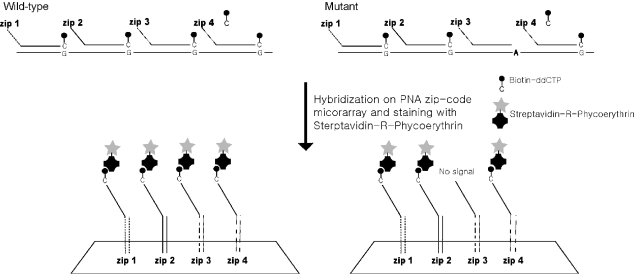
We examined the ability of a PNA zip-code microarray-based assay to detect human genetic mutations using multiplex SBE reactions. Figure 1 shows a schematic overview of the procedures. First, the PCR amplification of genomic region containing mutation sites was performed using allele-specific primers. The PCR products then served as templates for subsequent SBE reactions. The SBE primers were chimeric, containing a 5′-sequence that was complementary to a unique PNA zip-code immobilized on the array, and a 3′-sequence complementary to the genomic sequence and terminating one base before a mutation site. SBE primers corresponding to multiple mutation sites were added to a single reaction tube and single base extended in the presence of biotin-labeled ddNTPs. Next, the SBE reaction products were hybridized to the PNA zip-code microarray. To visualize the results, the hybridized PNA zip-code microarray was stained with streptavidin-R-phycoerythrin.
Figure 1.
Detection of mutations using PNA zip-code microarray with a multiplex SBE reaction.
Alternatively, a direct labeling method can be adopted to generate the labeled products using a fluorescently labeled ddNTP (e.g. Cy3-ddNTP). In this method, no further staining step is required. Biotin-labeled ddNTPs, however, are much less costly and better incorporated in the SBE reaction than Cy3-ddNTPs. Thus, the two-step strategy adopted in this study is considered to be more efficient and economical.
All zip-code sequences (Table 1) were randomly selected from a zip-code pool generated by shuffling three different tetramer sequences according to the method developed by Norman Gerry et al. (16). Each tetramer has a sequence that is different from all the others by at least two bases, and the resulting 12mer were designed to have similar Tm values.
PCR amplification with modified primers and immobilization of zip-code sequences
As a first step to test our strategy, we tried to perform PCR amplification of the exon 2 region of HNF-1α. With unmodified primers, we could not achieve efficient PCR amplification, but we reproducibly observed the improved amplification efficiency of the targeted region with a T7-linked forward primer and a T3-linked reverse primer. Particularly the specificity of the amplification reaction was enhanced. This can be attributed to the fact that the additional sequences require more specific annealing condition, eventually leading to the more specific amplification than the shorter unmodified primers. We also considered that T7 or T3 sequences could be a good candidate as a tag sequence because there is no sequence homology of the bacterial promoter sequences with human genome sequence.
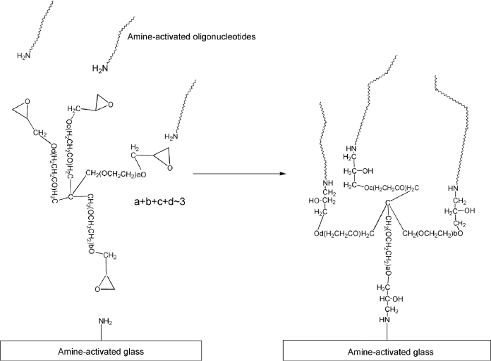
We also developed an epoxide-based strategy to covalently link the zip-code PNA molecules to an aminated glass surface. Using epichlorohydrin, we changed the hydroxyl groups of pentaerythritol ethoxylate to epoxide groups, which can react with amino groups. The resulting compound, which contained four epoxide groups within each molecule, was used as a linker between the aminohexyl–PNA or DNA and the amine-activated glass (Figure 2). Using this immobilization strategy, the binding capacity of the zip-code molecules was greatly improved in comparison with the conventional aldehyde-based direct immobilization without a linker. Furthermore, the reproducibility of the process was also improved, presumably due to the prevention of evaporation of the printed solution by the viscous epoxy compound (data not shown).
Figure 2.
Immobilization of a capture probe using an epoxide compound.
Hybridization strength and specificity of PNA–DNA duplexes
To verify these initial findings that PNA has advantageous features for use in zip-code sequences, we compared the hybridization strength and specificity of PNA and DNA molecules for their complementary DNA sequences. Three different zip-code sequences of 12mer PNA or DNA were immobilized as capture probes on the same glass surface, and a synthetic 25mer DNA (Cy3-labeled at the 5′ end) complementary to one of the three sequences was hybridized to the glass. To assess the hybridization strength, the fluorescence intensities of perfectly matched PNA–DNA and DNA–DNA duplexes were compared. To evaluate the specificity, we compared the discrimination ratio for PNA–DNA and DNA–DNA complexes, which was defined as the ratio of the signal intensity for a perfectly matched duplex (PM) to the average of the respective intensities obtained with the other two mismatched duplexes (MMavg). Because the duplex stabilities were found to be highly dependent on the ionic strength, we performed the experiments in a range of NaCl concentrations from 0.05 to 1.0 M, which correspond to 0.1× and 6× SSPE, respectively.
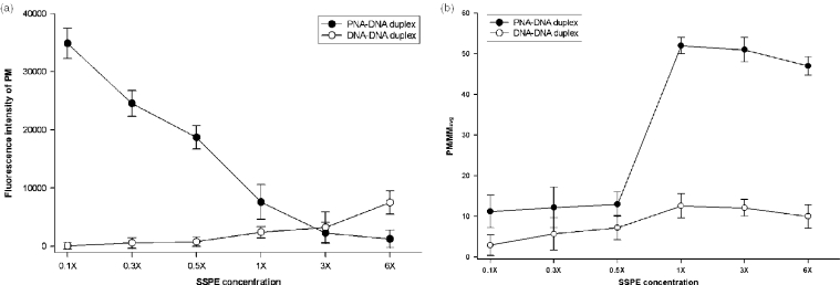
As shown in Figure 3a and b, as the ionic strength was decreased, the signal intensity of the PNA–DNA duplex increased, although its discrimination ratio decreased. At ionic strengths lower than 0.5× SSPE, significant non-specific hybridization was observed, even for the two non-complementary duplexes. In contrast, the DNA–DNA duplex showed increased signal intensity with increasing ionic strength, which is a well-known phenomenon. Up to an ionic strength of 1× SSPE, the PNA–DNA duplex showed much higher signal intensity than the DNA–DNA duplex, whereas the signals for the DNA–DNA duplex were higher than for PNA–DNA duplex above 3× SSPE. These dependencies of both duplexes on the ionic strength agree well with the findings of Tomac et al. (25). They explained that the stability of PNA–DNA duplexes decreases with increasing ionic strength due to counterion release upon duplex formation, whereas counterion association accompanies the formation of a DNA duplex. The higher stability of the PNA–DNA duplex compared with the DNA–DNA duplex was ascribed to more favorable entropic contributions, which is consistent with the counterion release that accompanies the PNA–DNA duplex formation.
Figure 3.
Effect of salt concentration on the strength and specificity of PNA–DNA duplexes and the corresponding DNA–DNA duplexes. Three different PNA or DNA zip sequences (Zip3, 7, and 9 in Table 1) were used to prepare the respective zip-code microarrays. This was followed by hybridization for 3 h with a 10 nM solution of 25mer 3′-Cy3-labeled DNA (5′-AAGAAGAAGGTCGATTTACCAAAGGA-Cy3-3′, the sequence complementary to the Zip7 sequence is bolded and underlined.) at various ionic strengths. (a) Fluorescence intensities of perfectly matched PNA–DNA and DNA–DNA duplexes. (b) The discrimination ratio for PNA–DNA and DNA–DNA complexes, which was defined as the ratio of the signal intensity for a perfectly matched duplex (PM) to the average of the respective intensities obtained with the other two mismatched duplexes (MMavg).
For the PNA–DNA duplex, there seems to be an optimal ionic strength that gives the best compromise of specificity and signal intensity of the PNA–DNA duplex. Specifically, within the tested range of ionic strengths, 1× SSPE appeared to be optimal and was therefore adopted for further diagnosis of HNF-1α mutations on PNA zip-code microarray. Under these conditions, the PNA–DNA duplex showed a little stronger intensity and a much higher specificity than the corresponding DNA–DNA duplex at its respective optimal condition. These results indicate that PNA molecules could be very promising for use as zip sequences in zip-code microarray-based assays.
Diagnosis of HNF-1α mutations using multiplex SBE genotyping
To demonstrate the usefulness of the PNA zip-code microarray, 10 selected HNF-1α mutation sites (Table 3) were genotyped for a wild-type and mutant samples using the multiplex SBE reaction. For wild-type sample, genomic DNA was isolated from the blood of an apparently healthy female, and direct sequencing confirmed that it had wild genotypes for all of the mutation sites. Mutant DNA was isolated from a MODY3 patient who fulfilled criteria for clinical MODY (3), and direct sequencing confirmed that it had two points of heterozygote mutant genotypes (CA at 1814 and CT at 1856 in Table 3) in exon 2 of HNF-1α. Since it is nearly impossible to find natural homozygote mutant samples in HNF-1α, homozygote mutant samples were artificially prepared by site-directed mutagenesis and the introduced mutations were also confirmed by direct sequencing.
Table 3.
HNF-1α mutations represented on the MODY3 chip
| Zip-code | Position | Wild-type | Mutation | Effect of mutation |
|---|---|---|---|---|
| 1 | 1821 | CGG | CAG | R131Q |
| 2 | 1814 | CCC | CAC | P129T |
| 3 | 1778 | GAA | GGA | K117E |
| 4 | 1810 | GCG | GTG | R131W |
| 5 | 1833 | GAT | GT | D135fsdelA |
| 6 | 1856 | CCA | CTA | H143Y |
| 7 | 1910 | CGC | CAC | A161T |
| 8 | 1869 | CAC | CGC | H147R |
| 9 | 1905 | CGG | CAG | R159Q |
| 10 | 1812 | ATC | AAC | I128N |
The mutation site is in bold.
The SBE reactions were carried out using chimeric primers containing both a mutation-specific sequence and a sequence complementary to the corresponding unique zip-code sequence. The SBE reaction involved a single base extension of the primer at a mutation point by DNA polymerase in the presence of biotin-labeled ddNTPs. First, we performed the experiments to select the optimum concentration of the primer for the final signal intensity on the PNA zip-code microarray. In the range from 500 pM to 1 μM, the signal intensity rather decreased with further increasing of the concentration over 250 nM presumably due to the competitive effect of unextended primers for the corresponding zip sequences on the microarray. Therefore, the primer concentration of 250 nM was adopted for the SBE reactions in this work.
Prior to the multiplex SBE reaction, we examined the ability of Thermosequenase to incorporate the correct ddNTP at the mutation site. For primer SBE02-01 (Table 2), only ddGTP should be added at the mutation site. However, there were significant fluorescence signals detected even when biotin-labeled ddATP and ddUTP was used, although the signal intensity caused by ddATP and ddUTP was lower than that caused by ddGTP, and a signal was not detected for biotin-labeled ddCTP. For the other primers in Table 2, similar non-specific incorporation was also observed.
This non-specific incorporation must be eliminated to achieve a reliable diagnosis with this strategy. We attempted to remove this problem by adjusting the extension conditions, such as the extension time. However, we did not obtain a substantial improvement in the reaction specificity in this way (data not shown). Therefore, it appeared that Thermosequenase was unable to effectively distinguish correct and incorrect ddNTP when only a single ddNTP was present in the reaction. We suspected that we could decrease the non-specific extension by adding the three ddNTPs as unlabeled molecules because the enzyme can make corrections. Therefore, we repeated the previous test with primer SBE02-01, but using all four ddNTPs, only one of which was labeled with biotin. Furthermore, we performed four separate reactions, one with each of the four different biotin-labeled ddNTPs. This time we found that a signal was obtained only with biotin-labeled ddGTP. With primers SBE02-02, SBE02-03 and SBE02-10, which have CC, AA and TT alleles, respectively, we were also able to obtain a signal only from the correct biotin-labeled ddNTP.
Using the improved strategy, we performed single SBE reactions for the detection of single point mutation at 1814 and 1856 position in Table 3 with a wild-type, a heterozygote mutant and two different homozygote mutant samples. The two homozygote mutant samples were prepared to have AA allele at 1814 and TT allele at 1856, respectively. For each mutation position, the two signal intensities resulting from a pair of two reactions (reactions with ddCTP and ddATP for 1814 and reactions with ddCTP and ddUTP for 1856) were measured and compared. As shown in Figure 4, three different kinds of alleles were successfully genotyped with this strategy. With both homozygote alleles (wild-type and mutant), non-specific signal intensities were not significant such that the signal-to-noise ratios were greater that 20 in all cases for the two mutation positions. With the heterozygote mutant samples, the signals were correctly generated from both reactions with corresponding two different ddNTPs even though the two signal intensities were not exactly same. The ratios of the two signal intensities were 1.2 and 1.5 for 1814 and 1856 mutation positions, respectively, and they are considered to be enough for the determination of the heterozygote allele type.
Figure 4.
Single point genotyping with a homozygote wild-type, homozygote mutant and heterozygote mutant sample. (a) Mutation position at 1814 (b) Mutation position at 1856. N-reaction indicates the signal resulted from the reaction with the corresponding biotin-labeled ddNTP (but T-reaction indicates the result from reaction with biotin-labeled ddUTP).
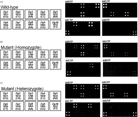
Finally, we achieved multiplex diagnoses of the 10 HNF-1α mutations with the wild-type, homozygote mutant and heterozygote mutant samples in a blinded manner. We added 10 chimeric primers corresponding to the 10 different mutation sites (Table 3) to a single tube and performed four different multiplex SBE reactions with each sample, one for each biotin-labeled ddNTP. As shown in Figure 5, correct genotyping determinations were successfully achieved with the all three samples for all 10 mutation sites, and significant non-specific extension was not detected. With the wild-type or homozygote mutant sample, the signals were generated only at the zip positions corresponding to the homozygote alleles (Figure 5a and b). The reaction of the wild-type sample with ddCTP resulted in the signal at the position for zip2, while the homozygote mutant sample with AA allele generated the signal at the zip2 position from the reaction with ddATP. When we tested the mutant sample with two heterozygote alleles at zip2 (CA) and zip6 (CT) positions, two signals were correctly generated at each of the two zip positions corresponding to the heterozygote alleles (Figure 5c). These results demonstrate that the strategy developed in this work could be reliably employed for the diagnosis of HNF-1α mutations.
Figure 5.
Multiplex diagnosis of the 10 HNF-1α mutations on a PNA zip-code microarray. In schematic representation of the PNA microarray on left side, each position in the 2 × 2 grid corresponds to an individual zip-code sequence and corresponding HNF-1α mutation. The corresponding allele is also indicated in parentheses. With each of three different samples, the right side images represent the fluorescence signals obtained from four different multiplex SBE reaction products, each containing a different biotin-labeled ddNTP. ddNTP above the right side images indicate that the image was obtained from reactions containing biotin-labeled ddATP, ddGTP, ddCTP and ddUTP, respectively. (a) homozygote wild-type sample (b) homozygote mutant sample (c) heterozygote mutant sample.
DISCUSSION
In the present study, we developed a SBE-based multiplex genotyping strategy utilizing PNA molecules as zip-code sequences. Owing to the inherent advantages of PNA molecules, the resulting PNA zip-code microarray showed a much higher specificity than the corresponding DNA microarray. Using this approach, several HNF-1α mutation sites were successfully identified on the microarray. A multiplex strategy within a single reaction is essential for high-throughput genotyping using DNA chips, but for the strategy developed in this work, four reactions are needed to diagnose multiple genetic mutations, regardless of the number of the mutations. This drawback could be overcome by using four different fluorescently labeled ddNTPs that have different emission wavelengths. With such a system, only a single multiplex SBE reaction would be required for the diagnosis of multiple mutations. However, this is currently difficult because of the availability and cost of four different fluorescent dyes that are suitable for labeling the four ddNTPs. Furthermore, because the incorporation efficiencies are different depending on the dye conjugated to the ddNTPs, the experiments should be carried out only after identifying conditions under which all four efficiencies are similar; however, it is very difficult and time-consuming to identify such conditions. In addition, a fluorescence scanner that can detect all four wavelengths is needed, and the scanning should be repeated one time for each of the four different wavelengths. Given these complexities, our strategy using four separate reactions for each of the ddNTPs appears to be simpler than the single reaction with four different dyes.
Although MODY represents only a small portion of type 2 diabetes, the number of young type 2 diabetes patients is increasing, particularly in Asian countries (2). Because the requirement for periodic testing and the prediction of diabetes development can be determined by identifying mutations in the MODY gene, the development of robust and efficient methods for the diagnosis of the mutations promises to improve clinical treatment and diagnosis. The current study covered only selected mutations in exon 2 of HNF-1α, but a higher degree of multiplexing can be achieved by simply increasing the number of SBE chimeric primers and PNA zip-code sequences on the microarray. Because of its excellent intensity and specificity, our PNA-based strategy should greatly help mutation screening and SNP genotyping technology. Furthermore, this work may help widen the application of PNA in biochip technology.
Acknowledgments
This work was supported by the Brain Korea 21 (BK21) Program and the Center for Ultramicrochemical Process Systems sponsored by KOSEF. Funding to pay the Open Access publication charges for this article was provided by the Brain Korea 21 (BK21).
REFERENCES
- 1.Hattersley A.T. Maturity onset diabetes of the young: clinical heterogeneity explained by genetic heterogeneity. Diabet. Med. 1998;15:15–24. doi: 10.1002/(SICI)1096-9136(199801)15:1<15::AID-DIA562>3.0.CO;2-M. [DOI] [PubMed] [Google Scholar]
- 2.Yamaga K., Oda N., Kaisaki P.J., Menzel S., Furuta H., Vaxillaire M., Southam L., Cox R.D., Lathrop G.M., Boriraj V.V., et al. Mutations in the hepatocyte nuclear factor-1α gene in maturity-onset diabetes of the young (MODY3) Nature. 1996;384:455–458. doi: 10.1038/384455a0. [DOI] [PubMed] [Google Scholar]
- 3.Velho G., Robert J.-J. Maturity-onset diabetes of the young (MODY): genetic and clinical characteristics. Horm. Res. 2002;57:29–33. doi: 10.1159/000053309. [DOI] [PubMed] [Google Scholar]
- 4.Shalon D., Smith S.J., Brown P.O. A DNA microarray system for analyzing complex DNA samples using two-color fluorescent probe hybridization. Genome Res. 1996;6:639–645. doi: 10.1101/gr.6.7.639. [DOI] [PubMed] [Google Scholar]
- 5.Xiang C.C., Chen Y. cDNA micorarray technology and its application. Biotechnol. Adv. 2000;18:35–46. doi: 10.1016/s0734-9750(99)00035-x. [DOI] [PubMed] [Google Scholar]
- 6.Epstein J.R., Biran I., Walt D.R. Fluorescence-based nucleic acid detection and microarrays. Anal. Chim. Acta. 2002;469:3–36. [Google Scholar]
- 7.Alexandre I., Hamels S., Defour S., Collet J., Zammatteo N., De Longueville F., Gala J.-L., Remecle J. Colorimetric silver detection of DNA microarrays. Anal. Biochem. 2001;295:1–8. doi: 10.1006/abio.2001.5176. [DOI] [PubMed] [Google Scholar]
- 8.Goto S., Takahashi A., Kamasango K., Matubara K. Single-nucleotide polymorphism analysis by hybridization protection assay on solid support. Anal. Biochem. 2002;307:25–32. doi: 10.1016/s0003-2697(02)00019-2. [DOI] [PubMed] [Google Scholar]
- 9.Sapolsky R.J., Hsie L., Berno A., Ghandour G., Mittman M., Fan J.-B. High-throughput polymorphism screening and genotyping with high-density oligonucleotides arrays. Genet. Anal. 1999;14:187–192. doi: 10.1016/s1050-3862(98)00026-6. [DOI] [PubMed] [Google Scholar]
- 10.Wang D.G., Fan J.-B., Siao C.-J., Berno A., Young P., Sapolsky R.J., Ghandour G., Perkins N., Winchester E., Spencer J., et al. Large-scale identification, mapping, and genotyping of single-nucleotide polymorphism in the human genome. Science. 1998;280:1077–1082. doi: 10.1126/science.280.5366.1077. [DOI] [PubMed] [Google Scholar]
- 11.Zabarovsky E.R., Petrenko L.P., Protopopov A., Vorontsova O., Kutsenko A.S., Zhao Y., Kilosanidze G., Zabarovska V., Rakhmanaliev E., Pettersson B., et al. Restriction site tagged (RST) microarrays: a novel technique to study the species composition of complex microbial systems. Nucleic Acids Res. 2003;31:e95. doi: 10.1093/nar/gng096. [DOI] [PMC free article] [PubMed] [Google Scholar]
- 12.Culter D.J., Zwick M.E., Carrasquillo M.M, Yohn C.T., Tobin K.P., Kashuk C., Mathews D.J., Shah N.A., Eichler E.E., Warrington J.A., Chakravariti A. High-throughput variation detection and genotyping using microarrays. Genome Res. 2001;11:1913–1925. doi: 10.1101/gr.197201. [DOI] [PMC free article] [PubMed] [Google Scholar]
- 13.Maitra M., Cohen Y., Gillespie S.E.D., Mambo E., Fukushima N., Hoque M.O., Shah N., Goggins M., Califano J., Sidransky D., Chakravarti A. The human MitoChip: a high-throughput sequencing microarray for mitochondrial mutation detection. Genome Res. 2004;14:812–819. doi: 10.1101/gr.2228504. [DOI] [PMC free article] [PubMed] [Google Scholar]
- 14.Bernstein J.A., Khodursky A.B., Lin P.-H., Lin-Chao S., Cohen S.N. Global analysis of mRNA decay and abundance in Escherichia coli at single-gene resolution using two-color fluorescent DNA microarray. Proc. Natl Acad. Sci. 2002;99:9697–9702. doi: 10.1073/pnas.112318199. [DOI] [PMC free article] [PubMed] [Google Scholar]
- 15.Relógio A., Schwager C., Richter A., Ansorge W., Valcárcel J. Optimization of oligonucleotide-based DNA microarrays. Nucleic Acids Res. 2002;30:e51. doi: 10.1093/nar/30.11.e51. [DOI] [PMC free article] [PubMed] [Google Scholar]
- 16.Gerry N.P., Witowski N.E., Day J., Hammer R.P., Barany G., Baraney F. Universal DNA microarray method for multiplex detection of low abundance point mutations. J. Mol. Biol. 1999;292:251–262. doi: 10.1006/jmbi.1999.3063. [DOI] [PubMed] [Google Scholar]
- 17.Fan J.B., Chen X., Halushka M.K., Berno A., Huang X., Ryder T., Lipshutz R.J., Lockhart D.J., Chakravarti A. Parallel genotyping of human SNPs using generic high-density oligonucleotide tag array. Genome Res. 2000;10:853–860. doi: 10.1101/gr.10.6.853. [DOI] [PMC free article] [PubMed] [Google Scholar]
- 18.Hirschhorn J.N., Skalr P., Lindblad-Toh K., Lim Y.M., Ruiz-Gutierrez M., Bolk S., Langhorst B., Schaffner S., Winchester E., Lander E.S. SBE-TAGS: an array base method for efficient single nucleotide polymorphism genotyping. Proc. Natl Acad. Sci. 2000;97:12164–12169. doi: 10.1073/pnas.210394597. [DOI] [PMC free article] [PubMed] [Google Scholar]
- 19.Nielsen P.E., Egholm M., Berg R.H., Buchardt O. Selective recognition of DNA by strand displacement with thymine-substituted polyamide. Science. 1991;254:1497–1500. doi: 10.1126/science.1962210. [DOI] [PubMed] [Google Scholar]
- 20.Nielsen P.E. Peptide nucleic acid. A molecule with two identities. Acc. Chem. Res. 1998;32:624–630. [Google Scholar]
- 21.Almarsson Ö., Bruice T.C. Peptide nucleic acid (PNA) conformation and polymorphism in PNA–DNA and PNA–RNA hybrids. Proc. Natl Acad. Sci. 1993;90:9542–9546. doi: 10.1073/pnas.90.20.9542. [DOI] [PMC free article] [PubMed] [Google Scholar]
- 22.Wittung P., Nielsen P.E., Burchardt O., Egholm M., Norden B. DNA-like double helix formed by peptide nucleic acid. Nature. 1994;368:561–563. doi: 10.1038/368561a0. [DOI] [PubMed] [Google Scholar]
- 23.Weiler J., Gausepohl H., Hauser H., Jensen O.N., Hoheisel J.D. Hybridisation based DNA screening on peptide nucleic acid (PNA) oligomer arrays. Nucleic Acids Res. 1997;25:2792–2799. doi: 10.1093/nar/25.14.2792. [DOI] [PMC free article] [PubMed] [Google Scholar]
- 24.Brandt O., Feldner J., Stephan A., Schröder M., Schnölzer M., Arlinghaus H.F., Hoheisel J.D., Jacob A. PNA microarrays for hybridisation of unlabelled DNA samples. Nucleic Acids Res. 2003;31:e119. doi: 10.1093/nar/gng120. [DOI] [PMC free article] [PubMed] [Google Scholar]
- 25.Tomac S., Sarkar M., Ratilainen T., Wittung P., Nielsen P.E., Norden B., Graslund A. Ionic effect on the stability and conformation of peptide nucleic acid complexes. J. Am. Chem. Soc. 1996;118:5544–5552. [Google Scholar]


