Abstract
Microalgal proteins are promising sources for functional nutrition and a sustainable candidate for nutraceutical formulations. They also gain importance due to emerging focus on a healthy nutrition and increase in the number of chronic diseases. In this study, dried dietary species of microalga, Chlorella vulgaris, and cyanobacterium Spirulina platensis were hydrolyzed with pancreatin enzyme to obtain protein hydrolysates. The hydrolysis yield of biomass was 55.1 ± 0.1 and 64.8 ± 3.6% for C. vulgaris and S. platensis; respectively. Digestibility, as an indicator for dietary utilization, was also investigated. In vitro protein digestibility (IVPD) values depicted that cell wall structure due to the taxonomical differences affected both hydrolysis and digestibility yield of the crude biomass (p < 0.05). Epithelial cells (Vero) maintained their viability around 70%, even in relatively higher concentrations of hydrolysates in the culture. The protein hydrolysates showed no any antimicrobial activities. This study clearly shows that the conventional protein sources in nutraceutical formulations such as soy, whey, and fish proteins can be replaced by enzymatic hydrolysates of microalgae, which shows elevated digestibility values as a sustainable and reliable source.
Keywords: Algae, Cyanobacterium, Enzymatic hydrolysis, Protein, Microalgae, Digestibility, Functional foods
Introduction
Recently, the importance of functional nutrition is gaining interest due to rapid increase on immune system deficiencies (Mayer et al. 2007) with increasing number of patients who suffer from chronic diseases as a result of reliance on processed foods (Morris et al. 2008). To supplement human nutrition, non-meal formulations are considered to be promising to elevate the health status of individuals. Thus, within the scope of functional foods and nutraceuticals obtained from grains, plants, dairy products, marine, and animal sources are trending (Mendis and Kim 2011).
Nutraceuticals are food formulations which are composed of bioactive compounds (Kim and Wijesekara 2010). These compounds can be named essential fatty acids, colorful pigments, proteins, and bioactive peptides (Gill et al. 1996). The interest on bioactive peptides and protein hydrolysates has recently gained importance due to aiding digestive dysfunction and malnutrition.
Protein hydrolysates are the forms of mixed proteins and peptides which show certain bioactivities. There are several ways to obtain protein hydrolysates; one of the most utilized methods is chemical treatment (acid or alkali catalysis) of materials with high protein content (Zhao et al. 2009). However, due to low yield, loss in activity and formation of d-amino acids are the main drawbacks of this method. Thus, lately, enzymatic hydrolysis is suggested which has opportunities as mild operation conditions and controlled environment for protein hydrolysis (Cian et al. 2012).
Within the search in novel protein sources, microalgae seem to be promising as for bioactive ingredients for several industries such as pharmaceuticals, cosmeceuticals, and nutraceuticals (Vo et al. 2013; Kim and Wijesekara 2010). There are some previous studies regarding anti-proliferative, anticoagulant (Indumathi and Mehta 2016), anti-hypertensive (Suetsuna and Chen 2001), and antioxidant (Sheih et al. 2009) peptides obtained from microalgae.
Microalgae are known to be a traditional food supplement especially in Asian culture (Oncel 2013). Among them, Chlorella species (Ko et al. 2012) and Spirulina platensis (Vo et al. 2013) have been the most utilized and approved as Generally Recognized as Safe (GRAS) categorized single cell protein nutritional supplement. Even though they have good nutritional quality, there are some undesirable features of these supplements, especially in western population (Morris et al. 2008). The fishy smell, green color, and odor of the microalgal supplements are obstacles for marketing and consumer’s choice. Another point is that human cannot digest the intact cell which is composed of cellulosic structure in microalgae and most of the biochemical component is unprocessed in gastrointestinal system (Morris et al. 2007). To increase digestibility, protein hydrolysates may be an effective solution to use microalgae in a more effective way for daily nutritional supplements.
The aim of this study is mainly to investigate whether microalgae and cyanobacteria can enzymatically be digested in vitro conditions prior to be utilized as a source for nutraceutical compound. The focus of this study is, therefore, to evaluate protein hydrolysis and in vitro protein digestibility. With this regard, Chlorella vulgaris and S. platensis are used as model commercial dietary supplements. Dried biomass is treated with digestive enzymes to display the digestibility of these species. The antimicrobial activity and in vitro cytotoxicity of the hydrolysates have also been tested.
Materials and methods
Materials
The dry powder of S. platensis cultivated in Zarrouk’s Medium is kindly provided by EGERT Inc., a spin-off company in Ege University and kept at +4 °C until experiments (Oncel and Vardar-Sukan 2008). Escherichia coli (ATCC 43895), Staphylococcus aureus (ATCC 6538), and Candida albicans (ATCC 10231) strains were kindly provided by the Industrial Microbiology Laboratory of Bioengineering Department of Ege University. The chemicals for BG-11 medium, l-tyrosine, casein, and solvents were purchased from Merck, Germany. Casein, trypsin, pancreatin from porcine pancreas and α-chymotrypsin, Bovine Serum Albumin (BSA), and MTT (3-(4,5-dimethyl-2-thiazolyl)-2.5-diphenyl-2H-tetrazolium bromide) were purchased from Sigma Chemical Co., USA. Gentamycin as a reference antibiotic, and animal cell culture chemicals were purchased from Biochrom, Germany and GreinerBio-one, Germany. Vero (kidney epithelial cells of African green monkey) cells were kindly provided from Animal Cell Culture and Tissue Engineering Laboratory (EgeReact) of Bioengineering Department at Ege University.
Microalgae cultivation
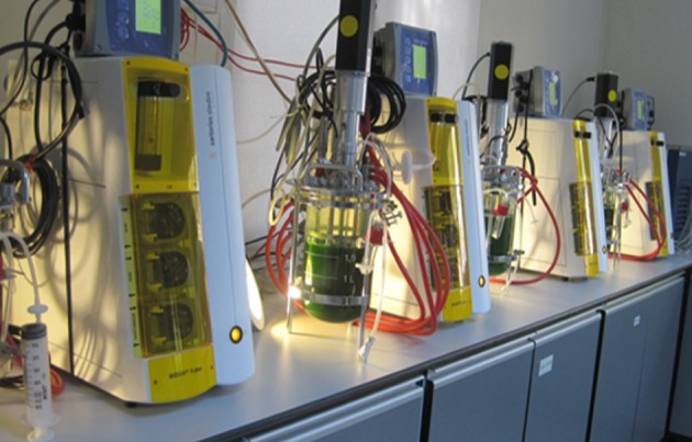
Green microalga C. vulgaris (SAG 211-12) is used in this study. BG-11 culture medium is supplemented with 5 g/L glucose and 1 g/L proteose peptone as organic carbon and nitrogen sources for photomixotrophic cultivation (Kose and Oncel 2015). The microalgae stock cultures are kept at 23 ± 2 °C as in the form of agar slants under continuous illumination of 30 µ/Em2 s. The cells on agar surface are suspended in BG-11 medium in 5 mL and monitored visually until a significant color change to a darker green has been observed and then transferred to 50 and 100 mL Erlenmayer flasks, respectively. The liquid cultures are cultivated and kept under continuous illumination of 70 µ E/m2 s in an orbital shaker (ZHWY-2112B, ZCICHENG) at 120 rpm for effective mixing and homogenous dispersion of light. 100 mL Erlenmayer cultures are used as inoculum. The biomass production of C. vulgaris is done in 2 L Continuous Stirred Tank Bioreactor (CSTR) system (Sartorious Biostat A-Plus, Germany) supplemented with external illumination at intensity of 70 µ E/m2 s (Fig. 1). The culture is inoculated with an initial cell concentration of 1 × 10⁴ mg/L at mid-logarithmic growth phase. The operational conditions are 1 vvm air flow, 23 ± 2 °C and 120 rpm agitation. At late-logarithmic growth phase, where biomass concentration (g/L) has reached the maximum level (approximately 7 days), the culture is harvested at 3000×g for 8 min in centrifuge (Universal 32R, Hettich, Germany). The cells are then suspended in distilled water and re-centrifuged for at least two times. The harvested biomass is kept at −20 °C and freeze dried. The dried biomass is kept at +4 °C until use.
Fig. 1.

Bioreactors used in production of Chlorella vulgaris
Enzyme activity
Prior to digestion experiments, enzyme activities of pancreatin from porcine pancreas, trypsin, and α-chymotrypsin are determined using sodium caseinate as a reference protein. The standard curve is prepared using 5 mM tyrosine solution with an increasing concentration of 10, 50, 100, 150, 200, 250, 350, and 450 nmol. The proteolytic activity of the enzyme is determined according to the amount of enzyme to catalyze the release of 1 nM tyrosine from 6% of casein solution at 37 °C in 1 min. The temperature is controlled with a water bath (Memmert, Germany).
Enzymatic hydrolysis
The crude biomass is hydrolyzed using proteolytic enzyme, pancreatin from porcine pancreas (Sigma-Aldrich, USA). The E/S ratio is used at 8% level as previously described (Kose and Oncel 2015). The reaction is stopped using heat treatment at 85 °C for 20 min. The mixture is centrifuged at 3000×g for 10 min at +4 °C. Supernatant which contains protein hydrolysates is kept at −20 °C until freeze drying. The freeze-dried samples of protein hydrolysates and residual biomass are also preserved at +4 °C for further experiments.
Determination of in vitro protein digestibility (IVPD) values
In vitro protein digestibility (IVPD) is done to determine apparent digestibility of microalgal samples. The assay is modified according to a study done by Hsu et al. (1977). The samples are treated with pancreatin before the IVPD assay is done with trypsin and α-chymotrypsin. 5 mg of dried untreated biomass (UTS), protein hydrolysates, and residual biomass of C. vulgaris and S. platensis are suspended in 5 mL distilled water at pH 8.184 U/mL α-chymotrypsin and 157 U/mL trypsin is dissolved in 1 mL of distilled water at pH 8. The experiments are done at 37 °C and pH drop is recorded for 10 min with 1 min of intervals. Sodium caseinate is used as reference protein for good digestibility features. The digestibility of sodium caseinate is accepted as 100%. The IVPD values are calculated according to the following equation:
| 1 |
where pHS0 and pHS10 correspond to the initial and final pH values for sample, respectively; pHC0 and pHC10 correspond to the initial and final pH values for casein, respectively.
Antimicrobial activity
Gentamicin and the samples are dissolved and suspended in distilled water with a concentration of 1 mg/mL. The cell suspensions of the organisms are prepared from solid agar slants and suspended in sterile serum physiologic. For antimicrobial activity determination liquid samples of E. coli (ATCC 43895), S. aureus (ATCC 6538) are inoculated onto Mueller–Hinton Agar (MHA) medium agar plates and C. albicans (ATCC 10231) to Sabouraud Dextrose Agar (SDA) medium agar plates. 20 µg of gentamicin, untreated samples (UTS), and protein hydrolysates are absorbed onto sterile disks for disc diffusion test. The disks are placed at the center of the agar plates (90 mm). The plates are incubated first +4 °C for 2 h and transferred to 37 °C incubator and the zones are visually observed after 24 h of incubation. The zone diameter (cm) is measured for dose-independent antimicrobial activity.
Vero cell cultures
Cytotoxicity of different concentrations of hydrolysates from S. platensis and C. vulgaris (0–100 µg/mL) on Vero cells was determined with MTT assay (Mosmann 1983). Cells were cultured using RPMI-1640 medium supplemented with 10% fetal bovine serum (FBS), 2 mM l-glutamine, and 10 µg/mL gentamycin. Cultures were incubated in a humidified atmosphere with 5% CO2 at 37 °C and sub-cultured using trypsin/EDTA solution, 0.05/0.2% (m/v) in phosphate buffer saline (PBS). With a cell seeding density of 1 × 10⁵ cell/mL, Vero cultures were prepared in wells of a 96-well cell culture plate with 100 µL/well for cytotoxicity determination (Deniz et al. 2016). After an overnight incubation, different amounts of hydrolysates, 0, 25, 50, and 100 µg/mL, were prepared using fresh cell culture medium (serum concentration was lowered to 4%) and filtered with syringe filters with 0.22 µm pore size for sterilization. A modified MTT assay was used to determine the viable cell percentage after incubation of cells with extracts for 24, 48, and 72 h. At the end of the incubation period with extracts, cell medium was replaced with a fresh medium supplemented with 10% MTT solution (5 µg/mL). Water insoluble formazan crystals were dissolved with 100 µL/well dimethyl sulfoxide (DMSO) and absorbance values were read with a spectrophotometer (Molecular Devices, UK) at 570 nm (with a reference filter, 690 nm). Viability values of cultures were calculated using the following equation (Yalci et al. 2014):
| 2 |
Analytical procedures
Light intensities were measured with a quantum sensor (Li-192SA) connected to a light meter (Licor, Lincoln, USA, Li-250A). Cells are counted using Thoma-type hemocytometer under microscope. Dry weight of the biomass is determined with direct weight measurement after freeze drying. Total soluble protein content is determined according to Lowry method (Lowry et al. 1951). BSA is used as reference protein. The solutions for Lowry assay are freshly prepared prior to utilization.
Statistical analysis
The data are provided as mean ± standard deviation. The data are analyzed using Microsoft Excel for statistics. All the experiments are done with triplicates. One-way ANOVA test is done for the experimental results with a 5% level of probability (p < 0.05).
Results and discussion
With the developments in microalgal biotechnology, the nutritional fact of microalgae has been one of the interests in marine biotechnology (Morris et al. 2007). Microalgae containing more than 50% of protein content on dry weight basis (mostly Chlorella and Spirulina species) have been utilized as single cell protein source. Even though in industrial biotechnology without considering the taxonomic differences, Spirulina is counted as a microalga (Pulz and Gross 2004), but it is actually a cyanobacteria with cell wall structure similar to prokaryotic bacteria, as Spirulina does not contain cellulosic cell wall which is an obstacle for a functional digestion in intestinal trait. Thus, pretreatment techniques are necessary to adopt increased digestibility of microalgae sources to gain benefit to fit the desires of the people with digestion dysfunctions.
Effect of enzymatic hydrolysis on microalgal biomass
All experiments with C. vulgaris are conducted in a series of 2 L photobioreactors (Fig. 1). Prior to any experiments, protein content of C. vulgaris and S. platensis biomass is determined.
The total protein values for untreated biomass of Chlorella vulgaris were 52.2 ± 0.9 and 52.9 ± 0.7% for S. platensis. The initial protein values of two samples are found to be quite similar. This can be due to photomixotrophic cultivation of C. vulgaris. The glucose and peptone supplement in C. vulgaris cultures are considered to have a significant effect on the increase of the total protein amount (Becker 2007). Metabolic pathway analysis of the supplemented organic sources is known to trigger the protein synthesis in microalgae, especially in the case of nitrogen addition (Chen 1996).
The dried biomass of C. vulgaris and S. platensis is hydrolyzed using pancreatin to obtain non-specific protein hydrolysate mixtures. The pancreatin enzyme is chosen considering the enzyme as a fundamental digestion enzyme in gastrointestinal tract. In enzymatic hydroloysis experiment, the % hydrolysis yields have been determined. The % hydrolysis value for C. vulgaris is 55.1 ± 0.1 and 64.8 ± 3.6% for S. platensis. The ANOVA result of the hydrolysis yields concludes that the microalgae species have an impact on the hydrolysis yield (p < 0.05). Cellulosic cell wall layers of C. vulgaris give a chance to survive in natural harsh conditions, however, when it comes to the protein hydrolysis; it has a negative effect on the utilization of Chlorella for nutritional formulations (Monks et al. 2013).
Figure 2 shows before and after images of microalgal biomass. The first picture (Fig. 2a) has a dark green color which is untreated sample of crude biomass. This form of the biomass can also be utilized in formulations for single cell protein supplements. However, in this study, we used pancreatic digestion to disrupt cell integrity and obtain protein hydrolysates. With this method, the algae-specific green color is lost and instead a yellowish powder is formed which is rich in proteins (Kose and Oncel 2015). Residual biomass can also be recovered for several industries as animal meals, aquaculture, and fertilizers. Considering the whole process, the left biomass is also worth to be input of several other industries.
Fig. 2.
Images of untreated sample (a), hydrolyzed biomass (b), and residual cell debris (c)
IVPD values
Digestibility of algal proteins as valuable functional food derivatives is an important characteristic for common use and preference. Thus, digestibility values affect its practical use in any product related to nutrition. Recently, plant materials are preferred in food formulation as protein sources (Kong et al. 2008). Among them, algae serve as a sustainable one and rather than utilizing the biomass for biofuel nutraceutical industry seems more feasible. However, the cell wall has a significant impact on the digestibility values among cyanobacteria and microalgae. The cell wall of cyanobacteria does not contain thick cellulosic layers but composed of peptidoglycan resembling Gram-negative bacteria cell wall with a thickness of 40–60 nm, which creates a mild environment in human digestive system. However, the cell wall of microalga, C. vulgaris as a model in this study, consists of thick cellulosic cell wall which corresponds to 10% of dry matter. Thus, the existence of this highly rigid cellulosic wall structure is considered to be a barrier to obtain high levels of digestibility ratios (Monks et al. 2013).
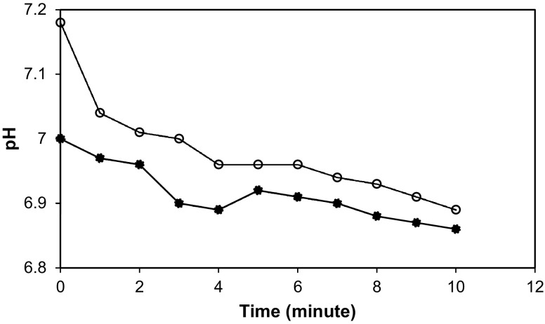
The digestibility of a protein source determines the availability of free amino acids which can be utilized within the body. To conduct IVPD test, pH drop method is adopted. Because pancreatin is used as a primary digestive enzyme for hydrolysis of microalgal proteins, the technique has been modified and only trypsin and α-chymotrypsin are used for IVPD assay. In this in vitro experimental system, pH drop is a sign for the release of free amino acids from protein. At very beginning, a rapid decrease occurs, and during 10 min of time period, the pH drop rate decreases (Fig. 3).
Fig. 3.
pH drop of the hydrolysates for 10 min according to the IVPD assay. Black circle, C. vulgaris; open circle, S. platensis
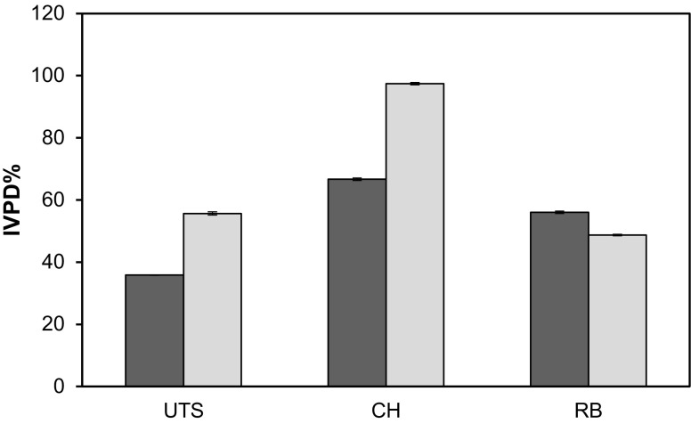
The digestibility values of samples are presented in Fig. 4. According to the results, the untreated sample of C. vulgaris biomass showed poor digestibility values around 35%. However, in S. platensis, the digestibility of crude biomass was found to be more than 50%. It is also discussed that cell wall composition decreases the availability of the biomass in digestive trait. Thus, in vitro values show good correlation with previous studies (Janczyk et al. 2007; Morris et al. 2008). IVPD values of the pancreatic hydrolysates showed a great increase in terms of apparent digestibility. Almost 70% of C. vulgaris biomass was found to be digestible and this value was even 97% for S. platensis. Considering casein which has a 100% of digestibility value, it can be said that S. platensis protein hydrolysates are good candidates to replace dairy based proteins in food and nutritional formulations. The low digestibility values of C. vulgaris when compared to S. platensis (p < 0.05) is thought to be a result of degree of hydrolysis due to cell wall structure which is also discussed in enzymatic hydrolysis “Effect of enzymatic hydrolysis on microalgal biomass”.
Fig. 4.
IVPD% values for C. vulgaris and S. platensis. The IVPD values show the in vitro protein digestibility of the samples (UTS, untreated sample, CH, cell hydrolysate, RB, residual biomass, C. vulgaris, dark grey square, and S. platensis, grey square)
Even if compared to S. platensis, C. vulgaris hydrolysates are lower in digestibility compared to S. platensis, and still have potential as a promising source for further processes.
Vero cell cultures
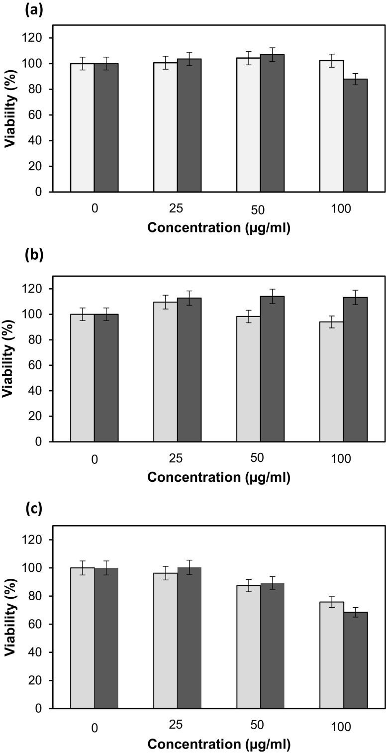
Vero cells were used to determine the cytotoxicity of different concentrations of hydrolysates (0, 25, 50, and 100 µg/mL) obtained from C. vulgaris and S. platensis biomass. According to results given in Fig. 5, cytotoxicity levels of the protein hydrolysates on Vero cells were 25% for S. platensis and 32% for C. vulgaris with 100 µg/mL concentration after 72 h. On the other hand, viability of cells was higher than that of 85% for 24 and 48 h cultures. Different extraction methods for the same/different molecules, harvested with different methods from microalgae, have been studied for their cytotoxic and anti-proliferative effects, meanwhile proliferative effects and wound healing (Thangam et al. 2013; Deniz et al. 2016). With the presented nutritional components from the hydrolysates, viability remained high, until the end of 72 h.
Fig. 5.
Viability of Vero cells incubated with different concentrations of protein hydrolysates harvested from C. vulgaris (dark grey square) and S. platensis (grey square) for 24 h (a), 48 h (b), and 72 h (c)
Antimicrobial activity of hydrolysates
Microalgal protein hydrolysates have also been screened for antimicrobial activities (Mayer et al. 2009). In this study, E. coli (Gram-negative bacteria), S. aureus (Gram-positive bacteria), and C. albicans (fungus) strains have been used in the frame of disc diffusion tests. The dose-independent antimicrobial activity of the microalgal samples is determined measuring the zone diameter. The zone diameter was 3.5 ± 0.5 cm for all of the test organisms which are exposed to reference antibiotic gentamicin. In the UTS, pancreatic hydrolysates and residual biomass were screened for antimicrobial activity; however, there were no observed zones on petri dishes. Even though some microalgae are known to synthesize antimicrobial compounds under stress conditions, in this study at least, pancreatic digests did not display any antimicrobial activity.
Other than a particular compound with antimicrobial activity, some previous studies report that solvent extracts in acetone, methanol, ethanol, or chloroform can show significant antimicrobial activity (Syed et al. 2015; Wan et al. 2014). The reason for this apparent activity is that because in solvent extraction methods, fatty acids and some algae-derived pigments are extracted. It is known that fatty acids like linolenic acid can show antimicrobial activity. In this study, due to enzymatic hydrolysis even if the microalgae species may have antimicrobial sequences, they may not be released within the enzyme hydrolysis environment.
Conclusion
The protein quality of a protein rich source is determined by the bioavailability values. With high protein content, antioxidant pigments, essential fatty acids, and algae-specific biochemical compounds, microalgae and cyanobacteria seem to be a promising feedstock for functional food formulation. Currently, the protein malnutrition is increasing and non-meal food formulations are marketed to provide sustainable and reliable protein sources. Recently, marketed products of Chlorella species are cell wall broken products with mechanical effects. Hydrolysates from S. platensis and C. vulgaris were used in the cultures of a mammalian cell line and they showed 25 and 32% cytotoxicity after 72 h, respectively. With an increase in the digestibility values and low cytotoxity, this study clearly states that microalgal proteins are potential for novel functional food formulation, especially for the ones who suffer from chronic disease with digestive malfunctions. As a final result, protein hydrolysates from microalgae and cyanobacteria can be replaced with conventional egg, dairy, and grain proteins as a reliable and sustainable source.
Acknowledgements
The authors are thankful to Prof. Fazilet Vardar-Sukan, Prof. Figen Zihnioglu, and also Dr. Zeliha Demirel from Ege University for their valuable discussions, advices, and shares on their expertise related to the subject; to the EGERT Inc. for their kind donation of Spirulina platensis biomass; to Prof. Ismet Deliloglu-Gurhan for supplying the cell line; and, finally, to Ege University Scientific Research Projects (BAP) for their financial support.
Compliance with ethical standards
Conflict of interest
All the authors declare that there is no conflict of interest.
References
- Becker EW. Microalgae as a source of protein. Biotechnol Adv. 2007;25(2):207–210. doi: 10.1016/j.biotechadv.2006.11.002. [DOI] [PubMed] [Google Scholar]
- Chen F. High cell density culture of microalgae in heterotrophic growth. Trends Biotechnol. 1996;14(11):421–426. doi: 10.1016/0167-7799(96)10060-3. [DOI] [Google Scholar]
- Cian RE, Martínez-Augustin O, Silvina RD. Bioactive properties of peptides obtained by enzymatic hydrolysis from protein byproducts of Porphyra columbina. Food Res Int. 2012;49(1):364–372. doi: 10.1016/j.foodres.2012.07.003. [DOI] [Google Scholar]
- Deniz I, Ozen MO, Yesil-Celiktas O. Supercritical fluid extraction of phycocyanin and investigation of cytotoxicity on human lung cancer cells. J Supercrit Fluids. 2016;108:13–18. doi: 10.1016/j.supflu.2015.10.015. [DOI] [Google Scholar]
- Gill I, López-Fandiño R, Jorba X, Vulfson EN. Biologically active peptides and enzymatic approaches to their production. Enzyme Microb Technol. 1996;18(3):162–183. doi: 10.1016/0141-0229(95)00097-6. [DOI] [PubMed] [Google Scholar]
- Hsu H, Vavak DL, Satterlee LD, Miller G. A multienzyme technique for estimating protein digestibility. J Food Sci. 1977;42(5):1269–1273. doi: 10.1111/j.1365-2621.1977.tb14476.x. [DOI] [Google Scholar]
- Indumathi P, Mehta A. A novel anticoagulant peptide from the nori hydrolysate. J Funct Food. 2016;20:606–617. doi: 10.1016/j.jff.2015.11.016. [DOI] [Google Scholar]
- Janczyk P, Franke H, Souffrant WB. Nutritional value of Chlorella vulgaris: effects of ultrasonication and electroporation on digestibility in rats. Anim Feed Sci Tech. 2007;132:163–169. doi: 10.1016/j.anifeedsci.2006.03.007. [DOI] [Google Scholar]
- Kim SK, Wijesekara I. Development and biological activities of marine-derived bioactive peptides: a review. J Funct Food. 2010;2(1):1–9. doi: 10.1016/j.jff.2010.01.003. [DOI] [Google Scholar]
- Ko SC, Kang N, Kim EA, Min CK, Lee SH, Kang SM, Lee JB. A novel angiotensin I-converting enzyme (ACE) inhibitory peptide from a marine Chlorella ellipsoidea and its antihypertensive effect in spontaneously hypertensive rats. Process Biochem. 2012;47(12):2005–2011. doi: 10.1016/j.procbio.2012.07.015. [DOI] [Google Scholar]
- Kong X, Guo M, Hua Y, Cao D, Zhang C. Enzymatic preparation of immunomodulating hydrolysates from soy proteins. Biores Technol. 2008;99(18):8873–8879. doi: 10.1016/j.biortech.2008.04.056. [DOI] [PubMed] [Google Scholar]
- Kose A, Oncel SS. Properties of microalgal enzymatic protein hydrolysates: biochemical composition, protein distribution and FTIR characteristics. Biotechnol Rep. 2015;6:137–143. doi: 10.1016/j.btre.2015.02.005. [DOI] [PMC free article] [PubMed] [Google Scholar]
- Lowry OH, Rosebrough NJ, Farr AL, Randall RJ. Protein measurement with the folin phenol reagent. J Biol Chem. 1951;193(1):265–275. [PubMed] [Google Scholar]
- Mayer AMS, Rodríguez AD, Berlinck RGS, Hamann MT. Marine pharmacology in 2003–4: marine compounds with anthelminthic, antibacterial, anticoagulant, antifungal, anti-inflammatory, antimalarial, antiplatelet, antiprotozoal, antituberculosis, and antiviral activities; affecting the cardiovascular, immune and nervous systems, and other miscellaneous mechanisms of action. Comp Biochem Physiol C Toxicol Pharmacol. 2007;145(4):553–581. doi: 10.1016/j.cbpc.2007.01.015. [DOI] [PMC free article] [PubMed] [Google Scholar]
- Mayer AMS, Rodríguez AD, Berlinck RGS, Hamann MT. Marine pharmacology in 2005–6: marine compounds with anthelmintic, antibacterial, anticoagulant, antifungal, anti-inflammatory, antimalarial, antiprotozoal, antituberculosis, and antiviral activities; affecting the cardiovascular, immune and nervous systems, and other miscellaneous mechanisms of action. Biochim Biophys Acta. 2009;1790(5):283–308. doi: 10.1016/j.bbagen.2009.03.011. [DOI] [PMC free article] [PubMed] [Google Scholar]
- Mendis E, Kim SK. Present and future prospects of seaweeds in developing functional foods. Adv Food Nutr Res. 2011;64:1–15. doi: 10.1016/B978-0-12-387669-0.00001-6. [DOI] [PubMed] [Google Scholar]
- Monks LM, Rigo A, Mazutti MA, Oliveira JV, Valduga E. Use of chemical, enzymatic and ultrasound-assisted methods for cell disruption to obtain carotenoids. Biocatal Agric Biotechnol. 2013;2(2):165–169. [Google Scholar]
- Morris HJ, Carrillo O, Almarales A, Bermúdez RC, Lebeque Y, Fontaine R, Llauradó G, Beltrán Y. Immunostimulant activity of an enzymatic protein hydrolysate from green microalga Chlorella vulgaris on undernourished mice. Enzyme Microb Technol. 2007;40(3):456–460. doi: 10.1016/j.enzmictec.2006.07.021. [DOI] [Google Scholar]
- Morris HJ, Almarales A, Carrillo O, Bermúdez RC. Utilisation of Chlorella vulgaris cell biomass for the production of enzymatic protein hydrolysates. Biores Technol. 2008;99(16):7723–7729. doi: 10.1016/j.biortech.2008.01.080. [DOI] [PubMed] [Google Scholar]
- Mosmann T. Rapid colorimetric assay for cellular growth and survival: application to proliferation and cytotoxicity assays. J Immunol Methods. 1983;65(1–2):55–63. doi: 10.1016/0022-1759(83)90303-4. [DOI] [PubMed] [Google Scholar]
- Oncel SS. Microalgae for a macroenergy world. Renew Sust Energ Rev. 2013;26:241–264. doi: 10.1016/j.rser.2013.05.059. [DOI] [Google Scholar]
- Oncel S, Vardar-Sukan F. Comparison of two different pneumatically mixed column photobioreactors for the cultivation of Artrospira platensis (Spirulina platensis)”. Biores Technol. 2008;99(11):4755–4760. doi: 10.1016/j.biortech.2007.09.068. [DOI] [PubMed] [Google Scholar]
- Pulz O, Gross W. Valuable products from biotechnology of microalgae. Appl Microbiol Biotechnol. 2004;65(6):635–648. doi: 10.1007/s00253-004-1647-x. [DOI] [PubMed] [Google Scholar]
- Sheih IC, Wu TK, Fang TJ. Antioxidant properties of a new antioxidative peptide from algae protein waste hydrolysate in different oxidation systems. Biores Technol. 2009;100(13):3419–3425. doi: 10.1016/j.biortech.2009.02.014. [DOI] [PubMed] [Google Scholar]
- Suetsuna K, Chen JR. Identification of antihypertensive peptides from peptic digest of two microalgae, Chlorella vulgaris and Spirulina platensis. Mar Biotechnol. 2001;3(4):305–309. doi: 10.1007/s10126-001-0012-7. [DOI] [PubMed] [Google Scholar]
- Syed S, Arasu A, Ponnuswamy I. The uses of Chlorella vulgaris as antimicrobial agent and as a diet: the presence of bio-active compounds which caters the vitamins, minerals in general. Int J Bio Sci Bio Technol. 2015;7(1):185–190. doi: 10.14257/ijbsbt.2015.7.1.19. [DOI] [Google Scholar]
- Thangam R, Suresh V, Asenath Princy W, Rajkumar M, Senthilkumar N, Gunasekaran P, Rengasamy R, Anbazhagan C, Kaveri K, Kannan S. C-Phycocyanin from Oscillatoria tenuis exhibited an antioxidant and in vitro antiproliferative activity through induction of apoptosis and G 0/G1 cell cycle arrest. Food Chem. 2013;140(1–2):262–272. doi: 10.1016/j.foodchem.2013.02.060. [DOI] [PubMed] [Google Scholar]
- Vo TS, Ryu BM, Kim SK. Purification of Novel anti-inflammatory peptides from enzymatic hydrolysate of the edible microalgal Spirulina maxima. J Funct Food. 2013;5(3):1336–1346. doi: 10.1016/j.jff.2013.05.001. [DOI] [Google Scholar]
- Wan HDM, Chen CC, Huynh P, Chang JS. Exploring the potential of using algae in cosmetics. Biores Technol. 2014;31:355–362. doi: 10.1016/j.biortech.2014.12.001. [DOI] [PubMed] [Google Scholar]
- Yalci HT, Ozen MO, Gocmen B, Nalbantsoy A. Effect of Ottoman viper [Montivipera xanthina (Gray, 1849)] venom on various cancer cells and on microorganisms”. Cytotechnology. 2014;66(1):87–94. doi: 10.1007/s10616-013-9540-z. [DOI] [PMC free article] [PubMed] [Google Scholar]
- Zhao X, Peng F, Cheng K, Liu D. Enhancement of the enzymatic digestibility of sugarcane bagasse by alkali–peracetic acid pretreatment. Enzyme Microb Technol. 2009;44(1):17–23. doi: 10.1016/j.enzmictec.2008.07.011. [DOI] [Google Scholar]


