Abstract
We establish a powerful poly(4-styrenesulfonate) (PSS)-treated strategy for sulfur vacancy healing in monolayer MoS2 to precisely and steadily tune its electronic state. The self-healing mechanism, in which the sulfur vacancies are healed spontaneously by the sulfur adatom clusters on the MoS2 surface through a PSS-induced hydrogenation process, is proposed and demonstrated systematically. The electron concentration of the self-healed MoS2 dramatically decreased by 643 times, leading to a work function enhancement of ∼150 meV. This strategy is employed to fabricate a high performance lateral monolayer MoS2 homojunction which presents a perfect rectifying behaviour, excellent photoresponsivity of ∼308 mA W−1 and outstanding air-stability after two months. Unlike previous chemical doping, the lattice defect-induced local fields are eliminated during the process of the sulfur vacancy self-healing to largely improve the homojunction performance. Our findings demonstrate a promising and facile strategy in 2D material electronic state modulation for the development of next-generation electronics and optoelectronics.
Two-dimensional MoS2 homojunctions, considered potential building blocks for next generation flexible electronics, currently suffer from quick degradation. Here, Zhang and co-workers use a self-healing sulfur vacancy mechanism to produce a MoS2 photodiode that possesses long term stability.
Because of its reduced dimensions, chemical stability1, proper direct band gap, highly efficient light absorption and piezoelectricity, two-dimensional (2D) molybdenum disulfide MoS2 has the potential in developing next-generation flexible, transparent and wearable nanodevices2,3. As an important research aspect, many researchers have focused on creating MoS2 homojunction, the fundamental building block of modern electronics4. Because of its identical crystal structure and continuous band alignments in the interface, the MoS2 homojunctions display ideal current rectifying behaviour and highly efficient photoresponse than those of heterojunctions5.
So far, it is the key issue to find a precise, stable and facile strategy to develop a steady and effective homojunction. The crucial process of building 2D homojunction is creating a graded junction by controlling the intrinsic carrier concentration and the work function. Conventional controls include classical doping and surface transfer doping6. Classical doping is realized by incorporating various atoms into 2D materials via thermal annealing. However, 2D materials obtained from this approach is only suitable for fabricating vertical homojunctions which always suffer from large contact resistance7. On the other hand, surface transfer doping is induced by strong electron-donating or withdrawing chemical species attachment on the 2D materials to achieve an effective coupling8. A significant limitation for surface transfer doping is the presence of inert dangling bond-free surface on 2D materials, which will reduce the doping efficiency of dopants9. Moreover, the adsorbed dopants, such as O2, BV and AuCl3 (refs 4, 5, 10), could desorb on 2D materials surface and react with reactive molecules in surroundings, leading to the short-term stability. As-mentioned two methods will induce additional lattice defects, which tend to introduce the local field or Coulomb’s scattering sites. As a result, the electronic and optoelectronic characteristics of 2D materials degrade9.
In this work, a lateral chemical vapour deposition (CVD) monolayer MoS2 homojunction is constructed by precise selected-area sulfur vacancy self-healing (SVSH) via nonoxidizing acids poly(4-styrenesulfonate) (PSS). The self-healing mechanism is that the sulfur vacancies are healed spontaneously by the sulfur adatom clusters on MoS2 surface through a PSS-induced hydrogenation process. Healing intrinsic lattice defects is a fundamental and efficient approach to control the work function without introducing additional local fields. Simultaneously, a work function difference of ∼150 meV between the as-grown and self-healed MoS2 was achieved to construct the homojunction. The rectifying performance of the homojunction shows no degradation after two months storing under ambient conditions. The homojunction shows perfect diode behaviour and excellent photoresponsivity of ∼308 mA W−1 at zero bias. Our findings pave a powerful strategy to control the 2D materials work functions and develop its homogeneous diodes for ultrathin, flexible, transparent and wearable electronics and optoelectronics.
Results
Complex characterization of sulfur vacancy self-healing
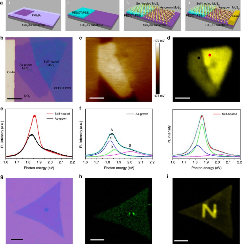
The lateral MoS2 monolayer homojunction was fabricated by PSS-induced selected-area SVSH. Figure 1a describes the building process of the homojunction device A1 (Supplementary Methods and Supplementary Figs 1, 2 and 3). To characteristic the PSS-induced SVSH, a Kelvin probe force microscopy (KPFM) was employed to verify the work function variation of monolayer MoS2. The contact potential difference (CPD) between the AFM tip (Pt/Ir coated tips) and the sample is defined as11,12
Figure 1. Complex characterization of sulfur vacancy self-healing.
(a) Construction process of the monolayer MoS2 homojunction. (b) Optical microscopy (OM) image of the device A1. Scale bar, 5 μm. (c) Corresponding 2D surface potential image. Scale bar, 5 μm. (d) Photoluminescence (PL) intensity mapping. Scale bar, 5 μm. (e) PL spectrums acquired from different regions highlighted in d. (f) Comparison of the deconvoluted PL spectrum features in e. The experimental results are reproduced by the sum (cyan) of three peaks (trion X−, blue; exciton X0, green; exciton B, purple) assumed by Lorentzian functions. (g–i) OM image, Raman mapping constructed by integrating E12g mode and PL intensity mapping for a monolayer MoS2 patterned by e-beam lithography (EBL) into the shape of the uppercase letter ‘N’ and PEDOT:PSS solution induced SVSH. Scale bar, 10 μm.
 |
where φtip, φsample and q are the work functions of the tip sample and the elementary charge, respectively. So the resulting KPFM image maps the variation of surface potential corresponding to the work function of the sample surface. Similar to the optical microscopy (OM) image (Fig. 1b), the 2D surface potential image intuitively depicts the triangle morphology of CVD monolayer MoS2, PEDOT:PSS and Cr/Au electrode (Fig. 1c). Importantly, there is an apparent brightness difference near the boundary between the self-healed and as-grown MoS2. On the other hand, Supplementary Fig. 4b,c indicates there is no work function difference between the self-healed MoS2 and PEDOT:PSS electrode, which suggests no potential barrier between them exists. Actually, a lateral n-p-n junction device can be also fabricated through the PSS-induced SVSH, in which the conductive channel of MoS2 lays across the PEDOT:PSS. The n-p-n junction device shows double Schottky rectifying characteristic (Supplementary Fig. 4d)13.
The lower the surface potential is, the higher the work function is and the lower the electron concentration is. According to the relevant literature14,15, the change of the MoS2 electron concentration generally will bring about the fluctuation of the photoluminescence (PL) spectrum. Similar to its 2D surface potential image, PL intensity mapping of the device also depicts the triangle morphology of CVD monolayer MoS2 (Fig. 1d). Besides, compared to the as-grown region, the PL spectrum intensity of the self-healed region was significantly enhanced, thus forming a clear dividing line between the as-grown and self-healed regions.
In fact, not only the PL spectrum intensity of the self-healed one is drastically enhanced, but also the peak energy is obviously blue shifted about 22 meV by PSS-induced SVSH (Fig. 1e). Previous studies reveal PL spectrum of monolayer MoS2 is composed of A exciton and B exciton (∼2.0 eV, purple). The prominent A exciton peak could be further evolved into exciton (X0; ∼1.86 eV; green) and trion (X−; ∼1.82 eV; blue) peaks. A negative trion is a quasiparticle composed of two electrons and a hole and formed through binding a neutral excition (a photogenerated electron–hole pair) to an electron, the process consumes energy of ∼40 meV (refs 8, 16, 17). By analysing the exciton peaks of the trion (X−) and exciton (X0), it explicitly manifests that the trion intensity is independent of PSS-induced SVSH (Fig. 1f). This phenomenon is ascribed to the large trion binding energy in monolayer MoS2 (ref. 14). Simultaneously, the exciton intensity is almost twofold after PSS treatment, which is correlated with the decrease of the intrinsic heavy electron (n-type) doping in CVD MoS2 (refs 14, 17, 18). This fact strongly suggests that the neutral excitons recombine rather than forming negative trions due to the decrease of the electron concentration. In our case, such electron concentration decrease is caused by the SVSH of the self-healed MoS2. Our experimental results are consistent with the pronounced PL spectrum change induced by HBr treatment or gate doping14,18. In a word, from the PL enhancement, we can conclude that PSS-induced SVSH could dramatically tune the intrinsic electrons concentration, which is important for us to construct MoS2 homojunction.
To further verify the PSS-induced self-healing effect and eliminate the PEDOT interference, the surface PSS of PEDOT:PSS film in device B was removed by 98% H2SO4 treatment19,20, while the other device structure is unchanged. Neither the surface potential nor the intensity of the PL spectrum intensity changes between the overlapped and as-grown region in MoS2 triangle (Supplementary Fig. 5b,c). Besides, Ohmic characteristic is observed between the overlapped MoS2 and the PEDOT electrode (Supplementary Fig. 5d), indicating there is no work function difference between the overlapped and as-grown region. These experimental result suggests PEDOT itself has no impact on MoS2 after PSS removal, and PEDOT:PSS-induced sulfur vacancy self-healing effect on MoS2 does not originate from PEDOT but is derived from PSS.
On the basis of the above experimental results, PEDOT:PSS solution was also used to heal sulfur vacancies (Methods). PEDOT:PSS solution is a water-soluble solution and therefore is easily washed with water. The Raman spectra of the as-grown and self-healed MoS2 before and after SVSH (Supplementary Fig. 6) did not change in the relative intensity or peak position. Thus, the structure of MoS2 was not altered during healing, MoS2 did not form any chemical bond with any other materials21. Meanwhile, compared to the Raman spectrum (Supplementary Fig. 2a), PEDOT: PSS Raman peak was not found from the data, and we can obtain that there was no PEDSOT: PSS residue on the MoS2 film surface. Besides, the frequency of E2g1 vibrational mode is sensitive to strain22, Raman mapping of the E2g1 peak is quite uniform, indicating the lattice was not subjected to any induced strain from PEDOT:PSS (Fig. 1h). However, the letter ‘N’ was vividly engraved on a monolayer MoS2 (Fig. 1i), and it attests to the advantages of our methodology for complicated pattern generation for monolithic system construction.
Sulfur vacancy self-healing mechanism
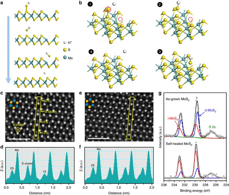
Thus the PSS-induced SVSH mechanism we proposed is that the hydrogenation of PSS guides sulfur adatom clusters on the as-grown MoS2 surface to heal sulfur vacancies (Fig. 2a,b). Note that the sulfur vacancies are sufficiently shallow to act as electron donation defect in n-type monolayer MoS2 (ref. 23). Contrary to sulfur vacancy, the sulfur adatom cluster is found to be an electrically neutral defect, even though its concentration is expected to be high24. In a word, the electrically neutral sulfur adatom clusters are used to fulfil the sulfur vacancies to precisely tune the electron concentration in monolayer MoS2. Similar vacancy healing by superacid TFSI and hydracids (HCl, HBr, HI) has been used to enhance PL intensity of monolayer transition metal dichalcogenides (TMDs)15,18,25. However, as-mentioned overpowered acids corrode the metal electrodes, damage the electrode contact and display unsuitable in constructing homojunction. PSS shows great advantages in low-cost and mild acidic nature26. Moreover, dissociated polymers PSS− has so large molecular weight that it would not dope the sulfur vacancies and hinder the hydrogenation process27.
Figure 2. Sulfur vacancy self-healing (SVSH) mechanism.
(a,b) 2D/3D chemical structure change showing the PSS-induced SVSH effect. (c–f) The HAADF images before c and after e PSS-induced SVSH, together with the Z-contrast mapping done before d and after f in the areas marked with yellow rectangles, reveal that the sulfur vacancies (1S) are healed spontaneously by the sulfur adatom clusters on MoS2 surface through a PSS-induced hydrogenation process. The cyan and yellow dots indicate the Mo and S atoms, respectively. Scale bar, 1 nm. (g) High-resolution XPS for Mo 3d before (top) and after (bottom) PSS treatment of MoS2. Red and blue lines represent the intrinsic MoS2 (i-MoS2) and defective MoS2 (d-MoS2), respectively.
To further confirm the SVSH mechanism, spherical aberration-corrected STEM was employed to obtain a direct vision of the atomic structure of the as-grown and self-healed MoS2. Recently, the scanning transmission electron microscopy (STEM) technique has been proved to be powerful in providing comprehensive information of monolayer MoS2 defects at the atomic scale. We visualized the films via chemical analysis using atomic-resolution Z-contrast imaging with high-angle annular-dark-field (HAADF) STEM. As the intensity of STEM images is directly related to the atomic number (Z-contrast)9,18, sulfur vacancies (1S) and sulfur adatom clusters can be easily recognized and differentiated from the three-fold coordinated two sulfur atoms (Fig. 2c). The corresponding line profiles were extracted to give a clearer picture for sulfur atomic amounts (Fig. 2d). The three kinds of imaging contrasts, which corresponded to sulfur vacancies (1S), three-fold coordinated two sulfur atoms (2S) and sulfur adatom clusters, were presented obviously in the as-grown MoS2. However, the self-healed MoS2 displayed uniform intensity, which implicated the PSS-induced SVSH could effectively decrease the sulfur vacancies and sulfur adatom clusters (Fig. 2e). Other low magnification of the STEM images was displayed in Supplementary Fig. 7. We can draw the conclusion that the sulfur vacancies are healed spontaneously by the sulfur adatom clusters on MoS2 surface through a PSS-induced hydrogenation process.
XPS was also used to identify whether sulfur vacancies were healed by the PSS-induced SVSH. The XPS spectra of Mo 3d consisted of two sets of peaks that can be respectively assigned to intrinsic MoS2 (i-MoS2) and defective MoS2 (d-MoS2) (Fig. 2g). The deconvoluted Mo4+ 3d5/2 and Mo4+ 3d3/2 doublet peaks depict the contributions of i-MoS2 (doublets located at 232.70 and 229.55 eV) and d-MoS2 (peaks at 233.05 and 229.85 eV). When the as-grown (as-grown) MoS2 are healed by PEDOT:PSS solution treatment, the contribution of the intrinsic MoS2 increases, whereas the defective MoS2 component decreases. As a result, the doublets were shifted to the higher binding energy side. This reveals that the density of sulfur vacancies was diminished by the PSS treatment, as the d-MoS2 peak is directly associated with sulfur vacancies28. In addition, The XPS spectra of S 2p also confirmed the sulfur vacancies healing (Supplementary Fig. 8). A similar behaviour was previously observed in sulfur vacancy healed MoS2 through sulfurization annealing and molecular chemisorption15,23,29,30. To quantify the XPS information, we measured the XPS peak area ratio of S 2p to Mo 3d states for the as-grown and self-healed MoS2. The value of S:Mo ratio was increased from ∼1.67 to ∼1.86 by the PSS-induced SVSH (Supplementary Note 1).
Construction and electrical properties of MoS2 homojunction
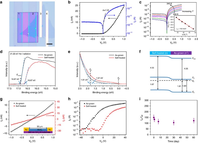
I–V curve test was first conducted in another homojunction device A2, which is on the basis of the device A1 added with the EBL and the PEDOT: PSS solution induced SVSH process. The as-fabricated homojunction shows typical rectifying behaviour (Fig. 3b). To confirm the diode barrier was formed only in self-healed/as-grown junction, the electrical transport properties of other contact types were characterized. Ohmic characteristics are all observed among the following two contact types: the as-grown MoS2 and Cr/Au electrode, and the self-healed MoS2 and PEDOT: PSS electrode (Supplementary Fig. 9). So, the existence of potential barrier in other contact positions is excluded31.
Figure 3. Construction and electrical properties of MoS2 homojunction.
(a) OM image of the device A2. PEDOT:PSS electrodes 1–2 define an self-healed MoS2 FET, 3–4 define an as-grown MoS2 FET, and 2–3 define the homojunction device A2. Scale bar, 5 μm. (b) Output characteristic on linear/logarithmic scale (black/blue) of the device A2. (c) Output characteristic of the homojunction under a series of temperatures. Inset: linear fitting result of the relationship between ln(IDS/T3/2) and e/(kBT). The red line fit is drawn to yield the Schottky barrier height. (d,e) Secondary-edge and valence-band spectrum of the ultraviolet photoelectron spectroscopy (UPS) measurement from as-grown and self-healed monolayer MoS2. (f) Band diagram of the monolayer MoS2 homojunction obtained from UPS measurements. (g,h) Output characteristics and transfer characteristics of a monolayer MoS2 transistor both before and after PSS-induced SVSH. (i) Ion/Ioff ratio of the homojunction measured during 60 days of storage under ambient conditions.
The rectifying performance of the homojunction is further quantitatively analysed under bias voltage by fitting to the diode equation4,32,33,34:
 |
where A is the area of the Schottky junction, A* is the effective Richardson constant, q is the elementary charge, kB is the Boltzmann constant, T is the temperature, and n is the ideality factor. Thus, the n can be calculated from linearly fitting the natural logarithm plot of current and voltage, as depicted by the blue curve in Fig. 3a. Through equation (2), the ideality factor of our device is obtained as 1.6 from the black fitting line, which slightly deviate from the ideal value of 1. The reason is probably the large resistance of organic electrode PEDOT:PSS, which provides series resistance effect7. Quantitative analysis of the Schottky barrier height φB can be done by investigating the temperature dependence of the diode current in the reverse bias saturation regime (exp(qVD/nkBT)<<1)35. Here, the diode current becomes insensitive to VD and Isat∝T2 exp(−qφB/kBT). Figure 3c inset shows a plot of ln(Isat/T2) versus q/kBT in the reverse bias saturation regime. The Schottky barrier height φB was estimated about 150 meV from the slope of the red curve.
The work function variation of the monolayer MoS2 was also carefully double-checked by ultraviolet photoelectron spectroscopy (UPS). The work function can be calculated using36,37
 |
where hν is the incident photon energy (20.22 eV) and Eonset is the onset level related to the secondary electrons (Fig. 3d). Hence, the ϕ for the as-grown and self-healed MoS2 is 4.35 and 4.55 eV, respectively. Note that the work function value obtained for the as-grown MoS2 is consistent with several other reports36,37. The valence band (Ev) for the as-grown and self-healed MoS2 is, respectively, located at 1.81 and 1.67 eV below the Fermi energy EF by linearly extrapolating the leading edge of the spectrum to the baseline (Fig. 3e). The work function difference between the as-grown and self-healed region is 140–200 meV, which was close to ∼150 meV obtained by the variable temperature measurements of the homojunction diode behaviour. In addition, the optical band gaps of the CVD monolayer MoS2 are determined to be ∼1.84 eV from the PL spectrum (Fig. 1e). On the basis of the above results, the well-aligned energy band diagram, which has the same band gap but different Fermi level, is constructed to show the band bending behaviour at the interface of the as-grown and self-healed monolayer MoS2 (Fig. 3f). The energy separation ΔE between the conduction band and EF of the self-healed MoS2 is ∼170 meV, indicating that self-healed monolayer MoS2 is still n-doped. However, the energy separation ΔE in as-grown MoS2 is only ∼30 meV. Then, the as-grown MoS2 region acted as an n+ type, and the self-healed region acted as an n-type. An n+-n monolayer MoS2 homojunction was formed at the as-grown/self-healed junction.
We also investigated the effect of the SVSH on the electrical properties of a back-gated MoS2 transistor at room T. Current decrease can be observed in the output characteristic curve and the threshold voltage dramatically shifted toward zero after the SVSH (Fig. 3g,h). The only constant is the Ohmic contact of the Au-MoS2, which can be attributed to the transistor channel is long enough to ignore the changes in electrode contact. Besides, Supplementary Fig. 10 suggests the decrease of sulfur vacancies bring about the about 643 times decrease of electron concentration ranging from 5.56 × 10¹⁹ to 8.65 × 1016 cm−3 (Supplementary Note 2), which can be comparable to the long-term sulfurization annealing23. These changes indicated that the electrons or sulfur vacancies in the as-grown MoS2 was removed. An improvement in the subthreshold slope indicated that the SVSH reduces interface trap states. Similar phenomenon was previously observed in sulfur vacancy healed MoS2 through sulfurization annealing and molecular chemisorption, and could be explained by a hopping transport model23,29. From another perspective, unipolar n-type electrical transport behaviour is observed in the self-healed MoS2, which is consistent with the UPS measurements (Fig. 3d,e). Besides, the homojunction diode also behaves n-type behaviour (Supplementary Fig. 11), which again confirms the n+-n homojunction structure.
In addition, the durability of the device also was investigated. As the PSS-induced SVSH is environmental-independent, the homojunction should be reliable under long-term operations. The rectifying behaviour or Ion/Ioff ratio of the homojunction have no degradation after two months storing under ambient conditions (Supplementary Fig. 12).
The photovoltaic effect of MoS2 homojunction
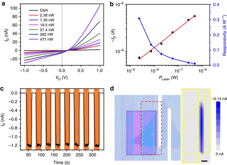
The responsivity test of the photodiode was performed under variable incident light intensity (Fig. 4a). The as-fabricated homojunction shows an open circuit voltage of about 150 mV, which does not change significantly with different illumination power. The ∼150 meV open circuit voltage is very close to the as-mentioned barrier height of variable temperature diode behaviour and UPS measurements. Certainly, the actual barrier height of our homojunction should be greater than this open-circuit voltage. Different from the open circuit voltage, the short circuit current increased with incident power (Fig. 4b). This indicates that the intensity of the light determines the number of photogenerated charge carriers, but not the homojunction band offset9. The responsivity decreases nonlinearly with the increasing light intensity, which is correlated to the decrease of unoccupied states in the conduction band of MoS2 as light intensity increases38. The excellent responsivity of ∼308 mA W−1 at zero bias is much larger than that of other 2D homojunctions by chemical doping (Supplementary Table 1)4,5,9,10,39,40. It can be attributed to the wide space charge regions of the homojunction diode of ∼150 meV barrier height.
Figure 4. The photovoltaic effect of the MoS2 homojunction.
(a) The photovoltaic effect of the monolayer MoS2 homojunction under different 575 nm illumination intensities. (b) Dependence of photocurrent and responsivity on incident light intensity at zero bias. (c) Time-resolved photoresponse of the homojunction upon light illumination (575 nm, 19.5 nW) being turned on and off at zero bias. (d) Photocurrent map of the region indicated by the red rectangle in the OM image at zero bias. Scale bar, 2 μm. The largest photocurrent (blue region) originates from the boundary line of the as-grown and self-healed MoS2 film.
Discussion
The time-resolved photoresponse characteristics revealed a reliable photoresponse with a stabilized photocurrent ON/OFF ratio of ∼200. In addition, the rise time (0–90%) and recovery time (10–100%) are 810 and 750 ms, respectively (Fig. 4c). The response speed is much faster than the CVD-grown MoS2-based photoconductive photodetectors3,41,42, which generally have a long response time. To further explore the photoresponse origin, photocurrent map was performed to spatially investigate the local photoresponse. During this process, a focused laser beam is employed to illuminate a series of special points in the device, while the current is recorded as a function of position (Methods). The photocurrent maximum locates at the boundary between the self-healed and as-grown MoS2, indicating the photoresponse arises from the homojunction rather than the MoS2/PEDOT:PSS or MoS2/metal contacts (Fig. 4d). In other words, the photogenerated electron–hole pairs are efficiently separated in the homojunction region and then transferred to the source and drain electrodes to generate photocurrent.
In conclusion, a lateral CVD monolayer MoS2 homojunction was successfully fabricated by PSS-induced SVSH in selected region. We systematically proposed and demonstrated the self-healing mechanism, in which the sulfur vacancies (electron donation defect) are healed spontaneously by the sulfur adatom clusters (electrically neutral defect) on MoS2 surface through a PSS-induced hydrogenation process. The SVSH preserved the original structure without additional local fields resulting the stable and efficient work function enhancement of ∼150 meV. The electron concentration of the self-healed MoS2 dramatically decreased by 643 times from 5.56 × 10¹⁹ to 8.65 × 1016 cm−3. By using the SVSH process on an individual MoS2, a homojunction was constructed at the interface of the as-grown and self-healed MoS2. The diode presented perfect rectifying characteristic and excellent photoresponsivity of ∼308 mA W−1 at zero bias, which was much larger than that of other 2D homojunctions. The homojunction maintained an outstanding air-stability in the rectifying behaviour and photocurrent for more than two months. In addition, the cost-effective method showed more environment-independent than widely investigated chemical doping. Therefore, our findings paved a powerful strategy to control the 2D materials work functions and develop their homogeneous diodes for ultrathin, flexible, transparent and wearable electronic and optoelectronic nanodevices.
Methods
Growth of monolayer MoS2
This monolayer MoS2 films were grown on the Si substrate with a 300 nm SiO2 insulation layer by the chemical vapour deposition method. MoO3 (Sigma-Aldrich, ≥99.5% purity) and sulfur (Sigma-Aldrich, ≥99.5% purity) were applied as precursor and reactant materials respectively. MoO3 powder (25 mg) was placed in a quartz boat at the center of furnace. A 2 × 2 cm2 SiO2 substrates were put face down at top of the MoO3 powder. S powder was heated to 180 °C by heating belt and carried through Ar flow of 500 s.c.c.m. The experiments were implemented at a reaction temperature of 850 °C for 30 min. Finally, the samples were taken out only if the furnace has naturally cooled down to room temperature.
Monolayer MoS2 transfer
As-grown MoS2 films were spin-coated with poly(methyl methacrylate) (PMMA) and submerged in 5% NaOH solution at 80 °C for 2 h. The PMMA/MoS2 stacks were lifted from the solution, diluted in DI water, and then transferred onto target substrates only with PEDOT:PSS (Sigma-Aldrich, 1.0 wt%) or PEDOT. Subsequently, the substrates were annealed on the hotplate at 60 °C for 30 min to remove DI water and induce PSS to heal defects.
PEDOT preparation by 98% H2SO4 treatment
The substrate only with PEDOT:PSS electrode shown in Fig. 1a(2) was immersed into 98% H2SO4 for 15 min at room T, next sufficiently washed by DI water, and then dried at 100 °C for 10 min to remove residual DI water. Actually, there is still residual PSS− connected with PEDOT by hydrogen in 98% H2SO4 treated PEODT:PSS film20, but for simplicity, the 98% H2SO4 treated PEDOT:PSS is referred to as PEDOT in this entire study.
PEDOT:PSS solution induced sulfur vacancy self-healing
Firstly, the MoS2 sample was immersed in the PEDOT:PSS solution, after standing for 5 min, and then immersed in plenty of DI water to wash the PEDOT:PSS solution for 10 min. Further, the residual DI water was dried with nitrogen, finally the sample was dried at 100 °C for 10 min to remove the residual DI water of PEDOT:PSS electrode if the sample has PEDOT:PSS electrode.
Measurements
The KPFM measurements, AFM images and electrical curve of vertical junction were taken on a commercially available AFM (Nanoscope IIID, Multimode). The PL and Raman spectrum measurements were performed with a confocal microscopy (JY-HR800) under 514 nm laser with a power of 20 mW at room temperature. The spot size of the laser is about 1 μm2. The step size for Raman and PL map is about 0.5 μm. All TEM samples were baked at 160 °C for 5 h under vacuum before the microscopy experiment. STEM imaging were performed on a JEM-ARM200F TEM. XPS was conducted with a Thermo Scientific ESCA Lab 250Xi XPS with a monochromatic KR Al X-ray line. Ultraviolet photoelectron spectroscopy (UPS) was performed in an ultrahigh vacuum chamber using a helium lamp source emitting (AXIS ULTRA DLD) at 21.2 eV. The photocurrent versus position curve used 514 nm laser as light source. The electrical characteristics and the photoresponse properties were implemented by a semiconductor analysis system (Keithley 4200). All electrical and optical signals were recorded in the ambient atmosphere, except variable temperature measurement.
Data availability
The data that support the findings of this study are available from the corresponding author on request.
Additional information
How to cite this article: Zhang, X. et al. Poly(4-styrenesulfonate)-induced sulfur vacancy self-healing strategy for monolayer MoS2 homojunction photodiode. Nat. Commun. 8, 15881 doi: 10.1038/ncomms15881 (2017).
Publisher’s note: Springer Nature remains neutral with regard to jurisdictional claims in published maps and institutional affiliations.
Supplementary Material
Acknowledgments
This work was supported by the National Major Research Program of China (No. 2013CB932602), the National Key Research and Development Program of China 2016YFA0202701, the Program of Introducing Talents of Discipline to Universities (B14003), the National Natural Science Foundation of China (Nos 51527802, 51232001, 51602020, 51672026 and 51372020), China Postdoctoral Science Foundation (2015M580981, 2016T90033) Beijing Municipal Science & Technology Commission, and the State Key Laboratory for Advanced Metals and Materials (No. 2016Z-06), the Fundamental Research Funds for the Central Universities (FRF-TP-15-075A1, FRF-BR-15-036A, FRF-AS-15-002).
Footnotes
The authors declare no competing financial interests.
Author contributions X.Z. and S.Z. deposited MoS2 films by CVD. X.Z., S.L. and Z.Z. performed the device fabrication, data collection and analysis. J.D. assisted in carrying out the film fabrication and characterizations, and J.X. and Z.K. assisted in the device performance measurements. F.L. and B.L. carried out KPFM and AFM measurements. X.Z., Z.K. and Y.O. performed part of the Raman, PL and UPS characterization. K.G. and X.L. carried out STEM experiments. X.Z., Q.L., Z.Z. and Y.Z. initiated and supervised the project. All authors discussed the results, and prepared and commented on the manuscript.
References
- Radisavljevic B., Radenovic A., Brivio J., Giacometti V. & Kis A. Single-layer MoS2 transistors. Nat. Nanotechnol. 6, 147–150 (2011). [DOI] [PubMed] [Google Scholar]
- Koppens F. H. et al. Photodetectors based on graphene, other two-dimensional materials and hybrid systems. Nat. Nanotechnol. 9, 780–793 (2014). [DOI] [PubMed] [Google Scholar]
- Lopez-Sanchez O., Lembke D., Kayci M., Radenovic A. & Kis A. Ultrasensitive photodetectors based on monolayer MoS2. Nat. Nanotechnol. 8, 497–501 (2013). [DOI] [PubMed] [Google Scholar]
- Choi M. S. et al. Lateral MoS2 p-n junction formed by chemical doping for use in high-performance optoelectronics. ACS Nano 8, 9332–9340 (2014). [DOI] [PubMed] [Google Scholar]
- Li H. M. et al. Ultimate thin vertical p-n junction composed of two-dimensional layered molybdenum disulfide. Nat. Commun. 6, 6564 (2015). [DOI] [PMC free article] [PubMed] [Google Scholar]
- Ristein J. Surface transfer doping of semiconductors. Science 313, 1057–1058 (2006). [DOI] [PubMed] [Google Scholar]
- Jin Y. et al. A Van Der Waals Homojunction: ideal p-n Diode Behavior in MoSe2. Adv. Mater. 27, 5534–5540 (2015). [DOI] [PubMed] [Google Scholar]
- Joo P. et al. Functional polyelectrolyte nanospaced MoS2 multilayers for enhanced photoluminescence. Nano Lett. 14, 6456–6462 (2014). [DOI] [PubMed] [Google Scholar]
- Lei S. et al. Surface functionalization of two-dimensional metal chalcogenides by Lewis acid-base chemistry. Nat. Nanotechnol. 11, 465–471 (2016). [DOI] [PubMed] [Google Scholar]
- Lee S. Y. et al. Large work function modulation of monolayer MoS2 by ambient gases. ACS Nano 10, 6100–6107 (2016). [DOI] [PubMed] [Google Scholar]
- Yoo H. et al. Spatial charge separation in asymmetric structure of Au nanoparticle on TiO2 nanotube by light-induced surface potential imaging. Nano Lett. 14, 4413–4417 (2014). [DOI] [PubMed] [Google Scholar]
- Wang Z. et al. Size dependence and UV irradiation tuning of the surface potential in single conical ZnO nanowires. RSC Adv. 5, 42075–42080 (2015). [Google Scholar]
- Baumgartner P., Engel C., Abstreiter G., Böhm G. & Weimann G. Fabrication of lateral npn-phototransistors with high gain and sub-μm spatial resolution. Appl. Phys. Lett. 66, 751 (1995). [Google Scholar]
- Mak K. F. et al. Tightly bound trions in monolayer MoS2. Nat. Mater. 12, 207–211 (2013). [DOI] [PubMed] [Google Scholar]
- Amani M. et al. Near-unity photoluminescence quantum yield in MoS2. Science 350, 1065–1068 (2015). [DOI] [PubMed] [Google Scholar]
- Eda G. et al. Photoluminescence from Chemically Exfoliated MoS2. Nano Lett. 11, 5111–5116 (2011). [DOI] [PubMed] [Google Scholar]
- Mouri S., Miyauchi Y. & Matsuda K. Tunable photoluminescence of monolayer MoS2 via chemical doping. Nano Lett. 13, 5944–5948 (2013). [DOI] [PubMed] [Google Scholar]
- Han H. V. et al. Photoluminescence enhancement and structure repairing of monolayer MoSe2 by hydrohalic acid treatment. ACS Nano 10, 1454–1461 (2016). [DOI] [PubMed] [Google Scholar]
- Xia Y., Sun K. & Ouyang J. Solution-processed metallic conducting polymer films as transparent electrode of optoelectronic devices. Adv. Mater. 24, 2436–2440 (2012). [DOI] [PubMed] [Google Scholar]
- Kim N. et al. Highly conductive PEDOT:PSS nanofibrils induced by solution-processed crystallization. Adv. Mater. 26, 2268–2272 (2014). [DOI] [PubMed] [Google Scholar]
- Cho K. et al. Electrical and optical characterization of MoS2 with sulfur vacancy passivation by treatment with alkanethiol molecules. ACS Nano 9, 8044–8053 (2015). [DOI] [PubMed] [Google Scholar]
- Li H. et al. Activating and optimizing MoS2 basal planes for hydrogen evolution through the formation of strained sulphur vacancies. Nat. Mater. 15, 364 (2016). [DOI] [PubMed] [Google Scholar]
- Kim I. S. et al. Influence of stoichiometry on the optical and electrical properties of chemical vapor deposition derived MoS2. ACS Nano 8, 10551–10558 (2014). [DOI] [PMC free article] [PubMed] [Google Scholar]
- Noh J.-Y., Kim H. & Kim Y.-S. Stability and electronic structures of native defects in single-layer MoS2. Phys. Rev. B 89, 205417 (2014). [Google Scholar]
- Amani M. et al. Recombination kinetics and effects of superacid treatment in sulfur- and selenium-based transition metal dichalcogenides. Nano Lett. 16, 2786–2791 (2016). [DOI] [PubMed] [Google Scholar]
- Alemu D., Wei H.-Y., Ho K.-C. & Chu C.-W. Highly conductive PEDOT:PSS electrode by simple film treatment with methanol for ITO-free polymer solar cells. Energy Environ. Sci. 5, 9662 (2012). [Google Scholar]
- Cai Y. et al. Constructing metallic nanoroads on a MoS2 monolayer via hydrogenation. Nanoscale 6, 1691–1697 (2014). [DOI] [PubMed] [Google Scholar]
- Addou R. et al. Impurities and electronic property variations of natural MoS2 crystal surfaces. ACS Nano 9, 9124–9133 (2015). [DOI] [PubMed] [Google Scholar]
- Sim D. M. et al. Controlled doping of vacancy-containing few-layer MoS2 via highly stable Thiol-based molecular chemisorption. ACS Nano 9, 12115–12123 (2015). [DOI] [PubMed] [Google Scholar]
- Li H. et al. Activating and optimizing MoS2 basal planes for hydrogen evolution through the formation of strained sulphur vacancies. Nat. Mater. 15, 364 (2016). [DOI] [PubMed] [Google Scholar]
- Zhang Y. et al. Performance and service behavior in 1-D nanostructured energy conversion devices. Nano Energy 14, 30–48 (2015). [Google Scholar]
- Liu S. et al. Strain modulation in Graphene/ZnO nanorod film Schottky junction for enhanced photosensing performance. Adv. Funct. Mater. 26, 1347–1353 (2016). [Google Scholar]
- Zhang Z., Liao Q., Yu Y., Wang X. & Zhang Y. Enhanced photoresponse of ZnO nanorods-based self-powered photodetector by piezotronic interface engineering. Nano Energy 9, 237–244 (2014). [Google Scholar]
- Zhang Y. et al. Scanning probe study on the piezotronic effect in ZnO nanomaterials and nanodevices. Adv. Mater. 24, 4647–4655 (2012). [DOI] [PubMed] [Google Scholar]
- Yang H. et al. Graphene barristor, a triode device with a gate-controlled Schottky barrier. Science 336, 1140–1143 (2012). [DOI] [PubMed] [Google Scholar]
- Chang Y.-H. et al. Monolayer MoSe2 grown by chemical vapor deposition for fast photodetection. ACS Nano 8, 8582–8590 (2014). [DOI] [PubMed] [Google Scholar]
- Tsai M.-L. et al. Monolayer MoS2 heterojunction solar cells. ACS Nano 8, 8317–8322 (2014). [DOI] [PubMed] [Google Scholar]
- Xue Y. et al. Scalable production of a few-layer MoS2/WS2 vertical heterojunction array and its application for photodetectors. ACS Nano 10, 573–580 (2016). [DOI] [PubMed] [Google Scholar]
- Yu X., Zhang S., Zeng H. & Wang Q. J. Lateral black phosphorene P–N junctions formed via chemical doping for high performance near-infrared photodetector. Nano Energy 25, 34–41 (2016). [Google Scholar]
- Najmzadeh M., Ko C., Wu K., Tongay S. & Wu J. Multilayer ReS2 lateral p-n homojunction for photoemission and photodetection. Appl. Phys. Express 9, 055201 (2016). [Google Scholar]
- Xie C., Mak C., Tao X. & Yan F. Photodetectors based on two-dimensional layered materials beyond graphene. Adv. Funct. Mater. 27, 1603886 (2017). [Google Scholar]
- Jariwala D., Marks T. J. & Hersam M. C. Mixed-dimensional van der Waals heterostructures. Nat. Mater. 16, 170–181 (2017). [DOI] [PubMed] [Google Scholar]
Associated Data
This section collects any data citations, data availability statements, or supplementary materials included in this article.
Supplementary Materials
Data Availability Statement
The data that support the findings of this study are available from the corresponding author on request.


