Abstract
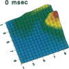
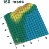
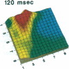
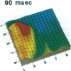
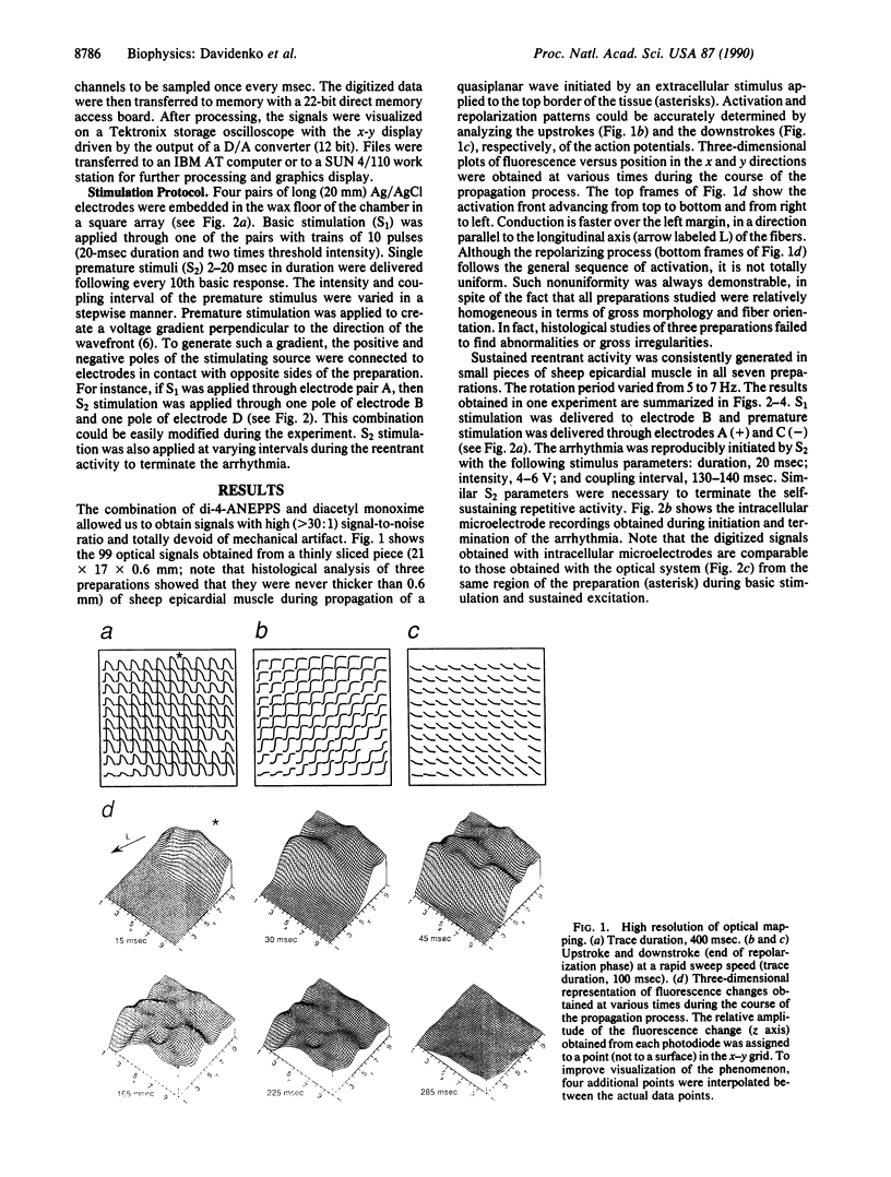
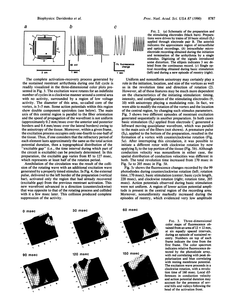
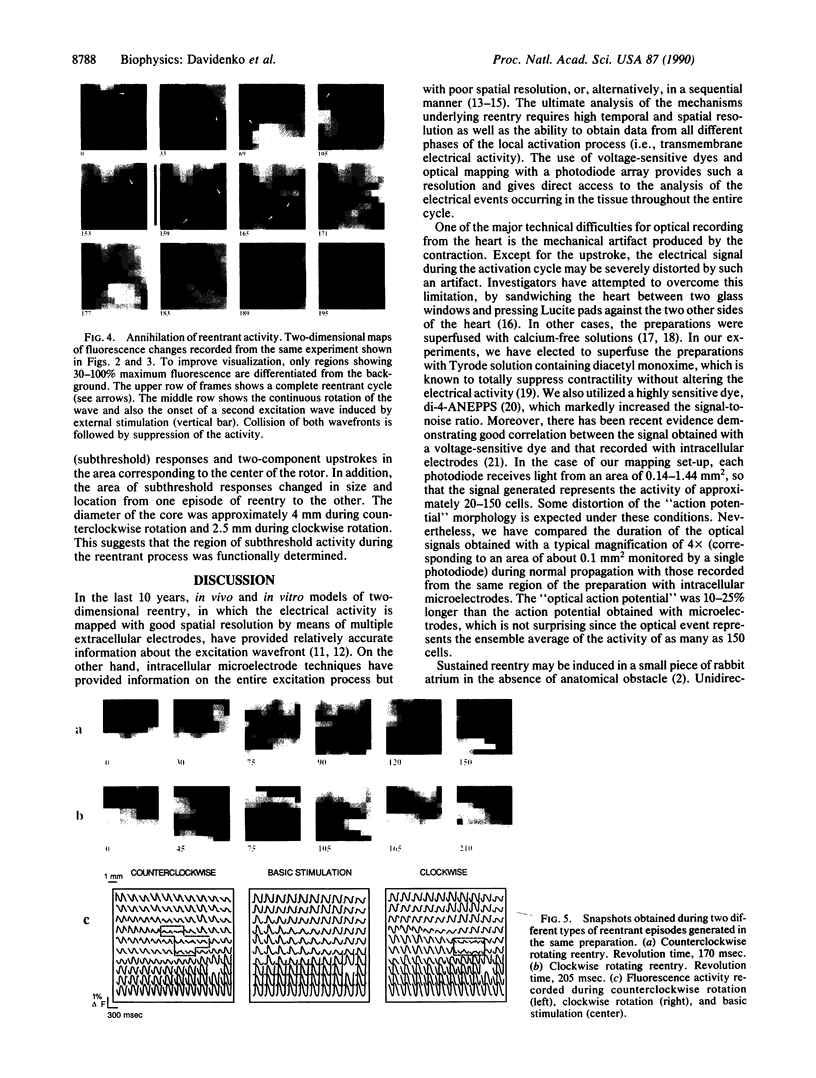
Sustained reentrant excitation may be initiated in small (20 x 20 x less than 0.6 mm) preparations of normal ventricular muscle. A single appropriately timed premature electrical stimulus applied perpendicularly to the wake of a propagating quasiplanar wavefront gives rise to circulation of self-sustaining excitation waves, which pivot at high frequency (5-7 Hz) around a relatively small "phaseless" region. Such a region develops only very low amplitude depolarizations. Once initiated, most episodes of reentrant activity last indefinitely but can be interrupted by the application of an appropriately timed electrical stimulus. The entire course of the electrical activity is visualized with high temporal and spatial resolution, as well as high signal-to-noise ratio, using voltage-sensitive dyes and optical mapping. Two- and three-dimensional graphics of the fluorescence changes recorded by a 10 x 10 photodiode array from a surface of 12 x 12 mm provide sequential images (every msec) of voltage distribution during a reentrant vortex. The results suggest that two-dimensional vortex-like reentry in cardiac muscle is analogous to spiral waves in other biological and chemical excitable media.
Full text
PDF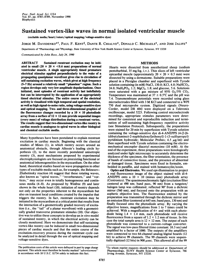
Images in this article
Selected References
These references are in PubMed. This may not be the complete list of references from this article.
- Allessie M. A., Bonke F. I., Schopman F. J. Circus movement in rabbit atrial muscle as a mechanism of tachycardia. II. The role of nonuniform recovery of excitability in the occurrence of unidirectional block, as studied with multiple microelectrodes. Circ Res. 1976 Aug;39(2):168–177. doi: 10.1161/01.res.39.2.168. [DOI] [PubMed] [Google Scholar]
- Allessie M. A., Bonke F. I., Schopman F. J. Circus movement in rabbit atrial muscle as a mechanism of tachycardia. III. The "leading circle" concept: a new model of circus movement in cardiac tissue without the involvement of an anatomical obstacle. Circ Res. 1977 Jul;41(1):9–18. doi: 10.1161/01.res.41.1.9. [DOI] [PubMed] [Google Scholar]
- Dillon S. M., Allessie M. A., Ursell P. C., Wit A. L. Influences of anisotropic tissue structure on reentrant circuits in the epicardial border zone of subacute canine infarcts. Circ Res. 1988 Jul;63(1):182–206. doi: 10.1161/01.res.63.1.182. [DOI] [PubMed] [Google Scholar]
- El-Sherif N., Smith R. A., Evans K. Canine ventricular arrhythmias in the late myocardial infarction period. 8. Epicardial mapping of reentrant circuits. Circ Res. 1981 Jul;49(1):255–265. doi: 10.1161/01.res.49.1.255. [DOI] [PubMed] [Google Scholar]
- Frazier D. W., Wolf P. D., Wharton J. M., Tang A. S., Smith W. M., Ideker R. E. Stimulus-induced critical point. Mechanism for electrical initiation of reentry in normal canine myocardium. J Clin Invest. 1989 Mar;83(3):1039–1052. doi: 10.1172/JCI113945. [DOI] [PMC free article] [PubMed] [Google Scholar]
- Gupta R. K., Salzberg B. M., Grinvald A., Cohen L. B., Kamino K., Lesher S., Boyle M. B., Waggoner A. S., Wang C. H. Improvements in optical methods for measuring rapid changes in membrane potential. J Membr Biol. 1981 Feb 15;58(2):123–137. doi: 10.1007/BF01870975. [DOI] [PubMed] [Google Scholar]
- Hart T. N., Trainor L. E. Geometrical aspects of surface morphogenesis. J Theor Biol. 1989 Jun 8;138(3):271–296. doi: 10.1016/s0022-5193(89)80195-x. [DOI] [PubMed] [Google Scholar]
- Hill B. C., Courtney K. R. Design of a multi-point laser scanned optical monitor of cardiac action potential propagation: application to microreentry in guinea pig atrium. Ann Biomed Eng. 1987;15(6):567–577. doi: 10.1007/BF02364249. [DOI] [PubMed] [Google Scholar]
- Janse M. J., Wit A. L. Electrophysiological mechanisms of ventricular arrhythmias resulting from myocardial ischemia and infarction. Physiol Rev. 1989 Oct;69(4):1049–1169. doi: 10.1152/physrev.1989.69.4.1049. [DOI] [PubMed] [Google Scholar]
- Kamiyama A., Eguchi K., Shibayama R. Circus movement tachycardia induced by a single premature stimulus on the ventricular sheet--evaluation of the leading circle hypothesis in the canine ventricular muscle. Jpn Circ J. 1986 Jan;50(1):65–73. doi: 10.1253/jcj.50.65. [DOI] [PubMed] [Google Scholar]
- Li T., Sperelakis N., Teneick R. E., Solaro R. J. Effects of diacetyl monoxime on cardiac excitation-contraction coupling. J Pharmacol Exp Ther. 1985 Mar;232(3):688–695. [PubMed] [Google Scholar]
- Mines G. R. On dynamic equilibrium in the heart. J Physiol. 1913 Jul 18;46(4-5):349–383. doi: 10.1113/jphysiol.1913.sp001596. [DOI] [PMC free article] [PubMed] [Google Scholar]
- Müller W., Windisch H., Tritthart H. A. Fast optical monitoring of microscopic excitation patterns in cardiac muscle. Biophys J. 1989 Sep;56(3):623–629. doi: 10.1016/S0006-3495(89)82709-2. [DOI] [PMC free article] [PubMed] [Google Scholar]
- Salama G., Lombardi R., Elson J. Maps of optical action potentials and NADH fluorescence in intact working hearts. Am J Physiol. 1987 Feb;252(2 Pt 2):H384–H394. doi: 10.1152/ajpheart.1987.252.2.H384. [DOI] [PubMed] [Google Scholar]
- Sawanobori T., Hirano Y., Hirota A., Fujii S. Circus-movement tachycardia in frog atrium monitored by voltage-sensitive dyes. Am J Physiol. 1984 Aug;247(2 Pt 2):H185–H194. doi: 10.1152/ajpheart.1984.247.2.H185. [DOI] [PubMed] [Google Scholar]
- Winfree A. T., Strogatz S. H. Organizing centres for three-dimensional chemical waves. Nature. 1984 Oct 18;311(5987):611–615. doi: 10.1038/311611a0. [DOI] [PubMed] [Google Scholar]
- Wit A. L., Hoffman B. F., Cranefield P. F. Slow conduction and reentry in the ventricular conducting system. I. Return extrasystole in canine Purkinje fibers. Circ Res. 1972 Jan;30(1):1–10. doi: 10.1161/01.res.30.1.1. [DOI] [PubMed] [Google Scholar]


