Surface K–dosed (Li0.8Fe0.2OH)FeSe provides new clues to understand the mechanism of FeSe-based superconductors.
Keywords: iron-based superconductors, FeSe, Fermi surface topology, scanning tunneling microscopy
Abstract
In iron-based superconductors, understanding the relation between superconductivity and electronic structure upon doping is crucial for exploring the pairing mechanism. Recently, it was found that, in iron selenide (FeSe), enhanced superconductivity (Tc of more than 40 K) can be achieved via electron doping, with the Fermi surface only comprising M-centered electron pockets. By using surface K dosing, scanning tunneling microscopy/spectroscopy, and angle-resolved photoemission spectroscopy, we studied the electronic structure and superconductivity of (Li0.8Fe0.2OH)FeSe in the deep electron-doped regime. We find that a Γ-centered electron band, which originally lies above the Fermi level (EF), can be continuously tuned to cross EF and contribute a new electron pocket at Γ. When this Lifshitz transition occurs, the superconductivity in the M-centered electron pocket is slightly suppressed, and a possible superconducting gap with a small size (up to ~5 meV) and a dome-like doping dependence is observed on the new Γ electron pocket. Upon further K dosing, the system eventually evolves into an insulating state. Our findings provide new clues to understand superconductivity versus Fermi surface topology and the correlation effect in FeSe-based superconductors.
INTRODUCTION
In high-Tc iron-based superconductors, carrier doping is one of the principal routes to induce superconductivity. Many factors, such as the density of states (DOSs), Fermi surface topology and nesting condition, and correlation strength, may vary significantly with carrier concentration. Detailed knowledge of the electronic structure versus doping is critical for understanding the pairing mechanism. Recently, it was found that through heavy electron doping, the Tc of FeSe can be enhanced from the bulk value of 8 K to more than 40 K. The doping can be achieved via interlayer intercalation [AxFe2−ySe2 (A = K, Rb, …) (1, 2), (Li,NH3)FeSe (3), (Li1−yFexOH)FeSe (4)], interface charge transfer (FeSe/SrTiO3) (5), surface K dosing (6), and ionic-liquid gating (7–9). Angle-resolved photoemission spectroscopy (ARPES) studies show that Tc enhancement in these systems is universally accompanied by a vanishing of the Γ hole pockets and that the superconducting gap on the M electron pockets is nodeless (10–14). Meanwhile, scanning tunneling microscopy (STM) studies suggest that the pairing symmetries of single-layer FeSe/SrTiO3 and (Li0.8Fe0.2OH)FeSe are plain s-wave (15, 16), which differs from the s±-wave of bulk FeSe and FeTexSe1−x (17, 18), and that double-dome–like superconductivity is observed in FeSe films upon K dosing (19). These results indicate that the high-Tc phase in heavily electron-doped FeSe may be quite different from that in undoped FeSe, with changes in Fermi surface topology likely playing a crucial role.
Despite the Tc enhancement, the detailed phase diagram of electron-doped FeSe, particularly in the region beyond “optimal” doping, is still not fully understood. Recent ARPES results show that after FeSe films enter the high-Tc phase via surface K dosing, the electron correlation anomalously increases upon further doping, and eventually, an insulating phase emerges (20). This indicates remarkable complexity and new physics in the “overdoped” region. Here, by using low-temperature STM and ARPES, we studied the detailed evolution of the superconductivity and electronic structure of (Li0.8Fe0.2OH)FeSe via surface K dosing. (Li0.8Fe0.2OH)FeSe is already heavily electron-doped with a Tc of ~40 K (4, 16). Surface K dosing can further increase the doping level of the surface FeSe layer. We observe that an unoccupied, Γ-centered electron band shifts significantly to the Fermi level (EF) with increasing K coverage (Kc), whereas the double superconducting gap on M-centered electron pockets gets suppressed slightly. At certain Kc, the Γ-centered band crosses EF, resulting in a Lifshitz transition of the Fermi surface. Shortly after the transition, a superconducting-like gap (up to 5 meV) opens at EF, showing a dome-like dependence on Kc. This represents a new Fermi surface topology for iron-based superconductors, which has sizable electron Fermi pockets at both the Brillouin zone center and the zone corner. At even higher Kc, the system eventually evolves into an insulating phase, characterized by a large, asymmetric gap in excess of 50 meV. The presence of a novel Fermi surface topology, anomalous insulating phase, and the continuous tunability make (Li0.8Fe0.2OH)FeSe a unique platform for gaining insight into the mechanism of iron-based superconductors.
RESULTS
Characterization of the as-cleaved FeSe surface
(Li0.8Fe0.2OH)FeSe single crystals with a Tc of ~42 K (see fig. S1) were grown by hydrothermal reaction method (4, 21). Details of the sample preparation and STM measurement are described in Materials and Methods. There are two possible surface terminations in a cleaved sample, namely, Li0.8Fe0.2OH-terminated and FeSe-terminated surfaces, as reported previously (16). Here, we focus on the FeSe surface with K dosing (see Materials and Methods for details). Figure 1A shows a topographic image of an as-cleaved FeSe surface. The square Se lattice (inset) and some dimer-shaped defects can be resolved. The dI/dV spectrum of this surface taken near EF shows a double superconducting gap (Fig. 1B). For comparison, the topographic image and scanning tunneling spectroscopy (STS) of the Li0.8Fe0.2OH surface are shown in fig. S2, which are distinct from the FeSe surface. The gap sizes of the FeSe surface determined from the two sets of coherence peaks are Δ1 = 14.2 meV and Δ2 = 8.9 meV, similar to previous reports (16, 22). As shown by ARPES studies (13, 14), these superconducting gaps are from M-centered electron pockets, whereas the double-peaked structure could be due to gap anisotropy (23) or band hybridization (22). The gap is found to be spatially homogeneous on the FeSe surface (see fig. S3), confirming the high quality of the sample.
Fig. 1. Topographic image, tunneling, and ARPES spectra of as-cleaved (Li0.8Fe0.2OH)FeSe.
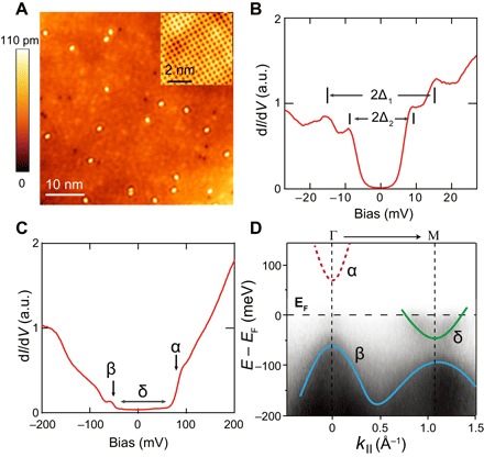
(A) Topographic image of as-cleaved, FeSe-terminated surface (Vb = 100 mV and I = 50 pA); inset shows the surface lattice. (B) Low-energy dI/dV spectrum of as-cleaved FeSe surface, which displays double superconducting gaps of size Δ1 = 15 meV and Δ2 = 9 meV. a.u., arbitrary units. (C) Larger energy scale dI/dV spectrum. Arrows indicate the onset of the α and β bands (see text). Horizontal bar indicates the range of the δ band. (D) ARPES measurement of as-cleaved (Li0.8Fe0.2OH)FeSe. Solid curves track the dispersion of the β and δ bands, whereas the α band above EF is sketched with red dashed curve.
Figure 1C shows the typical dI/dV spectrum of the FeSe surface on a larger energy scale (±200 meV). The tunneling conductance is relatively low near EF but increases rapidly above 70 mV and below −55 mV. The double superconducting gap is not observable on this scale. We note that Huang et al. (24) observed similar dI/dV spectra in single-layer FeSe/SrTiO3. They revealed that an unoccupied, Г-centered electron band gives the steep dI/dV upturn at positive bias. This band is well reproduced in density functional theory (DFT) calculations (24, 25). The dI/dV upturn at negative bias is from the onset of a Г hole band below EF. As explained by Huang et al. (24), the relatively low dI/dV near EF is due to the M-centered electron bands (which dominate the DOS at EF here) having a shorter decay length into the vacuum compared to Г-centered bands, resulting in much lower tunneling probability. The ARPES data of as-cleaved (Li0.8Fe0.2OH)FeSe, as presented in Fig. 1D, display a similar band structure as single-layer FeSe/SrTiO3. Hence, we would expect the resemblance in their tunneling spectra (on both FeSe surfaces). Below, we refer to the Г-centered electron-like band as the α band, Г-centered hole-like bands as β bands, and the M-centered electron-like band as the δ band.
Evolution of the electronic states after K dosing
Next, K atoms were deposited on the sample surface (see Materials and Methods for details). Figure 2 shows typical topographic images of the FeSe surface with Kc from 0.008 to 0.306 ML. Here, we define one monolayer (ML) as the areal density of Fe atoms in single-layer FeSe (1.41 × 10¹⁵/cm2). At small Kc, K atoms are randomly distributed on the surface (Fig. 2, A and B). At certain coverages like 0.098 and 0.124 ML, K atoms can form locally ordered structures, such as √5 × √5 [with respect to the FeSe unit cell (UC); Fig. 2C], or a sixfold close-packed lattice with an inter-atom spacing of 0.78 nm (Fig. 2D; see also fig. S4A). There are different rotational domains observed in Fig. 2D (as marked by the arrows) because of the different symmetry of the K lattice and underlying FeSe lattice. When Kc > 0.15 ML, K atoms begin to form clusters, and no ordered surface structures can be observed (see fig. S4, C and D, for larger-scale images).
Fig. 2. Topographic images of the FeSe surface with a different Kc.
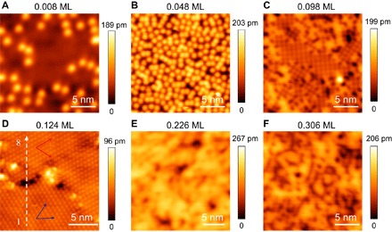
(A) Kc = 0.008 ML. (B) Kc = 0.048 ML. (C) Kc = 0.098 ML. (D) Kc = 0.124 ML. (E) Kc = 0.226 ML. (F) Kc = 0.306 ML. Typical imaging parameters are Vb = 0.5 V and I = 50 pA. The red and blue arrows in (D) indicate the orientation of two different rotational domains. The white dashed arrow marks the position where the STS in Fig. 5E is taken.
Figure 3 (A and B) shows the detailed evolution of the dI/dV spectra as a function of Kc. At low coverage (Kc < 0.080 ML), it is seen from Fig. 3A that the onset of the α band gradually moves to lower energy. However, the β band does not shift together with α, instead moving slightly to higher energy. This anomalous behavior is possibly due to correlation effects in FeSe (20). In Fig. 3B, one sees that double superconducting gaps barely change at Kc ≤ 0.048 ML. When Kc reaches 0.062 to 0.075 ML, the bottom of the α band approaches EF; thus, the corresponding spectra in Fig. 3B tilt up at positive bias. However, the double coherence peaks at negative bias are still observable, which indicates that the gap on the δ band still exists. The corresponding gap size is only slightly suppressed (Δ1 = 13.9 meV and Δ2 = 8.6 meV at Kc = 0.075 ML). This indicates that the superconductivity in the δ band is only weakly sensitive to additional electron doping.
Fig. 3. Evolution of dI/dV spectra taken on the FeSe surface with various Kc as labeled.
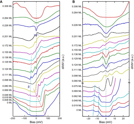
(A) Typical dI/dV spectra taken within large energy range (±200 meV). Red and blue dashed lines track the onsets of the α and β bands. The zero positions of the spectra at Kc = 0.306, 0.264, and 0.226 ML are marked by short horizontal bars. (B) Typical dI/dV spectra taken near EF (±27 meV). Two blue dashed lines track the superconducting coherence peaks at negative bias. The curves at Kc ≤ 0.075 ML are normalized by the dI/dV value at Vb = −27 mV, and the curves at Kc > 0.075 ML are normalized by the value at Vb = 27 mV. EF (Vb = 0) is indicated by gray dashed lines. At Kc = 0.111, 0.124, and 0.129 ML, the gap edge positions (defined as Δ3) are marked by short dashed lines.
When Kc reaches 0.080 ML, the α band begins to cross EF, as seen in Fig. 3 (A and B). The tunneling conductance near EF is now greatly enhanced and dominated by the α band. The spectral weight from the δ band is overwhelmed, and the double coherence peaks are no longer observable (note that the normalization scheme of Fig. 3B changes at this point to make all spectra appear with a similar scale; see fig. S5 for unnormalized dI/dV spectra near this Lifshitz transition). There is no gap-like feature near EF at Kc = 0.080 or 0.098 ML, or the gap is much smaller than our experimental resolution (~1 meV). This indicates that the pairing is weak on the α band as it crosses EF. In Fig. 4A, we summarize the energy shifts of the α and β bands as a function of Kc, by tracing the band bottom or top. We note that the sensitivity of the band position of α to surface K dosing is consistent with recent DFT calculations (25). It was shown that the α band has both Se 4p and Fe 3d orbital characters, which makes it sensitive to Fe-Se distance or Se height (hSe) (24). K dosing could significantly affect the hSe of the surface Se layer.
Fig. 4. Doping dependence of the energy band position and the DOS near EF.
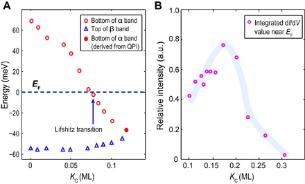
(A) The doping dependence of the band bottom (top) energy of the α (β) band. At Kc = 0.080 ML, the α band begins to cross EF. (B) Integrated dI/dV values within the bias range of ±8 meV as a function of Kc, which reflects the DOS near EF.
The Fermi surface of α will be a new electron pocket at Γ. To look for this pocket, we performed quasi-particle interference (QPI) mapping at Kc = 0.124 ML. As shown in Fig. 2D, for this coverage, the K atoms form a close-packed structure with a relatively smooth, ordered surface, which is suitable for QPI measurements. The mapping was carried out in an area of 100 × 100 nm2 (Fig. 5A). Figure 5 (B and C) shows a typical dI/dV map taken at Vb = 10 mV and its fast Fourier transform (FFT). A complete set of dI/dV maps and FFTs taken within ±50 mV of EF can be found in fig. S6. All FFTs display an isotropic scattering ring centered at q = (0, 0), with the radius increasing with energy. In Fig. 5D, we summarize the FFT linecuts through the center of the scattering ring, taken at various energies. An electron-like dispersion can be clearly seen, which is fully consistent with the presence of the α band. By assuming q = 2k for the intraband backscattering condition, a parabolic fit yields the Fermi crossing at kF = 0.075 Å−1 and the band bottom at −37 meV (this value is also marked in Fig. 4A). Such a sizable electron pocket has not been observed before in iron-based superconductors at the Γ point [for comparison, the kF of δ band for (Li0.8Fe0.2OH)FeSe is 0.21 Å−1 at Kc = 0; see the study of Yan et al. (16)].
Fig. 5. QPI measurement of the α band and the spatial and temperature dependence of its gap.
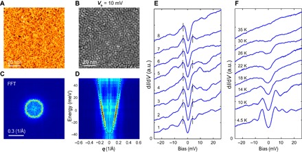
(A) Topographic image of the mapping area of size 100 × 100 nm2 (Kc = 0.124 ML). (B) Typical dI/dV map taken at Vb = 10 mV. The set point for dI/dV map is as follows: Vb = 50 mV, I = 150 pA, and ΔV = 3 mV. (C) FFT image of (B). (D) Intensity plot of the FFT linecuts through q = (0, 0); dashed curve is the parabolic fit. Note that the small gap is not observable here because of the large modulation (ΔV). (E) A dI/dV linecut taken along the dashed arrow in Fig. 2D, showing a spatially uniform gap. Bars indicate the coherence peaks. (F) Temperature dependence of the gap taken on a different sample with Kc ~ 0.12 ML.
Shortly after the α band begins being occupied, starting from Kc = 0.111 ML, one sees a small gap open at EF. We define the gap size by the peak or kinks on the gap edge and refer it to Δ3 below. Δ3 reaches 3.5 to 4 meV at Kc = 0.124 ML and closes at about Kc = 0.136 ML. In Fig. 5E, we show an STS linecut taken on the surface in Fig. 2D (Kc = 0.124 ML)—the small gap is spatially uniform, with coherence peaks in most locations. We have checked this gap in several different samples and found that it can reach ~5 meV at the optimal Kc near 0.12 ML. Figure 5F shows the temperature dependence of the gap at the optimal Kc, with clearly defined coherence peaks. It becomes less prominent as the temperature increases, vanishing at T = 35 K, close to the bulk Tc of the sample (~42 K). Therefore, it is likely that a possible superconducting gap opens on the α band, having a dome-like doping dependence. There could be other possibilities such as a charge density wave–induced gap; however, we did not observe any additional spatial modulation in the topographic image (Fig. 2D and fig. S4A), QPI maps (Fig. 5 and fig. S6), and their FFTs (fig. S4B). The gap has significant nonzero dI/dV at Vb = 0, which could be due to gap anisotropy and/or thermal broadening effects. Measurements at lower temperature and high magnetic field would further clarify the nature of this gap.
The small gap disappears at Kc = 0.136 and 0.155 ML, but starting from Kc = 0.172 ML, another gap-like feature develops at EF. This time, the gap size keeps increasing upon further K dosing, and eventually at Kc = 0. 306 ML, it exceeds 50 meV in width with a nearly flat bottom (Fig. 3B). We note that at Kc = 0.201 or 0.226 ML, the gap has a comparable size with the possible superconducting gap (Δ3) at Kc = 0.124 ML, but the feature is broader (bigger than Δ3 with weak or no coherence peak). Furthermore, at Kc = 0.306 ML, the gap is asymmetric with respect to EF, and STM imaging is not possible for bias voltages inside the gap. Therefore, the gap opening starting from Kc = 0.172 ML likely evidences that the system enters an insulating state, with gradually depleted DOS at EF. To illustrate this more quantitatively, in Fig. 4B, we integrated the dI/dV values extracted from Fig. 3A over the bias range of ±8 meV, as a function of Kc (>0.1 ML). This will give an estimation of the DOS of the α band near EF (note that the integration window is larger than Δ3). It is clear that when Kc < 0.172 ML, the DOS increases with Kc, although it quickly drops thereafter, indicating a metal-insulator transition. This finding is consistent with the insulating state observed in K-dosed FeSe films by ARPES (20) and in ionic liquid–gated (Li1−xFexOH)FeSe (26). Note that the topographic image of Kc = 0.306 ML in Fig. 2F and fig.S4D only shows a disordered structure. This suggests that the insulating phase is not due to the formation of some impurity phase (such as K2Fe4Se5) but is intrinsic to deeply electron-doped FeSe. Moreover, the emergence of the insulating phase also indicates that K atoms do not form a surface metallic layer by themselves up to Kc = 0.306 ML. The STS in Fig. 3 will reflect the electron states of doped FeSe layer.
To facilitate the understanding of the STM data, we performed ARPES measurements on K-dosed (Li0.8Fe0.2OH)FeSe (experiment details are described in Materials and Methods). Figure 6 (A and B) shows ARPES intensity along the cuts crossing Γ and M (Fig. 6C) as the function of Kc. Note that the Kc here is estimated from K flux and deposition time (t) (see Materials and Methods). As seen in Fig. 6B, the size of the δ Fermi pocket increases with K dosing (at Kc ≤ ~0.27 ML), indicating the electron doping. Meanwhile, near the Γ point (Fig. 6A), there is a noticeable spectral intensity that shows up and increases near EF upon K dosing (at Kc < ~0.27 ML). To illustrate it more quantitatively, we plot the corresponding momentum distribution curve (MDC) and energy distribution curve (EDC) (taken near EF and k = 0) for various Kc in Fig. 6 (D and E) (see figure captions). The spectral intensity at Γ evidences the emergence of an electron pocket, although the band dispersion is not clear, which could be due to small pocket size and/or limited resolution here. To have a comparison with the STM result, in the Kc ~ 0.12 ML panel of Fig. 6A, we superposed the band dispersion of α, which is derived from the QPI of Kc = 0.124 ML (Fig. 5D). There is a qualitative match between QPI band dispersion and ARPES intensity at Γ. Furthermore, it is notable that at high dosing (Kc ~ 0.45 ML and t = 302 s), the bands at both Γ and M near EF became unresolvable, which is also consistent with a metal-insulator transition suggested by the STM data. In Fig. 6F, we show symmetrized EDC taken near the kF of the δ band (marked in Fig. 6B), which displays the evolution of the superconducting gap on the δ band. The gap size was ~13 meV at Kc = 0 and ~0.06 ML, which decreased to ~9 meV at Kc ~ 0.12 ML and disappeared at Kc ~ 0.27 ML. The disappearance of superconductivity on the δ band before entering the insulating phase is also observed in K-dosed FeSe films (20).
Fig. 6. ARPES measurement of the band structure of surface K–dosed (Li0.8Fe0.2OH)FeSe.
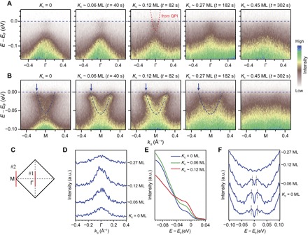
(A) ARPES intensity along cut #1 shown in (C), as a function of Kc and deposition time (t). Red dashed line in the third panel (Kc ~ 0.12 ML) represents the band dispersion of α that derived from QPI (Fig. 5D). (B) ARPES intensity along cut #2 shown in (C), as a function of Kc and t. Dashed lines track the dispersion of the δ band. (C) Sketch of the Brillouin zone of (Li0.8Fe0.2OH)FeSe. (D) Evolution of the MDC along cut #1 upon K dosing, integrated over ±14 meV at EF (curves are shifted vertically for clarity). The intensity at Γ increases up to Kc ~ 0.12 ML. The decreased intensity at Kc ~ 0.27 ML could be due to approaching to the insulating phase (consistent with Fig. 4B). (E) Evolution of the EDC taken around k = 0 (Γ point) upon K dosing (Kc = 0 to 0.12 ML). The increased intensity between −0.04 and 0 eV is consistent with the emergence of an electron pocket. (F) Symmetrized EDC showing the evolution of the superconducting gap on the δ band, as a function of Kc. The momenta of individual spectra are indicated by the arrows in (B).
We noted that the ARPES signal should come from both FeSe and Li0.8Fe0.2OH surfaces (the light spot is of millimeter size here). Our previous STM study found a small electron pocket at Γ for the Li0.8Fe0.2OH surface (16), and it may account for the weak spectral weight at Γ near EF for the Kc = 0 case in Fig. 6A (also indicated in Fig. 6, D and E). We note that a recent μSR (muon spin spectroscopy) study reported proximity-induced superconducting gap in the Li1−xFexOH layers, which also suggest that the Li1−xFexOH layer is conductive (27).
Figure 7 summarizes the observed electronic states from the STS in Fig. 3, as a function of Kc. This phenomenological phase diagram contains four distinct regimes. In regime I (0 ≤ Kc ≤ 0.075 ML), the Fermi surface only comprises the M-centered δ band, and its superconducting gap (Δ1 and Δ2) is only gradually suppressed. In regime II (0.080 ML ≤ Kc ≤ 0.172 ML), the α band crosses EF, introducing a new electron pocket at Γ (illustrated in the inset). A possible new superconducting dome on the α band exists in the middle of this regime (green squares represent the gap size of Δ3). As a complement, the ARPES measured gap sizes on the δ band (from Fig. 6F) are also marked here by gray circles. It appears that the gap persists in the left part of regime II; thus, STM measured Δ1 and Δ2 should also extend to regime II (indicated by two short dashed lines). In regime III (0.172 ML < Kc ≤ 0.26 ML), the DOS near EF begins to decrease as the system approaches a metal-insulator transition. Finally, in regime IV (Kc > 0.26 ML), the DOS near EF is depleted, and the system enters an insulating state.
Fig. 7. Summarized phase diagram of surface K–dosed (Li0.8Fe0.2OH)FeSe.
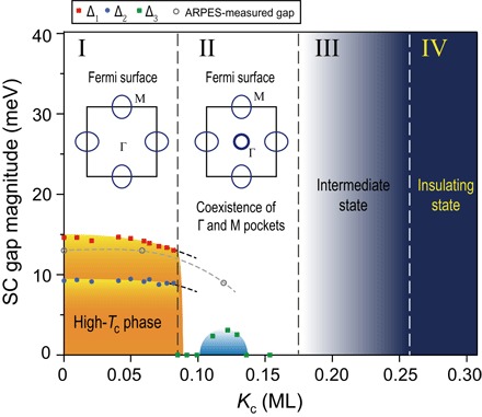
The insets in regimes I and II sketch the Fermi surface before and after the Lifshitz transition. The red, blue, and green dots represent the value of Δ1, Δ2, and Δ3, respectively. Gray circles represent the ARPES measured gap size on the δ band (gray dashed line traces its variation). ARPES measurement suggests that Δ1 and Δ2 would not suddenly disappear when entering regime II, as illustrated by the short black dashed lines. SC, superconducting.
We noted that the Fermi surface of AxFe2−ySe2 at the kz = π plane (10) is similar to the one shown in regime II of Fig. 7. However, the center electron pocket does not exist at Γ (kz = 0) in AxFe2−ySe2, reflecting its significant three-dimensional (3D) character. In (Li0.8Fe0.2OH)FeSe, the interlayer spacing between two FeSe layers (~0.932 nm) (4) is significantly larger than that of AxFe2−ySe2 (~0.702 nm) (1). This makes the Fermi surface of (Li0.8Fe0.2OH)FeSe rather 2D (14).
DISCUSSION
Surface K–dosed (Li0.8Fe0.2OH)FeSe provides several unique opportunities to understand superconductivity in Fe-based superconductors. First, the emergence of the Г-centered electron pocket will introduce a new pairing channel. For most known iron-based superconductors, there are two typical types of Fermi surface topology: one with hole pockets at the zone center and electron pockets at the zone corner and the other with only electron pockets at the zone corner. The scattering between different Fermi pockets has direct consequences on the pairing symmetry (28–31). It was suggested that the interband interactions (spin fluctuations) between the Г-hole and M-electron pockets with wave vector Q = (π, 0) are the main pairing glue, which will lead to s±-wave pairing symmetry (28, 29). However, the absence of a Г pocket in electron-doped FeSe-based systems seriously challenges this scenario. Later, it was suggested that the interaction between neighboring M-electron pockets with Q = (π, π) would dominate pairing in these cases and lead to a d-wave pairing symmetry (29–31), but this picture lacks direct experimental support. Recently, some theoretical work shows that the “incipient” band (a band that is close to but does not cross EF) may still play an important role in pairing, with a significant pairing potential (32–34), and a large “shadow gap” feature was observed in the incipient Г band in LiFe1−xCoxAs (35). Here, by surface K dosing (Li0.8Fe0.2OH)FeSe, we are able to continuously tune the α band to approach and cross EF, which is expected to enable the interaction between two electron bands at Г and M with Q near (π, 0) (for AxFe2−ySe2, these interactions may exist but would be weakened by the strong 3D character of its central electron pocket, as aforementioned). We did not observe gap opening on the α band near its Lifshitz transition (0.062 ML ≤ Kc ≤ 0.098 ML), although the gap on the δ band is slightly suppressed. This would suggest that such a Г-M interaction does not promote superconductivity at the onset of the transition and that the dominant pairing interaction must still lie in the δ band. When the α band does develop a gap in regime II, assuming that the observed gap is possibly a superconducting gap, the small gap size (compared to that on the δ band) also suggests a weak pairing potential on the α band. Because the gap-closing temperature is quite high, this gap could be induced by the δ band through normal interband scattering, as the latter band remains superconducting, as indicated in Figs. 6F and 7. Nevertheless, the dome-like behavior suggests that the α band gradually participates in the pairing. Because of the close competition of various pairing channels, the new type of Fermi surface topology found here may help facilitate a novel superconducting pairing state. In addition, orbital-selective pairing (36, 37), as recently evidenced in bulk FeSe (38), may also relate to our results. Band calculation of single-layer FeSe shows that the major orbital component of α is dx2−y2 (24), which differs from the dxy and dxz/dyz orbitals that comprise the δ band (29). Further theoretical work considering all possible inter- and intraband interactions and orbital structures will be needed to understand the electron pairing in such a case.
Second, the metal-insulator transition observed here provides more clues as to the unusual doping-driven insulating phase in FeSe. In particular, our result shows that the DOS near EF is gradually depleted during the transition, over a relatively wide doping range (from Kc = 0.172 to ~0.26 ML). This differs from transport measurements in ionic liquid–gated (Li1−xFexOH)FeSe, where a sharp, first-order–like transition is observed (26). The smooth transition is consistent with the ARPES result on K-dosed FeSe, where a gradual suppression of spectral weight accompanied by an increasing effective mass is observed (20), suggesting a correlation-driven transition (39). We note that a similar insulating phase has been observed in RbxFe2−ySe2−zTez (40), which indicates that the correlation-driven metal-insulator transition might be universal in FeSe-derived superconductors.
Third, K dosing may be able to change the band topology of the top FeSe layer, inducing a topological phase transition. Recently, Wu et al. (41) proposed that the band topology of the Fe(Te)Se system is controlled by Se(Te) height, which affects the separation (Δn) between the electron and hole bands at Г, and suggested that if Δn is smaller than 80 meV, then spin-orbit coupling can induce band inversion and lead to a nontrivial Z2 topology. In our case, the separation between the α and β bands is continuously reduced from 120 meV (Kc = 0) to ~20 meV (Kc ~ 0.1 ML), as summarized in Fig. 4A. Therefore, such a topological phase transition may well be achievable. We noted that at Kc > 0.1 ML, the evolution of the α and β bands is hard to identify in STS (Fig. 3A); however, topological edge states may exist near step edges if the system enters a nontrivial phase, which deserves further investigation.
In summary, by dosing K on the surface of (Li0.8Fe0.2OH)FeSe, a new electron pocket can be introduced at the Г point. This Lifshitz transition creates a new type of Fermi surface topology and enables a new pairing channel via Г-M interactions. However, only a small gap feature was observed on the new Г pocket, indicating its weak pairing potential. Further doping eventually drives the system into an anomalous insulating state. In addition, nontrivial band topology might be realized by the K dosing–induced band shift. This singular combination of new opportunities makes K-dosed (Li0.8Fe0.2OH)FeSe an intriguing platform for studying the pairing interaction, correlation effects, and topological properties in iron-based superconductors.
Upon completing this work, we noticed an ARPES study on surface K–dosed 1-UC FeSe/SrTiO3 (42), which has similar band structure as (Li0.8Fe0.2OH)FeSe. An electron pocket at Г is also observed after K dosing. This suggests the broader applicability of our findings.
MATERIALS AND METHODS
Sample growth
(Li0.8Fe0.2OH)FeSe single crystals were grown by hydrothermal ion-exchange method described by Dong et al. (21). K0.8Fe1.6Se2 matrix crystal, LiOH·H2O, Fe, and CH4N2Se were used as starting materials. During the hydrothermal reaction, Li1−xFexOH layers were formed and replaced the K atoms in K0.8Fe1.6Se2 (21). Resistivity and magnetic susceptibility measurements (fig. S1, A and B) confirm the Tc of about 42 K. The optical image (fig. S1C) shows that the sample surface is composed of separated domains with the size of tens of micrometers. This morphology may be due to the ion-exchange process.
STM measurement
STM experiment was conducted in a commercial CreaTec STM at the temperature of 4.5 K. (Li0.8Fe0.2OH)FeSe samples were cleaved in ultrahigh vacuum at 78 K. Pt tips were used in all measurements after careful treatment on a Au(111) surface. The tunneling spectroscopy (dI/dV) was performed using a standard lock-in technique with modulation frequency f = 915 Hz and typical amplitude ΔV = 1 mV.
ARPES measurement
ARPES measurement was conducted in an in-house ARPES system with a helium discharge lamp (21.2-eV photons), at the temperature of 11 K, using Scienta R4000 electron analyzers. The energy resolution was 8 meV, and the angular resolution was 0.3°. (Li0.8Fe0.2OH)FeSe samples were cleaved in situ under ultrahigh vacuum. During measurements, the spectroscopy qualities were carefully monitored to avoid the sample aging issue.
K dosing
K atoms were evaporated from a standard SAES alkali metal dispenser, and the samples were kept at 80 K during K dosing. In the STM study, the Kc at low coverages was obtained by directly counting surface K atoms. Then, the K deposition rate was carefully calibrated, and the Kc at high coverage was calculated by deposition rate and time. The Kc dependence of the STS was obtained by repeated deposition of K atoms on one sample. After each deposition, the STM tip was nearly placed on the same surface domain, which is found to be mostly covered by the FeSe-terminated surface. In the ARPES study, Kc was estimated from the K flux rate (measured by a quartz crystal microbalance) and deposition time. Kc dependence of the ARPES spectra was obtained by repeated deposition of K atoms on one sample.
Supplementary Material
Acknowledgments
We thank D. C. Peets and B. Y. Pan for helpful discussions. Funding: This work was supported by the National Science Foundation of China and National Key R&D Program of the Ministry of Science and Technology of China (grant nos. 2016YFA0300200 and 2017YFA0303004) and Science Challenge Project (grant no. TZ2016004). Author contributions: M.Q.R., Y.J.Y., and R.T. performed the STM/STS measurement and analyzed the data. X.H.N. and R.P. performed the ARPES measurement and analyzed the data. D.H. synthesized the sample under the guidance of J.Z. T.Z. and D.-L.F. designed and coordinated the whole work and wrote the manuscript. All authors have discussed the results and the interpretation. Competing interests: The authors declare that they have no competing interests. Data and materials availability: All data needed to evaluate the conclusions in the paper are present in the paper and/or the Supplementary Materials. Additional data related to this paper may be requested from the authors.
SUPPLEMENTARY MATERIALS
Supplementary material for this article is available at http://advances.sciencemag.org/cgi/content/full/3/7/e1603238/DC1
fig. S1. Resistivity, dc magnetic susceptibility measurement, and optical microscopy image of (Li0.8Fe0.2)OHFeSe single crystal.
fig. S2. Topographic image and STS taken on the as-cleaved Li0.8Fe0.2OH surface.
fig. S3. Spatial distribution of the superconducting gap on the as-cleaved FeSe surface.
fig. S4. Additional topographic images of the FeSe surface after K dosing.
fig. S5. Unnormalized dI/dV spectra at the Kc near Lifshitz transition.
fig. S6. dI/dV maps and corresponding FFTs taken in an area of 100 × 100 nm2 of the FeSe-terminated surface at Kc = 0.124 ML.
REFERENCES AND NOTES
- 1.Guo J., Jin S., Wang G., Wang S., Zhu K., Zhou T., He M., Chen X., Superconductivity in the iron selenide KxFe2Se2 (0 ≤ x ≤ 1.0). Phys. Rev. B 82, 180520(R) (2010). [Google Scholar]
- 2.Wang A. F., Ying J. J., Yan Y. J., Liu R., Luo X. G., Li Z. Y., Wang X. F., Zhang M., Ye G. J., Cheng P., Xiang Z. J., Chen X. H., Superconductivity at 32 K in single-crystalline RbxFe2−ySe2. Phys. Rev. B 83, 060512(R) (2011). [Google Scholar]
- 3.Burrard-Lucas M., Free D. G., Sedlmaier S. J., Wright J. D., Cassidy S. J., Hara Y., Corkett A. J., Lancaster T., Baker P. J., Blundell S. J., Clarke S. J., Enhancement of the superconducting transition temperature of FeSe by intercalation of a molecular spacer layer. Nat. Mater. 12, 15–19 (2013). [DOI] [PubMed] [Google Scholar]
- 4.Lu X. F., Wang N. Z., Wu H., Wu Y. P., Zhao D., Zeng X. Z., Luo X. G., Wu T., Bao W., Zhang G. H., Huang F. Q., Huang Q. Z., Chen X. H., Coexistence of superconductivity and antiferromagnetism in (Li0.8Fe0.2)OHFeSe. Nat. Mater. 14, 325–329 (2015). [DOI] [PubMed] [Google Scholar]
- 5.Wang Q.-Y., Li Z., Zhang W.-H., Zhang Z.-C., Zhang J.-S., Li W., Ding H., Ou Y.-B., Deng P., Chang K., Wen J., Song C.-L., He K., Jia J.-F., Ji S.-H., Wang Y.-Y., Wang L.-L., Chen X., Ma X.-C., Xue Q.-K., Interface-induced high-temperature superconductivity in single unit-cell FeSe films on SrTiO3. Chin. Phys. Lett. 29, 037402 (2012). [Google Scholar]
- 6.Miyata Y., Nakayama K., Sugawara K., Sato T., Takahashi T., High-temperature superconductivity in potassium coated multilayer FeSe thin films. Nat. Mater. 14, 775–780 (2015). [DOI] [PubMed] [Google Scholar]
- 7.Hanzawa K., Sato H., Hiramatsu H., Kamiya T., Hosonoa H., Electric field-induced superconducting transition of insulating FeSe thin film at 35 K. Proc. Natl. Acad. Sci. U.S.A. 113, 3986–3990 (2016). [DOI] [PMC free article] [PubMed] [Google Scholar]
- 8.Lei B., Cui J. H., Xiang Z. J., Shang C., Wang N. Z., Ye G. J., Luo X. G., Wu T., Sun Z., Chen X. H., Evolution of high-temperature superconductivity from a low-Tc phase tuned by carrier concentration in FeSe thin flakes. Phys. Rev. Lett. 116, 077002 (2016). [DOI] [PubMed] [Google Scholar]
- 9.Shiogai J., Ito Y., Mitsuhashi T., Nojima T., Tsukazaki A., Electric-field-induced superconductivity in electrochemically etched ultrathin FeSe films on SrTiO3 and MgO. Nat. Phys. 12, 42–46 (2016). [Google Scholar]
- 10.Zhang Y., Yang L. X., Xu M., Ye Z. R., Chen F., He C., Xu H. C., Jiang J., Xie B. P., Ying J. J., Wang X. F., Chen X. H., Hu J. P., Matsunami M., Kimura S., Feng D. L., Nodeless superconducting gap in AxFe2Se2 (A=K, Cs) revealed by angle-resolved photoemission spectroscopy. Nat. Mater. 10, 273–277 (2011). [DOI] [PubMed] [Google Scholar]
- 11.Liu D., Zhang W., Mou D., He J., Ou Y.-B., Wang Q.-Y., Li Z., Wang L., Zhao L., He S. L., Peng Y., Liu X., Chen C., Yu L., Liu G., Dong X., Zhang J., Chen C., Xu Z., Hu J., Chen X., Ma X., Xue Q., Zhou X., Electronic origin of high-temperature superconductivity in single-layer FeSe superconductor. Nat. Commun. 3, 931 (2012). [DOI] [PubMed] [Google Scholar]
- 12.Tan S., Zhang Y., Xia M., Ye Z., Chen F., Xie X., Peng R., Xu D., Fan Q., Xu H., Jiang J., Zhang T., Lai X., Xiang T., Hu J., Xie B., Feng D., Interface-induced superconductivity and strain-dependent spin density waves in FeSe/SrTiO3 thin films. Nat. Mater. 12, 634–640 (2013). [DOI] [PubMed] [Google Scholar]
- 13.Niu X. H., Peng R., Xu H. C., Yan Y. J., Jiang J., Xu D. F., Yu T. L., Song Q., Huang Z. C., Wang Y. X., Xie B. P., Lu X. F., Wang N. Z., Chen X. H., Sun Z., Feng D. L., Surface electronic structure and isotropic superconducting gap in (Li0.8Fe0.2)OHFeSe. Phys. Rev. B 92, 060504(R) (2015). [Google Scholar]
- 14.Zhao L., Liang A., Yuan D., Hu Y., Liu D., Huang J., He S., Shen B., Xu Y., Liu X., Yu L., Liu G., Zhou H., Huang Y., Dong X., Zhou F., Liu K., Lu Z., Zhao Z., Chen C., Xu Z., Zhou X. J., Common electronic origin of superconductivity in (Li,Fe)OHFeSe bulk superconductor and single-layer FeSe/SrTiO3 films. Nat. Commun. 7, 10608 (2016). [DOI] [PMC free article] [PubMed] [Google Scholar]
- 15.Fan Q., Zhang W. H., Liu X., Yan Y. J., Ren M. Q., Peng R., Xu H. C., Xie B. P., Hu J. P., Zhang T., Feng D. L., Plain s-wave superconductivity in single-layer FeSe on SrTiO3 probed by scanning tunnelling microscopy. Nat. Phys. 11, 946–952 (2015). [Google Scholar]
- 16.Yan Y. J., Zhang W. H., Ren M. Q., Liu X., Lu X. F., Wang N. Z., Niu X. H., Fan Q., Miao J., Tao R., Xie B. P., Chen X. H., Zhang T., Feng D. L., Surface electronic structure and evidence of plain s-wave superconductivity in (Li0.8Fe0.2)OHFeSe. Phys. Rev. B 94, 134502 (2016). [Google Scholar]
- 17.Song C.-L., Wang Y.-L., Cheng P., Jiang Y.-P., Li W., Zhang T., Li Z., He K., Wang L. L., Jia J.-F., Hung H.-H., Wu C., Ma X., Chen X., Xue Q.-K., Direct observation of nodes and twofold symmetry in FeSe superconductor. Science 332, 1410–1413 (2011). [DOI] [PubMed] [Google Scholar]
- 18.Hanaguri T., Niitaka S., Kuroki K., Takagi H., Unconventional s-wave superconductivity in Fe(Se,Te). Science 328, 474–476 (2010). [DOI] [PubMed] [Google Scholar]
- 19.Song C.-L., Zhang H.-M., Zhong Y., Hu X.-P., Ji S.-H., Wang L., He K., Ma X.-C., Xue Q.-K., Observation of double-dome superconductivity in potassium-doped FeSe thin films. Phys. Rev. Lett. 116, 157001 (2016). [DOI] [PubMed] [Google Scholar]
- 20.Wen C. H. P., Xu H. C., Chen C., Huang Z. C., Lou X., Pu Y. J., Song Q., Xie B. P., Abdel-Hafiez M., Chareev D. A., Vasiliev A. N., Peng R., Feng D. L., Anomalous correlation effects and unique phase diagram of electron-doped FeSe revealed by photoemission spectroscopy. Nat. Commun. 7, 10840 (2016). [DOI] [PMC free article] [PubMed] [Google Scholar]
- 21.Dong X., Jin K., Yuan D., Zhou H., Yuan J., Huang Y., Hua W., Sun J., Zheng P., Hu W., Mao Y., Ma M., Zhang G., Zhou F., Zhao Z., (Li0.84Fe0.16)OHFe0.98Se superconductor: Ion-exchange synthesis of large single-crystal and highly two-dimensional electron properties. Phys. Rev. B 92, 064515 (2015). [Google Scholar]
- 22.Du Z., Yang X., Lin H., Fang D., Du G., Xing J., Yang H., Zhu X., Wen H.-H., Scrutinizing the double superconducting gaps and strong coupling pairing in (Li1−xFex)OHFeSe. Nat. Commun. 7, 10565 (2016). [DOI] [PMC free article] [PubMed] [Google Scholar]
- 23.Zhang Y., Lee J. J., Moore R. G., Li W., Yi M., Hashimoto M., Lu D. H., Devereaux T. P., Lee D.-H., Shen Z.-X., Superconducting gap anisotropy in monolayer FeSe thin film. Phys. Rev. Lett. 117, 117001 (2016). [DOI] [PubMed] [Google Scholar]
- 24.Huang D., Song C.-L., Webb T. A., Fang S., Chang C.-Z., Moodera J. S., Kaxiras E., Hoffman J. E., Revealing the empty-state electronic structure of single-unit-cell FeSe/SrTiO3. Phys. Rev. Lett. 115, 017002 (2015). [DOI] [PubMed] [Google Scholar]
- 25.Zheng F. W., Wang L.-L., Xue Q.-K., Zhang P., Band structure and charge doping effects of the potassium-adsorbed FeSe/SrTiO3 system. Phys. Rev. B 93, 075428 (2016). [Google Scholar]
- 26.Lei B., Xiang Z. J., Lu X. F., Wang N. Z., Chang J. R., Shang C., Zhang A. M., Zhang Q. M., Luo X. G., Wu T., Sun Z., Chen X. H., Gate-tuned superconductor-insulator transition in (Li,Fe)OHFeSe. Phys. Rev. B 93, 060501(R) (2016). [Google Scholar]
- 27.Khasanov R., Zhou H., Amato A., Guguchia Z., Morenzoni E., Dong X., Zhang G., Zhao Z., Proximity-induced superconductivity within the insulating (Li0.84Fe0.16)OH layers in (Li0.84Fe0.16)OHFe0.98Se. Phys. Rev. B 93, 224512 (2016). [Google Scholar]
- 28.Mazin I. I., Singh D. J., Johannes M. D., Du M. H., Unconventional superconductivity with a sign reversal in the order parameter of LaFeAsO1−xFx. Phys. Rev. Lett. 101, 057003 (2008). [DOI] [PubMed] [Google Scholar]
- 29.Hirschfeld P. J., Korshunov M. M., Mazin I. I., Gap symmetry and structure of Fe-based superconductors. Rep. Prog. Phys. 74, 124508 (2011). [Google Scholar]
- 30.Wang F., Yang F., Gao M., Lu Z.-Y., Xiang T., Lee D.-H., The electron pairing of KxFe2−ySe2. Europhys. Lett. 93, 57003 (2011). [Google Scholar]
- 31.Maier T. A., Graser S., Hirschfeld P. J., Scalapino D. J., d-wave pairing from spin fluctuations in the KxFe2−ySe2 superconductors. Phys. Rev. B 83, 100515 (2011). [Google Scholar]
- 32.Bang Y., A shadow gap in the over-doped (Ba1−xKx)Fe2As2 compound. New J. Phys. 16, 023029 (2014). [Google Scholar]
- 33.Chen X., Maiti S., Linscheid A., Hirschfeld P. J., Electron pairing in the presence of incipient bands in iron-based superconductors. Phys. Rev. B 92, 224514 (2015). [Google Scholar]
- 34.Linscheid A., Maiti S., Wang Y., Johnston S., Hirschfeld P. J., High Tc via spin fluctuations from incipient bands: Application to monolayers and intercalates of FeSe. Phys. Rev. Lett. 117, 077003 (2016) [DOI] [PubMed] [Google Scholar]
- 35.Miao H., Qian T., Shi X., Richard P., Kim T. K., Hoesch M., Xing L. Y., Wang X.-C., Jin C.-Q., Hu J.-P., Ding H., Observation of strong electron pairing on bands without Fermi surfaces in LiFe1−xCoxAs. Nat. Commun. 6, 6056 (2015). [DOI] [PubMed] [Google Scholar]
- 36.Arakawa N., Ogata M., Orbital-selective superconductivity and the effect of lattice distortion in iron based superconductors. J. Phys. Soc. Jpn. 80, 074704 (2011). [Google Scholar]
- 37.Kreisel A., Andersen B. M., Sprau P. O., Kostin A., Davis J. C. S., Hirschfeld P. J., Orbital selective pairing and gap structures of iron-based superconductors. arXiv:1611.02643 (2016). [Google Scholar]
- 38.Sprau P. O., Kostin A., Kreisel A., Böhmer A. E., Taufour V., Canfield P. C., Mukherjee S., Hirschfeld P. J., Andersen B. M., Davis J. C. S., Discovery of orbital-selective cooper pairing in FeSe. arXiv:1611.02134 (2016). [DOI] [PubMed] [Google Scholar]
- 39.Imada M., Fujimori A., Tokura Y., Metal-insulator transitions. Rev. Mod. Phys. 70, 1039–1263 (1998). [Google Scholar]
- 40.Niu X. H., Chen S. D., Jiang J., Ye Z. R., Yu T. L., Xu D. F., Xu M., Feng Y., Yan Y. J., Xie B. P., Zhao J., Gu D. C., Sun L. L., Mao Q., Wang H., Fang M., Zhang C. J., Hu J. P., Sun Z., Feng D. L., A unifying phase diagram with correlation-driven superconductor-to-insulator transition for the 122 series of iron chalcogenides. Phys. Rev. B 93, 054516 (2016). [Google Scholar]
- 41.Wu X., Qin S., Liang Y., Fan H., Hu J., Topological characters in Fe(Te1−xSex) thin films. Phys. Rev. B 93, 115129 (2016). [Google Scholar]
- 42.Shi X., Han Z.-Q., Peng X.-L., Richard P., Qian T., Wu X.-X., Qiu M.-W., Wang S. C., Hu J. P., Sun Y.-J., Ding H., Enhanced superconductivity accompanying a Lifshitz transition in electron-doped FeSe monolayer. Nat. Commun. 8, 14988 (2017). [DOI] [PMC free article] [PubMed] [Google Scholar]
Associated Data
This section collects any data citations, data availability statements, or supplementary materials included in this article.
Supplementary Materials
Supplementary material for this article is available at http://advances.sciencemag.org/cgi/content/full/3/7/e1603238/DC1
fig. S1. Resistivity, dc magnetic susceptibility measurement, and optical microscopy image of (Li0.8Fe0.2)OHFeSe single crystal.
fig. S2. Topographic image and STS taken on the as-cleaved Li0.8Fe0.2OH surface.
fig. S3. Spatial distribution of the superconducting gap on the as-cleaved FeSe surface.
fig. S4. Additional topographic images of the FeSe surface after K dosing.
fig. S5. Unnormalized dI/dV spectra at the Kc near Lifshitz transition.
fig. S6. dI/dV maps and corresponding FFTs taken in an area of 100 × 100 nm2 of the FeSe-terminated surface at Kc = 0.124 ML.


