Abstract
Cervical human papillomavirus (HPV) infection may increase HIV risk. Since other genital infections enhance HIV susceptibility by inducing inflammation, we assessed the impact of HPV infection and clearance on genital immunology and the cervico-vaginal microbiome. Genital samples were collected from 65 women for HPV testing, immune studies and microbiota assessment; repeat HPV testing was performed after 6 months. All participants were HIV-uninfected and free of bacterial STIs. Cytobrush-derived T cell and dendritic cell subsets were assessed by multiparameter flow cytometry. Undiluted cervico-vaginal secretions were used to determine cytokine levels by multiplex ELISA, and to assess bacterial community composition and structure by 16S rRNA gene sequence analysis. Neither HPV infection nor clearance were associated with broad differences in cervical T cell subsets or cytokines, although HPV clearance was associated with increased Langerhans cells and HPV infection with elevated IP-10 and MIG. Individuals with HPV more frequently had a high diversity cervico-vaginal microbiome (community state type IV) and were less likely to have an L. gasseri predominant microbiome. In summary, HPV infection and/or subsequent clearance was not associated with inflammation or altered cervical T cell subsets, but associations with increased Langerhans cells and the composition of the vaginal microbiome warrant further exploration.
Keywords: female genital tract, HPV, sexually transmitted infections, mucosal immunology, microbiome, cytokines, CD4+ T cells, dendritic cells
INTRODUCTION
Human papillomavirus is the most common sexually transmitted infection (STI) globally.1 The majority of HPV infections are cleared quickly by the host,2 but high-risk subtypes (HR-HPV) can persist and can cause cervical and other types of cancer.3 Recent meta-analysis has associated HPV infection with a 2-fold increased risk of HIV acquisition,4 possibly due to the recruitment of cells highly-susceptible to HIV infection (HIV target cells) or other changes in the immunology and composition of bacterial communities within the genital tract.5
Many STIs induce robust host mucosal immune responses and genital inflammation,6 in order to facilitate pathogen clearance and in some cases protect against re-infection.7 However, this inflammatory response may render individuals more susceptible to HIV infection. Not only do activated CD4 T cells express elevated levels of the HIV co-receptor CCR5,8 making them preferential HIV targets,9 but STIs also increase pro-inflammatory cytokine levels and neutrophil protease levels10-12 which are associated with perturbed epithelial cell differentiation, cell–cell contacts, and epithelial barrier function and integrity.11 In keeping with this, macaque models have shown that levels of mucosal CCR5+13 CD4 T cells are predictive of subsequent SIV infection risk, and HIV acquisition has been linked to increased genital pro-inflammatory cytokines in human cohorts.14, 15
Classical bacterial STIs such as gonorrhea and chlamydia cause genital inflammation14 and an increase in activated mucosal CD4+ T cells,16 and have been associated with a 3-5 fold increase in HIV acquisition. Interestingly, although herpes simplex virus type-2 (HSV-2) is not associated with elevated pro-inflammatory genital cytokines,17 HSV-2 infection has been consistently associated with elevated levels of activated CD4+ T cells in the genital mucosa18, 19 and also with a three-fold increase in HIV acquisition risk.17 Even in the absence of classical STIs, HIV risk is increased 60% in the context of bacterial vaginosis (BV), defined as the absence of vaginal Lactobacillus spp. and an increase in wide array of strict and facultative anaerobic bacteria.20 Further, BV-associated bacteria have been associated with genital inflammation.10, 14 However, the mucosal immune impact of HPV has not been well defined, despite its high global prevalence.
Limited data have linked host immune clearance of HPV with a Th1 pro-inflammatory response in the female genital tract,5 with increased dendritic cell density in the foreskin,21 and with peripheral blood CD8 T cell responses to HPV proteins in vitro.22 These observations led us to hypothesize that an inflammatory host mucosal immune response to HPV is necessary for host immune clearance, but that this inflammatory response would recruit highly HIV-susceptible T cells to the genital tract, thereby enhancing HIV susceptibility.
In Ontario, the African/Caribbean and other black (ACB) community bears a disproportionate burden of HIV infection,23 and also has a high prevalence of HPV infection.23 Therefore, we performed a longitudinal observational study in ACB women to define the mucosal immune impact of prevalent HPV infection and subsequent host immune clearance. The primary study endpoints were the overall number of endocervical CD4+ T cells, as well as of CD4+ T cell subsets expressing CCR5 and the immune activation markers CD69 and CD38/HLA-DR. Analyses compared HPV uninfected and infected women, as well as women who went on to clear HPV in the short term vs. maintaining persistent HPV infection. Differences in the absolute number of various dendritic cell (DC) subsets, cervico-vaginal levels of pro-inflammatory and chemoattractant cytokines, and the associated composition and structure of the vaginal microbiota were also assessed.
RESULTS
Participant demographics
A total of 65 African/Caribbean women were recruited and enrolled into the study. Insufficient cells were recovered from cytobrushes for 6 participants, and these participants were excluded from subsequent analysis. Of the remaining 59 participants, 36 (61.0%) were HPV negative and 23 (39.0%) were HPV positive at the time of baseline sampling, with 13/23 (56.5%) infected with at least one HR-HPV type. Among HPV-infected participants, 21/23 (91.3%) attended the 6 month follow up visit; 11/21 (52.4%) were HPV negative (clearance group), 8/21 (38.1%) were infected with the same HPV type(s) (persistent group), and 2/21 (9.5%) had cleared one HPV subtype but were newly or persistently infected by another subtype (excluded from analyses of clearance vs. persistence). Among HPV negative participants, 30/36 (83.3%) attended the 6 month follow up visit 24/30 (80.0%) remained HPV negative and 6/30 (20.0%) acquired an HPV infection.
There were no differences between the HPV negative and HPV positive groups in terms of age (median 37.5 years, IQR 28.5-46.5; vs. 33 years; IQR 27.0-39.0) or number of women reporting regular menstrual cycles (30/36; 83.3% vs. 21/23; 91.3%; Table 1), nor were there differences in the number reporting sexual intercourse in the past week (13/36; 36.1% vs. 10/23; 43.5%). Among those sexually active in the past week the median time since previous intercourse was similar (3; 1-6 vs. 5; 3-7 days), although HPV-uninfected women were less likely to have used a condom (2/13; 15.4% vs. 7/10; 70.0%, p=0.008). There were similar rates of hormonal contraceptive use and intravaginal washing between the HPV negative and positive participants (5/36; 13.9% vs. 1/23; 4.3% and 4/36; 11.1% vs. 3/23; 13.0%, respectively). In terms of genital co-infections, there was a similar prevalence of BV and HSV-2 in HPV negative and positive women (2/36, 5.6% vs. 5/23, 21.7%, p=0.098; and 22/36, 61.1% vs. 16/23, 69.6%, p=0.585, respectively); however HPV negative women had higher rates of HSV-1 infection than their HPV positive counterparts (36/36; 100% vs. 18/23; 78.3%, p=0.003). Yeast detected on vaginal smears also did not differ between HPV positive (2/23; 8.7%) and HPV negative participants (4/36; 11.1%, p=0.765). No participants were infected with HIV-1/2, N. gonorrhoeae, or C. trachomatis.
Table 1.
Participant Demographics, by HPV status (total n=59).
| Variables | HPV Negative (n = 36) | HPV Positive (n = 23) | p-value* | HR-HPV Positive (n = 13)** | Clear HPV (n = 11)** | Persist HPV (n = 8)** |
|---|---|---|---|---|---|---|
| Demographics | ||||||
| Age (y; [IQR]) | 37.5 (28.5-46.5) | 33 (27.0-39.0) | 0.250 | 37 (25.5-48.5) | 37 (33-41) | 29 (20.5-37.5) |
| Postmenopausal (n [%]) | 6 (16.7) | 2 (8.7) | 0.464 | 2 (15.4) | 1 (9.1) | 1 (12.5) |
| Time since menses (d; [IQR])*** | 15 (12-17) | 15 (12.5-16) | 0.530 | 15 (13-16) | 15.5 (12.75-16.25) | 14 (10-15) |
| Behavioural characteristics | ||||||
| Sex in the past week (n [%]) | 13 (36.1) | 10 (43.5) | 0.596 | 4 (30.8) | 5 (45.5) | 4 (50.0) |
| Days since intercourse (y; [IQR]) | 3 (1-6) | 5 (3-7) | 0.238 | 4.5 (2-6.5) | 6 (3-7) | 6 (3-7) |
| Condom use (n [%]) | 2 (15.4) | 7 (70.0)**** | 0.013 | 1 (25.0) | 2 (40.0) | 2 (25.0) |
| Hormonal contraceptive use (n [%]) | 5 (13.9) | 1 (4.3) | 0.389 | 0 (0) | 1 (9.1) | 0 (0) |
| Intravaginal practices (n [%]) | 4 (11.1) | 3 (13.0) | 1.000 | 2 (15.4) | 0 (0) | 3 (37.5) |
| Clinical characteristics (n [%]) | ||||||
| Bacterial vaginosis (Nugent≥7) | 2 (5.6) | 5 (21.7) | 0.098 | 3 (23.1) | 2 (18.2) | 2 (25.0) |
| HSV-1 | 36 (100) | 18 (78.3)**** | 0.007 | 11 (84.6)**** | 9 (81.8)**** | 5 (62.5) |
| HSV-2 | 22 (61.1) | 16 (69.6) | 0.585 | 11 (84.6) | 8 (72.7) | 5 (62.5) |
| Yeast | 4 (11.1) | 2 (8.7) | 0.765 | 0 (0) | 1 (9.1) | 1 (12.5) |
p-value associated with the HPV Negative vs. HPV Positive comparison
These groups represent sub-groups of the “HPV Positive” group.
Among women reporting regular menstrual cycles
p<0.05 vs. HPV Negative group (except those with persistent HPV which were compared to those who cleared HPV)
Among the HPV positive participants, those infected with HR-HPV and those who went on to clear HPV infection had a lower prevalence of HSV-1 infection than HPV negative participants (11/13; 84.6% vs. 36/36; 100%, p=0.016 and 9/11; 81.8% vs. 36/36; 100%, p=0.009, Table 1). No other demographic factors that were assessed varied between HPV negative and HPV positive/HR-HPV positive/those that cleared HPV infection, nor were there any differences between those with persistent HPV infection and those who cleared HPV.
HPV infection and immune cell subsets
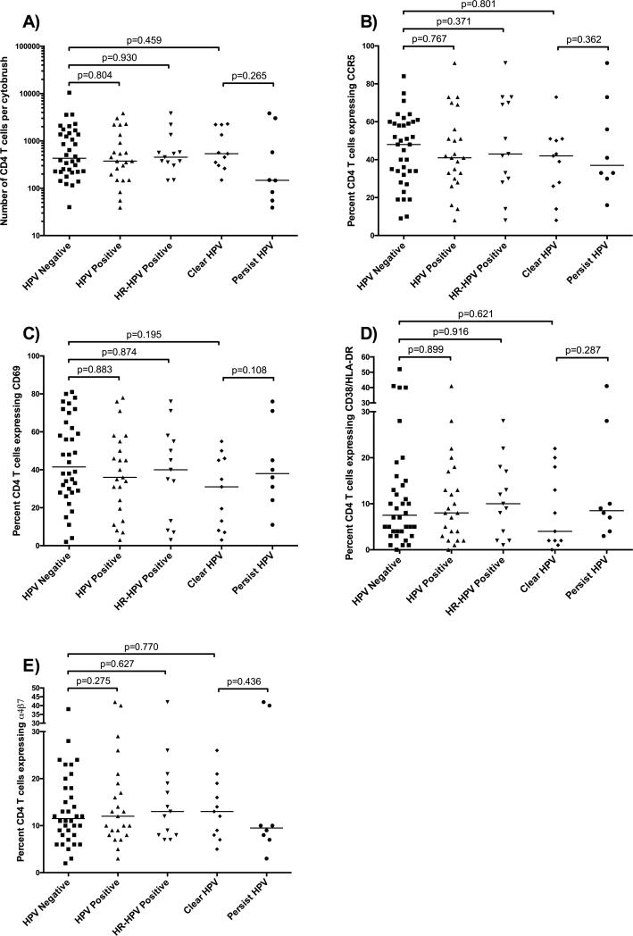
The overall endocervical CD4 T cell number did not differ between HPV negative participants (mean 1153 CD4+ T cells /cytobrush) and HPV positive participants (889 cells/cytobrush, p=0.804), or the subset of participants infected by HR-HPV (863 cells/cytobrush, p=0.930). Furthermore, among participants with baseline HPV infection, cervical CD4+ T cells numbers were similar in those who cleared HPV over the next 6 months (970 cells/cytobrush, p=0.459) and those with persistent HPV infection (988 cells/cytobrush; p=0.265, Figure 1A). There were no associations observed in the female genital tract or blood between HPV infection and the proportion or number of CD4+ T cells expressing the activation markers CD69 (early activation) or CD38/HLA-DR (chronic activation), or the mucosal homing integrin α4β7 (Figure 1B-E).
Figure 1. HPV infection status and T cell subsets in the female genital tract.
Association of HPV infection as well as HR-HPV infection and subsequent clearance or persistence with the (A) absolute number endocervical CD4 T cells; and the proportion of these cells expressing: (B) CCR5, (C) CD69, (D) CD38 and HLA-DR, and (E) α4β7. The “HR-HPV Positive,” “Clear HPV,” and “Persist HPV” groups represent subgroups of the “HPV Positive” group. Statistical comparisons were performed using independent samples t-test and confirmed with a multivariate general linear model, with p-values for the latter indicated.
Interestingly, participants who cleared HPV had a higher absolute number of endocervical Langerhans cells (5018 cells/cytobrush) than either HPV negative women (635 cells/cytobrush, p=0.015) and those with persistent HPV infection (180 cells/cytobrush, p=0.023, Figure 2A). However, no association with prevalent HPV infection was observed: HPV positive (2488 cells/cytobrush) and negative (635 cells/cytobrush) participants had similar numbers of endocervical Langerhans cells (p=0.608). Further, there were no differences in the absolute numbers of mDCs or monocytes (Figure 2B-C), nor were there differences in the expression of C-type lectin receptors (mannose receptor/DC-SIGN) on these subsets between groups (data not shown).
Figure 2. HPV infection status and DC subsets in the female genital tract.
Association of HPV and HR-HPV infection as well as subsequent clearance or persistence with the overall number of (A) Langerhans cells, (B) mDCs, and (C) monocytes in the endocervix. The “HR-HPV Positive,” “Clear HPV,” and “Persist HPV” groups represent subgroups of the “HPV Positive” group. Statistical comparisons were performed using independent samples t-test and confirmed with a multivariate general linear model, with p-values for the latter indicated.
HPV infection and soluble mediators
Having observed no association between HPV infection status and T cell subsets in the female genital tract, we explored potential associations between HPV infection or clearance and pro-inflammatory cytokine levels in cervicovaginal secretions (median mass collected by Instead Softcup 0.26g, range 0.09-1.36g). In order to reduce concerns around multiple comparisons, our primary endpoint was the presence or absence of elevated pro-inflammatory cytokines, using a pre-specified aggregate score in which genital inflammation was defined as having at least 3/7 of the following cytokines in the upper quartile for all participants: IL-1α, IL-1β, IL-8, MIP-1β, MIP-3α, RANTES, and TNF-α.11
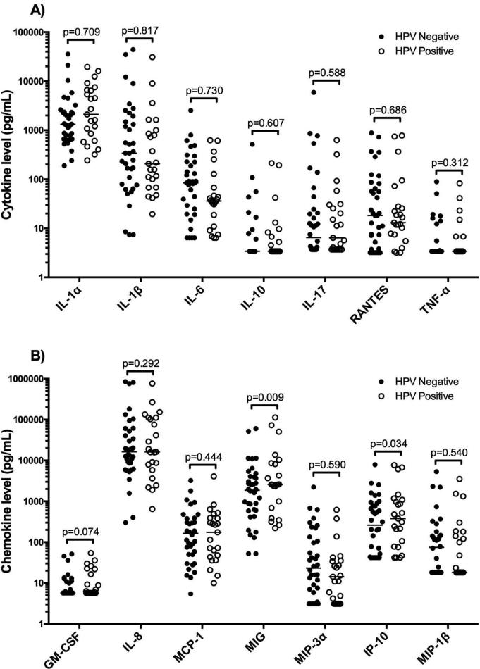
Using this aggregate score, 16/59 participants (27.1%) were defined as having genital inflammation. No association with genital inflammation was observed between those with prevalent HPV infection (8/23; 34.8%) versus HPV negative participants (8/36; 22.2%, p=0.371), neither was genital inflammation associated with those who subsequently cleared HPV infection (4/11; 36.4%), those with persistent HPV infection (3/8; 37.5%, p=0.960) or HPV negative participants (8/36; 22.2%, p=0.435). In addition, no associations were observed between prevalent or clearance of HPV infection and the individual pro-inflammatory cytokines measured (IL-1α, IL-1β, IL-6, IL-10, IL-17, RANTES, and TNFα, Figure 3A). Although levels of the chemokines MIG and IP-10 (which were not included in the aggregate inflammation score) were significantly higher in participants with prevalent HPV infection (MIG mean 3.5log10 and IP-10 mean 2.6log pg/mL) as compared to HPV negative participants (MIG mean 3.2log10 and IP-10 mean 2.4log pg/mL, p=0.009 and p=0.034, respectively, Figure 3B), other chemokine levels (GM-CSF, IL-8, MCP-1, MIP-3α and MIP-1β) were not associated with prevalent HPV infection or subsequent clearance.
Figure 3. HPV infection and cytokines in the female genital tract.
Association between (A) pro-inflammatory cytokine levels and (B) chemokines in undiluted cervicovaginal secretions between HPV negative (full circles) and HPV positive (empty circles) participants. Statistical comparisons were performed using independent samples t-test and confirmed with a multivariate general linear model, with p-values for the latter indicated.
HPV infection and the vaginal microbiota
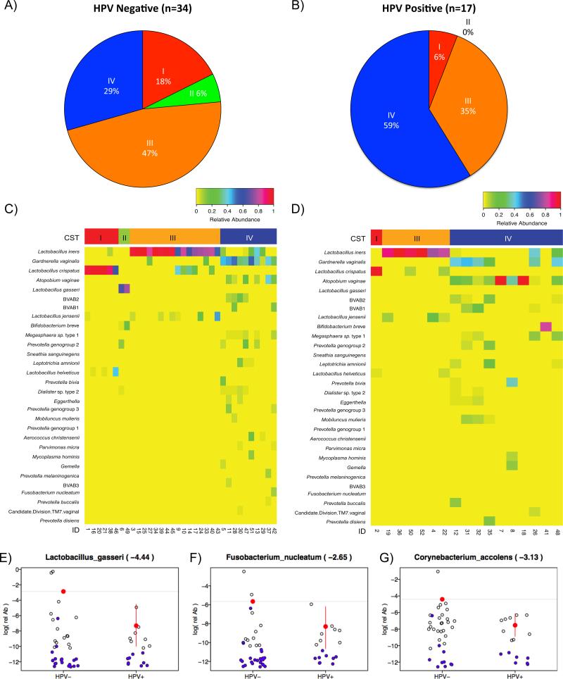
Samples for 8 participants were unavailable for 16S rRNA gene sequencing. The cervico-vaginal microbiota of the remaining 51 participants were assigned to one of five distinct community state types (CSTs) based on the relative abundance of bacterial taxa as previously described by Gajer et al.:24 CST-I (most often dominated by Lactobacillus crispatus), CST-II (most often dominated by Lactobacillus gasseri), CST-III (most often dominated by Lactobacillus iners), CST-IV (characterized by a paucity of Lactobacillus spp. and a wide array of facultative and strict anaerobes), and CST-V (most often dominated by Lactobacillus jensenii). Overall there was a high correlation between CST distribution and BV status, with 5/6 (83.3%) of participants with BV (defined as Nugent score ≥7) classified as CST-IV (Likelihood Ratio=5.73, p=0.017). The distribution of vaginal CSTs in HPV negative vs. HPV positive women were: 6/34 vs. 1/17 CST-I, 2/34 vs. 0/17 CST-II, 16/34 vs. 6/17 CST-III, 10/34 vs. 10/17 CST-IV; no participants in either group had a microbiome consistent with CST-V (Figure 4A-B).
Figure 4. HPV infection and the composition and structure of the vaginal microbiota.
The distribution of community state types (CSTs) in (A) HPV negative and (B) HPV positive women, a heatmap of the relative abundance of the 30 most common bacterial phylotypes in (C) HPV negative and (D) HPV positive women grouped by CST distribution, and log relative abundances of specific bacterial phylotypes by HPV status: (E) Lactobacillus gasseri, (F) Fusobacterium nucleatum, (G) Corynebacterium accolens. Samples with relative abundances of 0 were set to a small random number (blue circles). Number in parentheses indicate the difference of log mean relative abundances of each taxa in HPV positive versus HPV negative participants. Red circles indicate the log of the mean relative abundance of samples with red bars in the HPV positive group indicating the 95% credible interval of the comparison of the two log mean relative abundances. A significant difference is observed when the 95% credible interval in the HPV positive group does contain the log mean relative abundance of the HPV negative group (grey line).
Since CST-IV correlated most closely with a high Nugent score, and since both BV and HPV have been associated with increased HIV acquisition, we examined whether this group had higher rates of HPV infection relative to other CSTs. Interestingly, more participants with HPV infection had a cervico-vaginal microbiome consistent with CST-IV, compared to HPV-negative women (58.8% vs. 29.4%; p=0.043, Figure 4A-B). While significance was lost when post-menopausal women were excluded from the analysis (p=0.084), this likely relates to the reduced sample size, since menopausal status was not associated with CST status or other immune parameters in univariate analysis (data not shown). When we evaluated individual bacterial taxa (Figure 4 C-D), participants with HPV had a substantially lower relative abundance of Lactobacillus gasseri (p=0.009; Figure 4E), and also of the less abundant bacteria Fusobacterium nucleatum (p=0.019; Figure 4F), Cornybacterium accolens (p=0.001; Figure 4G), Peptoniphilus harei, Anaerococcus tetradius, Finegoldia magna, and Raoultella planticola (all P<0.05; data not shown). Finally, HPV-infected participants had a higher overall bacterial load (median 9.22log10) than their uninfected counterparts (median 8.95log10, p=0.018).
DISCUSSION
HPV infection has been consistently associated with an increased risk of HIV acquisition,4 but the underlying biological mechanism(s) behind this association are unclear. We hypothesized that this increased risk might relate to the mucosal recruitment of HIV-susceptible CD4+ target cells, as is seen during chronic HSV-2 infection.18, 19, 25 Furthermore, we hypothesized that this recruitment might be exaggerated in women who go on to clear HPV, since a Th1 cytokine milieu may predominate during host immune clearance5 and Th1 CD4+ T cells are highly HIV susceptible. However, we found no association between prevalent HPV infection and genital inflammatory cytokines, nor with endocervical CD4+ T cell numbers or highly susceptible CD4+ T cell subsets such as those expressing CCR5 or CD69.26 Furthermore, among women with prevalent HPV infection, none of these immune parameters differed between participants who subsequently cleared the virus and those who remained persistently infected. However, we did find an increased absolute number of Langerhans cells in women who cleared HPV, and a skewing towards vaginal CST-IV, characterized by a paucity of Lactobacillus spp., in participants with prevalent HPV infection. These results are broadly in keeping with previous reports.27, 28
Given the consistent association between HPV infection and HIV acquisition,4 particularly in the context of host HPV clearance,29 we were surprised by the lack of association with both HIV-susceptible target cells and pro-inflammatory cytokines in the female genital tract, although other groups also found no association of HPV or its clearance with elevated pro-inflammatory cytokines.30 HPV infection does not result in cell lysis, and is thought to maintain a regulatory state by evading recognition through the impairment of cytokine signaling.31 This state of immune quiescence induced by HPV could explain the lack of both a pro-inflammatory cytokine as well as CD4 T cell response to the virus within the local immune milieu of the female genital tract.
Given the lack of association between CD4 T cell subsets and pro-inflammatory cytokines and HPV infection, if the link between HPV infection and HIV acquisition is a causal one, then another mechanism is likely to be at play. It has been suggested that innate immune responses may be vital to clearing HPV infections through recognition via pattern recognition receptors.32 Imiquimod, a TLR-7 agonist that induces Langerhans cell recruitment and antigen presentation, increases the clearance of HPV-associated genital warts.33 This mirrors our results, with increased numbers of Langerhans cells being associated with HPV clearance. Further, Langerhans cells are reduced in individuals who develop HPV-positive cervical intraepithelial lesions34 and a previous study showed that HPV clearance is associated with increased CD1a+ dendritic cell density in the male foreskin.21 Since Langerhans cells may play a direct role in enhancing HIV susceptibility via binding HIV gp120 and facilitating transmission to CD4 T cells in trans,35 this constitutes a potential immune mechanism linking HPV clearance with HIV susceptibility. In addition, HPV-infected participants had elevated genital levels of the chemokines IP-10 and MIG (which were not components of the pro-inflammatory cytokine score). These chemokines are both IFN-γ inducible and bind CXCR3,36 meaning that they may indicate immune activation; interestingly, genital levels of both are reduced in women who remained HIV-uninfected despite frequent HIV exposure,37 and elevated levels of IP-10 were strongly associated with increased HIV acquisition in South African women.38 The exact mechanism(s) by which they enhance HIV susceptibility is not known, although we found both to be moderately correlated with an increased numbers of cervical CCR5+CD4+ T cells (both correlation coefficients 0.3, P>0.05).
Furthermore, the vaginal microbiota of participants with HPV infection were more likely to be classified as CST-IV, a state characterized by increased relative abundances of facultative and strict anaerobic bacterial species,39 consistent with previous findings demonstrating that women with HR-HPV infection have decreased abundance of Lactobacillus spp.27 Interestingly, we also observed a decrease in the relative abundance of certain bacterial phylotypes in HPV-infected women, most notably in the generally abundant Lactobacillus gasseri. Other studies have specifically linked L. gasseri with HPV clearance,27 and our results support a potential role for these bacteria in defense against HPV. The other bacterial phylotypes that were reduced in the context of HPV infection were of much lower overall abundance and their importance in the genital tract is less clear, although Fusobacterium nucleatum may act as a pathogen.40 Although our analysis only found significant reductions in specific bacterial phyla among HPV-infected women, anaerobes as a whole tended to be increased in HPV positive participants, in keeping with a higher proportion of CST-IV. While these differences in the composition and structure of the vaginal microbiota were not associated with alterations in HIV-susceptible CD4 T cells in the female genital tract, they could feasibly play a role in promoting the recruitment of Langerhans cells to sites of HIV exposure, or enhance HIV susceptibility through other mechanisms such as decreased integrity of the vaginal epithelial barrier.41
Our study does have limitations, including a relatively modest sample size. While robust associations were demonstrated between HSV-2 serostatus and CD4+ T cell subsets in a smaller study,19 and between HPV infection and specific immune and microbiologic parameters in this one, our sample size could have precluded us from finding true but less robust associations of HPV with other immune parameters. However, any causal effect of HPV infection or its clearance on CD4+ subsets or inflammation would be considerably weaker than the effect of HSV-2. While there was variability in the time of genital sampling in relation to the menstrual cycle and menopause, neither factor was associated with immune parameters or the microbiome in univariate analysis, and their incorporation into our multivariable model did not substantively impact the results. Longitudinal immunological data were not available, with cell populations and cytokines measured only at baseline, and follow up HPV infection data were only obtained after 6 months. Therefore, we may have missed a short-lived immune signature of clearance, and/or immune changes that are induced after (rather than before) HPV clearance. Future studies with more frequent sampling may help to understand the sequence of immune changes that culminates in HPV clearance. Our ability to assess the association of vaginal CST-I/II/V with HPV infection was limited, since these vaginal microbiota types are relatively underrepresented in the ACB community.39 Secretions for microbiome analysis were collected by Softcup, and therefore represent a mixture of secretions from the proximal vagina and cervix, while previous studies have used mid-vaginal swabs. However, recent work from our group demonstrates no more variability in the composition of the microbiota as assessed from a cervical and vaginal swab, than was seen between two independent vaginal swabs (Jacques Ravel, personal communication). Finally, limitations in cell numbers obtained by endocervical cytobrush sampling meant that we were not able to assess functional alterations in mucosal T cells that may be linked to HPV infection or clearance, and it is possible that by focusing on the endocervix our study could miss HPV-associated cellular changes limited to the ectocervix or transformation zone. Future studies that incorporate ectocervical biopsy might be considered to explore this possibility.
In summary, neither cervico-vaginal HPV infection nor HPV clearance were associated with cervical T cell alterations or a pro-inflammatory cytokine signature. However, HPV infection was associated with alterations in the composition and structure of the vaginal microbiota and with local elevations in the chemokines IP-10 and MIG, while HPV clearance was associated with increased numbers of cervical Langerhans cell, and each of these could potentially enhance HIV susceptibility. Further research should confirm these observations and characterize the impact of HPV on other immune parameters that may modulate HIV susceptibility.
METHODS
Participant enrollment and inclusion criteria
Participants were recruited through a flyer at a primary care clinic, Women's Health in Women's Hands Community Health Centre (WHIWH) located in Toronto, Canada. Briefly, ethnic African/Caribbean women living in the Toronto area were recruited, enrolled, and provided informed written consent as per the study protocol approved by the HIV Research Ethics Board at the University of Toronto. Participants both completed a questionnaire and underwent testing for common genital infections. Exclusion criteria for the study included: current infection with HIV-1 or 2/ Neisseria gonorrhoeae, or Chlamydia trachomatis, under 16 years of age, current pregnancy, or reporting a symptomatic genital infection within the previous 3 months.
Study protocol and sampling
Pre-menopausal women visited the clinic 10-18 days after the last day of bleeding from their previous menstrual period. After completing a clinical and sexual behaviour questionnaire; blood, urine, and a vaginal swab were collected for STI diagnostics. Instead Softcups (Evofem, San Diego, CA) were used to self-collect undiluted cervico-vaginal secretions for one minute and used for cytokine and microbiota analysis. Two physician-collected endocervical cytobrushes were collected for immune cell phenotyping. Each was gently inserted into the cervical os, rotated 360°, placed in R10 medium (RPMI 1640 with 10% heat inactivated fetal bovine serum (Sigma-Aldrich, Carlsbad, CA), 100mg/mL streptomycin, 100 U/mL penicillin, and 1x GlutaMAX-1 (Gibco, Grand Island, NY) media), stored at 4°C, transferred to the lab within 3 hours, filtered (100μm), washed, and divided evenly into two aliquots for staining. Peripheral blood mononuclear cells (PBMCs) isolated by ficoll-hypaque density centrifugation were washed twice in R10 and two 1 million cell aliquots were used for staining.
Coinfection diagnostics
Gram stain was performed on vaginal smears and evaluated for the presence of yeast and by Nugent criteria for BV diagnostic.42 Neisseria gonorrhoeae and Chlamydia trachomatis were detected by nucleic acid amplification test (NAAT; ProbeTech Assay, BD, Sparks, MD) on first-void urine. HerpeSelect® gG-1 and gG-2 ELISA (Focus Technologies, Cypress, CA; adjusted threshold of 3.543) was used to determine participant's HSV-2 serostatus. Self-collected vaginal swabs were used to identify 37 HPV types using Roche Linear Array (Roche Molecular Systems, Basel, Switzerland) as previously described.44 High-risk types were defined as: HPV16,18,31,33,35,39,45,51,52,56,58,59,68 and 69.44
Immune cell phenotyping
Endocervical cells collected by cytobrush and PBMCs were stained with two monoclonal antibody panels: one for T cells and the other DCs. The former consisted of: α4-FITC (Miltenyi Biotec, Bergisch Gladbach, Germany), CD4-ECD (Beckman Coulter, Marseille, France), CCR5-PE, β7-APC, CD38-AlexaFluor700, HLA-DR-APC-Cy7, CD69-eFluor450 (BD Biosciences, Franklin Lakes, NJ), Live/Dead Aqua (Invitrogen), CD25-PerCP-Cy5.5, CD39-PE-Cy7, and CD3-eFluor650 (eBiosciences, San Diego, CA). The DC panel included: BDCA2-FITC (Miltenyi), CD207-PE (Beckman Coulter), DC-SIGN-PerCP-Cy5.5, CD206-APC (BD Biosciences), CD83-Streptavidin, CD123-PE-Cy7, CD11c-AlexaFluor700, CD14-AlexaFluor780, CD1a-v450, CD3-eFluor650 (eBiosciences), and Live/Dead Aqua (Invitrogen). Cells were enumerated with a BD LSR-2 flow cytometer (BD Systems) and analyzed using FlowJo 9.3.2 software (Treestar, Ashland, OR) by a single, blinded researcher. Immune cell populations from the cervix are reported both as a proportion (%) and as the total number of cells per cytobrush after the entire contents of cytobrush-collected samples were acquired by flow cytometry (as above). DC subsets were defined as: Myeloid-derived DCs (mDCs; CD3-CD11c+), Monocytes (CD3-CD14+), and Langerhans cells (LCs; CD3-CD1a+langerin/CD207+). Langerhans cells were gated as per Supplementary Figure 1. Other cellular subsets were gated as previously described.19
Cytokine and chemokine assays
Aliquoted and cryopreserved cervico-vaginal secretions were used to assay levels of 14 cytokines (GM-CSF, IL-1α, IL-8, MCP-1, MIG, MIP-3α, RANTES, IL-10, IL-17, IL-1β, IL-6, IP-10, MIP-1β and TNFα) using the Meso Scale Discovery (MSD; Rockville, MD) electro-chemiluminescent ELISA system. Secretions were plated at 50μl per well and run in duplicate. A standard curve was used to determine the concentration (pg/ml). The dilution at which the coefficient of variation exceeded 30% was defined as the lower limit of quantification (LLOQ). Any sample out of range of the standard curve was repeated following dilution. All samples were assayed and analyzed by blinded lab personnel and the original undiluted concentrations of cytokines in the secretions were calculated.
DNA extraction, 16S rRNA gene amplification and sequencing
Genital secretions (100μl) collected by Instead Softcup (Evofem, San Diego, CA) underwent enzymatic cell wall digest and bead beating for total DNA extraction, as previously described.24, 39 A dual-barcode system with fusion primers 338F and 806R was used to amplify the V3-V4 regions of the 16S rRNA gene according to Fadrosh et al.45 and subsequently sequenced on an Illumina MiSeq instrument (Illumina, San Diego, CA) using the 300bp paired-end protocol at the Genomic Resource Center at the University of Maryland School of Medicine, Institute for Genome Sciences. If the average phred quality score of 4 consecutive bp was below 15 the sequence reads were trimmed and only retained if their length was at least 75% of their original length after trimming. FLASH46 was used to assemble paired reads. De-multiplexing by binning sequences with the same barcode and quality trimming were performed in QIIME (version 1.8.0)47 (see Fadrosh et. al45 for additional details). Detection of de novo and reference-based chimera was conducted in UCHIME (v5.1)48 using greengenes database of 16S rRNA gene sequences (Aug, 2013 vers.)49 as reference. The processed 16S rRNA gene amplicon sequences were assigned to genera and species, and vaginal microbial communities were assigned to a community state types (CSTs) using taxonomic composition and abundance and the Jensen-Shannon divergence metrics as previously described.24 A table providing the relative abundance of identified taxa and the CST assignments for each sample was generated and is provided as Supplementary Table S1 with metadata and a legend in Supplementary Tables S2-S3. The 16S rRNA gene sequence data has been deposited into NCBI SRA under accession number SRA362820. Overall bacterial load was measured using an assay previously described by Liu et al.50
Statistical analysis
Baseline characteristics and outcomes were summarized with frequencies and proportions for categorical variables and median and range for continuous variables. Differences were assessed using Pearson's χ2 test for categorical variables or an independent samples t-test for continuous variables. Cytokine levels and the number of cells per cytobrush were log-transformed to normalize the data (and verified using the Shapiro-Wilk normality test), although the latter is presented in absolute numbers within the text. Univariate immune associations were confirmed in a multivariate general linear model, controlling for demographic factors. Participants producing elevated levels of pro-inflammatory cytokines were defined using a scoring system previously described;11 briefly, individuals with elevated levels of pro-inflammatory cytokines were defined as producing at least 3 out of 7 cytokines (IL-1α, IL-8, MIP-3α, RANTES, IL-1β, MIP-1β, and TNF-α) in the upper quartile of all participants. Associations between specific bacterial taxa and HPV status were assessed with Bayesian zero-inflated negative binomial models.
All statistical analyses were performed using SPSS version 22 (IBM, New York, NY) and the statistical package R (R Foundation for Statistical Computing, Vienna, Austria).
Supplementary Material
ACKNOWLEDGEMENTS
The investigators acknowledge the kind assistance of the staff at the Women's Help in Women's Hands Clinic, and the time and cooperation of all study participants.
Funding. This study was supported by the Canadian Institutes of Health Research (RK, grants #TMI-138656 and #OCH-131579; BS, studentship) and Ontario HIV Treatment Network (studentship, TJY). RK is supported by a University of Toronto – OHTN Endowed Chair in HIV Research. JR, BM, MSH and PG were supported by the National Institute of Allergy and Infectious Diseases of the National Institute of Health under award number U19AI084044 and R01AI116799. The content is solely the responsibility of the authors and does not necessarily represent the official views of the National Institute of Health.
Footnotes
AUTHOR CONTRIBUTIONS
All authors have contributed to the study. BS, WT, JR, AR, and RK were involved in the conception and design of the study. BS, TJY, SP, PG, BM, MH, JT, LC, PJ, MS, SH, and KS were involved in the execution of the study. BS, PG, BM, MH, JR, AR, and RK were involved in the analysis and interpretation of the study.
DISCLOSURE
The authors declare no conflict of interest.
REFERENCES
- 1.Bruni L, Diaz M, Castellsague X, Ferrer E, Bosch FX, de Sanjose S. Cervical human papillomavirus prevalence in 5 continents: meta-analysis of 1 million women with normal cytological findings. J Infect Dis. 2010;202(12):1789–1799. doi: 10.1086/657321. [DOI] [PubMed] [Google Scholar]
- 2.Rosa MI, Fachel JM, Rosa DD, Medeiros LR, Igansi CN, Bozzetti MC. Persistence and clearance of human papillomavirus infection: a prospective cohort study. Am J Obstet Gynecol. 2008;199(6):617, e611–617. doi: 10.1016/j.ajog.2008.06.033. [DOI] [PubMed] [Google Scholar]
- 3.Munoz N, Bosch FX, de Sanjose S, Herrero R, Castellsague X, Shah KV, et al. Epidemiologic classification of human papillomavirus types associated with cervical cancer. N Engl J Med. 2003;348(6):518–527. doi: 10.1056/NEJMoa021641. [DOI] [PubMed] [Google Scholar]
- 4.Houlihan CF, Larke NL, Watson-Jones D, Smith-McCune KK, Shiboski S, Gravitt PE, et al. Human papillomavirus infection and increased risk of HIV acquisition. A systematic review and meta analysis. AIDS. 2012;26(17):2211–2222. doi: 10.1097/QAD.0b013e328358d908. [DOI] [PMC free article] [PubMed] [Google Scholar]
- 5.Scott M, Stites DP, Moscicki AB. Th1 cytokine patterns in cervical human papillomavirus infection. Clin Diagn Lab Immunol. 1999;6(5):751–755. doi: 10.1128/cdli.6.5.751-755.1999. [DOI] [PMC free article] [PubMed] [Google Scholar]
- 6.Patel P, Borkowf CB, Brooks JT, Lasry A, Lansky A, Mermin J. Estimating per act HIV transmission risk: a systematic review. AIDS. 2014;28(10):1509–1519. doi: 10.1097/QAD.0000000000000298. [DOI] [PMC free article] [PubMed] [Google Scholar]
- 7.Kaul R, Pettengell C, Sheth PM, Sunderji S, Biringer A, MacDonald K, et al. The genital tract immune milieu: an important determinant of HIV susceptibility and secondary transmission. J Reprod Immunol. 2008;77(1):32–40. doi: 10.1016/j.jri.2007.02.002. [DOI] [PubMed] [Google Scholar]
- 8.McKinnon LR, Nyanga B, Chege D, Izulla P, Kimani M, Huibner S, et al. Characterization of a human cervical CD4+ T cell subset coexpressing multiple markers of HIV susceptibility. J Immunol. 2011;187(11):6032–6042. doi: 10.4049/jimmunol.1101836. [DOI] [PubMed] [Google Scholar]
- 9.Joag VR, McKinnon LR, Liu J, Kidane ST, Yudin MH, Nyanga B, et al. Identification of preferential CD4 -T cell targets for HIV infection in the cervix. Mucosal Immunol. 2015 doi: 10.1038/mi.2015.28. [DOI] [PubMed] [Google Scholar]
- 10.Anahtar MN, Byrne EH, Doherty KE, Bowman BA, Yamamoto HS, Soumillon M, et al. Cervicovaginal bacteria are a major modulator of host inflammatory responses in the female genital tract. Immunity. 2015;42(5):965–976. doi: 10.1016/j.immuni.2015.04.019. [DOI] [PMC free article] [PubMed] [Google Scholar]
- 11.Arnold KB, Burgener A, Birse K, Romas L, Dunphy LJ, Shahabi K, et al. Increased levels of inflammatory cytokines in the female reproductive tract are associated with altered expression of proteases, mucosal barrier proteins, and an influx of HIV-susceptible target cells. Mucosal Immunol. 2015 doi: 10.1038/mi.2015.51. [DOI] [PubMed] [Google Scholar]
- 12.Kaul R, Rebbapragada A, Hirbod T, Wachihi C, Ball TB, Plummer FA, et al. Genital levels of soluble immune factors with anti-HIV activity may correlate with increased HIV susceptibility. AIDS. 2008;22(15):2049–2051. doi: 10.1097/QAD.0b013e328311ac65. [DOI] [PMC free article] [PubMed] [Google Scholar]
- 13.Carnathan DG, Wetzel KS, Yu J, Lee ST, Johnson BA, Paiardini M, et al. Activated CD4+CCR5+ T cells in the rectum predict increased SIV acquisition in SIVGag/Tat vaccinated rhesus macaques. Proc Natl Acad Sci U S A. 2015;112(2):518–523. doi: 10.1073/pnas.1407466112. [DOI] [PMC free article] [PubMed] [Google Scholar]
- 14.Masson L, Mlisana K, Little F, Werner L, Mkhize NN, Ronacher K, et al. Defining genital tract cytokine signatures of sexually transmitted infections and bacterial vaginosis in women at high risk of HIV infection: a cross-sectional study. Sex Transm Infect. 2014;90(8):580–587. doi: 10.1136/sextrans-2014-051601. [DOI] [PubMed] [Google Scholar]
- 15.Prodger JL, Shannon B, Shahabi K, Kong X, Liu CM, Price L, et al. CROI. Boston, MA: 2014. Penile Cytokines Correlate with HIV Target Cells and Decrease after Circumcision in Rakai Uganda. vol. Abstract #965. [Google Scholar]
- 16.Levine WC, Pope V, Bhoomkar A, Tambe P, Lewis JS, Zaidi AA, et al. Increase in endocervical CD4 lymphocytes among women with nonulcerative sexually transmitted diseases. J Infect Dis. 1998;177(1):167–174. doi: 10.1086/513820. [DOI] [PubMed] [Google Scholar]
- 17.Freeman EE, Weiss HA, Glynn JR, Cross PL, Whitworth JA, Hayes RJ. Herpes simplex virus 2 infection increases HIV acquisition in men and women: systematic review and meta-analysis of longitudinal studies. AIDS. 2006;20(1):73–83. doi: 10.1097/01.aids.0000198081.09337.a7. [DOI] [PubMed] [Google Scholar]
- 18.Prodger JL, Gray R, Kigozi G, Nalugoda F, Galiwango R, Nehemiah K, et al. Impact of asymptomatic Herpes simplex virus-2 infection on T cell phenotype and function in the foreskin. AIDS. 2012;26(10):1319–1322. doi: 10.1097/QAD.0b013e328354675c. [DOI] [PMC free article] [PubMed] [Google Scholar]
- 19.Shannon B, Yi TJ, Thomas-Pavanel J, Chieza L, Janakiram P, Saunders M, et al. Impact of asymptomatic herpes simplex virus type 2 infection on mucosal homing and immune cell subsets in the blood and female genital tract. J Immunol. 2014;192(11):5074–5082. doi: 10.4049/jimmunol.1302916. [DOI] [PubMed] [Google Scholar]
- 20.Atashili J, Poole C, Ndumbe PM, Adimora AA, Smith JS. Bacterial vaginosis and HIV acquisition: a meta-analysis of published studies. AIDS. 2008;22(12):1493–1501. doi: 10.1097/QAD.0b013e3283021a37. [DOI] [PMC free article] [PubMed] [Google Scholar]
- 21.Tobian AA, Grabowski MK, Kigozi G, Redd AD, Eaton KP, Serwadda D, et al. Human papillomavirus clearance among males is associated with HIV acquisition and increased dendritic cell density in the foreskin. J Infect Dis. 2013;207(11):1713–1722. doi: 10.1093/infdis/jit035. [DOI] [PMC free article] [PubMed] [Google Scholar]
- 22.Farhat S, Nakagawa M, Moscicki AB. Cell-mediated immune responses to human papillomavirus 16 E6 and E7 antigens as measured by interferon gamma enzyme-linked immunospot in women with cleared or persistent human papillomavirus infection. Int J Gynecol Cancer. 2009;19(4):508–512. doi: 10.1111/IGC.0b013e3181a388c4. [DOI] [PMC free article] [PubMed] [Google Scholar]
- 23.Remis RS, Liu J, Loutfy M, Tharao W, Rebbapragada A, Perusini SJ, et al. The epidemiology of sexually transmitted co-infections in HIV-positive and HIV-negative African Caribbean women in Toronto. BMC Infect Dis. 2013;13:550. doi: 10.1186/1471-2334-13-550. [DOI] [PMC free article] [PubMed] [Google Scholar]
- 24.Gajer P, Brotman RM, Bai G, Sakamoto J, Schutte UM, Zhong X, et al. Temporal dynamics of the human vaginal microbiota. Sci Transl Med. 2012;4(132):132ra152. doi: 10.1126/scitranslmed.3003605. [DOI] [PMC free article] [PubMed] [Google Scholar]
- 25.Rebbapragada A, Wachihi C, Pettengell C, Sunderji S, Huibner S, Jaoko W, et al. Negative mucosal synergy between Herpes simplex type 2 and HIV in the female genital tract. AIDS. 2007;21(5):589–598. doi: 10.1097/QAD.0b013e328012b896. [DOI] [PubMed] [Google Scholar]
- 26.McKinnon LR, Kaul R. Quality and quantity: mucosal CD4+ T cells and HIV susceptibility. Curr Opin HIV AIDS. 2012;7(2):195–202. doi: 10.1097/COH.0b013e3283504941. [DOI] [PubMed] [Google Scholar]
- 27.Brotman RM, Shardell MD, Gajer P, Tracy JK, Zenilman JM, Ravel J, et al. Interplay between the temporal dynamics of the vaginal microbiota and human papillomavirus detection. J Infect Dis. 2014;210(11):1723–1733. doi: 10.1093/infdis/jiu330. [DOI] [PMC free article] [PubMed] [Google Scholar]
- 28.Dareng EO, Ma B, Famooto AO, Akarolo-Anthony SN, Offiong RA, Olaniyan O, et al. Prevalent high-risk HPV infection and vaginal microbiota in Nigerian women. Epidemiol Infect. 2016;144(1):123–137. doi: 10.1017/S0950268815000965. [DOI] [PMC free article] [PubMed] [Google Scholar]
- 29.Smith-McCune KK, Shiboski S, Chirenje MZ, Magure T, Tuveson J, Ma Y, et al. Type specific cervico-vaginal human papillomavirus infection increases risk of HIV acquisition independent of other sexually transmitted infections. PLoS One. 2010;5(4):e10094. doi: 10.1371/journal.pone.0010094. [DOI] [PMC free article] [PubMed] [Google Scholar]
- 30.Kriek JM, Jaumdally SZ, Masson L, Little F, Mbulawa Z, Gumbi PP, et al. Female genital tract inflammation, HIV co-infection and persistent mucosal Human Papillomavirus (HPV) infections. Virology. 2016;493:247–254. doi: 10.1016/j.virol.2016.03.022. [DOI] [PubMed] [Google Scholar]
- 31.Kaul R, Hirbod T. Genital epithelial cells: foot soldiers or fashion leaders? J Leukoc Biol. 2010;88(3):427–429. doi: 10.1189/jlb.0410230. [DOI] [PubMed] [Google Scholar]
- 32.Daud, Scott ME, Ma Y, Shiboski S, Farhat S, Moscicki AB. Association between toll like receptor expression and human papillomavirus type 16 persistence. Int J Cancer. 2011;128(4):879–886. doi: 10.1002/ijc.25400. [DOI] [PMC free article] [PubMed] [Google Scholar]
- 33.Beutner KR, Spruance SL, Hougham AJ, Fox TL, Owens ML, Douglas JM., Jr. Treatment of genital warts with an immune-response modifier (imiquimod). J Am Acad Dermatol. 1998;38(2 Pt 1):230–239. doi: 10.1016/s0190-9622(98)70243-9. [DOI] [PubMed] [Google Scholar]
- 34.Mota F, Rayment N, Chong S, Singer A, Chain B. The antigen-presenting environment in normal and human papillomavirus (HPV)-related premalignant cervical epithelium. Clin Exp Immunol. 1999;116(1):33–40. doi: 10.1046/j.1365-2249.1999.00826.x. [DOI] [PMC free article] [PubMed] [Google Scholar]
- 35.Turville SG, Arthos J, Donald KM, Lynch G, Naif H, Clark G, et al. HIV gp120 receptors on human dendritic cells. Blood. 2001;98(8):2482–2488. doi: 10.1182/blood.v98.8.2482. [DOI] [PubMed] [Google Scholar]
- 36.Hsieh MF, Lai SL, Chen JP, Sung JM, Lin YL, Wu-Hsieh BA, et al. Both CXCR3 and CXCL10/IFN-inducible protein 10 are required for resistance to primary infection by dengue virus. J Immunol. 2006;177(3):1855–1863. doi: 10.4049/jimmunol.177.3.1855. [DOI] [PubMed] [Google Scholar]
- 37.Lajoie J, Juno J, Burgener A, Rahman S, Mogk K, Wachihi C, et al. A distinct cytokine and chemokine profile at the genital mucosa is associated with HIV-1 protection among HIV-exposed seronegative commercial sex workers. Mucosal Immunol. 2012;5(3):277–287. doi: 10.1038/mi.2012.7. [DOI] [PubMed] [Google Scholar]
- 38.Masson L, Passmore JA, Liebenberg LJ, Werner L, Baxter C, Arnold KB, et al. Genital inflammation and the risk of HIV acquisition in women. Clin Infect Dis. 2015;61(2):260–269. doi: 10.1093/cid/civ298. [DOI] [PMC free article] [PubMed] [Google Scholar]
- 39.Ravel J, Gajer P, Abdo Z, Schneider GM, Koenig SS, McCulle SL, et al. Vaginal microbiome of reproductive-age women. Proc Natl Acad Sci U S A. 2011;108(Suppl 1):4680–4687. doi: 10.1073/pnas.1002611107. [DOI] [PMC free article] [PubMed] [Google Scholar]
- 40.Citron DM. Update on the taxonomy and clinical aspects of the genus fusobacterium. Clin Infect Dis. 2002;35(Suppl 1):S22–27. doi: 10.1086/341916. [DOI] [PubMed] [Google Scholar]
- 41.Borgdorff H, Gautam R, Armstrong SD, Xia D, Ndayisaba GF, van Teijlingen NH, et al. Cervicovaginal microbiome dysbiosis is associated with proteome changes related to alterations of the cervicovaginal mucosal barrier. Mucosal Immunol. 2016;9(3):621–633. doi: 10.1038/mi.2015.86. [DOI] [PubMed] [Google Scholar]
- 42.Nugent RP, Krohn MA, Hillier SL. Reliability of diagnosing bacterial vaginosis is improved by a standardized method of gram stain interpretation. J Clin Microbiol. 1991;29(2):297–301. doi: 10.1128/jcm.29.2.297-301.1991. [DOI] [PMC free article] [PubMed] [Google Scholar]
- 43.Biraro S, Mayaud P, Morrow RA, Grosskurth H, Weiss HA. Performance of commercial herpes simplex virus type-2 antibody tests using serum samples from Sub Saharan Africa: a systematic review and meta analysis. Sex Transm Dis. 2011;38(2):140–147. doi: 10.1097/OLQ.0b013e3181f0bafb. [DOI] [PubMed] [Google Scholar]
- 44.Shannon B, Perusini S, Liu J, Chieza L, Saunders M, Green-Walker L-A, et al. Community-specific Prevalence and Type Distribution of HPV Infection among African/Caribbean Women in Toronto, Canada (Abstract PH.PP02.82).. 29th International Papillomavirus Conference; Seattle, WA. 2014. [Google Scholar]
- 45.Fadrosh DW, Ma B, Gajer P, Sengamalay N, Ott S, Brotman RM, et al. An improved dual-indexing approach for multiplexed 16S rRNA gene sequencing on the Illumina MiSeq platform. Microbiome. 2014;2(1):6. doi: 10.1186/2049-2618-2-6. [DOI] [PMC free article] [PubMed] [Google Scholar]
- 46.Magoc T, Salzberg SL. FLASH: fast length adjustment of short reads to improve genome assemblies. Bioinformatics. 2011;27(21):2957–2963. doi: 10.1093/bioinformatics/btr507. [DOI] [PMC free article] [PubMed] [Google Scholar]
- 47.Caporaso JG, Kuczynski J, Stombaugh J, Bittinger K, Bushman FD, Costello EK, et al. QIIME allows analysis of high-throughput community sequencing data. Nat Methods. 2010;7(5):335–336. doi: 10.1038/nmeth.f.303. [DOI] [PMC free article] [PubMed] [Google Scholar]
- 48.Edgar RC, Haas BJ, Clemente JC, Quince C, Knight R. UCHIME improves sensitivity and speed of chimera detection. Bioinformatics. 2011;27(16):2194–2200. doi: 10.1093/bioinformatics/btr381. [DOI] [PMC free article] [PubMed] [Google Scholar]
- 49.McDonald D, Price MN, Goodrich J, Nawrocki EP, DeSantis TZ, Probst A, et al. An improved Greengenes taxonomy with explicit ranks for ecological and evolutionary analyses of bacteria and archaea. ISME J. 2012;6(3):610–618. doi: 10.1038/ismej.2011.139. [DOI] [PMC free article] [PubMed] [Google Scholar]
- 50.Liu CM, Aziz M, Kachur S, Hsueh PR, Huang YT, Keim P, et al. BactQuant: an enhanced broad-coverage bacterial quantitative real time PCR assay. BMC Microbiol. 2012;12:56. doi: 10.1186/1471-2180-12-56. [DOI] [PMC free article] [PubMed] [Google Scholar]
Associated Data
This section collects any data citations, data availability statements, or supplementary materials included in this article.


