Abstract
Many psychiatric diseases such as post-traumatic stress disorder (PTSD) are characterized by abnormal processing of emotional stimuli particularly fear. The medial prefrontal cortex (mPFC) is critically involved in fear expression. However, the molecular mechanisms underlying this process are largely unknown. Neuregulin-1 (NRG1) reportedly regulates pyramidal neuronal activity via ErbB4 receptors, which are abundant in parvalbumin (PV)-expressing interneurons in the PFC. In this study, we aimed to determine how NRG1/ErbB4 signaling in the mPFC modulates fear expression and found that tone-cued fear conditioning increased NRG1 expression in the mPFC. Tone-cued fear conditioning was inhibited following neutralization of endogenous NRG1 and specific inhibition or genetic ablation of ErbB4 in the prelimbic (PL) cortex but not in the infralimbic cortex. Furthermore, ErbB4 deletion specifically in PV neurons impaired tone-cued fear conditioning. Notably, overexpression of ErbB4 in the PL cortex is sufficient to reverse impaired fear conditioning in PV-Cre;ErbB4−/− mice. Together, these findings identify a previously unknown signaling pathway in the PL cortex that regulates fear expression. As both NRG1 and ErbB4 are risk genes for schizophrenia, our study may shed new light on the pathophysiology of this disorder and help to improve treatments for psychiatric disorders such as PTSD.
Introduction
Patients with psychiatric diseases such as post-traumatic stress disorder (PTSD) have difficulty in processing emotional stimuli. Pavlovian fear conditioning, which in some respects resembles PTSD,1, 2 is a classical animal model used for the study of anxiety disorders.3, 4 An understanding of the mechanisms underlying fear conditioning-induced memory formation and expression is critical for understanding the neurobiology of fear inhibition and for the treatment of some anxiety disorders.
The medial prefrontal cortex (mPFC) consists of the medial agranular, anterior cingulate, prelimbic (PL) and infralimbic (IL) cortices. A wealth of evidence shows that the mPFC has an important part in fear expression.5, 6, 7, 8, 9, 10 In particular, the PL and IL cortices seemingly have different and even opposite roles in modulating fear expression. For example, stimulation of the PL cortex increases and stimulation of the IL cortex decreases conditioned fear responses.5 Other studies have shown that pharmacological inactivation of the PL cortex but not the IL cortex with muscimol (a gamma-aminobutyric acid A (GABAA) receptor agonist) or tetrodotoxin (a sodium channel blocker) reduces the expression of conditioned fear.6 The difference may be due to the fact that different subregions of the mPFC contain different cell types that project to different targets.11 In particular, the activity of long-range-projecting pyramidal neurons is under strict control by locally projecting GABAergic neurons.12, 13 Thus, the GABAergic activity of mPFC is also critically involved in fear learning and expression.5, 8 However, the molecular mechanisms by which GABAergic activity regulates fear expression in the mPFC remain unknown.
Neuregulin-1 (NRG1), which belongs to a family of growth factors that contains the epidermal growth factor-like domain,14, 15, 16 has a critical role in neuronal survival, synaptic transmission and plasticity15 through activating ErbB tyrosine kinases (ErbB2-4), among which ErbB4 is the only tyrosine kinase that can both bind to NRG1 and become a functionally active homodimer.15, 16, 17 Interestingly, ErbB4 is specifically expressed in interneurons, in particular in parvalbumin (PV)-expressing neurons.18, 19, 20, 21, 22, 23, 24, 25, 26 NRG1-ErbB4 signaling modulates the activity of pyramidal neurons in the corticolimbic system, including the PFC, hippocampus and amygdala, by promoting GABA release.25, 27 Moreover, ErbB4 null knockout mice show impaired tone-cued and contextual fear conditioning.28, 29, 30 These studies demonstrate that NRG1-ErbB4 signaling has a critical role in regulating fear learning and expression. However, little is known about the roles of NRG1-ErbB4 signaling in the mPFC in regulating fear expression.
In the present study, we addressed this issue by showing that ErbB4 signaling in the PL cortex but not the IL cortex is critical for fear expression. Tone-cued fear conditioning, which largely depends on the mPFC, was inhibited following neutralization of endogenous NRG1 and the specific inhibition or genetic ablation of ErbB4 in the PL cortex but not in the IL cortex. Specific deletion of ErbB4 in PV neurons impaired fear conditioning. Notably, overexpression of ErbB4 in the PL cortex suffices to normalize impaired fear conditioning in PV-ErbB4−/− mice. Together, these findings indicated an essential role for ErbB4 signaling in the PL cortex in controlling fear conditioning.
Materials and methods
Animals
Mice were maintained on a 12-h light/12-h dark cycle. Water and food were available ad libitum. LoxP-flanked ErbB4,31 PV-Cre,27 CaMKII-Cre32 and ErbB4 reporter33 mice were described previously. Behavioral testing was performed during the light cycle between 1000 and 1700 hours. Procedures were conducted with the approval of the Institutional Animal Care and Use Committee in accordance with the Chinese Council on Animal Care Guidelines.34 Efforts were made to minimize animal suffering and to reduce the number of animals used.
Reagents
A recombinant polypeptide containing the entire epidermal growth factor domain of the β-type NRG1 (rHRG β177–244) was used.35 Ecto-ErbB4 was prepared from stable HEK293 cells using a previously described method.25 AG1478 was obtained from Calbiochem (San Diego, CA, USA). When dimethylsulphoxide (Sigma, St. Louis, MO, USA) was used to prepare solutions, its final concentration was ⩽0.05%.
Western blot analysis
Tissue homogenates isolated from mPFC were prepared in radioimmunoprecipitation assay buffer containing 50 mM Tris-HCl, pH 7.4, 150 mM NaCl, 2 mM EDTA, 1% sodium deoxycholate, 1% SDS, 1 mM phenylmethylsulfonyl fluoride, 50 mM sodium fluoride, 1 mM sodium vanadate, 1 mM dithiothreitol and protease inhibitors. Homogenates or bound proteins were resolved on SDS-PAGE gels and transferred to nitrocellulose membranes, which were incubated in TBS buffer containing 0.1% Tween-20 and 5% milk for 1 h at room temperature prior to overnight incubation with primary antibodies at 4 °C. After being washed, the membranes were incubated with horseradish peroxidase-conjugated secondary antibodies in TBS for 1 h at room temperature and visualized using the Quantitative FluorChemSP Imaging System (Alpha Innotech, San Leandro, CA, USA). Band intensities were quantified using FluorChem SP software. All values from each band were normalized to corresponding β-actin controls. The following primary antibodies were used: rabbit polyclonal anti-ErbB4 (SC283; 1:500; Santa Cruz Biotechnology, Dallas, TX, USA), rabbit polyclonal anti-NRG1 (SC28916; 1:500; Santa Cruz Biotechnology) and monoclonal mouse anti-β-actin (1:1000; Bostor, China). n=3 mice each group.
Immunofluorescence
Mice were anesthetized with 1% pentobarbital sodium and perfused intracardially with saline and then with 4% formaldehyde. After cryoprotection in 30% sucrose, 40 μm-thick slices were cut on a freezing microtome (CM-1950, Leica, Nussloch, Germany). Slices were blocked with 10% normal goat serum in PBS with 1% Triton-100, and subsequently incubated with the rabbit anti-PV primary antibody (1:5000; PV25; Swant, Marly, Switzerland) overnight at 4 °C. The signals were visualized with the corresponding AlexaFluor 488-conjugated secondary antibody (1:500; A11034; Invitrogen, Carlsbad, CA, USA) at room temperature for 1 h. The slices were coverslipped with fluoroshield mounting medium with 4', 6-diamidino-2-phenylindole (ab104139; Abcam, Cambridge, MA, USA). Fluorescent images were captured using a fluorescence microscope (A1R, Nikon, Tochigi, Japan). n=3 mice each group.
Guide cannula placement and intracerebral infusions
All surgeries were performed under aseptic conditions with stereotaxic guidance. Mice were anesthetized with 1% pentobarbital sodium and mounted onto a stereotaxic instrument (Stoelting, Wood Dale, IL, USA). All coordinates are reported relative to the bregma in mm. A small burr hole (1 mm in diameter) in the skull was opened with a dental drill at the following coordinates: PL +1.75 mm anteroposterior (AP), ±0.4 mm mediolateral (ML) and –2.25 mm dorsoventral (DV) or IL 15° angle, +1.75 mm AP, ±0.75 mm ML and −1.5 mm DV, according to The Mouse Brain in Stereotaxic Coordinates.36 A guiding cannula (Plastics One; C315G/SPC; length, 3 mm) was implanted through the hole and then secured in place with glass ionomer cement. A dummy cannula (Plastics One; C315DC/SPC, with lengths matching the guide cannulas) was placed inside the guide cannula to prevent occlusion. The animals were kept warm with an electric blanket and placed back in their home cages after recovery. The behavioral tests were conducted 7 days after surgery. All unilateral manipulations were counterbalanced across hemispheres.
Intrasubventricular injections of NRG1 (500 nM), ecto-ErbB4 (200 μg ml−1) and AG1478 (50 μM;) were performed 30 min before fear response test. The dummy cannulas were replaced with the infusion cannulas (Plastics One; C315I/SPC, with length matching the guide cannula), which were connected to 5 μl microsyringes (Hamilton, Reno, NV, USA) mounted on a microinfusion pump (RWD200, Shenzhen, China) via polyethylene tubing (Plastics One; C313C). For each mouse, 0.3 μl of drug was injected within 6 min. This volume was selected according to the size and structure of these nuclei. To allow the drug to diffuse, the infusion cannulas were maintained in place for an additional 5 min before being replaced with the dummy cannulas.
At the end of the experiment, mice were perfused for further histological verification. Only mice with correct cannula placement were used.
Virus packaging and stereotaxic injection
The recombinant adeno-associated viral (AAV) vectors were serotyped with AAV5 coat proteins and packaged by NeuronBiotech (Shanghai, China). Viral titers were 2 × 10¹² particles per ml. The expression of Cre recombinase and green fluorescent protein (GFP) was driven by a truncated chimeric cytomegalovirus (CMV)-chicken β-actin (smCBA) promoter. An internal ribosomal re-entry site connected the GFP sequence and the Cre sequence.
Lentiviral (LV) vectors were produced by University of Florida Research Foundation using a previously described method.37 Flag-tagged ErbB4 was subcloned into the pFUGM LV vector. The pFU GM-ErbB4, pCMV DR8.92 and pVSVG vectors were co-transfected into HEK293FT cells, and LV-ErbB4 was harvested with 109 particles per ml. Expression in both LV-ErbB4 and LV-GFP was controlled by a CMV promoter.38 A 5 μl Hamilton syringe was used to deliver AAV or LV solution using a microinjector pump (KDS, Stoelting). 0.3 μl of virus solution was injected at a rate of 0.05 μl min−1. After the injection was completed, the needle was raised 0.1 mm and maintained in place for an additional 10 min to allow the virus to diffuse at the injection site; the needle was then slowly withdrawn.
At the end of the experiment, mice were perfused for further histological verification. Only mice with virus expression restricted to the target region were used.
Tone-cued fear conditioning
Two kinds of fear-conditioning shock chambers (Chamber A: 25 × 25 × 31 cm, with plastic walls and numerous parallel stainless-steel grid bars in the floor connected to a scrambled shocker; Chamber B: 25 × 25 × 31 cm, with plastic walls and floor) and multiparameter activity monitors (The FreezeFrame System, Coulbourn Instruments, Woonsocket, RI, USA) were used. The mice (2 to 3 months of age, n⩾8 mice each group) were handled twice a day for 3 days before the experiments. The mice were alternatively habituated to the manipulation, transported to an experimental room, removed from their cages, handled, weighed and returned to their home cages. The conditioned stimulus (CS) used in this study was a 75 dB sound at 2800 Hz, and the unconditioned stimulus (US) was a one time-continuous scrambled foot shock at 0.7 mA for 1 s. On the day of conditioning, mice were transported from the housing room and individually placed in the fear-conditioning chamber A. Animals were left undisturbed for a 3 min acclimation period (pre-shock period), followed by four CS (30 s duration; 80 s intershock interval) that were each terminated with a US (1 s duration). Mice remained in the chamber for an additional 2 min (post-shock period) to test immediate freezing behaviors. They were then returned to their home cages in the colony room.
Testing for tone-cued fear conditioning was performed 24 h after training. The behavior of each mouse was continuously videotaped to score freezing during the entire testing period. Each mouse was placed into novel chamber B, monitored for 3 min (pre-tone freezing) and then subjected to 3 min of CS tone exposure (tone-cued freezing). The total time spent freezing in each period was quantified and expressed as the percentage of total time (in seconds) using Actimetrics FreezeFrame software (version 2.2, Coulbourn Instruments).
The luminal current intensities required to elicit flinching, vocalization and jumping were compared among all groups to determine whether the significant differences in the freezing responses were due to impaired fear memory induced by gene deletion or to the decrease in nociceptive responses to shock by gene deletion.
Pain threshold test
A mouse was placed into chamber A and received 11 repeated scrambled shocks with various intensities (0.10, 0.15, 0.20, 0.25, 0.30, 0.35, 0.40, 0.45, 0.50, 0.55 and 0.60 mA). The shock lasted 1 s and the intershock intervals were at least 2 min. Two experimenters without prior knowledge of shock intensities or genotypes scored the flinching, vocalization or jumping response. A flinching event was defined as when the mouse curled up their feet, vocalization as when the mouse made an audible squeak and jumping as when the mouse propelled itself off the floor.39
Statistical analyses
The number of experimental animals is indicated by 'n'. An independent-sample t-test or repeated-measures analysis of variance followed by the least significant difference test for post hoc comparisons was used for statistical analyses throughout the study using SPSS software (SPSS, Chicago, IL, USA). For all results, the significance threshold was set to *P=0.05, **P<0.01, ***P<0.001. All data are presented as the mean as the means±s.e.m.
Results
Expression of NRG1 and ErbB4 in the mPFC after fear conditioning
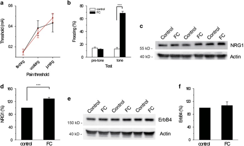
To see whether NRG1/ErbB4 signaling in the mPFC is involved in fear expression, we assessed the expression of the NRG1 and ErbB4 proteins in the mPFC of fear-conditioned mice. C57BL/6 mice were subjected to classical tone-cued fear conditioning, in which an initial neutral auditory stimulus (the CS) elicits a fear response after pairing with an aversive foot shock (the US). Fear-conditioned mice were trained with four CS–US pairs, whereas the CS was not paired with the US in the control group. Twenty-four hours after fear conditioning, exposure of the fear-conditioned mice to the CS in a different context led to an increase in fear behavior (as measured by freezing) compared with the control group (t (4)=1.396; P=0.0001; Figure 1b), with no significant difference in the pain threshold (as an index of pain sensitivity; F1,4=0.000; P=0.9996; Figure 1a) and pre-tone freezing behaviors (as an index of baseline startle response). Mice were killed 24 h after fear memory retrieval, and the mPFC was isolated for immunoblotting. We observed a significant increase in the NRG1 protein levels in the mPFC of fear-conditioned mice compared with controls (t(4)=7.153; P=0.002; Figures 1c and d). However, no significant difference in the ErbB4 protein levels was observed (t (4)=0.646; P=0.553; Figures 1e and f).
Figure 1.
Increased expression of NRG1 in the mPFC of fear-conditioned mice. (a) There were no significant differences in pain threshold. (b) Conditioned mice, but not unpaired controls, showed fear responses 24 h after training. (c, d) NRG1 levels in the mPFC are enhanced in FC mice. (e, f) Total ErbB4 levels did not change between the control and FC groups. FC, fear-conditioned; mPFC, medial prefrontal cortex; NRG1, Neuregulin-1.
NRG1 expressed in the PL cortex but not in the IL cortex is involved in fear expression
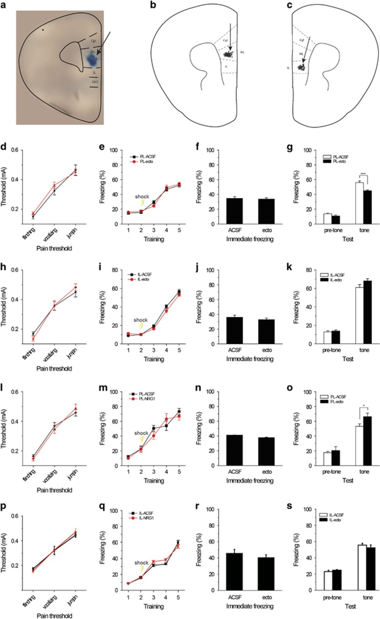
The mPFC consists of the medial agranular, anterior cingulate, PL and IL cortices. The IL and PL cortices have been suggested to exert opposite effects on fear expression.9, 40 To see whether NRG1 regulates fear expression in the mPFC, we examined the consequences of altering NRG1 activity in the PL or IL cortex on tone-cued fear conditioning using two strategies: neutralization of endogenous NRG1 and application of exogenous NRG1. Guide cannulas were implanted into the PL or IL cortex of normal healthy C57BL/6 mice 1 week before fear-conditioning training. Twenty-four hours after training, ecto-ErbB4 or NRG1 was applied through the guide cannulas. Thirty minutes later, the mice were subjected to a fear expression test. As shown in Figure 2, all cannulas were implanted in the PL (Figure 2b) or IL (Figure 2c) cortex, and the trypan blue staining demonstrates that the drugs spread within the PL cortex when targeted to the PL (Figure 2a).
Figure 2.
NRG1 signaling in the PL cortex but not in the IL cortex is involved for fear expression. (a) Representative infusion site (as indicated by the arrow) in the mouse PL cortex (approximately anterior +1.85 mm, lateral±0.4 mm and ventral −2.25 mm). The trypan blue staining that showed the spread of the drugs was only limited to the PL cortex. (b, c) Summary of all the correct probe placements for PL (b) and IL (c) cortices in this study. The approximate infusion sites in the PL cortex (n=58) were: anterior +1.75 mm, lateral ±0.4 mm and ventral −2.25 mm. Infusion sites in the IL cortex (n=38) were: 15° angle, anterior +1.75 mm, lateral ±0.75 mm and ventral −1.5 mm. (d–g) Infusion of ecto-ErbB4 (n=12) into the PL cortex decreased fear expression compared to the controls (n=10; g), with no difference in pain thresholds (d), training (e), immediate freezing behaviors (f) or pre-tone freezing behaviors (g). (h–k) Infusion of ecto-ErbB4 (n=9) into the IL cortex did not affect pain thresholds (h), training (i), immediate freezing behaviors (j), pre-tone freezing behaviors (k) or fear expression (k) compared to the controls (n=8). (l–o) NRG1 injections (n=12) into PL cortex increased fear expression (o), with no differences in pain thresholds (l), training (m), immediate freezing behaviors (n) or pre-tone freezing behaviors (o) compared to controls (n=12). (m–p) NRG1 injections (n=10) into IL cortex did not affect pain thresholds (p), training (q), immediate freezing behaviors (r), pre-tone freezing behaviors (s) or fear expression (s) compared to the controls (n=11). Data are presented as the means±s.e.m. *P<0.05, **P<0.01, ***P<0.001, two-tailed t-test. IL, infralimbic; PL, prelimbic.
To see whether endogenous NRG1 modulates fear expression, we managed to block endogenous NRG1 signaling by applying ecto-ErbB4, which is a soluble polypeptide that contains the full extracellular domain of ErbB4 and can bind to NRG1, thus blocking the mutual interaction between endogenous NRG1 and ErbB4 receptors.25 No differences were observed in pain thresholds (F1,20=0.359; P=0.556; Figure 2d), fear learning (F1,20=0.063; P=0.805; Figure 2e) and immediate freezing behaviors (t(20)=0.322; P=0.751; Figure 2f) among the different groups. However, injection of ecto-ErbB4 into the PL cortex reduced fear expression 24 h after training (t(20)=4.263; P=0.0003; Figure 2g), indicating that the behavior depended on endogenous NRG1. Surprisingly, infusion of ecto-ErbB4 into the IL cortex had no effect on fear expression (Figures 2h–k).
Next, we went on to see whether the application of exogenous NRG1 has any effect on fear expression. Using the same training protocol, both groups showed similar pain thresholds (F1,22=0.013; P=0.910; Figure 2l), fear learning (F1,22=0.235; P=0.632; Figure 2m) and immediate freezing behaviors (t(22)=1.671; P=0.109; Figure 2n). The NRG1 injections into the PL cortex increased fear expression (t(22)=2.139; P=0.043; Figure 2o) without affecting the baseline startle reflex. However, application of NRG1 into the IL cortex produced no significant effect on fear expression (Figures 2p–s).
Together, the results that neutralizing endogenous NRG1 in the PL cortex but not the IL cortex impaired fear expression, whereas application of exogenous NRG1-enhanced fear expression suggests that NRG1 signaling in the PL cortex is critically involved in regulating fear expression.
ErbB4 expression in the PL cortex is important for fear expression
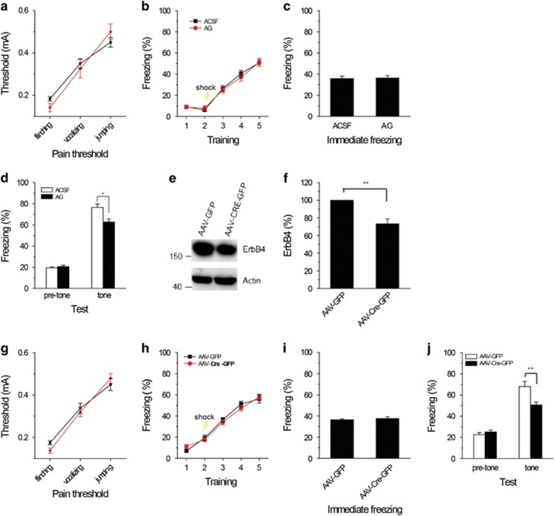
To see whether ErbB4 in the PL cortex is required for the modulation of fear expression, we infused the ErbB4 antagonist AG1478 into the PL cortex through the guide cannulas that had been implanted into the PL cortex 1 week earlier. Thirty minutes after application of AG1478, ErbB4 inhibition led to a significant reduction in freezing behaviors (t(10)=2.697; P=0.022; Figure 3d) without impairing the baseline activity. Thus, ErbB4 is involved in the modulation of fear expression.
Figure 3.
ErbB4 expressed in the PL cortex is required for fear expression. (a–d) Infusion of the ErbB4 antagonist (AG1478) into the PL cortex decreased fear expression (d), but no significant differences were observed in pain thresholds (a), fear training (b), immediate freezing behaviors (c) or pre-tone freezing behaviors (d). n=6 for each group. (e, f) The western blot images show the successful knockdown of the ErbB4 protein in the PL cortex. (g–j) Both the AAV-Cre-injected mice and AAV-GFP-injected mice showed comparable pain thresholds (g), fear training (h), immediate freezing behaviors (i) and pre-tone freezing behaviors (j). AAV-Cre-injected mice showed impaired fear expression (j). n=12 for each group. Data are presented as the means±s.e.m. *P<0.05, **P<0.01, ***P<0.001, two-tailed t-test. AAV, adeno-associated virus; GFP, green fluorescent protein; PL, prelimbic.
As AG1478 is a pan-antagonist for ErbB receptors, we then used a Cre recombinase-expressing AAV (AAV-Cre) to selectively ablate the loxP-flanked ErbB4 gene by taking advantage of ErbB4-loxP mice. AAV-GFP vectors without the Cre sequence were used as controls. AAV-Cre or AAV-GFP was injected selectively into the PL cortex of ErbB4-loxP mice. Two weeks later, western blot analysis revealed successful knockdown of ErbB4 in AAV-Cre-treated mice compared to AAV-GFP controls (t(4)=4.720; P=0.009; Figures 3e and f). We then examined the consequences of ErbB4 ablation on fear conditioning. ErbB4 knockdown mice exhibited similarities to the control mice in pain thresholds (F1,22=0.142; P=0.710; Figure 3g), fear learning (F1,22=2.964; P=0.870; Figure 3h) and immediate freezing behaviors (t(22)=0.697; P=0.493; Figure 3i) but showed a significant decrease in fear expression 24 h after training (t(22)=3.085; P=0.005; Figure 3j). These observations confirmed that ErbB4 expressed in the PL cortex is required for the modulation of fear expression.
ErbB4 expressed in PV-positive interneurons is important for fear expression
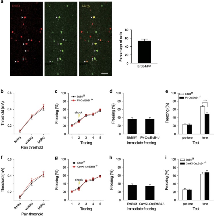
ErbB4 is expressed in PV-positive neurons in the PFC and hippocampus.21, 27, 33, 41 PV-positive neurons account for the majority of the GABAergic neurons in the PFC.42 To identify the subtype of nerve cells that express ErbB4 in the PL cortex, we generated ErbB4-reporter mice by crossing ErbB4-2A-CreERT2 mice, which express Cre recombinase under ErbB4 promoter in a tamoxifen-inducible manner, with Ai14 mice in which tdTomato transcription from the Rosa 26 locus was prevented by a loxP-flanked STOP cassette. These ErbB4-reporter mice were then treated with tamoxifen to induce the expression of tdTomato. The number of ErbB4-positive cells represented 53.34±4.42% of the PV-positive neurons. Thus, ErbB4 is expressed in a majority of PV-positive neurons in the PL cortex.
To investigate whether ErbB4 in PV cells is required for tone-cued fear expression, we generated PV-Cre;ErbB4−/− mice in which ErbB4 was conditionally knocked out in PV neurons and CaMKII-Cre;ErbB4−/− mice in which ErbB4 was specifically knocked out in pyramidal neurons. All groups showed similar immediate freezing behaviors (CaMKII-Cre;ErbB4−/− mice t(21)=0.314, P=0.757; PV-Cre;ErbB4−/− mice t(23)=0.212, P=0.834; Figures 4d and h), pre-tone freezing behaviors and shock thresholds (CaMKII-Cre;ErbB4−/− mice F1,21=0.001, P=0.836; PV-Cre; ErbB4−/− mice F1,23=0.126, P=0.726; Figures 4b and f). However, PV-Cre;ErbB4−/− mice (t(23)=4.144; P=0.0004; Figure 4e), but not CaMKII-Cre;ErbB4−/− mice (t(21)=−0.405; P=0.690; Figure 4i) showed impaired fear expression. These findings indicated that ErbB4 expressed in PV cells is necessary for fear expression.
Figure 4.
ErbB4 expressed in PV-positive neurons is required for fear expression. (a) Costaining of PV in tdTomato-expressing neurons. Sections were prepared from adult ErbB4-2A-CreERT2;Ai14 mice and stained for PV (green). Scale bar, 50 μm. (b–e) Pain thresholds (b), fear training (c), immediate freezing behaviors (d) and pre-tone freezing behaviors (e) were not different in the control (n=12) and PV-Cre;ErbB4−/− mice (n=13). PV-Cre;ErbB4−/− mice (n=11) showed impaired fear expression (e). (f–i) CaMKII-Cre;ErbB4−/− mice showed normal pain thresholds (f), fear learning (g), immediate freezing behaviors (h), pre-tone freezing behaviors (i) and fear expression (i) compared with the controls (n=12). Data are presented as the means±s.e.m. *P<0.05, **P<0.01, ***P<0.001, two-tailed t-test. PV, parvalbumin.
ErbB4 expression in the PL cortex rescues the deficits observed in PV-Cre;ErbB4−/− mice
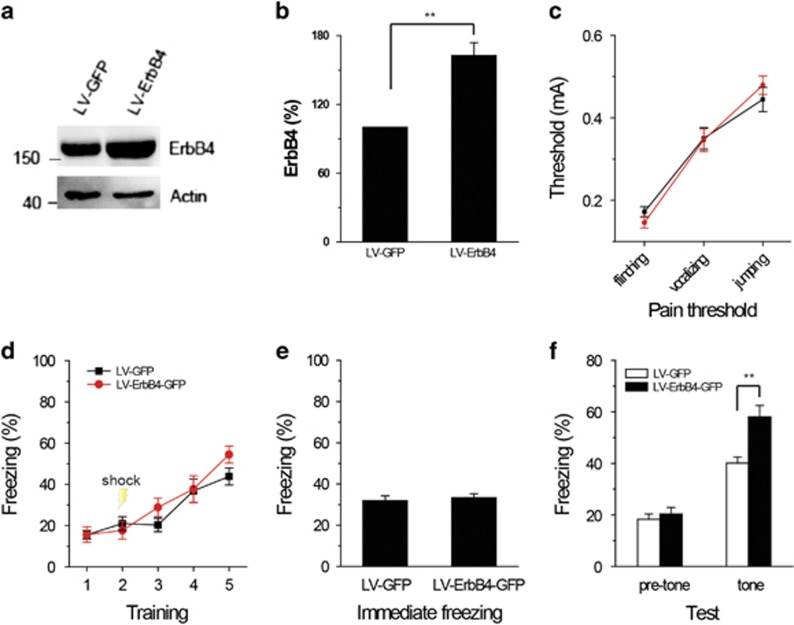
Given our observations that both the AG1478 treatment and the AAV-Cre-mediated ErbB4 deletion in the PL cortex led to deficits in fear memory expression, we hypothesized that ErbB4 expression in PV-expressing neurons in the PL cortex was required for fear expression. To test this hypothesis, we generated a LV ErbB4 expression vector (LV-ErbB4). LV-ErbB4 was bilaterally infused into the PL cortex of PV-Cre;ErbB4−/− mice, and tissues were harvested 2 weeks later. The western blots showed successful overexpression of the ErbB4 protein in the PL cortex (t(4)=5.525; P=0.005; Figures 5a and b). Then, we examined the consequences of ErbB4 overexpression on fear expression. No differences in fear learning (F1,19=1.038; P=0.321; Figure 5d), immediate freezing behaviors (t(19)=0.447; P=0.660; Figure 5e), pre-tone freezing behaviors or shock thresholds (F1,19=0.003; P=0.954; Figure 5c) were observed. The PV-Cre;ErbB4−/− mice exhibited impaired fear memory expression. LV-mediated ErbB4 overexpression in the PL cortex significantly rescued part of the fear memory on the second day (t(19)=3.289; P=0.004; Figure 5f). These findings demonstrated that re-expression of ErbB4 specifically in the PL cortex suffices to reverse fear expression in the PV-Cre;ErbB4−/− mice.
Figure 5.
ErbB4 overexpression in the PL cortex rescues the deficits in PV-Cre;ErbB4−/− mice. (a, b) The western blot images show the successful overexpression of the ErbB4 protein in the PL region. (c–f) LV-ErbB4 expressed within the PL cortex rescued the fear memory deficits in PV-Cre;ErbB4−/− mice (f). No differences were observed in pain thresholds (c), fear learning (d), immediate freezing behaviors (e) or pre-tone freezing behaviors (f). n=12 for each group. Data are presented as the means±s.e.m. *P<0.05, **P<0.01, ***P<0.001, two-tailed t-test. PL, prelimbic; PV, parvalbumin.
Discussion
Our major findings are as follows. The expression level of the NRG1 protein was increased in the mPFC of fear-conditioned mice and manipulation of NRG1 activity in the PL cortex affected fear-conditioning behaviors. Furthermore, the ErbB4 signaling pathway in the PL cortex was necessary to modulate fear expression. Tone-cued fear conditioning was inhibited following the specific inhibition or genetic ablation of ErbB4 in the PL cortex. More importantly, specific deletion of ErbB4 in PV-expressing cells impaired fear conditioning, and this impairment was rescued by ErbB4 overexpression in the PL. Although the ErbB4 protein levels did not exhibit significant changes, we cannot exclude the possibility of functional modifications in ErbB4, such as phosphorylation, during fear expression, and therefore further studies are required to clarify this issue. Together, these findings indicate an essential role for ErbB4 signaling in the PL cortex in controlling fear conditioning.
Despite the consensus that the mPFC is important in fear expression, few studies have assessed the molecular mechanisms underlying this process. NRG1 and ErbB4 have been shown to be involved in fear conditioning, and PV-Cre;ErbB4−/− mice show impaired contextual and tone-cued fear expression.28, 29, 30 Moreover, NRG1 heterozygous mice consistently display deficits in contextual fear expression.43 Together, these findings highlight a critical role for NRG1-ErbB4 signaling in regulating fear expression. However, very little is known about the brain regions involved. There is increasing evidence that specific subregions of the mPFC have different roles in fear expression. Previous studies have shown that inactivation of the PL cortex reduces fear expression,5, 10 and microstimulation of the PL cortex promotes fear expression.9 Consistent with these findings, sustained conditioned tone responses in the PL cortex were recently shown to parallel the time course of conditioned fear expression.7 Our present finding that manipulation of NRG1/ErbB4 signaling in the PL cortex but not in the IL cortex-regulated fear expression adds further support to the hypothesis that the PL cortex is critically involved in fear expression. The different effects of manipulating NRG1/ErbB4 signaling in the PL and IL cortices may be due to the different projections from these mPFC subregions.11 The IL cortex is generally believed to be a critical site of plasticity for fear extinction,44 which is the process by which the memories of irritating and fearful events are extinguished. Both the PL and IL cortices are potentially powerful targets for anxiety disorder treatments that manipulate traumatic memories within the fear circuit. Our findings identify ErbB4 signaling in the PL cortex as an interesting new modulator of fear expression.
Our previous studies showed that NRG1 signaling maintains high levels of GABAergic activity in the amygdala30 and suppresses long-term potentiation at hippocampus CA3–CA1 synapses by enhancing GABA release,29 thus regulating tone-cued and contextual fear memory. Similar to the amygdala and hippocampus, NRG1 also enhances depolarization-induced GABA release in the PFC through ErbB4 expressed in PV-positive neurons.25 Exogenous NRG1 promoted fear expression, whereas neutralization of endogenous NRG1 and the inhibition or ablation of ErbB4 expression in the PL cortex impaired fear expression. Notably, most PV-positive cells in the PL cortex expressed ErbB4. Specific deletion of ErbB4 in PV-expressing cells impaired fear conditioning, whereas ErbB4 overexpression rescued tone-cued fear conditioning in PV-Cre;ErbB4−/− mice. These findings suggest that ErbB4 expressed in PV-positive neurons has a critical role in fear expression. Further studies should be performed to clarify whether ErbB4 expressed in other cell types regulates fear expression and to elucidate how NRG1 specifically promotes fear expression in PL cortex.
As shown in our recent study, NRG1-ErbB4 signaling regulates fear memory by maintaining a high level of GABAergic activity in the amygdala that is under strict top-down control by mPFC pyramidal neurons.30 We extend these findings in this study by showing that the ErbB4 signaling pathway in the PL cortex, but not the IL cortex, was critical in modulating fear memory. Together, these studies suggest that targeting the ErbB4 signaling in the PL–amygdala circuit may represent a promising strategy for the treatments of psychiatric disorders characterized by aberrant processing of fear memory.
Acknowledgments
This work was supported by grants from the National Natural Science Foundation of China (31430032, 81329003, 81671356), the Program for Changjiang Scholars and Innovative Research Team in University (IRT_16R37) and Science and Technology Program of Guangzhou (201707020027).
Footnotes
The authors declare no conflict of interest.
References
- Pitman RK. Overview of biological themes in PTSD. Ann N Y Acad Sci 1997; 821: 1–9. [DOI] [PubMed] [Google Scholar]
- Hamner MB, Lorberbaum JP, George MS. Potential role of the anterior cingulate cortex in PTSD: review and hypothesis. Depress Anxiety 1999; 9: 1–14. [PubMed] [Google Scholar]
- Myers KM, Davis M. Behavioral and neural analysis of extinction. Neuron 2002; 36: 567–584. [DOI] [PubMed] [Google Scholar]
- Sullivan GM, Apergis J, Gorman JM, LeDoux JE. Rodent doxapram model of panic: behavioral effects and c-Fos immunoreactivity in the amygdala. Biol Psychiatry 2003; 53: 863–870. [DOI] [PubMed] [Google Scholar]
- Corcoran KA, Quirk GJ. Activity in prelimbic cortex is necessary for the expression of learned, but not innate, fears. J Neurosci 2007; 27: 840–844. [DOI] [PMC free article] [PubMed] [Google Scholar]
- Sierra-Mercado D Jr., Corcoran KA, Lebron-Milad K, Quirk GJ. Inactivation of the ventromedial prefrontal cortex reduces expression of conditioned fear and impairs subsequent recall of extinction. Eur J Neurosci 2006; 24: 1751–1758. [DOI] [PubMed] [Google Scholar]
- Burgos-Robles A, Vidal-Gonzalez I, Quirk GJ. Sustained conditioned responses in prelimbic prefrontal neurons are correlated with fear expression and extinction failure. J Neurosci 2009; 29: 8474–8482. [DOI] [PMC free article] [PubMed] [Google Scholar]
- Tang J, Ko S, Ding HK, Qiu CS, Calejesan AA, Zhuo M. Pavlovian fear memory induced by activation in the anterior cingulate cortex. Mol Pain 2005; 1: 6. [DOI] [PMC free article] [PubMed] [Google Scholar]
- Vidal-Gonzalez I, Vidal-Gonzalez B, Rauch SL, Quirk GJ. Microstimulation reveals opposing influences of prelimbic and infralimbic cortex on the expression of conditioned fear. Learn Memory 2006; 13: 728–733. [DOI] [PMC free article] [PubMed] [Google Scholar]
- Sierra-Mercado D, Padilla-Coreano N, Quirk GJ. Dissociable roles of prelimbic and infralimbic cortices, ventral hippocampus, and basolateral amygdala in the expression and extinction of conditioned fear. Neuropsychopharmacology 2011; 36: 529–538. [DOI] [PMC free article] [PubMed] [Google Scholar]
- Vertes RP. Differential projections of the infralimbic and prelimbic cortex in the rat. Synapse 2004; 51: 32–58. [DOI] [PubMed] [Google Scholar]
- Druga R. Neocortical inhibitory system. Folia Biol 2009; 55: 201–217. [PubMed] [Google Scholar]
- Markram H, Toledo-Rodriguez M, Wang Y, Gupta A, Silberberg G, Wu C. Interneurons of the neocortical inhibitory system. Nat Rev Neurosci 2004; 5: 793–807. [DOI] [PubMed] [Google Scholar]
- Falls DL. Neuregulins: functions, forms, and signaling strategies. Exp Cell Res 2003; 284: 14–30. [DOI] [PubMed] [Google Scholar]
- Mei L, Xiong WC. Neuregulin 1 in neural development, synaptic plasticity and schizophrenia. Nat Rev Neurosci 2008; 9: 437–452. [DOI] [PMC free article] [PubMed] [Google Scholar]
- Buonanno A. The neuregulin signaling pathway and schizophrenia: from genes to synapses and neural circuits. Brain Res Bull 2010; 83: 122–131. [DOI] [PMC free article] [PubMed] [Google Scholar]
- Sweeney C, Lai C, Riese DJ 2nd, Diamonti AJ, Cantley LC, Carraway KL 3rd. Ligand discrimination in signaling through an ErbB4 receptor homodimer. J Biol Chem 2000; 275: 19803–19807. [DOI] [PubMed] [Google Scholar]
- Lai C, Lemke G. An extended family of protein-tyrosine kinase genes differentially expressed in the vertebrate nervous system. Neuron 1991; 6: 691–704. [DOI] [PubMed] [Google Scholar]
- Huang YZ, Won S, Ali DW, Wang Q, Tanowitz M, Du QS et al. Regulation of neuregulin signaling by PSD-95 interacting with ErbB4 at CNS synapses. Neuron 2000; 26: 443–455. [DOI] [PubMed] [Google Scholar]
- Abe Y, Namba H, Kato T, Iwakura Y, Nawa H. Neuregulin-1 signals from the periphery regulate AMPA receptor sensitivity and expression in GABAergic interneurons in developing neocortex. J Neurosci 2011; 31: 5699–5709. [DOI] [PMC free article] [PubMed] [Google Scholar]
- Fazzari P, Paternain AV, Valiente M, Pla R, Lujan R, Lloyd K et al. Control of cortical GABA circuitry development by Nrg1 and ErbB4 signalling. Nature 2010; 464: 1376–1380. [DOI] [PubMed] [Google Scholar]
- Fox IJ, Kornblum HI. Developmental profile of ErbB receptors in murine central nervous system: implications for functional interactions. J Neurosci Res 2005; 79: 584–597. [DOI] [PubMed] [Google Scholar]
- Neddens J, Buonanno A. Expression of the neuregulin receptor ErbB4 in the brain of the rhesus monkey (Macaca mulatta. PLoS ONE 2011; 6: e27337. [DOI] [PMC free article] [PubMed] [Google Scholar]
- Vullhorst D, Neddens J, Karavanova I, Tricoire L, Petralia RS, McBain CJ et al. Selective expression of ErbB4 in interneurons, but not pyramidal cells, of the rodent hippocampus. J Neurosci 2009; 29: 12255–12264. [DOI] [PMC free article] [PubMed] [Google Scholar]
- Woo RS, Li XM, Tao Y, Carpenter-Hyland E, Huang YZ, Weber J et al. Neuregulin-1 enhances depolarization-induced GABA release. Neuron 2007; 54: 599–610. [DOI] [PubMed] [Google Scholar]
- Yau HJ, Wang HF, Lai C, Liu FC. Neural development of the neuregulin receptor ErbB4 in the cerebral cortex and the hippocampus: preferential expression by interneurons tangentially migrating from the ganglionic eminences. Cereb Cortex 2003; 13: 252–264. [DOI] [PubMed] [Google Scholar]
- Wen L, Lu YS, Zhu XH, Li XM, Woo RS, Chen YJ et al. Neuregulin 1 regulates pyramidal neuron activity via ErbB4 in parvalbumin-positive interneurons. Proc Natl Acad Sci USA 2010; 107: 1211–1216. [DOI] [PMC free article] [PubMed] [Google Scholar]
- Shamir A, Kwon OB, Karavanova I, Vullhorst D, Leiva-Salcedo E, Janssen MJ et al. The importance of the NRG-1/ErbB4 pathway for synaptic plasticity and behaviors associated with psychiatric disorders. J Neurosci 2012; 32: 2988–2997. [DOI] [PMC free article] [PubMed] [Google Scholar]
- Chen YJ, Zhang M, Yin DM, Wen L, Ting A, Wang P et al. ErbB4 in parvalbumin-positive interneurons is critical for neuregulin 1 regulation of long-term potentiation. Proc Natl Acad Sci USA 2010; 107: 21818–21823. [DOI] [PMC free article] [PubMed] [Google Scholar]
- Lu Y, Sun XD, Hou FQ, Bi LL, Yin DM, Liu F et al. Maintenance of GABAergic activity by neuregulin 1-ErbB4 in amygdala for fear memory. Neuron 2014; 84: 835–846. [DOI] [PubMed] [Google Scholar]
- Garcia-Rivello H, Taranda J, Said M, Cabeza-Meckert P, Vila-Petroff M, Scaglione J et al. Dilated cardiomyopathy in Erb-b4-deficient ventricular muscle. Am J Physiol Heart Circ Physiol 2005; 289: H1153–H1160. [DOI] [PubMed] [Google Scholar]
- Erdmann G, Schutz G, Berger S. Inducible gene inactivation in neurons of the adult mouse forebrain. BMC Neurosci 2007; 8: 63. [DOI] [PMC free article] [PubMed] [Google Scholar]
- Bean JC, Lin TW, Sathyamurthy A, Liu F, Yin DM, Xiong WC et al. Genetic labeling reveals novel cellular targets of schizophrenia susceptibility gene: distribution of GABA and non-GABA ErbB4-positive cells in adult mouse brain. J Neurosci 2014; 34: 13549–13566. [DOI] [PMC free article] [PubMed] [Google Scholar]
- Zhu XH, Yan HC, Zhang J, Qu HD, Qiu XS, Chen L et al. Intermittent hypoxia promotes hippocampal neurogenesis and produces antidepressant-like effects in adult rats. J Neurosci 2010; 30: 12653–12663. [DOI] [PMC free article] [PubMed] [Google Scholar]
- Holmes WE, Sliwkowski MX, Akita RW, Henzel WJ, Lee J, Park JW et al. Identification of heregulin, a specific activator of p185erbB2. Science 1992; 256: 1205–1210. [DOI] [PubMed] [Google Scholar]
- Franklin K, Paxinos G. The Mouse Brain in Stereotaxic Coordinates. Academic Press, 1997. [Google Scholar]
- Davidson BL, Stein CS, Heth JA, Martins I, Kotin RM, Derksen TA et al. Recombinant adeno-associated virus type 2, 4, and 5 vectors: transduction of variant cell types and regions in the mammalian central nervous system. Proc Natl Acad Sci USA 2000; 97: 3428–3432. [DOI] [PMC free article] [PubMed] [Google Scholar]
- Rattiner LM, Davis M, French CT, Ressler KJ. Brain-derived neurotrophic factor and tyrosine kinase receptor B involvement in amygdala-dependent fear conditioning. J Neurosci 2004; 24: 4796–4806. [DOI] [PMC free article] [PubMed] [Google Scholar]
- Soria-Gomez E, Busquets-Garcia A, Hu F, Mehidi A, Cannich A, Roux L et al. Habenular CB1 receptors control the expression of aversive memories. Neuron 2015; 88: 306–313. [DOI] [PubMed] [Google Scholar]
- Gilmartin MR, McEchron MD. Single neurons in the medial prefrontal cortex of the rat exhibit tonic and phasic coding during trace fear conditioning. Behav Neurosci 2005; 119: 1496–1510. [DOI] [PubMed] [Google Scholar]
- Neddens J, Fish KN, Tricoire L, Vullhorst D, Shamir A, Chung W et al. Conserved interneuron-specific ErbB4 expression in frontal cortex of rodents, monkeys, and humans: implications for schizophrenia. Biol Psychiatry 2011; 70: 636–645. [DOI] [PMC free article] [PubMed] [Google Scholar]
- Rudy B, Fishell G, Lee S, Hjerling-Leffler J. Three groups of interneurons account for nearly 100% of neocortical GABAergic neurons. Dev Neurobiol 2011; 71: 45–61. [DOI] [PMC free article] [PubMed] [Google Scholar]
- Ehrlichman RS, Luminais SN, White SL, Rudnick ND, Ma N, Dow HC et al. Neuregulin 1 transgenic mice display reduced mismatch negativity, contextual fear conditioning and social interactions. Brain Res 2009; 1294: 116–127. [DOI] [PMC free article] [PubMed] [Google Scholar]
- Peters J, Dieppa-Perea LM, Melendez LM, Quirk GJ. Induction of fear extinction with hippocampal-infralimbic BDNF. Science 2010; 328: 1288–1290. [DOI] [PMC free article] [PubMed] [Google Scholar]


