

Abstract
Porin spans the outer membrane of Escherichia coli with most of the protein embedded within the membrane. It lacks pronounced hydrophobic domains and consists predominantly of beta-pleated sheet. These observations require the accommodation of polar and ionizable residues in an environment that has a low dielectric constant. Owing to a currently limited understanding of the constraints governing membrane protein structure, a minimal approach to structure prediction is proposed that identifies segments causing polypeptides to reverse their direction (turn identification). The application of this procedure avoids hydrophobicity parameters and yields a model of porin which is in good agreement with all experimental data available. The presence of polar and ionizable residues within membrane boundaries implies a dense (saturating) network of hydrogen bond donor and acceptor groups. Application to a paradigm of hydrophobic membrane proteins, bacteriorhodopsin, reveals a pattern consistent with its alpha-helical folding. The postulated structure includes significantly more polar residues in the membrane domain than have been assumed previously, suggesting that there are also hydrogen bonding networks in bacteriorhodopsin. Extensive networks permeating protein interior and surfaces would explain the extraordinary stability and the tight interactions between functional units in the formation of crystalline arrays of both proteins.
Full text
PDF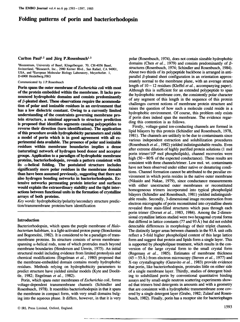
Selected References
These references are in PubMed. This may not be the complete list of references from this article.
- Argos P., Rao J. K., Hargrave P. A. Structural prediction of membrane-bound proteins. Eur J Biochem. 1982 Nov 15;128(2-3):565–575. doi: 10.1111/j.1432-1033.1982.tb07002.x. [DOI] [PubMed] [Google Scholar]
- Baker E. N., Hubbard R. E. Hydrogen bonding in globular proteins. Prog Biophys Mol Biol. 1984;44(2):97–179. doi: 10.1016/0079-6107(84)90007-5. [DOI] [PubMed] [Google Scholar]
- Charbit A., Clement J. M., Hofnung M. Further sequence analysis of the phage lambda receptor site. Possible implications for the organization of the lamB protein in Escherichia coli K12. J Mol Biol. 1984 May 25;175(3):395–401. doi: 10.1016/0022-2836(84)90355-3. [DOI] [PubMed] [Google Scholar]
- Chen R., Krämer C., Schmidmayr W., Henning U. Primary structure of major outer membrane protein I of Escherichia coli B/r. Proc Natl Acad Sci U S A. 1979 Oct;76(10):5014–5017. doi: 10.1073/pnas.76.10.5014. [DOI] [PMC free article] [PubMed] [Google Scholar]
- Chothia C. Hydrophobic bonding and accessible surface area in proteins. Nature. 1974 Mar 22;248(446):338–339. doi: 10.1038/248338a0. [DOI] [PubMed] [Google Scholar]
- Chou P. Y., Fasman G. D. Empirical predictions of protein conformation. Annu Rev Biochem. 1978;47:251–276. doi: 10.1146/annurev.bi.47.070178.001343. [DOI] [PubMed] [Google Scholar]
- Chou P. Y., Fasman G. D. Prediction of beta-turns. Biophys J. 1979 Jun;26(3):367–383. doi: 10.1016/S0006-3495(79)85259-5. [DOI] [PMC free article] [PubMed] [Google Scholar]
- Crawford J. L., Lipscomb W. N., Schellman C. G. The reverse turn as a polypeptide conformation in globular proteins. Proc Natl Acad Sci U S A. 1973 Feb;70(2):538–542. doi: 10.1073/pnas.70.2.538. [DOI] [PMC free article] [PubMed] [Google Scholar]
- Deisenhofer J., Epp O., Miki K., Huber R., Michel H. X-ray structure analysis of a membrane protein complex. Electron density map at 3 A resolution and a model of the chromophores of the photosynthetic reaction center from Rhodopseudomonas viridis. J Mol Biol. 1984 Dec 5;180(2):385–398. doi: 10.1016/s0022-2836(84)80011-x. [DOI] [PubMed] [Google Scholar]
- Dorset D. L., Engel A., Häner M., Massalski A., Rosenbusch J. P. Two-dimensional crystal packing of matrix porin. A channel forming protein in Escherichia coli outer membranes. J Mol Biol. 1983 Apr 25;165(4):701–710. doi: 10.1016/s0022-2836(83)80275-7. [DOI] [PubMed] [Google Scholar]
- Dorset D. L., Engel A., Massalski A., Rosenbusch J. P. Three Dimensional Structure of a Membrane Pore: Electron Microscopical Analysis of Escherichia coli Outer Membrane Matrix Porin. Biophys J. 1984 Jan;45(1):128–129. doi: 10.1016/S0006-3495(84)84135-1. [DOI] [PMC free article] [PubMed] [Google Scholar]
- Engelman D. M. An Implication of the Structure of Bacteriorhodopsin: Globular Membrane Proteins are Stabilized by Polar Interactions. Biophys J. 1982 Jan;37(1):187–188. doi: 10.1016/S0006-3495(82)84662-6. [DOI] [PMC free article] [PubMed] [Google Scholar]
- Engelman D. M., Henderson R., McLachlan A. D., Wallace B. A. Path of the polypeptide in bacteriorhodopsin. Proc Natl Acad Sci U S A. 1980 Apr;77(4):2023–2027. doi: 10.1073/pnas.77.4.2023. [DOI] [PMC free article] [PubMed] [Google Scholar]
- Engelman D. M., Steitz T. A. The spontaneous insertion of proteins into and across membranes: the helical hairpin hypothesis. Cell. 1981 Feb;23(2):411–422. doi: 10.1016/0092-8674(81)90136-7. [DOI] [PubMed] [Google Scholar]
- Engelman D. M., Zaccai G. Bacteriorhodopsin is an inside-out protein. Proc Natl Acad Sci U S A. 1980 Oct;77(10):5894–5898. doi: 10.1073/pnas.77.10.5894. [DOI] [PMC free article] [PubMed] [Google Scholar]
- Finney J. L. Volume occupation, environment, and accessibility in proteins. Environment and molecular area of RNase-S. J Mol Biol. 1978 Mar 5;119(3):415–441. doi: 10.1016/0022-2836(78)90223-1. [DOI] [PubMed] [Google Scholar]
- Fleming P. J., Dailey H. A., Corcoran D., Strittmatter P. The primary structure of the nonpolar segment of bovine cytochrome b5. J Biol Chem. 1978 Aug 10;253(15):5369–5372. [PubMed] [Google Scholar]
- Garavito R. M., Jenkins J., Jansonius J. N., Karlsson R., Rosenbusch J. P. X-ray diffraction analysis of matrix porin, an integral membrane protein from Escherichia coli outer membranes. J Mol Biol. 1983 Feb 25;164(2):313–327. doi: 10.1016/0022-2836(83)90079-7. [DOI] [PubMed] [Google Scholar]
- Garnier J., Osguthorpe D. J., Robson B. Analysis of the accuracy and implications of simple methods for predicting the secondary structure of globular proteins. J Mol Biol. 1978 Mar 25;120(1):97–120. doi: 10.1016/0022-2836(78)90297-8. [DOI] [PubMed] [Google Scholar]
- Henderson R. The structure of the purple membrane from Halobacterium hallobium: analysis of the X-ray diffraction pattern. J Mol Biol. 1975 Apr 5;93(2):123–138. doi: 10.1016/0022-2836(75)90123-0. [DOI] [PubMed] [Google Scholar]
- Henderson R., Unwin P. N. Three-dimensional model of purple membrane obtained by electron microscopy. Nature. 1975 Sep 4;257(5521):28–32. doi: 10.1038/257028a0. [DOI] [PubMed] [Google Scholar]
- Honig B. H., Hubbell W. L. Stability of "salt bridges" in membrane proteins. Proc Natl Acad Sci U S A. 1984 Sep;81(17):5412–5416. doi: 10.1073/pnas.81.17.5412. [DOI] [PMC free article] [PubMed] [Google Scholar]
- Inokuchi K., Mutoh N., Matsuyama S., Mizushima S. Primary structure of the ompF gene that codes for a major outer membrane protein of Escherichia coli K-12. Nucleic Acids Res. 1982 Nov 11;10(21):6957–6968. doi: 10.1093/nar/10.21.6957. [DOI] [PMC free article] [PubMed] [Google Scholar]
- Khorana H. G., Gerber G. E., Herlihy W. C., Gray C. P., Anderegg R. J., Nihei K., Biemann K. Amino acid sequence of bacteriorhodopsin. Proc Natl Acad Sci U S A. 1979 Oct;76(10):5046–5050. doi: 10.1073/pnas.76.10.5046. [DOI] [PMC free article] [PubMed] [Google Scholar]
- Kossiakoff A. A. Protein dynamics investigated by the neutron diffraction-hydrogen exchange technique. Nature. 1982 Apr 22;296(5859):713–721. doi: 10.1038/296713a0. [DOI] [PubMed] [Google Scholar]
- Kuntz I. D. Protein folding. J Am Chem Soc. 1972 May 31;94(11):4009–4012. doi: 10.1021/ja00766a060. [DOI] [PubMed] [Google Scholar]
- Kyte J., Doolittle R. F. A simple method for displaying the hydropathic character of a protein. J Mol Biol. 1982 May 5;157(1):105–132. doi: 10.1016/0022-2836(82)90515-0. [DOI] [PubMed] [Google Scholar]
- Levitt M. Conformational preferences of amino acids in globular proteins. Biochemistry. 1978 Oct 3;17(20):4277–4285. doi: 10.1021/bi00613a026. [DOI] [PubMed] [Google Scholar]
- Long M. M., Urry D. W., Stoeckenius W. Circular dichroism of biological membranes: purple membrane of Halobacterium halobium. Biochem Biophys Res Commun. 1977 Apr 11;75(3):725–731. doi: 10.1016/0006-291x(77)91532-7. [DOI] [PubMed] [Google Scholar]
- Madison V., Atreyi M., Deber C. M., Blout E. R. Cyclic peptides. IX. Conformations of a synthetic ion-binding cyclic peptide, cyclo-(pro-gly)3, from circular dichroism and 1H and 13C nuclear magnetic resonance. J Am Chem Soc. 1974 Oct 16;96(21):6725–6734. doi: 10.1021/ja00828a031. [DOI] [PubMed] [Google Scholar]
- Mathews F. S., Argos P., Levine M. The structure of cytochrome b 5 at 2.0 Angstrom resolution. Cold Spring Harb Symp Quant Biol. 1972;36:387–395. doi: 10.1101/sqb.1972.036.01.050. [DOI] [PubMed] [Google Scholar]
- Michel H. Characterization and crystal packing of three-dimensional bacteriorhodopsin crystals. EMBO J. 1982;1(10):1267–1271. doi: 10.1002/j.1460-2075.1982.tb00023.x. [DOI] [PMC free article] [PubMed] [Google Scholar]
- Mouritsen O. G., Bloom M. Mattress model of lipid-protein interactions in membranes. Biophys J. 1984 Aug;46(2):141–153. doi: 10.1016/S0006-3495(84)84007-2. [DOI] [PMC free article] [PubMed] [Google Scholar]
- Nikaido H., Nakae T. The outer membrane of Gram-negative bacteria. Adv Microb Physiol. 1979;20:163–250. doi: 10.1016/s0065-2911(08)60208-8. [DOI] [PubMed] [Google Scholar]
- Overbeeke N., Bergmans H., van Mansfeld F., Lugtenberg B. Complete nucleotide sequence of phoE, the structural gene for the phosphate limitation inducible outer membrane pore protein of Escherichia coli K12. J Mol Biol. 1983 Feb 5;163(4):513–532. doi: 10.1016/0022-2836(83)90110-9. [DOI] [PubMed] [Google Scholar]
- Parsons S. M., Raftery M. A. Ionization behavior of the catalytic carboxyls of lysozyme. Effects of ionic strength. Biochemistry. 1972 Apr 25;11(9):1623–1629. doi: 10.1021/bi00759a013. [DOI] [PubMed] [Google Scholar]
- Paul C. H. Building models of globular protein molecules from their amino acid sequences. I. Theory. J Mol Biol. 1982 Feb 15;155(1):53–62. doi: 10.1016/0022-2836(82)90491-0. [DOI] [PubMed] [Google Scholar]
- Rashin A. A., Honig B. On the environment of ionizable groups in globular proteins. J Mol Biol. 1984 Mar 15;173(4):515–521. doi: 10.1016/0022-2836(84)90394-2. [DOI] [PubMed] [Google Scholar]
- Robson B., Pain R. H. Analysis of the code relating sequence to conformation in globular proteins. The distribution of residue pairs in turns and kinks in the backbone chain. Biochem J. 1974 Sep;141(3):899–904. doi: 10.1042/bj1410899. [DOI] [PMC free article] [PubMed] [Google Scholar]
- Rose G. D., Young W. B., Gierasch L. M. Interior turns in globular proteins. Nature. 1983 Aug 18;304(5927):654–657. doi: 10.1038/304654a0. [DOI] [PubMed] [Google Scholar]
- Rosenbusch J. P. Characterization of the major envelope protein from Escherichia coli. Regular arrangement on the peptidoglycan and unusual dodecyl sulfate binding. J Biol Chem. 1974 Dec 25;249(24):8019–8029. [PubMed] [Google Scholar]
- Schindler H., Rosenbusch J. P. Matrix protein from Escherichia coli outer membranes forms voltage-controlled channels in lipid bilayers. Proc Natl Acad Sci U S A. 1978 Aug;75(8):3751–3755. doi: 10.1073/pnas.75.8.3751. [DOI] [PMC free article] [PubMed] [Google Scholar]
- Schindler H., Rosenbusch J. P. Matrix protein in planar membranes: clusters of channels in a native environment and their functional reassembly. Proc Natl Acad Sci U S A. 1981 Apr;78(4):2302–2306. doi: 10.1073/pnas.78.4.2302. [DOI] [PMC free article] [PubMed] [Google Scholar]
- Schindler M., Rosenbusch J. P. Structural transitions of porin, a transmembrane protein. FEBS Lett. 1984 Jul 23;173(1):85–89. doi: 10.1016/0014-5793(84)81022-4. [DOI] [PubMed] [Google Scholar]
- Steitz T. A., Goldman A., Engelman D. M. Quantitative application of the helical hairpin hypothesis to membrane proteins. Biophys J. 1982 Jan;37(1):124–125. doi: 10.1016/S0006-3495(82)84633-X. [DOI] [PMC free article] [PubMed] [Google Scholar]
- Steven A. C., Heggeler B., Müller R., Kistler J., Rosenbusch J. P. Ultrastructure of a periodic protein layer in the outer membrane of Escherichia coli. J Cell Biol. 1977 Feb;72(2):292–301. doi: 10.1083/jcb.72.2.292. [DOI] [PMC free article] [PubMed] [Google Scholar]
- Stoeckenius W., Bogomolni R. A. Bacteriorhodopsin and related pigments of halobacteria. Annu Rev Biochem. 1982;51:587–616. doi: 10.1146/annurev.bi.51.070182.003103. [DOI] [PubMed] [Google Scholar]
- Suck D., Saenger W., Rohde W. X-ray structure of thymidine 5'-carboxylic acid, an inhibitor of thymidine and thymidylate kinase: preferred nucleobase-carboxylic acid hydrogen bonding scheme. Biochim Biophys Acta. 1974 Aug 15;361(1):1–10. doi: 10.1016/0005-2787(74)90204-4. [DOI] [PubMed] [Google Scholar]
- Takagaki Y., Radhakrishnan R., Wirtz K. W., Khorana H. G. The membrane-embedded segment of cytochrome b5 as studied by cross-linking with photoactivatable phospholipids. II. The nontransferable form. J Biol Chem. 1983 Aug 10;258(15):9136–9142. [PubMed] [Google Scholar]
- Thornton J. M. Electrostatic interactions in proteins. Nature. 1982 Jan 7;295(5844):13–14. doi: 10.1038/295013a0. [DOI] [PubMed] [Google Scholar]
- Tokunaga H., Tokunaga M., Nakae T. Permeability properties of chemically modified porin trimers from Escherichia coli B. J Biol Chem. 1981 Aug 10;256(15):8024–8029. [PubMed] [Google Scholar]
- Tomita M., Furthmayr H., Marchesi V. T. Primary structure of human erythrocyte glycophorin A. Isolation and characterization of peptides and complete amino acid sequence. Biochemistry. 1978 Oct 31;17(22):4756–4770. doi: 10.1021/bi00615a025. [DOI] [PubMed] [Google Scholar]
- Ullrich A., Coussens L., Hayflick J. S., Dull T. J., Gray A., Tam A. W., Lee J., Yarden Y., Libermann T. A., Schlessinger J. Human epidermal growth factor receptor cDNA sequence and aberrant expression of the amplified gene in A431 epidermoid carcinoma cells. 1984 May 31-Jun 6Nature. 309(5967):418–425. doi: 10.1038/309418a0. [DOI] [PubMed] [Google Scholar]
- Wilson I. A., Skehel J. J., Wiley D. C. Structure of the haemagglutinin membrane glycoprotein of influenza virus at 3 A resolution. Nature. 1981 Jan 29;289(5796):366–373. doi: 10.1038/289366a0. [DOI] [PubMed] [Google Scholar]