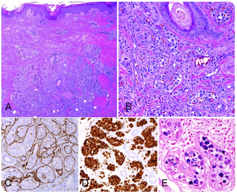
A 51-year-old HIV-negative male from El Salvador presented with neurologic symptoms and computed tomography findings of an acute/subacute cerebral infarction. Physical examination identified indurated and hyperpigmented cutaneous lesions on the legs. A skin biopsy revealed an intravascular infiltrate composed of large atypical lymphoid cells (panel A, original magnification ×20; hematoxylin and eosin stain; panel B, original magnification ×200; hematoxylin and eosin stain), which were delimited by CD31-positive endothelial cells (panel C, original magnification ×200). A cytotoxic T-cell derivation was demonstrated by immunohistochemical stains for CD3 (panel D, original magnification ×200), CD8, TIA-1, and granzyme B. The cells were negative for CD20, CD79a, CD56, and ALK-1. The neoplastic cells were positive for Epstein-Barr virus (EBV) by EBV encoded small RNA in situ hybridization (panel E, original magnification ×400). The cells were negative for LMP-1, indicative of latency I. Polymerase chain reaction identified a clonal T-cell receptor γ gene rearrangement, supporting a T-cell origin.
Intravascular large cell lymphomas (IVLs) are most often of B-cell origin and frequently present with neurological symptoms, including stroke, secondary to accumulation of tumor cells in the intravascular space. IVLs of natural killer (NK)/T-cell type are extremely rare. While it has been suggested that B-cell IVL presenting in the skin has a better prognosis, the prognosis in NK/T-cell cases is dire, with survival measured in weeks to months. NK/T-cell lymphomas are more common in Asian and Hispanic populations, a feature evident in this case.
Footnotes
For additional images, visit the ASH IMAGE BANK, a reference and teaching tool that is continually updated with new atlas and case study images. For more information visit http://imagebank.hematology.org.


