Abstract
Tyrosine site-specific recombinases (T-SSR) are polynucleotidyltransferases that catalyze cutting and joining reactions between short specific DNA sequences. We developed three systems for performing genetic modifications in Bacillus anthracis that use T-SSR and their cognate target sequences, namely Escherichia coli bacteriophage P1 Cre-loxP, Saccharomyces cerevisiae Flp-FRT, and a newly discovered IntXO-PSL system from B. anthracis plasmid pXO1. All three tyrosine recombinase systems were used for creation of a B. anthracis sporulation-deficient, plasmid-free strain deleted for ten proteases which had been identified by proteomic analysis as being present in the B. anthracis secretome. This strain was used successfully for production of various recombinant proteins, including several that are candidates for inclusion in improved anthrax vaccines. These genetic tools developed for DNA manipulation in B. anthracis were also used for construction of strains having chromosomal insertions of 1, 2, or 3 adjacent atxA genes. AtxA is a B. anthracis global transcriptional regulator required for the response of B. anthracis virulence factor genes to bicarbonate. We found a positive correlation between the atxA copy number and the expression level of the pagA gene encoding B. anthracis protective antigen, when strains were grown in a carbon dioxide atmosphere. These results demonstrate that the three T-SSR systems described here provide effective tools for B. anthracis genome editing. These T-SSR systems may also be applicable to other prokaryotes and to eukaryotes.
Introduction
Site-specific recombinases constitute a distinctive class of enzymes that possess the unique ability to both cleave and reseal DNA. Some of these recombinases function without cofactors and lead to precise, predictable and efficient genome modifications [1]. In particular, tyrosine site-specific recombinases (T-SSRs) have found widespread use in biotechnology and biomedical research. T-SSRs are DNA modifying enzymes that bind, cleave, exchange strands, and rejoin DNA at their respective, typically palindromic, target recognition sites [2]. Most T-SSRs are found in prokaryotes and bacteriophages where they perform a plethora of functions, including DNA integration and excision, plasmid copy number control, regulation of gene expression, chromosome segregation at cell division, and separation of multimeric circular DNAs [3]. In addition, T-SSRs are frequently encoded on yeast plasmids, where they complement the partitioning system to maintain high-copy numbers of the plasmids [4]. Among these, the bacteriophage P1 enzyme Cre and the yeast 2 μ- derived Flp recombinase are the enzymes most widely used in genome engineering. Using combinations of different T-SSRs in the same cell or organism has made it possible to establish increasingly sophisticated systems, and several different enzymes have been recently utilized in synthetic biology to build complex genetic circuits [2].
Accordingly, in the work continued here, we have created three T-SSR systems for performing genetic modifications in Bacillus anthracis: the Escherichia coli Cre-loxP and Saccharomyces cerevisiae Flp-FRT systems [5–7] and a newly developed B. anthracis IntXO-PSL system [8]. We previously used the Cre-loxP system to create the BH460 strain [9] having the following phenotypic characteristics: Spo0A-, pXO1-, pXO2-, NprB-, TasA-, Cam-, InhA1-, InhA2-, MmpZ-. This six protease-deficient, non-sporulating and avirulent strain was recommended as an effective host for recombinant protein production, typically yielding greater than 10 mg pure protein per liter of culture [9].
In this study, we describe work to further improve the BH460 strain by identification and deletion of additional secreted proteases. Functional and proteomic analysis of BH460 and its derivatives revealed four more proteases in the secretomes: CysP1, VpR, NprC and S41. The first three proteases were inactivated with the Flp-FRT system, producing the nine proteases mutant BH490. Deletion of the last S41 protease was performed with the IntXO-PSL system, yielding the ten proteases mutant BH500.
The three T-SSRs were also successfully used for sequential insertion of three copies of the atxA gene into the BH490 genome, as demonstrated here. The atxA gene encodes the pleiotropic regulator AtxA (anthracis toxin activator) [10] that regulates the expression of B. anthracis toxin genes in the presence of CO2. The two-step quantitative real-time PCR (qPCR) analysis demonstrated an increase in atxA and protective antigen (PA) gene pagA [11] transcription in the sequential recombinant strains, with maximum expression for the strain BH490A3 containing three atxA copies. Maximum PA production was also shown in the strain with three atxA genes. We plan to use the constructed multi-protease deficient strain BH490A3 for recombinant proteins overproduction with a promoter that responds to CO2 and AtxA activation.
Materials and methods
Materials
The B. anthracis strains, plasmids and genes inactivated or analyzed in this study are listed in Tables 1–3. All B. anthracis strains used here lack pXO2, are avirulent, are exempt from CDC Select Agent regulation, and are approved by NIAID to be used at BSL-2. Primers for PCR, primers and probes for qPCR, and T-SSR target sites that replaced the B. anthracis genes deleted in this study are listed in S1–S3 Tables.
Table 1. Bacterial strains used in this study.
| B. anthracis strains | Relevant characteristic(s) | Source or reference |
|---|---|---|
| Ames 35 | B. anthracis (pXO1+, pXO2-), from Ames strain by removal of pXO2. | [7] |
| Ames 35 ΔAtxA | Ames 35 with atxA gene deleted | [12] |
| BH460 | pXO1-, pXO2-, Spo0A-, NprB-, TasA-, Cam-, InhA1-, InhA2-, MmpZ-; total of five loxP sites in chromosome, six proteases inactivated | [9] |
| BH470 | BH460 with cysP1 knockout containing one FRT site | This work |
| BH480 | BH470 with vpR knockout containing second FRT site in chromosome | This work |
| BH490 | BH480 with nprC knockout containing third FRT site in chromosome | This work |
| BH490A | BH490 with the first atxA located upstream of the third FRT site | This work |
| BH490A2 | BH490A with the second atxA located upstream of the first atxA copy; sixth loxP site is located between two atxA | This work |
| BH490A3 | BH490A2 with the third atxA located downstream of the sixth loxP site and between two originally inserted atxA; contains one PSL site in chromosome | This work |
| BH500 | BH490 with s41 knockout containing one PSL site in chromosome | This work |
Table 3. B. anthracis Ames ancestor strain genes inactivated or analyzed in this study.
| Protein | Gene | Function/Name | Locus Tag |
|---|---|---|---|
| CysP1 | cysP1 | Putative cysteine protease | GBAA_1995 |
| NprC | nprC | Neutral metalloprotease | GBAA_2183 |
| VpR | vpR | Minor extracellular protease | GBAA_4584 |
| S41 | s41 | Serine protease | GBAA_5414 |
| HtrA | htrA | Serine protease | GBAA_3660 |
| AtxA | atxA | Transcriptional regulator | GBAA_RS29060 |
| EF | cya | Edema factor | GBAA_RS29035 |
| LF | lef | Lethal factor | GBAA_RS29135 |
| PA | pag | Protective antigen | GBAA_RS29110 |
| RpoB | rpoB | RNA polymerase subunit β | GBBA_0102 |
| DnaJ | dnaJ | Chaperone | GBBA_4538 |
| GyrB | gyrB | DNA gyrase subunit B | GBBA_0005 |
Bacterial growth conditions and phenotypic characterization
Escherichia coli strains were grown in Luria-Bertani (LB) broth and used as hosts for cloning. LB agar was used for selection of transformants [16]. B. anthracis strains were also grown in LB, FA [13] or NBY [12] liquid medium (ambient air). For growth with CO2, the NBY medium was supplemented with 0.8% (w/v) NaHCO3 and the air was supplemented with 15% CO2. Antibiotics (Sigma-Aldrich, St. Louis, MO) were added to the medium when appropriate to give the following final concentrations: ampicillin (Ap), 100 μg/ml (only for E. coli); erythromycin (Em), 400 μg/ml for E. coli and 10 μg/ml for B. anthracis; spectinomycin (Sp), 150 μg/ml for both E. coli and B. anthracis; kanamycin (Km), 15 μg/ml and tetracycline (Tc), 5 μg/ml for B. anthracis. SOC medium (Quality Biologicals, Inc., Gaithersburg, MD) was used for outgrowth of transformation mixtures prior to plating on selective media to isolate transformants.
DNA isolation and manipulation
Preparation of plasmid DNA from E. coli, transformation of E. coli, and recombinant DNA techniques were carried out by standard procedures [16]. E. coli SCS110 competent cells were purchased from Agilent Technologies (Santa Clara, CA), and E. coli TOP10 competent cells were purchased from Life Technologies (Grand Island, NY). Recombinant plasmid construction was carried out in E. coli TOP10 cells. Plasmid DNA from B. anthracis was isolated according to the Plasmid Protocol: Purification of Plasmid DNA from Bacillus subtilis (QIAGEN Inc., Valencia, Calif.). Chromosomal DNA from B. anthracis was isolated with a Wizard genomic purification kit (Promega, Madison, WI) in accordance with the protocol for isolation of genomic DNA from Gram-positive bacteria. B. anthracis was electroporated with unmethylated plasmid DNA isolated from E. coli SCS110 (dam-/dcm-) (Stratagene, San Diego, CA). Electroporation-competent B. anthracis cells were prepared and transformed as previously described [7].
Restriction enzymes, T4 ligase, T4 DNA polymerase, and alkaline phosphatase were purchased from New England BioLabs (Ipswich, MA). The pGEM-T Easy vector system (Promega) was applied for PCR fragment cloning. Phusion high-fidelity DNA polymerase from New England BioLabs was used for fragment PCR. OneTaq® 2X Master Mix with Standard Buffer (New England BioLabs) were used for routine DNA analysis. All constructs were verified by restriction enzyme digestion and/or DNA sequencing. Plasmid and chromosomal DNAs were sequenced using primers listed in S1 Table. Primers for sequencing and PCR were synthesized by Integrated DNA Technologies (IDT, Inc. Coralville, IA). Sequences were determined using a primer walking strategy (Macrogen, Rockville, MD). Sequence data were assembled using the Vector NTI software (Life Technologies, Carlsbad, CA). The National Center for Biotechnology Information (NCBI) BLAST and FASTA programs (http://www.ncbi.nlm.nih.gov) were used for homology searches in GenBank and nonredundant protein sequence databases. Predicted protein motifs were analyzed using SignalP software version 3.0 for prediction of signal sequences (http://www.cbs.dtu.dk/services/SignalP) and TMpred software for prediction of transmembrane regions and orientation (http://www.ch.embnet.org/software/TMPRED_form.html).
Recombinase systems used for preparation of mutants
Genetic modifications in the B. anthracis genome were generated with the Cre-loxP system as previously described [7], employing plasmids we designated generically as pSC, for single-crossover plasmid, together with pCrePAS2 for Cre recombinase production [6]. Deletions and insertions were also generated with the Flp-FRT system, employing plasmids pSCF (an analog of the pSC plasmid) and pFPAS (a plasmid with all the characteristics of pCrePAS2, but containing the flp gene instead of the cre gene) as previously described [6].
A new IntXO-PSL recombinase system based on the recently identified IntXO recombinase of B. anthracis [8] was created for use in a manner analogous to that of the Flp-FRT system described above. The plasmid pSCP is a shuttle plasmid with all the characteristics of pSCF, except that it contains repeats of the 37-bp PSL sequence instead of the FRT sequence. An analog of the pFPAS plasmid that we created and designated as pIntPAS is a shuttle vector with all the characteristics of pFPAS, except that it contains the IntXO recombinase gene instead of flp. All three T-SSRs described above were used for genetic deletions or insertions in accordance with the scheme presented in Fig 1, as will be detailed later. Sequences and maps of the plasmids described in this section are available upon request.
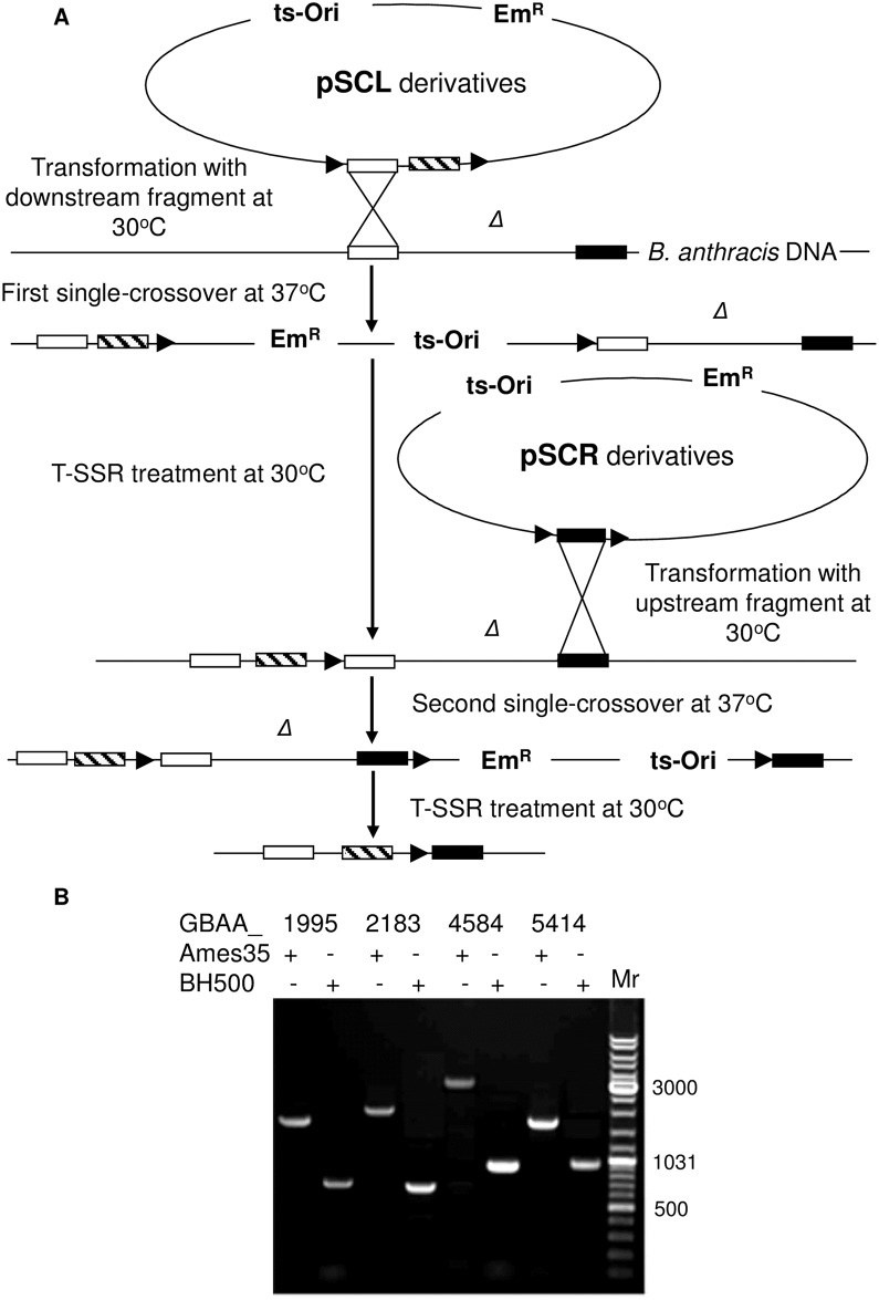
Fig 1. T-SSR system used for deletion/insertion of DNA fragments in B. anthracis genome.
(A) Scheme of the T-SSR application. White and black rectangles are fragments flanking Δ, the DNA fragment to be deleted, while the striped rectangle is the DNA fragment to be inserted in place of Δ. Black triangles represent target sequences loxP, FRT or PSL for Cre, Flp or IntXO T-SSR, respectively. The detailed description of the application of Cre-loxP, Flp-FRT and IntXO-PSL were described previously [6–8] and in the Results section. (B) PCR verification of deletions in cysP1, nprC, vpR and s41 protease genes with corresponding primer pairs 1995seqF/R, 2183 seqF/R, 4584 seqF/R and 5414 seqF/R. GenBank accession numbers for proteases and strains used to verify the retention of specific segments are indicated at the top of the gel. Mr, GeneRuler DNA ladder mix for size determination (e.g., 3,000 bp).
Identification of additional proteases in the protease-deficient strains BH460 and BH480
Both BH460 and BH480 strains (Table 1) were grown overnight in FA medium and centrifuged at 10,000 g for 15 min. The supernatants were filter sterilized using a Pellicon cassette system (EMD Millipore, Billerica, MA) with Durapore 0.22-mm membranes and concentrated with Ultracel-10K membrane (Amicon Ultra-4 centrifugal filters, Merck Millipore Ltd. Tullagreen, IRL). The concentrated supernatants were loaded onto a hydroxyapatite column (Bio-Rad, Hercules, CA) that was pre-equilibrated with 0.02 M potassium phosphate and 0.1 M NaCl buffer (pH 7.0). Proteins were eluted at 1 ml/min in a 0.02–0.25 M potassium phosphate gradient and protein-containing fractions were analyzed for proteolytic activity. Casein protease activities of the supernatants were quantified by the Enz Chek Protease Assay Kit E-6638 (Invitrogen, Thermo Fisher Scientific, Grand Island, NY). For measurement of the protease activity, each 100 μL reaction mixture contained 5 μg/ml of BODIPY FL casein and 5 μl supernatant in 10 mM Tris-HCl (pH 7.8). Reaction mixtures were incubated in the dark at 37°C for 60 min. Fluorescence was measured with a Wallace 1420 VICTOR 96-well plate reader (Perkin Elmer, Boston, MA) with excitation at 485 nm and emission at 530 nm. Fractions with proteolytic activities were loaded on 4–20% SDS PAGE (Life Technologies). Every band was marked and sent for LC MS-MS analysis at the Research Technologies Branch, NIAID/NIH Twinbrook I Facility, Rockville, MD.
Construction of vectors for protease gene inactivation and preparation of mutants
B. anthracis BH460 (derived from the pXO1-, pXO2- Ames 33 strain) was used for genetic manipulations. The GenBank database (GenBank Accession No. for the Ames strain is NC_003997) was analyzed for the identification of target genes and for the corresponding primer design. To inactivate CysP1, VpR and NprC proteases we amplified upstream and downstream fragments with corresponding primer pairs (S1 Table) and inserted them into pSCF to produce recombinant plasmids used for integration by homologous recombination with chromosomal DNA in accordance with the scheme (Fig 1). To inactivate the S41 gene, the same procedure was performed and amplified fragments were inserted into the pSCP plasmid for further integration. Subsequent transformation of the recombinant strains either with pFPAS or pIntPAS plasmid eliminated the protease genes and produced strains BH470, BH480, BH490 and finally BH500 (Table 1).
PCR fragments containing either FRT or PSL sites within mutated genes were amplified and sequenced using primers listed in S1 Table (1995seqF/1995seqR, 2183seqF/2183seqR, 4584seqF/4584seqR, and 5414seqF/5414seqR).
Protein purification and verification
B. anthracis toxin components and HtrA protease were expressed in strain BH500 from plasmids pSJ136EFOS (EF), pSJ115 (LF), pYS5 (PA) and pUTE29-htrA (Table 2). The strains containing plasmids were grown in FA medium containing 20 μg/ml of kanamycin (or 10 μg/ml tetracycline for pUTE29-htrA) at 37°C for 14 h, largely following procedures previously used for production of LF [14]. The cultures were cooled and, except in the case of HtrA, supplemented with 2 μg/ml of AEBSF [4-(2-Aminoethyl)-benzenesulfonylfluoride HCl] (US Biological, Swampscott, MA), and then centrifuged at 4550 g for 30 min. All subsequent steps were performed at 4°C. The supernatants were filter sterilized and supplemented with 5 mM EDTA. Solid ammonium sulfate was added to the supernatants to obtain 40% saturation. Phenyl-Sepharose Fast Flow (low sub) (GE Healthcare Life Sciences, Uppsala, Sweden) was added and supernatants gently mixed in 4°C for 1.5 h. The resins were collected on porous plastic funnels (BelArt Plastics, Pequannock, NJ) and washed with buffer containing 1.5 M ammonium sulfate, 10 mM Tris HCl, and 1 mM EDTA (pH 8.0). The proteins were eluted with 0.3 M ammonium sulfate, 10 mM Tris HCl, and 1 mM EDTA (pH 8.0), precipitated by adding an additional 30 g ammonium sulfate per 100 mL eluate, and centrifuged at 18,370 g for 20 min. The proteins were dissolved and dialyzed against 5 mM HEPES, 0.5 mM EDTA (pH 7.5). The dialyzed samples were applied to a Q-Sepharose Fast Flow column (GE Healthcare Life Sciences) and eluted with a 0–0.5 M NaCl gradient in 20 mM Tris–HCl, 0.5 mM EDTA (pH 8.0). The protein-containing fractions identified by SDS-PhastGel analysis were purified on a column of ceramic hydroxyapatite (Bio-Rad Laboratories) with a gradient of 0.02–1.0 M potassium phosphate containing 0.1 M NaCl (pH 7.0). The fractions containing proteins were dialyzed overnight against 5 mM HEPES and 0.5 mM EDTA, pH 7.5, concentrated as necessary, frozen, and stored at −80°C. The molecular masses of purified toxin components were confirmed by liquid chromatography-electrospray ionization mass spectrometry (LC-ESI-MS) using an HP/Agilent 1100 MSD instrument (Hewlett Packard, Palo Alto, CA) at the National Cancer Institute (Frederick, MD) as described previously [9]. The N-terminal sequence of HtrA was determined using a gas-phase sequencer at the Research Technologies Branch, NIAID/NIH Twinbrook I Facility (Rockville, MD). All purified proteins were quantified by Ultrospec 2100 UV/Visible spectrophotometer (Amersham Pharmacia Biotech Inc., Piscataway, NJ) at 280 nm.
Table 2. Plasmids used in this study.
| Plasmid | Relevant characteristic(s) | Source or reference |
|---|---|---|
| pSC | Contains multiple restriction site flanked by two direct loxP; pSC has permissive and restrictive temperatures of 30°C and 37°C for B. anthracis, respectively. Ampr in E. coli, Emr both in E. coli and B. anthracis | [7] |
| pSC-A | XhoI/ZraI PCR fragment containing atxA gene was inserted into pSC XhoI/SmaI sites | This work |
| pSCF | Contains multiple restriction site flanked by two direct FRT; pSCF has permissive and restrictive temperatures of 30°C and 37°C for B. anthracis, respectively, and can be easily isolated from E. coli strains grown at 37°C; Ampr in E. coli, Emr both in E. coli and B. anthracis. | [6] |
| pSCF-2183L | BstZ17I/XhoI PCR fragment containing upstream fragment of nprC gene was inserted into pSCF | This work |
| pSCF-2183LA | XhoI/ZraI PCR fragment containing atxA gene was inserted into pSCF-2183L XhoI/SmaI sites | This work |
| pSCF-2183R | XmaI/SacI PCR fragment containing downstream fragment of nprC gene was inserted into pSCF | This work |
| pSCP | Contains multiple restriction site flanked by two direct PSL; pSCP has permissive and restrictive temperatures of 30°C and 37°C for B. anthracis, respectively, and can be easily isolated from E. coli strains grown at 37°C; Ampr in E. coli, Emr both in E. coli and B. anthracis. | [8] |
| pSCP-A | XhoI/ZraI PCR fragment containing atxA gene was inserted into pSCF XhoI/ZraI sites | This work |
| pCrePAS2 | Contains entire Flp recombinase gene under the control of pagA promoter; pCrePAS2 has permissive and restrictive temperatures of 30°C and 37°C for B. anthracis, respectively, and can be easily isolated from E. coli strains grown at 37°C. | [6] |
| pFPAS | Contains entire Flp recombinase gene under the control of pagA promoter; pFPAS has permissive and restrictive temperatures of 30°C and 37°C for B. anthracis, respectively, and can be easily isolated from E. coli strains grown at 37°C; Spr both in E. coli and B. anthracis. | [6] |
| pIntPAS | Contains entire IntXO recombinase gene under the control of pagA promoter; pIntPAS has permissive and restrictive temperatures of 30°C and 37°C for B. anthracis, respectively, and can be easily isolated from E. coli strains grown at 37°C; Spr both in E. coli and B. anthracis. | [8] |
| pGEM-T Easy | Cloning vector for PCR products; Apr in E. coli. | Promega |
| pUC4-ΩKM2 | pUC4 carrying an Ω-kan element with kanamycin resistance marker; Kmr in E. coli and B. anthracis. | [6] |
| pYS5 | B. anthracis expression vector. Transcription of pag is under control of truncated pag promoter. | [13] |
| pPAGP | pYS5 with SnaBI/HindIII small fragment replaced by SnaBI/HindIII PCR fragment amplified with pagPF/pagPR primer pair from B. anthracis Ames 35 pXO1plasmid | This work |
| pPAGK | pYS5 derivative (neo gene replaced with Ω-kan from pUC4-ΩKM2, transcription of pag is under control of full length pag promoter). | This work |
| pSJ115 | Contains B. anthracis lef gene instead of the pag gene in pYS5. Retains the pag signal sequence gene. | [14] |
| pSJ136EFOS | Contains B. anthracis cya gene instead of the lef gene in pSJ115. Retains the pag signal sequence gene. | [9] |
| pUTE29-htrA | The B. anthracis htrA gene amplified with HAF/HAR primers pair was inserted into pGEM-T Easy. The SalI-BamHI fragment of pGEM-T Easy-htrA containing the htrA gene was inserted between SalI, BamHI sites of pUTE29 [15] producing pUTE29-htrA. | This work |
SDS-PAGE protein analysis
Proteins were analyzed by Bolt® Bis-Tris Plus 4–12% gradient gels (Life Technologies) with the Bolt® MES as running buffer (B0002, Life Technologies). Samples were prepared by mixing 10 μl of SDS loading buffer (161–0747, Bio-Rad) with 40 μl of protein samples and boiling for 5 min at 95°C. Samples of 5 μg of the protein were loaded on the gel (Fig 2). Electrophoresis was run using the Bolt Mini Gel tank (Life Technologies) at 165 volts for 35 min.
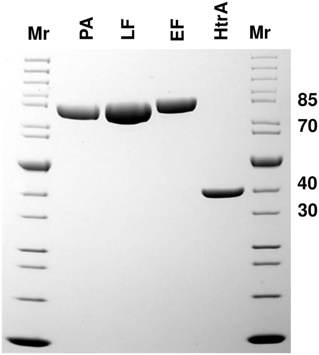
Fig 2. SDS-PAGE analysis of the PA, LF, EF and HtrA proteins (Coomassie stained).
Proteins were purified from BH500 strains containing pYS5, pSJ115, pSJ136EFOS and pUTE29-htrA plasmids, respectively. The strains were grown in FA medium with 20 μg/ml of kanamycin (or 10 μg/ml tetracycline for pUTE29-htrA) at 37°C for 14 h. Protein purification was performed following procedures described in the Materials and Methods section. Mr—PageRuler unstained protein ladder mix for size determination (e.g., 85 kDa).
Construction of vectors for atxA gene insertion and preparation of mutants
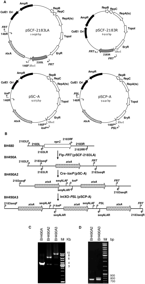
A PCR fragment containing the atxA promoter, ORF, and 3’ terminator was amplified with primer pair 146F/146R (S1 Table). The fragment was inserted along with the upstream fragment of the nprC gene amplified by primers 2183LL/2183LR (S1 Table) into the pSCF plasmid to produce the pSCF-LA construct. The same atxA fragment was inserted into the pSC and pSCP vectors to produce the pSC-A and pSCP-A plasmids, respectively (Fig 3A). The first insertion of the atxA gene was performed by the Flp-FRT T-SSR in accordance with the scheme of Fig 1. The plasmid pSCF-R was used to complete deletion of the nprC gene. Subsequent insertions of second and third copies of atxA were performed by homologous recombination within the atxA sequence using the Cre-loxP and IntXO-PSL T-SSRs. The scheme used in preparation of these BH490 strain variants containing one, two and three copies of the atxA gene and confirmation of structures by PCR analysis using the 2183seqF/2183seqR and seqALAF/seqALAR primer pairs is shown in Fig 3.
Fig 3. Creation of B. anthracis strains containing variable numbers of atxA genes replacing the nprC gene.
(A) Plasmids used for sequential atxA gene insertions. (B) Scheme indicating sequential atxA gene insertions: nprC gene was replaced by the first atxA gene using Flp-FRT system with pSCF-2183LA plasmid as a donor of atxA; the second atxA was added from pSC-A plasmid with Cre-loxP system; and the third atxA copy was inserted with IntXO-PSL system from pSCP-A plasmid. (C) PCR confirmation of atxA gene insertions into genome of B. anthracis (primers 2183seqF/2183seqR). Strains with one, two, and three copies of atxA are indicated on the top of the gel. (D) PCR confirmation of atxA intergenic regions in strains with double and triple atxA genes (primers seqALAF/ALARseqR).
Construction of pPAGK plasmid
To create plasmids containing the full length promoter of the pag gene (1260 bp) as originally identified by Uchida et al. [10] (Table 2), we first amplified that region with the pagPF/pagPR primer pair from the B. anthracis Ames 35 pXO1 plasmid and inserted it into the large SnaBI/HindIII fragment of pYS5. The resulting pPAGP plasmid was then cut with BbsI/SnaBI. The sticky BbsI site end was filled (blunted) with T4 DNA polymerase and the linearized plasmid was ligated to the SmaI fragment of pUC4-ΩKM2 containing Ω-kan element with the kanamycin resistance marker flanked by two bacteriophage T4 transcriptional terminators. The kan gene of the resulting pPAGK plasmid was inserted in opposite orientation to the pag gene and its promoter (Fig 4). As a result, the short pag promoter (162 bp) of the pYS5 plasmid was replaced with the full length pag promoter (1260 bp) in pPAGK.
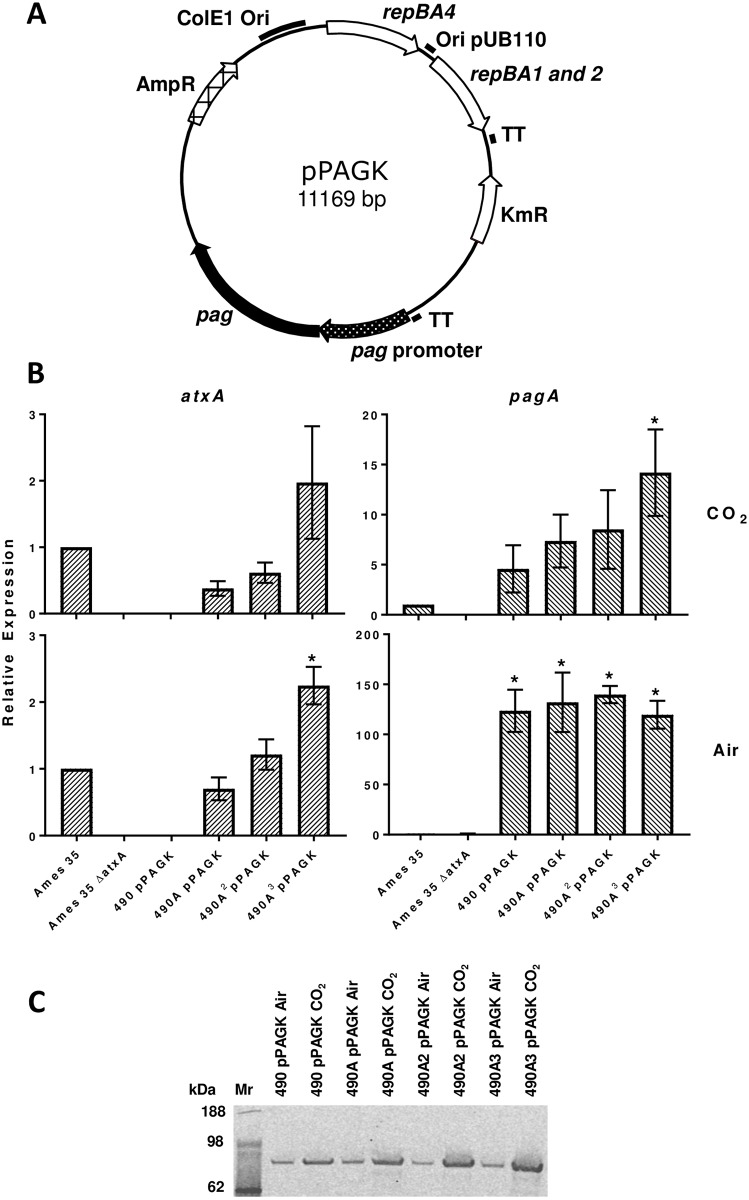
Fig 4. Increased atxA copy number enhances pagA gene transcription and PA content in the B. anthracis secretome.
(A) Genetic structure of pPAGK plasmid. TT—T4 phage transcription terminator. (B) Active transcription of atxA and pagA in BH490 derivatives containing the pPAGK plasmid. Two-step qPCR results of atxA and pagA transcription respectively from cultures grown in NBY broth with 0.8% NaHCO3 in 15% CO2 (top panels) and in air (bottom panels). Relative expression represents the 2-ΔΔCt (RQ) value normalized to Ames 35 and evaluated with three reference genes: rpoB, gyrB, and dnaJ. Values reported are the mean relative expression ± standard error of the mean calculated from ExpressionSuite and GraphPad Prism software. An asterisk indicates a significant difference (p<0.05) from an unpaired t-test compared to Ames 35. (C) Western blot analysis of PA production in strains grown in the same conditions as in panel B.
Preparation of samples for RNA and protein analysis
BH490 strain variants (A, A2, A3) containing the pPAGK plasmid were inoculated into NBY broth containing kanamycin and grown overnight at 37°C in air or with 0.8% (w/v) sodium bicarbonate in 15% CO2. The overnight cultures were diluted to A600 of 0.05 and grown at 37°C in the same media until A600 = 2.0. The bacterial cultures were then centrifuged at 10,000 g for 10 min at 4°C. Supernatants and pellets were separated, frozen on dry ice and stored at -80°C for further analysis of PA and RNA content. Ames 35 and Ames 35 ΔAtxA strains were grown in parallel as controls.
RNA isolation and purification
Bacterial cell pellets were suspended in RNAlater stabilization solution (Ambion, Life Technologies) and incubated at room temperature for 1 h. After incubation, suspensions were centrifuged at 12,000 g for 5 min at 4°C and pellets were flash frozen on dry ice and stored at -80°C. Pellets were thawed on ice and washed with RNase-free water and re-pelleted by centrifugation at 15,000 g for 5 min at 4°C. The pellets were re-suspended in Buffer RLT from the RNeasy Mini Kit (Qiagen, Germantown, MD) and shaken with Lysing Matrix B (MP Biomedicals, Santa Ana, CA) for 20 sec at 6.0 M/s using FastPrep-24 (MP Biomedicals). The bacterial lysates were treated according to manufacturer’s instructions and incubated with TURBO DNase (Ambion, Life Technologies), again following manufacturer’s instructions, for 1 h at 37°C. The partially cleaned RNAs were purified again using the RNeasy Mini Kit, treated again with TURBO DNase, purified once more with the RNeasy Mini Kit, and then cleaned up using the RNA Cleanup procedure from the RNeasy Mini Kit protocol. We established that our protocol requiring two TURBO DNase incubations ensured elimination of contaminating genomic and plasmid DNA (S5 Table). RNA integrity numbers were determined using an Agilent 2100 Bioanalyzer (Agilent Technologies). Purity and concentration of the RNA was determined using a Nandrop ND-1000 spectrophotometer (Thermo Scientific, Wilmington, DE). The RNA samples were stored at -80°C before cDNA synthesis.
qPCR analysis
Purified RNAs were converted to cDNA with Superscript™ Vilo™ cDNA Synthesis Kit (Invitrogen, Thermo Fisher Scientific) according to the manufacturer’s instructions with a minor modification: 12.5 μg of RNA in 100 μL of total reaction volume was incubated at room temperature for 10 min, 42°C for 2 h, and terminated at 85°C for 5 min. The synthesized cDNA was stored in -20°C until used for analysis.
The transcription levels of pagA and atxA genes were assayed by qPCR with three reference genes in accordance with the MIQE guidelines [17]: rpoB, gyrB, and dnaJ (Table 3). The cDNA was amplified using TaqMan Fast Advanced Master Mix (Applied Biosystems, Thermo Fisher Scientific) with IDT-prepared primers and Applied Biosystems-prepared probes (S2 Table). The final concentration of primers and probes were 300 nM and 250 nM, respectively. Reactions were plated in MicroAmp Fast Optical 96-Well Reaction Plates (Applied Biosystems) in 20 μL reactions with 20 ng of cDNA and in three technical replicates and assayed with Applied Biosystems 7500 Fast Real Time PCR System. Cycling conditions were as follows: 2 min at 50°C, 20 sec at 95°C and 40 cycles of 3 sec at 95°C and 30 sec at 60°C. Data were analyzed using the ΔΔCt method and normalized to Ames 35 using Applied Biosystems ExpressionSuite Version 1.0.3 software (https://www.thermofisher.com/us/en/home/technical-resources/software-downloads/expressionsuite-software.html). GraphPad Prism 7.01 software (http://www.graphpad.com/scientific-software/prism/) was used for graphing and statistical analysis. Data were analyzed by unpaired t-tests comparing all strains to the Ames 35 control and are presented as the mean ± standard error of the mean. Statistical significance was considered as p<0.05. All qPCR graphs represent combined data from three biological replicates.
Analysis of PA production under control of AtxA and carbon dioxide
Supernatant samples from bacterial culture samples grown either in air or CO2 were loaded onto Bolt 4–12% Bis-Tris Plus gels and run as described previously in the Materials and Methods section (paragraph “SDS-PAGE protein analysis”). Proteins were transferred to nitrocellulose paper using the iBlot 2 Dry Blotting System (Invitrogen, Thermo Fisher Scientific). After transferring, the blot was blocked for 45 min at room temperature in Odyssey Blocking Buffer (PBS) (LI-COR Biosciences) and incubated with anti-PA rabbit primary antibody 1:2000 dilution [18] in the blocking buffer + 0.05% Tween 20. After overnight incubation at room temperature, membrane was rinsed 3 times for 10 min in 1X PBST (PBS + 0.05% Tween 20) and washed another 3 times with 1X PBST for 5 min. An anti-rabbit HRP secondary antibody (Rockland, Limerick, PA) was diluted 1:10,000 in in the blocking buffer + 0.05% Tween 20 as before and membrane was incubated for 45 min at room temperature. Membrane was then rinsed as described before and imaged.
Results
Four additional proteases were identified in, and deleted from, strain BH460
Our previous work identified and then deleted several proteases secreted by B. anthracis, resulting in strain BH460, lacking 6 proteases (S3 Table). To extend this analysis, supernatants collected from BH460 cultures were separated by hydroxyapatite chromatography, and peaks having protease activity were analyzed by MS/MS. The two active fractions from BH460 were found to contain two caseinolytic proteases, CysP1 and VpR (S1 Fig). CysP1 belongs to a transglutaminase/protease-like superfamily having a conserved Cys/His/Asp catalytic triad. The minor extracellular protease VpR belongs to the S8 family of serine proteases characterized by the Asp/His/Ser catalytic triad found in trypsin-like proteases. The CysP1 and VpR proteases are predicted by SignalP analysis to contain intrinsic signal sequences, MGKTSKYVTAAALCSTIVMGGLHASSVSYA and MKKTTSILLSMALVFSSFGALSAHA, respectively. Deletion of these proteases by the procedure to be described below produced strain BH480 with genotype: spo0A-, pXO1-, pXO2-, nprB-, tasA-, cam-, inhA1-, inhA2, mmpZ-, cysP1-, vpR- (S3 Table). Confirmation of the deletions is shown in S1 Fig.
The same analysis was then applied to identify proteases in the BH480 secretome, leading to the identification of proteases NprC and S41 (Panel C in S1 Fig). NprC belongs to the M36 family of Zn-dependent metalloproteases and the C-terminal processing protease S41 belongs to serine protease family S41. The SignalP algorithm predicts that NprC contains an intrinsic signal sequence, MFNKKMVAMAMTVPLVMGTLSTVSA, whereas S41 contains a strong N-terminal transmembrane anchoring domain, MVVAFLIGAGGMFAGMSL. Deletion of these proteases produced the strain BH500 with genotype: spo0A-, pXO1-, pXO2-, nprB-, tasA-, cam-, inhA1-, inhA2, mmpZ-, cysP1-, vpR- s41- (S3 Table).
The mass spectrometric analyses of these strains used to identify and confirm the presence of the four new proteases are summarized in panel C of S1 Fig. Two biological replicas obtained from the BH460 and BH480 secretomes were analyzed by LC MS-MS. Peptides belonging to CysP1 and VpR were identified in the BH460 secretome as noted above, and were shown to be absent in BH480. Peptides from NprC and S41 were present in BH480, from which these proteases were deleted to produce strain BH500. No proteomic data was obtained for the BH500 strain, and instead, DNA sequencing was used to confirm deletion of these two proteases (Fig 1B), as noted below.
The scheme of deletion of the proteases described above is shown in Fig 1A. Temperature sensitive vectors containing a fragment flanking the upstream end of Δ (pSCL, pSCFL or pSCPL) were inserted into the genome of B. anthracis as the first single cross-over event at 37°C. Subsequent treatment with the corresponding T-SSR removed the vector, retaining the target sequence and a DNA fragment that should be inserted. Both of these elements are flanked by the upstream fragment and still contain the downstream located Δ fragment. Temperature sensitive vectors with the fragment flanking the downstream end of Δ (pSCR, pSCFR or pSCPR) were inserted into the genome of B. anthracis as the second single cross-over event at 37°C. The last treatment with the T-SSR produces a structure containing a DNA fragment that was inserted instead of deleted and a target for the T-SSR. Confirmation for the protease gene deletions is shown on Fig 1B.
B. anthracis strain BH500 produces recombinant proteins in high yield and purity
The B. anthracis toxin components PA, LF, and EF were purified from transformants of BH500 containing plasmids pYS5, pSJ115, and pSJ136EFOS, respectively (Fig 2), in excellent yield and purity. While useful amounts of these proteins could be obtained from strain BH460 [9], use of BH500 typically gave better results. For example, LF yields after purification were often > 50 mg of pure, functional protein per liter of culture. LF made from this strain has been used successfully in many projects, such as the ongoing tumor-targeting work our laboratory has previously described [19].
Strain BH500 was also used to express HtrA. This serine protease is a member of the better-known DegP family of Gram-negative cell-surface proteases that play a key role in protein secretion, folding, and quality control. In the case of B. anthracis, HtrA has been implicated as a virulence factor [20]. The gene and its promoter were cloned into pUTE29 and transformed into BH500. Surprisingly, we found strong expression of a protein in the supernatant of the BH500 (pUTE29-htrA) transformant, and this was easily purified. The calculated molecular mass of HtrA is 43.9 kDa. However, analysis of the gel (Fig 2), demonstrated that the secreted HtrA migrates as if it were less than 40 kDa. The purified protein was transferred to a polyvinylidene difluoride membrane and subjected to N-terminal sequence determination. The sequence of the first 12 residues was VNKAKNETDLPG, indicating the loss of a 76-amino acids pro sequence. This sequence contains a strong transmembrane domain (S2 Fig), consistent with the anchoring of Bacillus HtrA proteases by their N-terminal domains [21]. It appears that overexpression increases self-processing to release the mature enzyme of 337 amino acids (35.9 kDa) that we obtained. After several chromatographic purification steps, the pure HtrA protein was obtained in a yield of 4 mg per liter of culture supernatant.
T-SSRs as a tool for DNA insertion into the genome of B. anthracis
The T-SSRs can be used both for deletion of genes, as illustrated above, and for insertion of DNA into the genome of B. anthracis (Fig 1A). An example of using the T-SSRs to create B. anthracis strains containing variable numbers of atxA genes in the chromosome is shown in Fig 3. Four plasmids were constructed for atxA gene insertions: pSCF-LA, pSCF-2183R, pSC-A and pSCP-A (Fig 3A). The pSCF-LA plasmid was used to insert the first copy of atxA in place of nprC through Flp action as indicated in Fig 1B. In this case the 2183L PCR fragment flanking the nprC gene from the upstream side was used for the first single cross-over inserting the atxA gene. Subsequent treatment with Flp, the second cross-over with pSCF-2183R, and repeated Flp treatment produced strain BH490A containing a FRT-site downstream of the inserted atxA. The second copy of atxA gene was inserted with Cre-loxP. In this case the atxA gene of BH490A was used itself for the cross-over, inserting a second copy of the atxA gene. Subsequent treatment with Cre deleted the pSC vector and inserted a loxP between the two copies of atxA, producing strain BH490A2. The third copy of atxA was inserted with IntXO-PSL. In this case the atxA genes of BH490A2 were used for cross-over inserting a third copy of the atxA gene. Subsequent treatment with IntXO deleted the pSCP vector sequences and inserted PSL between two copies of atxA, producing strain BH490A3.
Confirmation of the sequential atxA gene insertions was obtained by PCR with primer pair 2183seqF/2183seqR (Fig 3B and 3C). BH490A, BH490A2 and BH40A3 produced PCR fragments with gel mobilities consistent with the expected sizes of 3,636, 6,726, and 9,837 bp. The presence of a small amount of an approximately 3.5-Kbp fragment in the BH490A2 and BH490A3 strains suggests that homologous recombination between atxA sequences occurs at a low frequency. The presence of three atxA gene copies in BH490A3 was also confirmed by PCR analysis and sequencing of the atxA intergenic regions with primer pair seqALAF/seqALAR (Fig 3B and 3D). The two seqALAF/seqALAR-generated PCR fragments of approximately 700 and 800 bp were found to contain the expected PSL and loxP sites, respectively (S3 Fig).
Increased atxA copy number enhances pagA expression in a CO2-dependent manner
To confirm that the atxA inserts act as a positive transcriptional regulator along with CO2, we assayed the transcription of both the atxA and pagA genes via qPCR for strains containing 0, 1, 2 and 3 copies of the atxA gene. Plasmid pPAGK was constructed so as to have the pagA gene in an environment like that in pXO1 (Fig 4A). Thus, the transcription of pagA in pPAGK is under control of the full-length native pag promoter, with other promoter activities not being expected to influence its transcription. Experiments were performed both in air and carbon dioxide conditions. RNA integrity numbers for preparations from cultures grown in CO2 and air and used in the qPCR reactions averaged 9.43 (± 0.46) and 8.98 (± 0.83), respectively.
We found that as the copy number of atxA increased, the transcription of the gene also increased for strains grown in both air and CO2 (Fig 4B, left-hand panels; S4 Table). As expected, no atxA transcription was detected in the negative control Ames 35 ΔAtxA or in the strain without a chromosomal atxA insert, BH490 pPAGK. Increasing the atxA copy number had a positive effect on pagA transcription, but only when bacteria were grown in CO2, with no increase seen for cultures grown in air (Fig 4B, right-hand panels). BH490A3 pPAGK had significantly higher levels of pagA expression in CO2 than Ames 35 (p<0.05). The pagA expression of BH490 derivatives when grown in air was determined to be significantly higher than in Ames 35 (p<0.05), presumably due to the high copy number of plasmid pPAGK, and residual (leaky) promoter activity of its promoter in air.
To determine how the chromosomal inserts of atxA impacted PA production we conducted a Western blot analysis with PA antibodies on proteins secreted by the same strains and in the same conditions as the qPCR analysis. When grown in CO2, an increased atxA copy number led to an increase in PA production, but when grown in air, PA production appeared to remain constant with increasing copy number (Fig 4C). This result agrees with transcriptomic data indicating that AtxA is an enhancer of the positive effect of carbon dioxide on pagA expression.
Discussion
This work demonstrates how sequential use of different T-SSRs can facilitate the deletion and insertion of DNA fragments into B. anthracis. These recombinases are shown to enable the creation of strains having improved behavior as hosts for recombinant protein production. Furthermore, strains having complex gene duplications can be created for study of transcriptional control, as occurs in B. anthracis in response to CO2.
Relying on a single T-SSR to make multiple gene knock-outs in a strain can produce problems. This became evident in our early work when loxP sites placed about 30 kbp apart led to deletion of the intervening region, to produce strain McrB3P-Mrr-LΔ30, which remained viable in spite of lacking many genes [5]. More recently, attempts to use Cre recombinase to further manipulate strain BH460, which contains five loxP sites [9], produced strains that grew poorly and generated undesirable deletions in the plasmids that were used for transformation. To resolve this problem, we brought into use two additional T-SSR systems: Flp-FRT and IntXO-PSL. The functionality of both systems was demonstrated previously [6, 8]. These enabled the inactivation of four additional proteases that were identified by proteomic analysis of the BH460 and BH480 culture supernatants. Successful application of the newly identified IntXO recombinase expanded the repertoire of available enzymes that are useful for advanced genome engineering of B. anthracis and that may find application in other microorganisms and in eukaryotes.
We also cloned and purified the HtrA protease that has been described as a major virulence determinant of B. anthracis [20]. However, the molecular mass of the secreted HtrA identified in this study (35.85 kDa) was lower than that of the HtrA secreted by strains described by Chitlaru et al. [20]. To clarify this discrepancy, we are working to produce antibodies to HtrA to facilitate analysis of the secretion, membrane anchoring, and release to the supernatant of this protease.
The development and implementation of three T-SSR systems for B. anthracis allowed us to produce sequential insertions of three copies of the atxA gene into the BH490 genome. The PCR product inserted was designed to include the dual promoter controlling the expression of atxA [22] and a 2.7-kb-long atxA mRNA that contains a terminator structure [23]. We found the atxA chromosomal inserts to be both transcriptionally and translationally active as demonstrated by the qPCR and Western blot data, proving the success of the T-SSRs in this complex genome editing procedure. The qPCR data also demonstrated the previously established cooperative relationship between AtxA and CO2 [12, 24]. The molecular mechanism by which AtxA and CO2 cooperatively regulate virulence gene expression in B. anthracis is still not known, but the strains and techniques presented here can serve as important tools to elucidate the relationship.
Furthermore, based on the protein production data presented here we propose that BH490A3 pPAGK is an appropriate host for production of recombinant proteins when grown in CO2, indicated by the increased level of PA production with more atxA inserts and the statistically significant increase in pagA gene expression. We also hypothesize that BH500 with three chromosomal inserts of atxA would be a more efficient host strain for recombinant protein production due to the tenth protease deletion.
Supporting information
Top panel (A and B)—Casein proteolytic activities of HTP-chromatographic fractions for BH460 (A) and BH480 (B) supernatants. Axis X—fraction numbers, axis Y—arbitrary units of the activities, FT—flow through the column. Bottom panel (A and B)—SDS-PAGE electrophoresis of proteins in the peaks revealed during HTP-chromatography. (C) Heatmaps of proteases identified in BH460 and BH480 secretomes. Number of peptides found for proteases in BH460 and BH480 strains are shown.
(PPTX)
Transmembrane domain identified by TMpred software (http://www.ch.embnet.org/software/TMPRED_form.html) is indicated in red. Secreted part of the HtrA is shown in bold, amino acids identified by N-terminal degradation method are underlined. Molecular mass is 43.9 kDa for the whole molecule, and 35.85 kDa for secreted form.
(PPTX)
Primers seqALAF and seqALAR were used for sequencing of PCR fragments amplified with the same primers and demonstrated on Fig 3D. (A)The PSL-site was identified in the sequence of top PCR fragment (indicated in bold blue). (B) The loxP-site was identified in the sequence of the bottom PCR fragment (indicated in bold red).
(PPTX)
The names and nucleotide sequences of all primers used in the current study.
(DOCX)
The names and nucleotide sequences of all qPCR primers and TaqMan probes used in the current study.
(DOCX)
LoxP, FRT and PSL indicate that gene is replaced by this sequence.
(DOCX)
Plasmid pYS5PAPK is the same plasmid as pPAGK.
(XLSX)
Quantitative real time PCR raw Ct values from BH490A3 pPAGK RNA isolated using the procedure detailed in Materials and Methods. Samples without reverse transcriptase are marked in red and include RT- in sample name. Ct values of samples without reverse transcriptase are generally between 16 and 19 cycles higher than samples with reverse transcriptase indicating successful elimination of DNA during RNA isolation.
(XLSX)
Acknowledgments
This research was supported by the intramural research program of the National Institute of Allergy and Infectious Diseases, National Institutes of Health. We thank Brian Martin for N-terminal sequencing of proteins and L. Renee Olano for proteomic analysis of secretomes (both at NIAID/NIH Research Technologies Branch) and Sergey Tarasov (Biophysics Resource in the Structural Biophysics Laboratory, NCI/NIH-Frederick) for LC-ESI-MS determination of protein exact masses.
Data Availability
All relevant data are within the paper and its Supporting Information files.
Funding Statement
This work was supported by the National Institutes of Health Intramural Program (1 ZIA ZI001030-04). The funders had no role in study design, data collection and analysis, decision to publish, or preparation of the manuscript.
References
- 1.Buchholz F. Engineering DNA processing enzymes for the postgenomic era. Curr Opin Biotechnol. 2009;20(4):383–9. doi: 10.1016/j.copbio.2009.07.005 [DOI] [PubMed] [Google Scholar]
- 2.Meinke G, Bohm A, Hauber J, Pisabarro MT, Buchholz F. Cre recombinase and other tyrosine recombinases. Chem Rev. 2016. [DOI] [PubMed] [Google Scholar]
- 3.Grindley ND, Whiteson KL, Rice PA. Mechanisms of site-specific recombination. Annu Rev Biochem. 2006;75:567–605. doi: 10.1146/annurev.biochem.73.011303.073908 [DOI] [PubMed] [Google Scholar]
- 4.Yen Ting L, Sau S, Ma CH, Kachroo AH, Rowley PA, Chang KM, et al. The partitioning and copy number control systems of the selfish yeast plasmid: an optimized molecular design for stable persistence in host cells. Microbiol Spectr. 2014;2(5). [DOI] [PMC free article] [PubMed] [Google Scholar]
- 5.Pomerantsev AP, Sitaraman R, Galloway CR, Kivovich V, Leppla SH. Genome engineering in Bacillus anthracis using Cre recombinase. Infect Immun. 2006;74(1):682–93. doi: 10.1128/IAI.74.1.682-693.2006 [DOI] [PMC free article] [PubMed] [Google Scholar]
- 6.Pomerantsev AP, Chang Z, Rappole C, Leppla SH. Identification of three noncontiguous regions on Bacillus anthracis plasmid pXO1 that are important for its maintenance. J Bacteriol. 2014;196(16):2921–33. doi: 10.1128/JB.01747-14 [DOI] [PMC free article] [PubMed] [Google Scholar]
- 7.Pomerantsev AP, Camp A, Leppla SH. A new minimal replicon of Bacillus anthracis plasmid pXO1. J Bacteriol. 2009;191(16):5134–46. doi: 10.1128/JB.00422-09 [DOI] [PMC free article] [PubMed] [Google Scholar]
- 8.Pomerantsev AP, Rappole C, Chang Z, Chahoud M, Leppla SH. The IntXO-PSL recombination system is a key component of the second maintenance system for Bacillus anthracis plasmid pXO1. J Bacteriol. 2016;198(14):1939–51. doi: 10.1128/JB.01004-15 [DOI] [PMC free article] [PubMed] [Google Scholar]
- 9.Pomerantsev AP, Pomerantseva OM, Moayeri M, Fattah R, Tallant C, Leppla SH. A Bacillus anthracis strain deleted for six proteases serves as an effective host for production of recombinant proteins. Protein Expr Purif. 2011;80(1):80–90. doi: 10.1016/j.pep.2011.05.016 [DOI] [PMC free article] [PubMed] [Google Scholar]
- 10.Uchida I, Hornung JM, Thorne CB, Klimpel KR, Leppla SH. Cloning and characterization of a gene whose product is a trans-activator of anthrax toxin synthesis. J Bacteriol. 1993;175(17):5329–38. [DOI] [PMC free article] [PubMed] [Google Scholar]
- 11.Vodkin MH, Leppla SH. Cloning of the protective antigen gene of Bacillus anthracis. Cell. 1983;34(2):693–7. [DOI] [PubMed] [Google Scholar]
- 12.McKenzie AT, Pomerantsev AP, Sastalla I, Martens C, Ricklefs SM, Virtaneva K, et al. Transcriptome analysis identifies Bacillus anthracis genes that respond to CO2 through an AtxA-dependent mechanism. BMC Genomics. 2014;15(1):229. [DOI] [PMC free article] [PubMed] [Google Scholar]
- 13.Singh Y, Chaudhary VK, Leppla SH. A deleted variant of Bacillus anthracis protective antigen is non-toxic and blocks anthrax toxin action in vivo. J Biol Chem. 1989;264(32):19103–7. [PubMed] [Google Scholar]
- 14.Park S, Leppla SH. Optimized production and purification of Bacillus anthracis lethal factor. Protein Expr Purif. 2000;18(3):293–302. doi: 10.1006/prep.2000.1208 [DOI] [PubMed] [Google Scholar]
- 15.Koehler TM, Dai Z, Kaufman-Yarbray M. Regulation of the Bacillus anthracis protective antigen gene: CO2 and a trans-acting element activate transcription from one of two promoters. J Bacteriol. 1994;176:586–95. [DOI] [PMC free article] [PubMed] [Google Scholar]
- 16.Sambrook J, Russell DW. Molecular Cloning A Laboratory Manual. 2 ed Cold Spring Harbor, New York: Cold Spring Harbor Laboratory Press; 2001. 2001. [Google Scholar]
- 17.Bustin SA, Benes V, Garson JA, Hellemans J, Huggett J, Kubista M, et al. The MIQE guidelines: minimum information for publication of quantitative real-time PCR experiments. Clin Chem. 2009;55(4):611–22. doi: 10.1373/clinchem.2008.112797 [DOI] [PubMed] [Google Scholar]
- 18.Moayeri M, Wiggins JF, Leppla SH. Anthrax protective antigen cleavage and clearance from the blood of mice and rats. Infect Immun. 2007;75(11):5175–84. doi: 10.1128/IAI.00719-07 [DOI] [PMC free article] [PubMed] [Google Scholar]
- 19.Liu S, Liu J, Ma Q, Cao L, Fattah RJ, Yu Z, et al. Solid tumor therapy by selectively targeting stromal endothelial cells. Proc Natl Acad Sci U S A. 2016;113(28):E4079–87. doi: 10.1073/pnas.1600982113 [DOI] [PMC free article] [PubMed] [Google Scholar]
- 20.Chitlaru T, Zaide G, Ehrlich S, Inbar I, Cohen O, Shafferman A. HtrA is a major virulence determinant of Bacillus anthracis. Mol Microbiol. 2011;81(6):1542–59. doi: 10.1111/j.1365-2958.2011.07790.x [DOI] [PubMed] [Google Scholar]
- 21.Krishnappa L, Dreisbach A, Otto A, Goosens VJ, Cranenburgh RM, Harwood CR, et al. Extracytoplasmic proteases determining the cleavage and release of secreted proteins, lipoproteins, and membrane proteins in Bacillus subtilis. J Proteome Res. 2013;12(9):4101–10. doi: 10.1021/pr400433h [DOI] [PubMed] [Google Scholar]
- 22.Bongiorni C, Fukushima T, Wilson AC, Chiang C, Mansilla MC, Hoch JA, et al. Dual promoters control the expression of the Bacillus anthracis virulence factor AtxA. J Bacteriol. 2008;190(19):6483–92. doi: 10.1128/JB.00766-08 [DOI] [PMC free article] [PubMed] [Google Scholar]
- 23.Bertin M, Chateau A, Fouet A. Full expression of Bacillus anthracis toxin gene in the presence of bicarbonate requires a 2.7-kb-long atxA mRNA that contains a terminator structure. Res Microbiol. 2010;161(4):249–59. doi: 10.1016/j.resmic.2010.03.003 [DOI] [PubMed] [Google Scholar]
- 24.Hoffmaster AR, Koehler TM. The anthrax toxin activator gene atxA is associated with CO2-enhanced non-toxin gene expression in Bacillus anthracis. Infect Immun. 1997;65:3091–9. [DOI] [PMC free article] [PubMed] [Google Scholar]
Associated Data
This section collects any data citations, data availability statements, or supplementary materials included in this article.
Supplementary Materials
Top panel (A and B)—Casein proteolytic activities of HTP-chromatographic fractions for BH460 (A) and BH480 (B) supernatants. Axis X—fraction numbers, axis Y—arbitrary units of the activities, FT—flow through the column. Bottom panel (A and B)—SDS-PAGE electrophoresis of proteins in the peaks revealed during HTP-chromatography. (C) Heatmaps of proteases identified in BH460 and BH480 secretomes. Number of peptides found for proteases in BH460 and BH480 strains are shown.
(PPTX)
Transmembrane domain identified by TMpred software (http://www.ch.embnet.org/software/TMPRED_form.html) is indicated in red. Secreted part of the HtrA is shown in bold, amino acids identified by N-terminal degradation method are underlined. Molecular mass is 43.9 kDa for the whole molecule, and 35.85 kDa for secreted form.
(PPTX)
Primers seqALAF and seqALAR were used for sequencing of PCR fragments amplified with the same primers and demonstrated on Fig 3D. (A)The PSL-site was identified in the sequence of top PCR fragment (indicated in bold blue). (B) The loxP-site was identified in the sequence of the bottom PCR fragment (indicated in bold red).
(PPTX)
The names and nucleotide sequences of all primers used in the current study.
(DOCX)
The names and nucleotide sequences of all qPCR primers and TaqMan probes used in the current study.
(DOCX)
LoxP, FRT and PSL indicate that gene is replaced by this sequence.
(DOCX)
Plasmid pYS5PAPK is the same plasmid as pPAGK.
(XLSX)
Quantitative real time PCR raw Ct values from BH490A3 pPAGK RNA isolated using the procedure detailed in Materials and Methods. Samples without reverse transcriptase are marked in red and include RT- in sample name. Ct values of samples without reverse transcriptase are generally between 16 and 19 cycles higher than samples with reverse transcriptase indicating successful elimination of DNA during RNA isolation.
(XLSX)
Data Availability Statement
All relevant data are within the paper and its Supporting Information files.


