Abstract
Vitrification reduces the fertilisation capacity and developmental ability of mammalian oocytes; this effect is closely associated with an abnormal increase of cytoplasmic free calcium ions ([Ca2+]i). However, little information about the mechanism by which vitrification increases [Ca2+]i levels or a procedure to regulate [Ca2+]i levels in these oocytes is available. Vitrified bovine oocytes were used to analyse the effect of vitrification on [Ca2+]i, endoplasmic reticulum Ca2+ (ER Ca2+), and mitochondrial Ca2+ (mCa2+) levels. Our results showed that vitrification, especially with dimethyl sulfoxide (DMSO), can induce ER Ca2+ release into the cytoplasm, consequently increasing the [Ca2+]i and mCa2+ levels. Supplementing the cells with 10 μM 1,2-bis (o-aminophenoxy)ethane-N,N,N′,N′-tetraacetic acid (BAPTA-AM or BAPTA) significantly decreased the [Ca2+]i level and maintained the normal distribution of cortical granules in the vitrified bovine oocytes, increasing their fertilisation ability and cleavage rate after in vitro fertilisation (IVF). Treating vitrified bovine oocytes with 1 μM ruthenium red (RR) significantly inhibited the Ca2+ flux from the cytoplasm into mitochondria; maintained normal mCa2+ levels, mitochondrial membrane potential, and ATP content; and inhibited apoptosis. Treating vitrified oocytes with a combination of BAPTA and RR significantly improved embryo development and quality after IVF.
Introduction
Cryopreservation of oocytes plays an important role in providing oocytes for assisted reproductive technologies, including in vitro fertilisation (IVF), intracytoplasmic sperm injection, and somatic cell nuclear transfer1–3. Oocyte cryopreservation also contributes to infertility treatment in humans by avoiding ethical and legal problems of human embryo freezing4. Currently, slow freezing and vitrification are two methods used for oocyte cryopreservation; of these, vitrification is considered to be better5–7 because the high concentration of cryoprotectants (CPAs) used and the extremely high cooling rates help to prevent the formation of ice crystals4, 8.
Meanwhile, vitrification decreases the fertilisation ability and developmental competence of oocytes3, 9–12, which greatly limits its wide application in embryonic biotechnology. This phenomenon is closely associated with the abnormal increase of cytoplasmic free calcium ions ([Ca2+]i) in vitrified oocytes9, which then triggers the premature release of cortical granules (CGs) to the zona pellucida (ZP) layers13, 14, resulting in abnormal ZP hardening before fertilisation. However, how vitrification increases the [Ca2+]i level in oocytes remains unclear.
Endoplasmic reticulum (ER) and mitochondria are important Ca2+ pools in oocytes15, and the [Ca2+]i increase at fertilisation is primarily derived from the ER15, 16. Mitochondria are responsible for Ca2+ absorption and release and play an important role in the conduction of [Ca2+]i signalling17. Until now, the effect of vitrification on ER Ca2+ and mitochondrial Ca2+ (mCa2+) levels, which would help to determine the mechanism by which vitrification increases [Ca2+]i level in oocytes, has not yet been fully investigated.
The Ca2+ chelator 1,2-bis(o-aminophenoxy)ethane-N,N,Nʹ,Nʹ-tetraacetic acid (BAPTA-AM or BAPTA) significantly reduces the level of [Ca2+]i18, 19. The mCa2+ uniporter ruthenium red (RR) can inhibit the influx of Ca2+ into the mitochondria20. A previous study reported that 10 μM BAPTA treatment obviously reduced ZP hardening in vitrified mouse oocytes9; another study reported that thawed vitrified porcine germinal vesicle oocytes treated with 1 μM RR or 10 μM BAPTA showed significantly improved survival and maturation rates21. However, little information is available regarding the mechanism by which RR and BAPTA can improve the development ability of vitrified oocytes.
Therefore, in the present study, we investigated the effect of vitrification on [Ca2+]i, ER Ca2+, and mCa2+ levels in vitrified bovine oocytes to determine the mechanism by which the [Ca2+]i level is increased in vitrified bovine oocytes. Based on our findings, we further investigated the effect of BAPTA and RR on the [Ca2+]i, ER Ca2+, and mCa2+ levels; CG distribution; mitochondrial function; apoptosis; and fertilisation ability of vitrified bovine oocytes to illustrate the means through which BAPTA and RR improve the developmental ability of vitrified oocytes.
Results
In our study, 7015 of 7702 (91.1 ± 6.2%) MII oocytes survived after vitrification.
Experiment 1: Effect of vitrification on Ca2+ levels in bovine oocytes
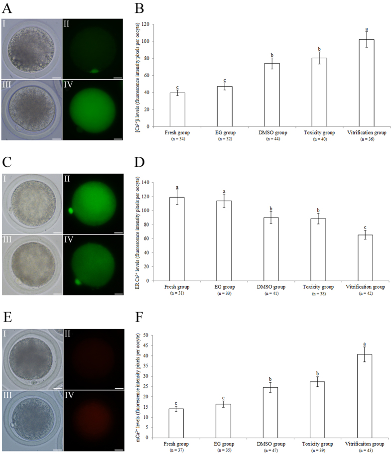
Figure 1A illustrates the [Ca2+]i staining of bovine oocytes. As shown in Fig. 1B, the [Ca2+]i level was significantly higher in the vitrification group than in the ethylene glycol (EG), dimethyl sulfoxide (DMSO), toxicity, and fresh groups (P < 0.05). The [Ca2+]i levels of the DMSO and toxicity groups were significantly higher than those of the EG and fresh groups (P < 0.05). The [Ca2+]i levels did not significantly differ between the EG and fresh groups (P > 0.05).
Figure 1.
Effect of vitrification on the Ca2+ levels of bovine oocytes. (A,C,E) Bovine oocytes stained with [Ca2+]i, ER Ca2+, and mCa2+ detection kits. (I, II) oocytes from the fresh group. (III, IV) oocytes from the vitrification group. Scale bar = 20 μm. (B,D,F) [Ca2+]i, ER Ca2+, and mCa2+ levels in bovine oocytes, respectively. a,b,c: Values with different superscripts differ significantly between groups (P < 0.05).
Figure 1C shows the ER Ca2+ staining of bovine oocytes. As shown in Fig. 1D, the ER Ca2+ level was significantly lower in the vitrification group than in the EG, DMSO, toxicity, and fresh groups (P < 0.05). The ER Ca2+ levels in the DMSO and toxicity groups were similar and were both significantly lower than those in the EG and fresh groups (P < 0.05). No significant difference was found in the ER Ca2+ levels between the EG and fresh groups (P > 0.05).
Figure 1E illustrates the mCa2+ staining of bovine oocytes. As shown in Fig. 1F, the mCa2+ level was significantly higher in the vitrification group than in the EG, DMSO, toxicity, and fresh groups (P < 0.05). The mCa2+ levels in the DMSO and toxicity groups were similar and were significantly higher than the levels in the EG and fresh groups (P < 0.05). No significant difference was found in mCa2+ levels between the EG and fresh groups (P > 0.05).
Experiment 2: Effect of BAPTA treatment on Ca2+ levels and developmental ability of vitrified bovine oocytes
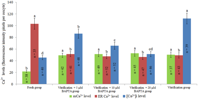
As shown in Fig. 2, the [Ca2+]i level of the vitrification + 10 μM BAPTA group was significantly lower than that of the vitrification + 5 μM BAPTA group and vitrification group (P < 0.05) but still significantly higher than that of the fresh group. Meanwhile, the [Ca2+]i level of the vitrification + 20 μM BAPTA group was similar to that of the fresh group. The mCa2+ levels did not significantly differ between the three vitrification + BAPTA groups (5, 10, and 20 μM) and the vitrification group (P > 0.05), and these levels were all significantly higher than that of the fresh group (P < 0.05). The ER Ca2+ levels of the three vitrification + BAPTA groups (5, 10, and 20 μM) were similar to that of the vitrification group (P > 0.05) and were all significantly lower than that of the fresh group (P < 0.05).
Figure 2.
Effect of BAPTA treatment on Ca2+ levels of vitrified bovine oocytes. a,b,c,dValues with different superscripts differ significantly between groups (P < 0.05).
Fig. S1 shows the CG staining of bovine oocytes. As shown in Table 1, the percentage of oocytes with peripheral distribution of CGs in the vitrification + 20 μM BAPTA group (63.5 ± 6.0%) was significantly higher than that in the vitrification + 5 μM, vitrification + 10 μM BAPTA and vitrification groups (39.3 ± 3.6%, 52.2 ± 4.8%, and 25.0 ± 2.2%, respectively; P < 0.05) and similar to that of the fresh group (70.6 ± 6.3%; P > 0.05). The percentages of oocytes with complete release and discontinuous peripheral distribution in the vitrification + 5 μM BAPTA (30.4 ± 2.7%, 19.6 ± 1.9%), vitrification + 10 μM BAPTA (19.4 ± 1.6%, 17.9 ± 1.0%), and vitrification + 20 μM BAPTA (11.1 ± 1.0%, 15.9 ± 1.4%) groups were significantly lower than the corresponding values in the vitrification group (39.1 ± 3.3%, 23.4 ± 2.1%; P < 0.05) but still higher than those in the fresh group (7.8 ± 0.6%, 11.8 ± 1.3%; P < 0.05). There was no significant difference in the percentages of oocytes with a homogeneous distribution of CGs among all groups (P > 0.05; Table 1).
Table 1.
Effect of BAPTA treatment on the CG distribution in vitrified bovine oocytes.
| Groups | Number of oocytes | Number of oocytes with peripheral distribution | Number of oocytes with discontinuous peripheral distribution | Number of oocytes with complete release of CGs | Number of oocytes with homogeneous distribution |
|---|---|---|---|---|---|
| Vitrification group | 64 | 16 (25.0 ± 2.2%)d | 15 (23.4 ± 2.1%)a | 25 (39.1 ± 3.3%)a | 8 (12.5 ± 1.1%)a |
| Vitrification + 5 μM BAPTA group | 56 | 22 (39.3 ± 3.6%)c | 11 (19.6 ± 1.9%)b | 17 (30.4 ± 2.7%)b | 6 (10.7 ± 0.9%)a |
| Vitrification + 10 μM BAPTA group | 67 | 35 (52.2 ± 4.8%)b | 12 (17.9 ± 1.0%)bc | 13 (19.4 ± 1.6%)c | 7 (10.5 ± 0.9%)a |
| Vitrification + 20 μM BAPTA group | 63 | 40 (63.5 ± 6.0%)a | 10 (15.9 ± 1.4%)c | 7 (11.1 ± 1.0%)d | 6 (9.5 ± 0.6%)a |
| Fresh group | 51 | 36 (70.6 ± 6.3%)a | 6 (11.8 ± 1.3%)d | 4 (7.8 ± 0.6%)e | 5 (9.8 ± 0.8%)a |
a,b,c,d,eValues with different superscripts differ significantly within the same column (P < 0.05).
Fig. S2 illustrates the fertilisation capacity of bovine oocytes after IVF (Supplementary Methods 1.6). As shown in Table 2, the percentage of monospermic oocytes in the vitrification + 10 μM BAPTA group (61.0 ± 5.8%) was significantly higher than that in the vitrification group (42.1 ± 4.0%; P < 0.05) and similar to that in the vitrification + 5 μM BAPTA (51.6 ± 4.3%), vitrification + 20 μM BAPTA (65.2 ± 6.0%), and fresh groups (69.8 ± 6.3%; P > 0.05). The percentages of polyspermic and unfertilised oocytes of the vitrification + 10 μM BAPTA group (15.3 ± 1.4%, 23.7 ± 2.3%) were significantly lower than those of the vitrification group (23.4 ± 1.9%, 34.6 ± 3.2%; P < 0.05) and similar to those of the vitrification + 20 μM BAPTA group (11.4 ± 0.7%, 21.4 ± 1.9%) and fresh group (10.5 ± 1.0%, 19.8 ± 1.7%; P > 0.05).
Table 2.
Effect of BAPTA treatment on the fertilisation capacity of vitrified bovine oocytes.
| Groups | Number of oocytes | Number of polyspermic oocytes | Number of monospermic oocytes | Number of unfertilised oocytes |
|---|---|---|---|---|
| Vitrification group | 107 | 25 (23.4 ± 1.9%)a | 45 (42.1 ± 4.0%)c | 37 (34.6 ± 3.2%)a |
| Vitrification + 5 μM BAPTA group | 95 | 19 (20.0 ± 1.8%)ab | 49 (51.6 ± 4.3%)bc | 27 (28.4 ± 2.2%)b |
| Vitrification + 10 μM BAPTA group | 118 | 18 (15.3 ± 1.4%)bc | 72 (61.0 ± 5.8%)ab | 28 (23.7 ± 2.3%)c |
| Vitrification + 20 μM BAPTA group | 112 | 15 (11.4 ± 0.7%)c | 73 (65.2 ± 6.0%)a | 24 (21.4 ± 1.9%)c |
| Fresh group | 86 | 9 (10.5 ± 1.0%)c | 60 (69.8 ± 6.3%)a | 17 (19.8 ± 1.7%)c |
a,b,cValues with different superscripts differ significantly within the same column (P < 0.05).
As shown in Table 3, the cleavage rate of the vitrification + 10 μM BAPTA group (65.7 ± 5.7%) was significantly higher than those of the vitrification (41.6 ± 3.8%) and vitrification + 20 μM BAPTA (49.7 ± 4.3%; P < 0.05) groups and similar to those of the vitrification + 5 μM BAPTA group (52.5 ± 4.2%) and fresh group (75.0 ± 6.2%; P > 0.05). For the blastocyst rate, no significant difference was found among the vitrification + 5 μM BAPTA (15.1 ± 1.4%), vitrification + 10 μM BAPTA (15.7 ± 1.2%), vitrification + 20 μM BAPTA (12.0 ± 0.9%), and vitrification (12.4 ± 1.1%; P > 0.05) groups, and they were all significantly lower than that of the fresh group (34.1 ± 2.8%; P < 0.05).
Table 3.
Effect of BAPTA treatment on the developmental ability of vitrified bovine oocytes after IVF.
| Groups | Number of oocytes | Number of cleaved oocytes (%) | Number of blastocysts (%) |
|---|---|---|---|
| Vitrification group | 233 | 97 (41.6 ± 3.8%)c | 12 (12.4 ± 1.1%)b |
| Vitrification + 5 μM BAPTA group | 177 | 93 (52.5 ± 4.2%)bc | 14 (15.1 ± 1.4%)b |
| Vitrification + 10 μM BAPTA group | 213 | 140 (65.7 ± 5.7%)ab | 22 (15.7 ± 1.2%)b |
| Vitrification + 20 μM BAPTA group | 185 | 92 (49.7 ± 4.3%)c | 11 (12.0 ± 0.9%)b |
| Fresh group | 176 | 132 (75.0 ± 6.2%)a | 45 (34.1 ± 2.8%)a |
a,b,cValues with different superscripts differ significantly within the same column (P < 0.05).
Experiment 3: Effect of RR treatment on Ca2+ levels and developmental potential of vitrified bovine oocytes
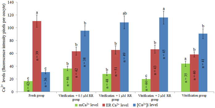
As shown in Fig. 3, the mCa2+ levels were significantly lower in the vitrification + RR (0.5, 1, and 2 μM) groups than in the vitrification group (P < 0.05). The mCa2+ levels were similar between the vitrification + 0.5 μM RR group and vitrification + 1 μM RR group, and these levels were significantly higher than those of the fresh and vitrification + 2 μM RR groups (P < 0.05). No statistically significant difference was observed in mCa2+ levels between the vitrification + 2 μM RR group and fresh group (P > 0.05).
Figure 3.
Effect of RR treatment on Ca2+ levels in vitrified bovine oocytes. a,b,cValues with different superscripts differ significantly between groups (P < 0.05).
The ER Ca2+ level of the vitrification + 0.5 μM RR group was similar to those of the vitrification + 1 μM RR group, vitrification + 2 μM RR group, and vitrification group but significantly lower than that of the fresh group (P < 0.05). The [Ca2+]i level of the vitrification + 2 μM RR group was similar to that of vitrification + 1 μM RR group, and this value was significantly higher than those of the 0.5 μM RR group, vitrification group, and fresh group (P < 0.05).
Figure S3 illustrates the mitochondrial membrane potential (∆Ψm) of bovine oocytes stained with JC-1, and Fig. S4 illustrates terminal deoxynucleotidyl transferase-mediated dUTP nick-end labelling (TUNEL)-stained bovine oocytes. As shown in Table 4, the ration of red to green fluorescence intensity and ATP content of the vitrification + 0.5 μM RR group (1.3 ± 0.1, 0.8 ± 0.1 pmol), vitrification + 1 μM RR group (1.7 ± 0.1, 1.0 ± 0.1 pmol), and vitrification + 2 μM RR group (1.4 ± 0.1, 0.8 ± 0.1 pmol) were significantly higher than those of the vitrification group (0.9 ± 0.1, 0.6 ± 0.0 pmol; P < 0.05). However, TUNEL-positive oocyte percentages in the vitrification + 0.5 μM RR group (16.7 ± 1.1%), vitrification + 1 μM RR group (8.2 ± 0.7%), and vitrification + 2 μM RR group (17.8 ± 1.1%) were significantly lower than that in the vitrification group (31.4 ± 2.7%; P < 0.05). The ratio of red to green fluorescence intensity, ATP content, and percentage of TUNEL-positive oocytes were all similar between the vitrification + 1 μM RR group (1.7 ± 0.1, 1.0 ± 0.1 pmol, 8.2 ± 0.7%) and fresh group (1.7 ± 0.2, 1.1 ± 0.1 pmol, 8.3 ± 0.6%; P > 0.05).
Table 4.
Effect of RR treatment on the ratio of red to green fluorescence intensity, ATP content, and apoptosis of vitrified bovine oocytes.
| Groups | Ratio of red to green fluorescence in oocytes | ATP content (pmol per oocyte) | Percentage of TUNEL-positive oocytes |
|---|---|---|---|
| Vitrification group | 0.9 ± 0.1c (n = 35) | 0.6 ± 0.0c (n = 40) | 31.4 ± 2.7%a (n = 51) |
| Vitrification + 0.5 μM RR group | 1.3 ± 0.1b (n = 41) | 0.8 ± 0.1b (n = 40) | 16.7 ± 1.1%b (n = 42) |
| Vitrification + 1 μM RR group | 1.7 ± 0.1a (n = 45) | 1.0 ± 0.1a (n = 40) | 8.2 ± 0.7%c (n = 49) |
| Vitrification + 2 μM RR group | 1.4 ± 0.1b (n = 42) | 0.8 ± 0.1b (n = 40) | 17.8 ± 1.1%b (n = 45) |
| Fresh group | 1.7 ± 0.2a (n = 37) | 1.1 ± 0.1a (n = 40) | 8.3 ± 0.6%c (n = 36) |
a,b,cValues with different superscripts differ significantly within the same column (P < 0.05).
As shown in Table 5, cleavage and blastocyst rates were significantly lower in the vitrification group (66.0 ± 5.7%, 22.9 ± 1.4%) than in the fresh group (82.1 ± 4.6%, 43.7 ± 4.1%; P < 0.05), and no significant difference was found between the vitrification + 0.5 μM RR group (72.9 ± 6.9%, 28.1 ± 2.4%) and vitrification group (66.0 ± 5.7%, 22.9 ± 1.4%; P > 0.05). However, the cleavage and blastocyst rates of the vitrification + 1 μM RR group (84.0 ± 7.2%, 39.7 ± 3.9%) were significantly higher than those of the vitrification group (66.0 ± 5.7%, 22.9 ± 1.4%; P < 0.05) and similar to those of the fresh group (82.1 ± 4.6%, 43.7 ± 4.1%; P > 0.05).
Table 5.
Effect of RR treatment on the development potential of vitrified bovine oocytes after PA.
| Groups | Number of oocytes | Number of cleaved oocytes (%) | Number of blastocysts (%) | Total cell number |
|---|---|---|---|---|
| Vitrification group | 212 | 140 (66.0 ± 5.7%)b | 32 (22.9 ± 1.4%)c | 57.4 ± 4.9b (n = 30) |
| Vitrification + 0.5 μM RR group | 210 | 153 (72.9 ± 6.9%)ab | 43 (28.1 ± 2.4%)bc | 64.6 ± 5.2b (n = 30) |
| Vitrification + 1 μM RR group | 225 | 189 (84.0 ± 7.2%)a | 75 (39.7 ± 3.9%)a | 75.8 ± 6.6a (n = 30) |
| Vitrification + 2 μM RR group | 183 | 133 (72.7 ± 5.2%)ab | 45 (33.8 ± 2.3%)ab | 62.4 ± 4.6b (n = 30) |
| Fresh group | 173 | 142 (82.1 ± 4.6%)a | 62 (43.7 ± 4.1%)a | 82.6 ± 7.7a (n = 30) |
a,b,cValues with different superscripts differ significantly within the same column (P < 0.05).
The total blastocyst cell number of the vitrification + 0.5 μM RR group (64.6 ± 5.2) was similar to that of the vitrification + 2 μM RR (62.4 ± 4.6) and vitrification (57.4 ± 4.9; P > 0.05) groups and significantly lower than that in the fresh group (82.6 ± 7.7; P < 0.05). However, the total blastocyst cell number in the vitrification + 1 μM RR group (75.8 ± 6.6) was significantly higher than that in the vitrification group (57.4 ± 4.9; P < 0.05) and similar to that in the fresh group (82.6 ± 7.7; P > 0.05).
Experiment 4: Effect of BAPTA and RR on the developmental ability of vitrified bovine oocytes after IVF
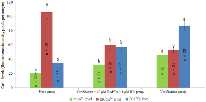
As shown in Fig. 4, the mCa2+ and [Ca2+]i levels of the vitrification + 10 μM BAPTA + 1 μM RR group were significantly lower than those of the vitrification group (P < 0.05) but still higher than those of the fresh group. Meanwhile, the ER Ca2+ level of the vitrification + 10 μM BAPTA + 1 μM RR group was similar to that of the vitrification group and lower than that of the fresh group (P < 0.05).
Figure 4.
Effect of BAPTA + RR on Ca2+ levels in vitrified bovine oocytes. a,b,cValues with different superscripts differ significantly between groups (P < 0.05).
Fig. S5 illustrates blastocysts stained with Hoechst-33342. As shown in Table 6, the percentages of cleaved oocytes and blastocysts and total cell number per blastocyst were higher in the vitrification + 10 μM BAPTA + 1 μM RR group (74.4 ± 5.2%, 30.1 ± 2.7%, 99.7 ± 8.4) than in the vitrification group (51.6 ± 4.4%, 10.4 ± 0.9%, 83.1 ± 6.4; P < 0.05); the values in the vitrification + 10 μM BAPTA + 1 μM RR group were similar to those in the fresh group (80.7 ± 7.8%, 34.4 ± 2.3%, 103.8 ± 9.6; P > 0.05).
Table 6.
Effect of BAPTA + RR treatment on the development of vitrified bovine oocytes after IVF.
| Groups | Number of oocytes | Number of cleaved oocytes (%) | Number of blastocysts (%) | Total cell number |
|---|---|---|---|---|
| Vitrification group | 896 | 462 (51.6 ± 4.4%)b | 48 (10.4 ± 0.9%)b | 83.1 ± 6.4b (n = 30) |
| Vitrification + 10 μM BAPTA + 1 μM RR group | 648 | 482 (74.4 ± 5.2%)a | 145 (30.1 ± 2.7%)a | 99.7 ± 8.4a (n = 30) |
| Fresh group | 612 | 494 (80.7 ± 7.8%)a | 170 (34.4 ± 2.3%)a | 103.8 ± 9.6a (n = 30) |
a,bValues with different superscripts differ significantly within the same column (P < 0.05).
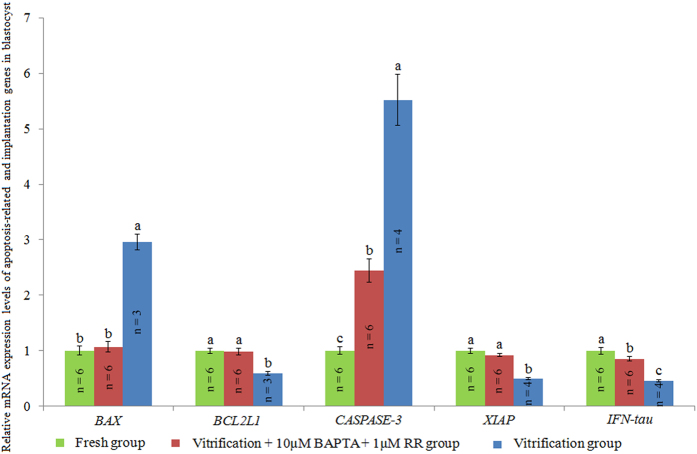
As shown in Fig. 5, the mRNA expression levels of anti-apoptotic genes BCL2L1 and XIAP and the pregnancy recognition signal gene IFN-tau were significantly higher in blastocysts of the vitrification + 10 μM BAPTA + 1 μM RR group than in those of the vitrification group, while the mRNA expression levels of pro-apoptosis genes BAX and CASPASE-3 were lower in blastocysts of the vitrification + 10 μM BAPTA + 1 μM RR group than in those of the vitrification group.
Figure 5.
Effect of BAPTA + RR on the mRNA expression levels of apoptosis-related and implantation genes in blastocysts. a,b,cValues with different superscripts differ significantly between groups (P < 0.05).
Discussion
EG is a widely used CPA in vitrification solution; it causes Ca2+ influx across the plasma membrane from the oocyte culture medium, which results in an increase in the [Ca2+]i level9, 22. Therefore, DPBS without Ca2+ was used as the base medium to prepare vitrification solution in the present study to relieve Ca2+ influx from the medium into the cytoplasm of oocytes. In the present study, vitrification was found to significantly increase the [Ca2+]i level in bovine oocytes (Fig. 1B); this finding is similar to previous results reported in murine9, 22, 23 and feline24 oocytes. As we used DPBS without Ca2+ to prepare vitrification solution in our study, the increase in the [Ca2+]i level may be partially due to Ca2+ release induced by vitrification from internal Ca2+ pools in oocytes. Furthermore, our results showed that DMSO, and not EG, induced the increase in the [Ca2+]i level in vitrified oocytes, indicating that DMSO increases [Ca2+]i by triggering Ca2+ release from the internal Ca2+ pools of oocytes, which confirmed the previous results of Larman et al.9, 22.
As shown in Fig. 1D, our experiment demonstrated that vitrification significantly decreased the ER Ca2+ level in bovine oocytes. Furthermore, our results proved that it was DMSO, and not EG, that induced Ca2+ release from ER in vitrified oocytes, which was due to the nonspecific effect of DMSO on the ER membrane. DMSO also triggered premature Ca2+ release from the ER9. Interestingly, our experiment showed that vitrification significantly increased the mCa2+ level in bovine oocytes (Fig. 1F). Theoretically, the mCa2+ level in vitrified oocytes should be reduced as DMSO has a nonspecific effect on the mitochondrial membrane, inducing the release of mCa2+ 9. The increased mCa2+ level in vitrified oocytes was due to Ca2+ influx from the ER to the cytoplasm, which promotes Ca2+ flow into the mitochondria25, 26. Taken together with these results, our experiments provided strong evidence showing that vitrification, especially with DMSO, significantly induced Ca2+ release from the ER, leading to abnormal increases of [Ca2+]i and mCa2+ in oocytes.
As shown in Fig. 2, our results showed that BAPTA did not influence mCa2+ or ER Ca2+ levels, but it significantly decreased [Ca2+]i levels in vitrified bovine oocytes because BAPTA is an effective chelator of [Ca2+]i27. During oocyte maturation, CGs translocate from the cytoplasm to the adjacent plasma membrane and represent the maturation of the cytoplasm28, 29. As shown in Table 1, our results showed that vitrification significantly decreased the percentage of oocytes with a peripheral distribution of CGs and increased the percentages of oocytes with discontinuous distribution and completed release of CGs. This effect is similar to a phenomenon observed previously in vitrified ovine oocytes30 and human oocytes31, 32. These results may be due to the increased [Ca2+]i level in vitrified bovine oocytes, which can trigger the release of CGs to the cytoplasm28, 33. Meanwhile, the percentage of vitrified oocytes with peripheral distribution of CGs was significantly increased in all three BAPTA groups because BAPTA is capable of decreasing the [Ca2+]i levels of vitrified oocytes.
With regard to fertilisation capacity, our results showed that vitrification significantly decreased the percentage of monospermic oocytes and thus increased the percentages of polyspermic and unfertilised oocytes (Table 2), and this result was closely associated with a lower percentage of oocytes with peripheral distribution of CGs in the vitrification group, as it is necessary for prevention of polyspermy28, 33. Meanwhile, the higher percentage of vitrified oocytes with peripheral distribution of CGs could partly explain why the 10 µM BAPTA group had a higher percentage of monospermic and cleaved oocytes after IVF (Tables 2 and 3). However, treatment with BAPTA only was not sufficient to improve blastocyst development from vitrified oocytes (Table 3).
As shown in Fig. 3, our results demonstrated that RR treatment significantly decreased the mCa2+ level in vitrified bovine oocytes because RR blocks the influx of increased [Ca2+]i into the mitochondria of vitrified oocytes20. ΔΨm and ATP content are indicators of mitochondrial function34, 35. Similar to previous results reported for bovine36–38, mouse39 and porcine40 oocytes, our results showed that ΔΨm was significantly decreased in bovine oocytes by vitrification (Table 4). Our experiment also revealed that vitrification significantly reduced the ATP content of bovine oocytes, confirming previous results in bovine36, human41, rabbit42, murine23, and porcine43 oocytes. The plausible reason for the decreased ΔΨm and ATP content of vitrified bovine oocytes could be that mCa2+ overload leads to membrane permeability transition, resulting in rupture of the mitochondrial membrane44. The ΔΨm and ATP content were significantly increased in the RR-treated oocytes because RR is capable of inhibiting mCa2+ overload and protecting mitochondrial function, as shown in Table 4.
Confirming previous reports37, 43, our experiment also showed that vitrification significantly increased the percentage of TUNEL-positive oocytes (Table 4), which was due to their lower ∆Ψm, since loss of ∆Ψm could release proteins such as cytochrome c that are located in the intermembrane space, which then activates the caspase cascade that executes the apoptotic program45, 46. Meanwhile, the percentage of TUNEL-positive oocytes was significantly decreased in the RR groups as RR inhibits the influx of [Ca2+]i to the mitochondria and maintains their ΔΨm levels (Table 4).
Similar to previous results in murine9, porcine47, ovine48, and bovine36 oocytes, our results showed the cleavage and blastocyst rates of vitrified bovine oocytes were significantly reduced after parthenogenetic activation (PA) (Supplementary Methods 1.5, Table 5), which was due to the damaged mitochondrial function of vitrified oocytes37, 39, 43, 49. Meanwhile, RR treatment was found to increase the cleavage and blastocyst rates of vitrified bovine oocytes in the present experiment (Table 5) due to the increase of ΔΨm and ATP content mediated by RR.
Furthermore, our results showed that BAPTA + RR treatment significantly improved the cleavage and blastocyst rates of vitrified bovine oocytes, which were similar to those of the fresh group (Table 6). According to the results of Experiments 2 and 3, one possible reason for this result is that the combined effects of BAPTA and RR can increase the incidence of normal fertilisation and protect mitochondrial functions simultaneously.
The BAX protein promotes apoptosis by enhancing the release of cytochrome c from mitochondria50, while the BCL2L1 protein prevents apoptosis by inhibiting the release of cytochrome c from mitochondria51. The CASPASE-3 protein is responsible for the breakdown of cytosolic/nuclear proteins, resulting in apoptosis52, while the XIAP protein inhibits apoptosis via the inhibition of the CASPASE-3 protein53. As shown in Fig. 5, BAPTA + RR treatment significantly increased the mRNA expression levels of the anti-apoptotic genes (BCL2L1 and XIAP) and decreased the mRNA expression levels of the pro-apoptotic genes (BAX and CASPASE-3), indicating that BAPTA + RR treatment significantly decreased the blastocysts’ apoptotic index. The IFN-tau protein plays an important role in regulating implantation in bovine blastocysts54, and its expression level is widely used to evaluate the developmental competence of embryos55, 56. In the present study, the IFN-tau gene mRNA level (Fig. 5) and the total cell number per blastocyst were significantly increased (Table 6), indicating that treatment with BAPTA + RR significantly improved the quality of blastocysts derived from vitrified oocytes after IVF.
In conclusion, our study illustrated that vitrification, especially when DMSO was used in vitrification solution, caused Ca2+ release from the ER into the cytoplasm, resulting in the increased [Ca2+]i and mCa2+ levels observed in vitrified bovine oocytes. Meanwhile, combined treatment of RR and BAPTA was found capable of chelating [Ca2+]i and inhibiting [Ca2+]i influx into the mitochondria, thus preventing the premature release of CGs and protecting mitochondrial function, finally improving the fertilisation capacity and developmental ability of vitrified oocytes. Our results will help to illustrate the approach by which vitrification increases [Ca2+]i in vitrified oocytes and improves their fertilisation capacity and developmental ability after IVF.
Materials and Methods
Unless otherwise indicated, all chemicals and media used in the present study were purchased from Sigma Chemical Co. (St. Louis, MO, USA), and all plasticware was obtained from Nunc-ware (Nunc; Nalge Nunc International, Roskilde, Denmark).
Ethics statement
All experimental procedures were conducted exactly according to the guidelines for the Care and Use of Laboratory Animals issued by the Animal Ethics Committee of the Institute of Special Animal and Plant Sciences, Chinese Academy of Agricultural Sciences (Permit Number: 2014-0035). The study protocol was approved by the same committee before the start of our experiments.
Measurement of [Ca2+]i, ER Ca2+, and mCa2+ levels
The [Ca2+]i level was detected by immunostaining as described in a previous report57. Briefly, oocytes were incubated in presence of 5 μM Fluo-3/AM (Invitrogen, NY, CA, USA) for 30 min at 38.5 °C, followed by three washes in Dulbecco’s phosphate-buffered saline (DPBS without Ca2+). Then, the oocytes were observed under an Olympus fluorescence microscope (Tokyo, Japan) equipped with a CoolSNAP HQ CCD camera (Photometrics/Roper Scientific, Inc., Tucson, AZ, USA). According to the method described by Kim et al.58, Nikon EZ-C1 FreeViewer software (Nikon, Tokyo, Japan) was used to analyse the [Ca2+]i fluorescence intensity of oocytes. The fluorescence pixel values within a constant area from ten different cytoplasmic regions were measured with background fluorescence values subtracted from the final values.
ER Ca2+ staining was performed according to the instructions of the ER Ca2+ detection kit (GMS10267, Genmed Scientifics Inc., Arlington, MA, USA). Briefly, oocytes were washed thrice in 100 μL cleaning medium, treated with permeable solution for 4 min at 38.5 °C, and then incubated in 100 μL staining medium for 1 h at 38.5 °C. After the oocytes were washed in 100 μL cleaning medium, they were examined under a camera-equipped fluorescence microscope, and fluorescence intensity was analysed as described above. The wavelength of excitation light was 490 nm, and the wavelength of emission light was 525 nm.
mCa2+ detection was performed according to the instructions of mCa2+ detection kits (GMS10153, Genmed Scientifics Inc., Arlington, MA, USA). The procedures for the mCa2+ staining and analysis were similar to those described for staining and analysis of ER Ca2+ levels. The wavelength of excitation light was 550 nm, and the wavelength of emission light was 590 nm.
Analysis of CG distribution
Briefly, oocytes were treated with 0.5% (w/v) pronase at 38.5 °C for 2 min to remove ZP, fixed in 3.7% (w/v) paraformaldehyde at room temperature for 30 min, and washed thrice in 0.5% bovine serum albumin (BSA) blocking solution. After being permeabilised in 0.1% (v/v) Triton X-100 in DPBS (without Ca2+) for 5 min, the oocytes were washed three times in blocking solution and stained with 50 μg/mL fluorescein isothiocyanate conjugated to lens culinaris agglutinin (FITC-LCA) at 38.5 °C for 30 min. Finally, the oocytes were washed three times in DPBS (without Ca2+), fixed, and observed using confocal microscopy (Nikon, Tokyo, Japan). The distribution of CGs was divided into four categories according to the method described by Tian et al.30: periphery, discontinuous periphery, completed release, and homogeneous distribution.
Determination of ΔΨm level and ATP content
JC-1 dye (Molecular Probes, Eugene, OR, USA) was used to measure the ΔΨm of the oocytes according to the procedure described by Smiley et al.59. Oocytes were incubated with 10 µg/mL JC-1 at 38.5 °C in 5% CO2 for 10 min, after which they were washed in DPBS (without Ca2+) to remove surface fluorescence, and the oocytes were observed under a confocal microscope (Nikon). Laser excitation was set at 488 nm, and emission channels of 515/30 and 650 LP were used to detect green and red fluorescence, respectively. The fluorescence intensity was quantified as described above, and ΔΨm was used as an indicator of mitochondrial function and analysed using the ratio of red to green fluorescence34.
A bioluminescence assay kit (ATP Bioluminescence Assay Kit HS II, Roche Diagnostics GmbH, Mannheim, Germany) was used to detect the ATP content of oocytes as described by Van Blerkom et al.60. Before analysis, oocytes were treated with 0.5% (w/v) pronase for 2 min to remove ZP. Then, 20-μL aliquots of cell lysis reagent were added to 0.5-mL centrifuge tubes containing 10 oocytes, and the ZP-free oocytes were homogenised and lysed by repeated pipetting. Meanwhile, a standard curve containing 0.01–10.0 pmol ATP was generated for each series of analyses. Then, 100 μL of ATP detection solution was added to wells of 96-well dishes and equilibrated for 3–5 min. Finally, standard solutions and samples (20 μL) were added to each well and luminescence was immediately measured using a luminometer (InfiniteM200, Tecan Group Ltd., Untersbergstrasse, Austria) for 10 s. The ATP content of the samples was determined from a standard curve.
Detection of DNA fragmentation by TUNEL
A TUNEL detection kit (Roche, Indianapolis, IN, USA) was used to detect DNA fragmentation according to the manufacturer’s instructions. After three washes in DPBS without Ca2+, oocytes were first fixed in 4% paraformaldehyde solution for 40 min and then treated with 0.1% Triton X-100 solution for 40 min at room temperature. After being exposed to blocking solution (0.5% BSA in DPBS without Ca2+) at 4 °C overnight, the oocytes were stained with TUNEL solution at 38.5 °C for 1 h, then incubated in 50 µg/mL propidium iodide (PI) solution for 10 min to stain the nuclei of membrane-damaged cells. Finally, they were examined under a confocal microscope (Nikon) with the nuclei of TUNEL-positive oocytes stained yellowish green and normal oocyte nuclei stained orange-red. For the positive control, oocytes were treated with 50 µg/mL of DNase I solution for 1 h and then stained with TUNEL.
Experimental design
Experiment 1: Effect of vitrification on Ca2+ levels in bovine oocytes
Oocytes were divided into five groups: the fresh group contained untreated oocytes; the toxicity group consisted of oocytes exposed to pretreatment solution (Supplementary Methods 1.3) for 30 s, vitrification solution for 25 s, rinsed in 0.25 M sucrose solution for 1 min at 38.5 °C, followed by rinsing in 0.15 M sucrose for 5 min; the EG group consisted of oocytes exposed to 10% EG for 10 min; the DMSO group consisted of oocytes exposed to 10% DMSO for 10 min; the vitrification group consisted of oocytes that were vitrified as described in the supplementary information (Supplementary Methods 1.2, 1.3 and 1.4). The [Ca2+]i, ER Ca2+, and mCa2+ levels of these five groups were then analysed. The experiments were all biologically repeated three times.
Experiment 2: Effect of BAPTA treatment on Ca2+ levels and the developmental ability of vitrified bovine oocytes
A stock solution of BAPTA (A1076, Sigma; 30 mM in DMSO) was diluted to the final concentration used in the experiment according to the experimental design. The medium was pre-incubated for 2 h after BAPTA addition. After in vitro maturation (IVM) (Supplementary Methods 1.1) for 20–22 h, cumulus-oocyte complexes (COCs) were treated with 0.1% (w/v) hyaluronidase for 1–2 min to remove cumulus cells; cultured in IVM medium (Supplementary Methods 1.1) supplemented with 5, 10, or 20 μM BAPTA for 2 h; and vitrified with vitrification solution (Supplementary Methods 1.3) containing 5, 10, or 20 μM BAPTA. Untreated fresh oocytes were used as the fresh group, and those that were vitrified without BAPTA were used as the vitrification group. Oocytes from these five groups (fresh, vitrification, 5 µM BAPTA, 10 µM BAPTA, and 20 µM BAPTA) were used to detect [Ca2+]i, ER Ca2+, and mCa2+ levels; CG distribution; and the fertilisation and developmental capacity of vitrified bovine oocytes after IVF (Supplementary Methods 1.6). The whole experiment was biologically repeated three times.
Experiment 3: Effect of RR on Ca2+ levels and developmental potential of vitrified bovine oocytes
A stock solution of RR (R2751, Sigma; 10 mM in DPBS without Ca2+) was diluted to the final concentration used in the experiment according to the experimental design. The medium was pre-incubated for 2 h after RR addition. To investigate the effect of RR on Ca2+ levels and the developmental potential of vitrified bovine oocytes, bovine oocytes were vitrified using vitrification solution containing 0.5, 1, or 2 μM RR and incubated in IVM medium containing 0.5, 1, or 2 μM RR for 0.5 h after thawing in a CO2 incubator (5% CO2, 38.5 °C, humidified air). Untreated fresh bovine oocytes were used as the fresh group, and those that were vitrified without RR belonged to the vitrification group. The [Ca2+]i, mCa2+, ER Ca2+, ATP, ΔΨm, DNA fragmentation, and developmental potential after PA (Supplementary Methods 1.5) of these five groups were examined. The experiments for Ca2+ staining, ΔΨm, DNA fragmentation and development potential of vitrified bovine oocytes were all biologically repeated three times, and the experiment for ATP content was biologically repeated four times.
Experiment 4: Combined effect of BAPTA and RR on the developmental ability of vitrified bovine oocytes after IVF
After IVM for 20–22 h, COCs were treated in 0.1% (w/v) hyaluronidase for 1–2 min to remove cumulus cells and cultured in IVM medium supplemented with 10 μM BAPTA for 2 h. Then, the oocytes were vitrified using vitrification solution containing 1 μM RR and 10 μM BAPTA and incubated in IVM medium containing 1 μM RR for 0.5 h after thawing. Untreated fresh bovine oocytes were used as the fresh group, and those that were vitrified without BAPTA and RR belonged to the vitrification group. The Ca2+ levels ([Ca2+]i, ER Ca2+, and mCa2+), fertilisation capacity and developmental ability (Supplementary Methods 1.6, 1.7 and 1.8) of vitrified bovine oocytes from these three groups were evaluated. The IVF experiment was biologically repeated six times, and the Ca2+ level analysis of oocytes, the total cell number counting and qRT-PCR experiments on blastocysts were all biologically repeated three times.
Statistical analysis
All the results are presented as the mean ± standard error. Percentage data were previously checked for normality and homogeneity of variance followed by Duncan’s test. A statistical package (SAS Software; SAS Institute, Cary, NC, USA) was utilized to analyse one-way analysis of variance. P < 0.05 was considered statistically significant.
Electronic supplementary material
Acknowledgements
This work was supported by the National Natural Science Foundation of China (31472100), the Foundation of Chinese Academy of Agricultural Sciences (Y2017JC54) and the Agricultural Science and Technology Innovation Program (ASTIP-IAS-TS-5, ASTIP-IAS06).
Author Contributions
X.-M. Zhao and H.-B. Zhu designed the study and drafted the paper. N. Wang, H.-S. Hao, C.-Y. Li, Y.-H. Zhao, H.-Y. Wang, C.-L. Yan, W.-H. Du, D. Wang, Y. Liu, and Y.-W. Pang performed the experiment, analysed the data and wrote the paper. All authors read and approved the final version of the manuscript.
Competing Interests
The authors declare that they have no competing interests.
Footnotes
Na Wang, Hai-Sheng Hao and Chong-Yang Li contributed equally to this work.
Electronic supplementary material
Supplementary information accompanies this paper at doi:10.1038/s41598-017-10907-9
Publisher's note: Springer Nature remains neutral with regard to jurisdictional claims in published maps and institutional affiliations.
References
- 1.Wininger JD, Kort HI. Cryopreservation of immature and mature human oocytes. Semin Reprod Med. 2002;20:45–49. doi: 10.1055/s-2002-23519. [DOI] [PubMed] [Google Scholar]
- 2.Chang CC, et al. Nuclear transfer and oocyte cryopreservation. Reprod Fertil Dev. 2009;21:37–44. doi: 10.1071/RD08218. [DOI] [PubMed] [Google Scholar]
- 3.Canesin HS, et al. Blastocyst development after intracytoplasmic sperm injection of equine oocytes vitrified at the germinal-vesicle stage. Cryobiology. 2017;75:52–59. doi: 10.1016/j.cryobiol.2017.02.004. [DOI] [PubMed] [Google Scholar]
- 4.Kim BY, et al. Alterations in calcium oscillatory activity in vitrified mouse eggs impact on egg quality and subsequent embryonic development. Pflugers Arch. 2011;461:515–526. doi: 10.1007/s00424-011-0955-0. [DOI] [PubMed] [Google Scholar]
- 5.Levi Setti PE, et al. Human oocyte cryopreservation with slow freezing versus vitrification. Results from the National Italian Registry data, 2007-2011. Fertil Steril. 2014;102:90–95. doi: 10.1016/j.fertnstert.2014.03.052. [DOI] [PubMed] [Google Scholar]
- 6.Paramanantham J, Talmor AJ, Osianlis T, Weston GC. Cryopreserved oocytes: update on clinical applications and success rates. Obstet Gynecol Surv. 2015;70:97–114. doi: 10.1097/OGX.0000000000000152. [DOI] [PubMed] [Google Scholar]
- 7.Gunnala V, Schattman G. Oocyte vitrification for elective fertility preservation: the past, present, and future. Curr Opin Obstet Gynecol. 2017;29:59–63. doi: 10.1097/GCO.0000000000000339. [DOI] [PubMed] [Google Scholar]
- 8.Leibo SP, Pool TB. The principal variables of cryopreservation: solutions, temperatures, and rate changes. Fertil Steril. 2011;96:269–276. doi: 10.1016/j.fertnstert.2011.06.065. [DOI] [PubMed] [Google Scholar]
- 9.Larman MG, Sheehan CB, Gardner DK. Calcium-free vitrification reduces cryoprotectant-induced zona pellucida hardening and increases fertilization rates in mouse oocytes. Reproduction. 2006;131:53–61. doi: 10.1530/rep.1.00878. [DOI] [PubMed] [Google Scholar]
- 10.Combelles CM. & Chateau, G. The use of immature oocytes in the fertility preservation of cancer patients: current promises and challenges. Int J Dev Biol. 2012;56:919–929. doi: 10.1387/ijdb.120132cc. [DOI] [PubMed] [Google Scholar]
- 11.Fesahat F, et al. Vitrification of mouse MII oocytes: Developmental competency using paclitaxel. Taiwan J Obstet Gynecol. 2016;55:796–800. doi: 10.1016/j.tjog.2016.05.010. [DOI] [PubMed] [Google Scholar]
- 12.Gu R, et al. Improved cryotolerance and developmental competence of human oocytes matured in vitro by transient hydrostatic pressure treatment prior to vitrification. Cryobiology. 2017;75:144–150. doi: 10.1016/j.cryobiol.2016.12.009. [DOI] [PubMed] [Google Scholar]
- 13.Kline D, Kline JT. Repetitive calcium transients and the role of calcium in exocytosis and cell cycle activation in the mouse egg. Dev Biol. 1992;149:80–89. doi: 10.1016/0012-1606(92)90265-I. [DOI] [PubMed] [Google Scholar]
- 14.Sun QY. Cellular and molecular mechanisms leading to cortical reaction and polyspermy block in mammalian eggs. Microsc Res Tech. 2003;61:342–348. doi: 10.1002/jemt.10347. [DOI] [PubMed] [Google Scholar]
- 15.Wakai T, Fissore RA. Ca(2 + ) homeostasis and regulation of ER Ca(2 + ) in mammalian oocytes/eggs. Cell Calcium. 2013;53:63–67. doi: 10.1016/j.ceca.2012.11.010. [DOI] [PMC free article] [PubMed] [Google Scholar]
- 16.Kim B, et al. The role of MATER in endoplasmic reticulum distributeon and calcium homeostasis in mouse oocytes. Dev Biol. 2014;386:331–339. doi: 10.1016/j.ydbio.2013.12.025. [DOI] [PMC free article] [PubMed] [Google Scholar]
- 17.Duchen MR. Mitochondria and calcium: from cell signalling to cell death. J Physiol. 2000;529:57–68. doi: 10.1111/j.1469-7793.2000.00057.x. [DOI] [PMC free article] [PubMed] [Google Scholar]
- 18.Ricci AJ, Wu YC, Fettiplace R. The endogenous calcium buffer and the time course of transducer adaptation in auditory hair cells. J Neurosci. 1998;18:8261–8277. doi: 10.1523/JNEUROSCI.18-20-08261.1998. [DOI] [PMC free article] [PubMed] [Google Scholar]
- 19.Baliño P, Monferrer L, Pastor R, Aragon CM. Intracellular calcium chelation with BAPTA-AM modulates ethanol-induced behavioral effects in mice. Exp Neurol. 2012;234:446–453. doi: 10.1016/j.expneurol.2012.01.016. [DOI] [PubMed] [Google Scholar]
- 20.Bae JH, Park JW, Kwon TK. Ruthenium red, inhibitor of mitochondrial Ca2+ uniporter, inhibits curcumin-induced apoptosis via the prevention of intracellular Ca2 + depletion and cytochrome c release. Biochem Biophys Res Commun. 2003;303:1073–1079. doi: 10.1016/S0006-291X(03)00479-0. [DOI] [PubMed] [Google Scholar]
- 21.Nakagawa S, Yoneda A, Hayakawa K, Watanabe T. Improvement in the in vitro maturation rate of porcine oocytes vitrified at the germinal vesicle stage by treatment with a mitochondrial permeability transition inhibitor. Cryobiology. 2008;57:269–275. doi: 10.1016/j.cryobiol.2008.09.008. [DOI] [PubMed] [Google Scholar]
- 22.Larman MG. Katz-, M. G., Sheehan, C. B. & Gardner, D. K. 1, 2- propanediol and the type of cryopreservation procedure adversely affect mouse oocyte physiology. Hum Reprod. 2007;22:250–259. doi: 10.1093/humrep/del319. [DOI] [PubMed] [Google Scholar]
- 23.Salehnia M, Töhönen V, Zavareh S, Inzunza J. Does cryopreservation of ovarian tissue affect the distribution and function of germinal vesicle oocytes mitochondria? Biomed Res Int. 2013;2013 doi: 10.1155/2013/489032. [DOI] [PMC free article] [PubMed] [Google Scholar]
- 24.Herrick JR, Wang C, Machaty Z. The effects of permeating cryoprotectants on intracellular free-calcium concentrations and developmental potential of in vitro-matured feline oocytes. Reprod Fertil Dev. 2016;28:599–607. doi: 10.1071/RD14233. [DOI] [PubMed] [Google Scholar]
- 25.Murphy MP. How mitochondria produce reactive oxygen species. Biochem J. 2009;417:1–13. doi: 10.1042/BJ20081386. [DOI] [PMC free article] [PubMed] [Google Scholar]
- 26.Dolai S, Pal S, Yadav RK, Adak S. Endoplasmic reticulum stress-induced apoptosis in Leishmania through Ca2 + -dependent and caspase-independent mechanism. J Biol Chem. 2011;286:13638–13646. doi: 10.1074/jbc.M110.201889. [DOI] [PMC free article] [PubMed] [Google Scholar]
- 27.Takahashi T, et al. Lowering intracellular and extracellular calcium contents prevents cytotoxic effects of ethylene glycol-based vitrification solution in unfertilized mouse oocytes. Mol Reprod Dev. 2004;68:250–258. doi: 10.1002/mrd.20073. [DOI] [PubMed] [Google Scholar]
- 28.Tahara M, et al. Dynamics of cortical granule exocytosis at fertilization in living mouse eggs. Am J Physiol. 1996;270:C1354–1361. doi: 10.1152/ajpcell.1996.270.5.C1354. [DOI] [PubMed] [Google Scholar]
- 29.Mo X, et al. Leukemia inhibitory factor enhances bovine oocyte maturation and early embryo development. Mol Reprod Dev. 2014;81:608–618. doi: 10.1002/mrd.22327. [DOI] [PubMed] [Google Scholar]
- 30.Tian SJ, et al. Vitrification solution containing DMSO and EG can induce parthenogenetic activation of in vitro matured ovine oocytes and decrease sperm penetration. Anim Reprod Sci. 2007;101:365–371. doi: 10.1016/j.anireprosci.2007.01.007. [DOI] [PubMed] [Google Scholar]
- 31.Gualtieri R, et al. Ultrastructure and intracellular calcium response during activation in vitrified and slow-frozen human oocytes. Hum Reprod. 2011;26:2452–2460. doi: 10.1093/humrep/der210. [DOI] [PubMed] [Google Scholar]
- 32.Palmerini MG, et al. Ultrastructure of immature and mature human oocytes after cryotop vitrification. J Reprod Dev. 2014;60:411–420. doi: 10.1262/jrd.2014-027. [DOI] [PMC free article] [PubMed] [Google Scholar]
- 33.Ducibella T, Fissore R. The roles of Ca2 + , downstream protein kinases, and oscillatory signaling in regulating fertilization and the activation of development. Dev Biol. 2008;315:257–279. doi: 10.1016/j.ydbio.2007.12.012. [DOI] [PMC free article] [PubMed] [Google Scholar]
- 34.Xu JS, Chan ST, Ho PC, Yeung WS. Coculture of human oviductal cells maintains mitochondrial function and decreases caspase activity of cleavage stage mouse embryos. Fertil Steril. 2003;80:178–83. doi: 10.1016/S0015-0282(03)00570-3. [DOI] [PubMed] [Google Scholar]
- 35.Park J, et al. Involvement of S6K1 in mitochondria function and structure in HeLa cells. Cell Signal. 2016;28:1904–1915. doi: 10.1016/j.cellsig.2016.09.003. [DOI] [PubMed] [Google Scholar]
- 36.Zhao XM, et al. Effect of cyclosporine pretreatment on mitochondrial function in vitrified bovine mature oocytes. Fertil Steril. 2011;95:2786–2788. doi: 10.1016/j.fertnstert.2011.04.089. [DOI] [PubMed] [Google Scholar]
- 37.Zhao XM, et al. Melatonin inhibits apoptosis and improves the developmental potential of vitrified bovine oocytes. J Pineal Res. 2016;60:132–141. doi: 10.1111/jpi.12290. [DOI] [PubMed] [Google Scholar]
- 38.Liang S, et al. Effect of antifreeze glycoprotein 8 supplementation during vitrification on the developmental competence of bovine oocytes. Theriogenology. 2016;86:485–494. doi: 10.1016/j.theriogenology.2016.01.032. [DOI] [PubMed] [Google Scholar]
- 39.Yan CL, et al. Mitochondrial behaviors in the vitrified mouse oocyte and its parthenogenetic embryo: effect of Taxol pretreatment and relationship to competence. Fertil Steril. 2010;93:959–966. doi: 10.1016/j.fertnstert.2008.12.045. [DOI] [PubMed] [Google Scholar]
- 40.Niu Y, et al. The application of apoptotic inhibitor in apoptotic pathways of MII stage porcine oocytes after vitrification. Reprod Domest Anim. 2016;51:953–959. doi: 10.1111/rda.12772. [DOI] [PubMed] [Google Scholar]
- 41.Manipalviratn S, et al. Effect of vitrification and thawing on human oocyte ATP concentration. Fertil Steril. 2011;95:1839–1841. doi: 10.1016/j.fertnstert.2010.10.040. [DOI] [PubMed] [Google Scholar]
- 42.Salvetti P, et al. Structural, metabolic and developmental evaluation of ovulated rabbit oocytes before and after cryopreservation by vitrification and slow freezing. Theriogenology. 2010;74:847–855. doi: 10.1016/j.theriogenology.2010.04.009. [DOI] [PubMed] [Google Scholar]
- 43.Dai J, et al. Changes in mitochondrial function in porcine vitrified MII-stage oocytes and their impacts on apoptosis and developmental ability. Cryobiology. 2015;71:291–298. doi: 10.1016/j.cryobiol.2015.08.002. [DOI] [PubMed] [Google Scholar]
- 44.Hajnóczky G, et al. Mitochondrial calcium signalling and cell death: approaches for assessing the role of mitochondrial Ca2 + uptake in apoptosis. Cell Calcium. 2006;40:553–560. doi: 10.1016/j.ceca.2006.08.016. [DOI] [PMC free article] [PubMed] [Google Scholar]
- 45.Green DR, Kroemer G. The pathophysiology of mitochondrial cell death. Science. 2004;305:626–629. doi: 10.1126/science.1099320. [DOI] [PubMed] [Google Scholar]
- 46.Suen DF, Norris KL, Youle RJ. Mitochondrial dynamics and apoptosis. Genes Dev. 2008;22:1577–1590. doi: 10.1101/gad.1658508. [DOI] [PMC free article] [PubMed] [Google Scholar]
- 47.Somfai T, et al. Developmental competence of in vitro-fertilized porcine oocytes after in vitro maturation and solid surface vitrification: effect of cryopreservation on oocyte antioxidative system and cell cycle stage. Cryobiology. 2007;55:115–126. doi: 10.1016/j.cryobiol.2007.06.008. [DOI] [PubMed] [Google Scholar]
- 48.Succu S, et al. Calcium concentration in vitrification medium affects the developmental competence of in vitro matured ovine oocytes. Theriogenology. 2011;75:715–721. doi: 10.1016/j.theriogenology.2010.10.012. [DOI] [PubMed] [Google Scholar]
- 49.Lei T, Guo N, Tan MH, Li YF. Effect of mouse oocyte vitrification on mitochondrial membrane potential and distribution. J Huazhong Univ Sci Technolog Med Sci. 2014;34:99–102. doi: 10.1007/s11596-014-1238-8. [DOI] [PubMed] [Google Scholar]
- 50.Adams JM, Cory S. The Bcl-2 protein family: arbiters of cell survival. Science. 1998;281:1322–1326. doi: 10.1126/science.281.5381.1322. [DOI] [PubMed] [Google Scholar]
- 51.Tan Y, Demeter MR, Ruan H, Comb MJ. BAD Ser-155 phosphor-ylation regulates BAD/Bcl-XL interaction and cell survival. J Biol Chem. 2000;275:25865–25869. doi: 10.1074/jbc.M004199200. [DOI] [PubMed] [Google Scholar]
- 52.Thornberry NA, Lazebnik Y. Caspases: enemies within. Science. 1998;281:1312–1316. doi: 10.1126/science.281.5381.1312. [DOI] [PubMed] [Google Scholar]
- 53.Deveraux QL, Takahashi R, Salvesen GS, Reed JC. X-linked IAP is a direct inhibitor of cell-death proteases. Nature. 1997;388:300–304. doi: 10.1038/40901. [DOI] [PubMed] [Google Scholar]
- 54.Roberts RM, Cross JC, Leaman DW. Interferons as hormones of pregnancy. Endocrine Rev. 1992;13:432–452. doi: 10.1210/edrv-13-3-432. [DOI] [PubMed] [Google Scholar]
- 55.Rizos D, Pintado B, de la Fuente J, Lonergan P, Gutiérrez-Adán A. Development and pattern of mRNA relative abundance of bovine embryos cultured in the isolated mouse oviduct in organ culture. Mol Reprod Dev. 2007;74:716–723. doi: 10.1002/mrd.20652. [DOI] [PubMed] [Google Scholar]
- 56.Yao N, et al. Expression of interferon-tau mRNA in bovine embryos derived from different procedures. Reprod Domest Anim. 2009;44:132–139. doi: 10.1111/j.1439-0531.2007.01009.x. [DOI] [PubMed] [Google Scholar]
- 57.Liang SL, et al. Dynamic analysis of Ca2 + level during bovine oocytes maturation and early embryonic development. J Vet Sci. 2011;12:133–142. doi: 10.4142/jvs.2011.12.2.133. [DOI] [PMC free article] [PubMed] [Google Scholar]
- 58.Kim JM, Liu H, Tazaki M, Nagata M, Aoki F. Changes in histone acetylation during mouse oocyte meiosis. J Cell Biol. 2003;162:37–46. doi: 10.1083/jcb.200303047. [DOI] [PMC free article] [PubMed] [Google Scholar]
- 59.Smiley ST, et al. Intracellular heterogeneity in mitochondrial membrane potentials revealed by a J-aggregate-forming lipophilic cation JC-1. Proc Natl Acad Sci U S A. 1991;88:3671–3675. doi: 10.1073/pnas.88.9.3671. [DOI] [PMC free article] [PubMed] [Google Scholar]
- 60.Van B, Davis J. P. W. & Lee, J. ATP content of human oocytes and developmental potential and outcome after in-vitro fertilization and embryo transfer. Hum Reprod. 1995;10:415–424. doi: 10.1093/oxfordjournals.humrep.a135954. [DOI] [PubMed] [Google Scholar]
Associated Data
This section collects any data citations, data availability statements, or supplementary materials included in this article.


