Abstract
Litchi is a tasty fruit that is commercially grown for food consumption and nutritional benefits in various parts of the world. Due to its biological activities, the fruit is becoming increasingly known and deserves attention not only for its edible part, the pulp, but also for its peel and seed that contain beneficial substances with antioxidant, cancer preventive, antimicrobial, and anti-inflammatory functions. Although literature demonstrates the biological activity of Litchi components in reducing tumor cell viability in in vitro or in vivo models, data about the biochemical mechanisms responsible for these effects are quite fragmentary. This review specifically describes, in a comprehensive analysis, the antitumor properties of the different parts of Litchi and highlights the main biochemical mechanisms involved.
Keywords: Litchi chinensis fruit extracts, nutraceutical properties, antitumor activity
1. Introduction
Litchi chinensis Sonnerat (also known as Chinese cherry, lychee nut, leechee, etc.) is a fruit tree belonging to Sapindaceae family. It was originally cultivated in China for more than 2300 years and in northern Vietnam. Some cultivars in the west of Guangdong region in China have a long history of cultivation, while others are relatively new. It has been reported that cultivars such as Sum Yee Hong, Haak Yip, Kwai May, No Mai Chee, Wai Chee, and Seong Sue Wai date back to 500 or 600 years or more, while others, such as Bah Lup, Heong Lai and Tim Naan, or Souey Tung are relatively young cultivars (about 100–300 years old) [1,2,3].
Traditionally, Litchi cultivar characterization was based on morphological studies focused on traits such as floral and fruit characteristics and the harvest season [4]. However, despite Litchi cultivation being diffused in many tropical and sub-tropical regions in the world, this approach is limited because it does not evaluate the interaction of morphological traits with environmental conditions [5]. Only recently, a study was undertaken to identify Litchi cultivars and their genetic relationships using new generation molecular approaches such as single nucleotide polymorphism (SNP) markers [6]. This strategy has allowed for cultivar standardization nomenclature facilitating the integration and interpretation of Litchi germoplasm.
Today, Litchi is mainly present in many countries of Southeast Asia, the Indian subcontinent, and South Africa, and in other tropical or sub-tropical areas around the world even if China, India, Thailand, and Vietnam are the leading Litchi-producing countries in the world. Recently, its cultivation, together with other sub-tropical orchards [7], has been also launched in Southern Europe, such as in Spain and more recently in Sicily, a Southern Italian island, where the climatic conditions (cold winter, especially at the beginning of flowering and the hot humid climate during the growth of the fruit) have been proved to be favorable for this crop. In Sicily region the most diffused cultivars are Wai-Chee, Kwai May, and Mclaine [8].
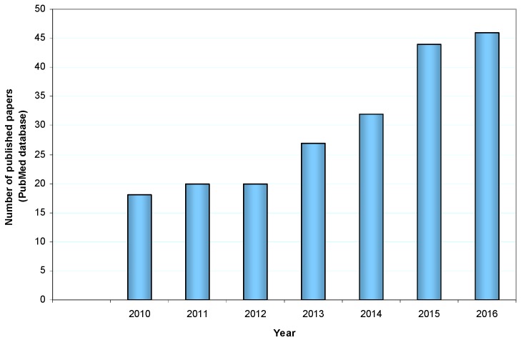
The interest for this crop is associated to the goodness of its fruits and the high nutritional value. Investigations demonstrated a long series of beneficial health compounds including antioxidant, cancer preventive, antimicrobial, anti-inflammatory activities, and so on, so that in 2012 Litchi chinensis was inserted in the list of functional foods [9]. In parallel to the identification of the bioactive components of Litchi fruit (but also of the other portions of the plant), in recent years the number of published articles showing the different biological activities of Litchi components has greatly increased (Figure 1).
Figure 1.
Number of papers on Litchi chinensis biological activities published in the last seven years (font PubMed database, https://www.ncbi.nlm.nih.gov/pubmed).
Beyond their healthy potential, some studies have also indicated that some parts of the fruit could find application in other commercial sectors. Discarded fruit peels can prove to be good source for dyeing textile material, a sort of application that may have a great commercial impact. Litchi peel extract has also been proven to act as a potential green inhibitor in the corrosion of mild steel [10].
2. The Plant
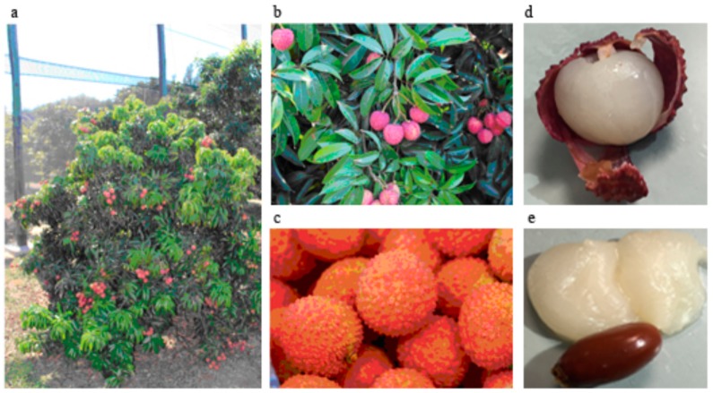
Litchi chinensis is an arboreal and evergreen tree; the bark is grey-black, the branches present shiny, lanceolate leaves of deep green color, and a brownish-red, dense and rounded-shape crown, which can be very large, up to 15–20 m in origin countries, while in other areas, such as in Sicily, comes to 6–8 m in height (Figure 2a). Flowers, which grow on a terminal inflorescence, are small, without corolla, and yellowish-white. Litchi fruits, which are similar in volume to a strawberry, are in pendulous clusters, roundish, green, and, once mature, become pinkish or reddish (Figure 2b,c). The fruits of Litchi have a thin and rigid peel that easily comes off to show a pearl-white jelly-like pulp with excellent flavor due to the combination of acids and sugars (Figure 2d,e). In tropical countries, the fruit reaches the maturation in the late autumn, while in Sicily it ripens in August.
Figure 2.
Sicilian Litchi chinensis tree, Kway May cultivar (a), Sicilian Litchi fruit. Kway May (b) and Way Chee (c) cultivars. The details of the pulp and seed are reported in (d,e), respectively (with the permission of Azienda Siciliana Cupitur, Caronia, Sicily).
Litchi fruit is rich in carbohydrates and fibers while lipids and proteins are scarce (Table 1). It is also appreciated for its nutritional properties: in fact the fruit is very rich in nutraceuticals, fundamental compounds with extra health benefits in addition to the basic nutritional value found in foods. Beyond the differences between cultivars of various origins, the beneficial effects of fruits have been partly related to their high contents in micronutrients including vitamins (B1, B2, B3, B6, C, E, K), carotenoids, minerals (potassium, copper, iron, magnesium, phosphorus, calcium, sodium, zinc, manganese, and selenium), and polyphenols (Table 1), which are highly represented in comparison with other tropical fruits [11,12].
Table 1.
Principal macronutrients and micronutrients of Litchi fruit.
| CALORIES | 66 kcal/100 g |
|---|---|
| MACROCOMPONENTS (g/100 g) | |
| Carbohydrates | 16.53 |
| Lipid | 0.44 |
| Protein | 0.83 |
| Dietary fiber | 1.30 |
| Water | 81.76 |
| MICROCOMPONENTS | |
| Total carotenoid content (μg beta-carotene equivalent/100 g) | 571.4 ± 117.2 |
| Vitamin C content (mg ascorbic acid equivalent/100 g) | 10.1 ± 2.2 |
| Total polyphenol content (mg gallic acid equivalent/100 g) | 178.0 ± 34.7 |
| Total flavonoid content (mg quercetin equivalent/100 g) | 53.3 ± 5.9 |
Nutritive value and the amounts of macrocomponents, referring to fresh fruit, are from the National Nutrient Database for Standard Reference (United States Department of Agriculture, Washington, DC, USA) [13], while those concerning the microcomponent amounts are from Septembre-Malaterre [12].
Studies evidenced that Litchi fruit from different cultivars may show significant qualitative and/or quantitative differences in their composition. By analyzing the total content of phenolics, flavonoids, anthocyanins, and procyanidins in the pericarp of nine commercially available Litchi cultivars, Li et al. [14] found a 3.2-fold difference in phenolic content between the highest and lowest Litchi varieties, Heiye and Chanchutou, respectively. Similar results were obtained comparing the levels of flavonoids and anthocyanins, while no difference seemed to be in the individual procyanidin composition of the different Litchi varieties analyzed. These results indicate that the significant differences in phytochemical profiles among the varieties should be considered for their potential application to promote health.
3. Litchi Fruit as a Functional Food
In recent years, medicine has started to pay great attention to functional food, which displays an additional function related to health promotion or disease prevention. Since it is well known that particular dietary factors and lifestyle can promote cancer development, research is increasingly betting on some protective components of vegetables and fruits or phytocompounds enriched with antitumor properties that can exert cancer prevention or even antitumor properties. The tumor-protective effects of dietary factors are most likely mediated by multiple signaling pathways, consistent with the heterogeneous nature of the disease and the distinct genetic profiles of different tumors. The effects induced by molecules with antitumor activity are often related to cell cycle arresting ability or tumor targeting pro-apoptotic action which might be dependent on unbalanced redox equilibrium. Even though the relationship between the redox state in tumor cells and anticancer response is quite complicated and sometimes controversial, it is known that some agents, that are capable of inducing oxidative stress, can promote tumor cell death [15]. On the other hand, there is ample evidence that phytochemicals with antioxidant properties can behave as potent antitumor agents [16,17]. In this scenario, Litchi fruit is currently emerging as a potential functional food due to its important nutraceutical properties and chemical composition, which includes specific components endowed with antioxidant as well as anticancer activities. It is important to emphasize that not only the pulp, which is the only edible part of the fruit, contains bioactive compounds that can exert anti-proliferative effects in tumor cells, but also the peel and the seed are enriched with substances that are potentially beneficial and endowed with antitumor properties. Their isolation and characterization can thus open up the possibility to produce novel active principles with a potential application in cancer therapy.
Recently, Ibrahim and Mohamed [18] exhaustively reviewed chemical constituents and pharmacological activities of Litchi chinensis, listing the single constituents identified over the past few decades. Here, among the multiple biological activities of the different portions of the Litchi fruit, we specifically highlight the major findings in the literature on the anticancer properties. In particular, the next sections of this review describe the composition of the different parts of Litchi fruit and report the effects of their components in different in vitro and in vivo tumor models. It has to be considered, however, that in many papers antitumor properties have been attributed to Litchi crude extracts of the different portions and only in some cases to single biochemical compounds that have been isolated and characterized. Moreover, a certain part of the literature only refers to anti-proliferative effects of Litchi extracts but poorly describes the biochemical mechanisms involved. We thus made an effort to critically summarize in this review the most significant contributions to the elucidation of the biochemical effects induced by particular components of the Litchi portions.
4. Antitumor Properties of Litchi Pulp-Derived Components
The fresh Litchi fruit has a delicate whitish pulp with a floral smell and a fragrant, sweet flavor. This portion is enriched with a blend of components with interesting nutraceutical properties. Specifically, many studies have shown that Litchi pulp contains a great percentage of bioactive polysaccharides with strong antioxidant activity [19,20,21]. In addition, these compounds display antitumor and immunomodulatory effects in vitro [22]. Specifically, as regards the anti-proliferative effects of polysaccharides contained in the pulp fraction, these authors assert that dried Litchi pulp, which contains more proteins, uronic acid, arabinose, galactose, and xylose, exerts greater effects than the fresh pulp in different tumor cell lines, including hepatocarcinoma HepG2, HeLa, and lung adenocarcinoma A549 cells. In addition, dried Litchi pulp was shown to be a better stimulator of spleen lymphocyte proliferation, NK cell cytotoxicity, and macrophage phagocytosis. The authors thus concluded that drying enhances the bioactivity of polysaccharides contained in Litchi pulp [22]. Moreover, they have shown that the anti-proliferative effect of Litchi pulp in tumor cells was higher when extraction of polysaccharides was carried out with 80% ethanol, which contained more galactose and mannose compared to the fractions obtained with 40% and 60% ethanol solutions [23]. Immunomodulatory and antioxidant effects of Litchi pulp polysaccharides have been also described in vivo in a cyclophosphamide-induced immunosuppression model by the same authors [21].
The antioxidant and anti-tumor activity of Litchi pulp extracts has been attributed not only to polysaccharides but also to bioactive phenolic compounds, in particular polyphenols, a structural class of natural, organic chemicals characterized by the presence of multiple phenol structural units. These compounds are abundant micronutrients with antioxidant properties in our diet, and evidence is emerging for their role in cancer prevention [24]. Zhang et al. [25] have detected six individual phenolics (gallic acid, chlorogenic acid, (+)-catechin, caffeic acid, (−)-epicatechin, and rutin) in litchi pulp by high-performance liquid chromatography (HPLC), and reported their antioxidant activity by Frap assay.
More recently, analysis by reverse-phase preparative HPLC has revealed that the three-polyphenol components with major antioxidant activity in Litchi pulp fraction were quercetin 3-rut-7-rha, rutin, and epicatechin [26]. Phenolic-rich Litchi pulp extracts administered at a dosage of 200 mg/kg/day for 3 consecutive weeks have been also shown to protect in vivo the liver against restraint stress-induced damage by increasing the activity of free radicals scavenger enzymes (glutathione peroxidase, superoxide dismutase, catalase) and reducing mitochondrial reactive oxygen species (ROS) production [27].
A wide literature refers to the anticancer properties of natural catechins [28,29] and in particular epigallocatechin has been used as a potent chemopreventive agent [30,31]. Also, rutin has revealed antitumor properties; inhibiting proliferation, attenuating superoxide anion production, and affecting migration of human cancer cells, as reported by Ben Sghaier et al. [32]. From a biochemical point of view, both cathechins and rutin given at different doses (0.02%, 0.04%, 0.08%, 0.16%, or 0.32%) for 6 weeks as the sole source of drinking fluid to tumor-bearing mice increased the levels of E-cadherin on the plasma membrane, and decreased those of nuclear β-catenin, c-Myc, phospho-AKT, and phospho-ERK1/2 in the tumors [33]. Moreover, epigallocatechin-3-gallate has been shown to decrease IGF1 and restore IGF binding protein 3 (IGFBP3) levels in hepatocellular carcinoma cells. The modulation of IGF1/IGFR3 axis, which plays a critical role in the development of hepatocarcinoma, was associated with reduced levels of phosphoinositide 3-kinase (PI3K) as well as phospho-AKT and phospho-ERK1/2 [34]. In addition, gallic acid, a bioactive phenol compound present in the pulp, has been shown to induce apoptosis in tumor cells by reducing the activation of EGFR, ERK1/2, and AKT proteins and downregulating the expression of Cyclin D and Bcl-2 genes [35].
These studies and many others not cited here demonstrate that the dietary consumption of the fruit might be of great benefit. Further studies are needed to highlight the anti-tumor molecular mechanisms induced by isolated components of the Litchi pulp. In this regard, we are now carrying out research on the specific components of Litchi pulp, which behave as anti-tumor and pro-apoptotic agents in in vitro cancer models.
5. Antitumor Properties of Litchi Peel-Derived Components
Litchi peel (pericarp), although not edible, is an important portion of the fruit that can be considered as a source of biologically interesting compounds. In particular, the pericarp has been shown to contain bioactive flavonoids and anthocyanins. The major flavonoids contained in this portion of the fruit are proanthocyanidin B2, proanthocyanidin B4, and epicatechin, while cyanindin-3-rutinside, cyanidin-3-glucoside, quercetin-3-rutinoside, and quercetin-3-glucoside represent important anthocyanins isolated by Litchi pericarp [36].
Both flavonoids and anthocyanins display antioxidant properties and can exert anticancer effects. Clear evidence of the antitumor action of Litchi pericarp extracts was obtained in human breast cancer cells where oligonucleotide microarray analysis revealed the upregulation of genes predominantly involved in cell cycle regulation, apoptosis, and signal transduction and the downregulation of those mainly associated with invasion and malignancy of cancer cells [37]. The same authors evidenced a significant tumor mass reduction in vivo using breast cancer mouse xenograft treated for 10 weeks with 0.3 mg/mL of Litchi fruit pericarp extracts; the effects were accompanied with remarkable increase of caspase-3 protein expression [37]. However, the results reported by Wang were obtained using a water-soluble ethanolic extract of Litchi fruit pericarp without the identification of the specific components. Successively, Zhao et al. [38] evidenced anti-breast cancer activities of epicatechin, proanthocyanidin B2, proanthocyanidin B4 extracted by Litchi pericarp together with immunomodulatory properties examined by evaluating proliferation of mouse splenocytes. Interestingly, epicatechin and proanthocyanidin B2 have been shown to display lower cytotoxicity in human breast cancer MCF-7 cells and human embryonic lung fibroblasts than paclitaxel [38]. A limit of this research, as well as in other cases, is that the anticancer activity is only reported as cell viability reduction measured by MTT assay and no biochemical mechanism is proposed.
Although the literature referring to the potential antitumor properties of Litchi pericarp is quite dated, we believe that this part of the fruit represents a proper material to extract active principles that can be used in cancer research. This consideration is based on our current research that indicates a potent antitumor action of Litchi pericarp extracts.
6. Antitumor Properties and Biochemical Aspects of Litchi Seed-Derived Components
The seed (endocarp) is a non-edible part of Litchi fruit, rather it can even be toxic to humans. The toxicity of Litchi seed is most likely due to the presence of methylene cyclopropyl-alanine (MCPA), also known as hypoglycin A, and its analogue methylene cyclopropyl-glycine (MCPG), both toxins causing hypoglycaemic encephalopathy [39,40]. Despite potential toxicity, Litchi seed extracts are widely used in Chinese popular medicine to relieve pain in different diseases, and there is increasing evidence that some components of this portion possess multiple activities, such as modulation of blood glucose and lowering of blood lipids [41,42,43], preventing liver injury [44], and exerting anti-oxidative and antiviral effects [45]. There is literature that refers to the potent antitumor properties exerted by Litchi seed extracts or specific isolated components from this portion. This paragraph critically summarizes the recent findings on this subject.
Xiao et al. [46] evaluated the anticancer effects of Litchi seed extracts in vivo and provided the first evidence of the anti-tumor action of this portion of the fruit. Subsequently, other authors have focused on human hepatocellular carcinoma HepG2 cells, demonstrating reduced proliferation and the appearance of apoptotic morphological features following treatment with Litchi seed preparation [47]. More recently, Hsu et al. [48] provided evidence that seed extracts (25–50 μg/mL) affect the levels of cyclins determining G2/M cell cycle arrest, and induce a canonic apoptotic pathway which is accompanied with upregulation of the pro-apoptotic Bax protein and caspase-3 activation with PARP cleavage. In this study, the authors used hydro-alcoholic Litchi seed extracts and attribute the effects observed to polyphenols. Their analysis, in fact, identified the extract as polyphenol-rich fraction with flavonoids and condensed tannins as dominant components [48].
Actually, since Litchi seed contain a complex blend of components, it remains to be elucidated which specific compounds are endowed with antitumor properties. In this regard, Lin N. et al. [49] have shown that saponins, a class of terpenic glycosides extracted from the Litchi seed, inhibit hyperplasia of mammary gland tissue and influence estrogen-mediated signaling pathways in rats at the dose of 0.1 g/kg and 0.2 g/kg, respectively. More recently, these molecules have been proven to improve the cognitive function preventing neuronal injury [50]. Moreover, it has been shown that specific flavonoid glycosides purified from Litchi seeds, including litchioside D, taxifolin-glucopyranoside, and kaempferol neohesperidoside, exert strong anti-proliferative effects in different tumor cell lines including hepatocellular carcinoma HepG2, cervix cancer HeLa, lung carcinoma A549, and LAC cells [51].
With regard to the biochemical antitumor mechanisms induced by Litchi seed, most of the papers present in the literature refer to crude extracts of this portion. Lin et al. [52] showed that Litchi seed extracts regulate the ratio between Bcl-2 and Bax (two proteins of bcl-2 family with anti- and pro-apoptotic action, respectively) both in vitro and in vivo tumor models. More recent evidences have been provided that Litchi seed extracts (25–150 μg/mL) induce cell cycle arrest and apoptosis, suppressing cyclins and Bcl-2 and elevating the levels of Kip1/p27, Bax, and caspase -8, -9 and -3 activities in non-small cell lung carcinoma cells [53]. In the same paper, the authors showed that seed extracts inhibit epidermal growth factor receptor (EGFR) and its downstream Akt and Erk1/2 signaling.
In line with these observations, recently, in an elegant study, Guo et al. [54] demonstrated the effects of Litchi seed n-butyl alcohol extracts (60–120 μg/mL) in in vitro and in vivo models of prostate cancer. The authors analyzed in detail the proteins involved in cell proliferation and apoptosis as well as those involved in migration and invasion. They demonstrated activation of mitochondrial caspase-dependent apoptotic cascades, up-regulation of cyclin-dependent kinase (CDK) inhibitors, and inhibition of the correlated cyclin/CDK network and correlated the increased expression of E-cadherin, β-catenin, vimentin, and snail with the decrease in cell migration and invasion potential. The research on oncogenic signaling cascades underlying the observed effects is noteworthy. In fact, the authors identified the Akt/GSK3β signaling pathway as the target of Litchi seed extracts [49]. However, although the study analyzes new aspects of Litchi seed extract action, in our opinion, the direct involvement of this pathway should be better clarified.
Based on our specific background regarding programmed cell death activated by different compounds in tumor cell lines [55,56,57,58,59,60], we have recently analyzed the effects of Sicilian Litchi fruit extracts in our experimental tumor models. This project, in collaboration with the “Sicilian local action group” (GAL) from “Castellammare”, aims to prompt cultivation and consumption of Litchi fruit in the Sicilian territory. In addition, it aims to consider the sustainability of using non-edible Litchi parts (seed and peel, at present, a waste product from Litchi processing) as a source of anti-tumor compounds.
7. Other Parts of the Litchi Plant That Contain Antitumor Compounds
Other parts of Litchi have been used to extract antitumor compounds. For instance, Litchi leaf, which is a good resource of phenolics as well, can be considered a part of the plant with potential anticancer properties. In this regard, Wen et al. [61] extracted three phenolic compounds among which cinnamtannin B1 showed antiproliferative and antioxidant activities in tumor cells. In addition, new δ-tocotrienols and meroditerpene chromane were isolated from the leaves of Litchi and tested against human gastric adenocarcinoma and hepatocarcinoma cell lines [62].
Also, the Litchi flower contains bioactive compounds some of which have been shown to inhibit lipopolysaccharide-induced expression of pro-inflammatory mediators [63,64]. The analysis of inhibitory effects on lipopolysaccharide-induced expression of pro-inflammatory mediators induced by Litchi flower showed the involvement of NF-κB, ERK, and JAK2/STAT3 pathways in tumor cells [64].
Overall, the list of biologically active compounds isolated from Litchi chinensis is very long. The in vitro analysis of IC50 values showed that many of these components display a similar or even higher antitumor efficacy than chemotherapeutics used as positive controls. Moreover, some classes of compounds are noteworthy, such as chromanes extracted from Litchi leaves, which exhibited much lower IC50 values than 5-fluorouracil (IC50 62.9 μM), a well-known chemotherapeutic drug [62]. Therefore, studies aimed at the identification of specific compounds with high antitumor efficacy could pave the way to a new generation of chemotherapeutics.
8. Oligonol
Among the Litchi components, Oligonol deserves particular attention. Oligonol is a polyphenol-rich Litchi extract processed to convert the high-molecular weight proanthocyanidins into low-molecular compounds to improve bioavailability [65,66]. Oligonol has shown favorable effects on various chronic diseases [67,68]. It has been shown to display an ameliorative effect on diabetes-induced alterations and renal disorders associated with gluco-lipotoxicity-mediated oxidative stress, inflammation, and apoptosis in type 2 diabetic db/db mice [66]. These effects could be ascribed to proanthocyanidins which have been reported to exhibit beneficial bioactivities in many studies [69]. In addition, some papers attribute antitumor properties to Oligonol [70,71]. In this regard, Yum et al. [72] have shown that Oligonol can behave as a tumor preventive agent since it inhibited azoxymethane-initiated and dextran sulfate sodium-promoted adenoma formation in the mouse colon.
Specifically, the induction of apoptosis by Oligonol observed in MCF7 and MDA-MB-231 breast cancer cells is related to the regulation of Bcl-2 family members and the inactivation of ERK/MEK signaling [73]. Moreover, inhibitory effects of Oligonol on phorbol ester-induced tumor promotion and COX-2 expression have been reported in mouse skin and are associated with reduced NF-κB and C/EBP DNA binding [71]. Oligonol has been also shown to inhibit melanoma-derived lung metastasis in mice [70]. More recently, it has been shown that Oligonol treatment attenuates the production of inflammatory mediators and suppresses NF-κB and ERK phosphorylation in HepG2 cells and in in vivo models [74].
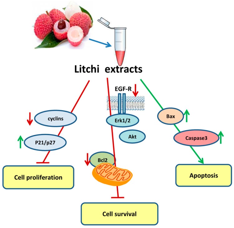
Overall, the main biochemical mechanisms induced by Litchi extracts involved in anticancer effects are summarized in Figure 3 and described in Table 2.
Figure 3.
Scheme of the effects of Litchi extracts on cell proliferation, cell survival, and apoptosis. Red arrows indicate downregulated factors or processes, and green arrows indicate increased factors or processes.
Table 2.
Specific antitumor effects of Litchi fruit portions and principal involved biochemical pathways.
| Cancer Model | Extracts or Specific Components | Antitumor Effect | Biochemical Pathways | References | ||
|---|---|---|---|---|---|---|
| Pulp | In vitro(cancer cell lines) | Lung adenocarcinoma, cervical cancer, hepatocellular carcinoma | Polysaccharides | Antiproliferative | Cell viability reduction | [22] |
| Immunomodulatory | Induction of mouse splenocyte proliferation | |||||
| Gastric cancer, hepatocellular and lung carcinoma | Galactose and mannose | Antiproliferative | Cell viability reduction | [23] | ||
| Antioxidant | Increase in cellular antioxidant activity | |||||
| In vivo(mice) | Chemical-induced liver injury | Pulp extract | Hepatoprotective | Decreased serum ALT and AST levels | [27] | |
| Antioxidant | Changes in antioxidant enzyme levels | |||||
| Peel | In vitro(cancer cell lines) | Hepatocellular carcinoma | Water-soluble crude ethanolic extract | Antiproliferative | Cell viability reduction, clonogenic growth decrease | [75] |
| Apoptosis induction | Pre G0/G1 pro-apoptotic peak in cell cycle profile | |||||
| Breast cancer cells | Water-soluble crude ethanolic extract | Antiproliferative | Cell viability reduction, Clonogenic growth decrease | [37] | ||
| Apoptosis induction | Up and down-regulation of gene clusters involved in cell death | |||||
| Breast cancer cells | Specific flavonoid components | Antiproliferative | Cell viability reduction | [36,38] | ||
| In vivo | Murine hepatoma bearing-mice | Inhibition of tumor growth | Reduction in cell proliferation | [75] | ||
| Nude mice bearing human breast infiltrating duct carcinoma | Water-soluble crude ethanolic extract | Tumor mass reduction | Reduction in cell proliferation | [37] | ||
| Apoptosis induction | Caspase-3 activation | |||||
| Seed | In vitro(cancer cell lines) | Lung adenocarcinoma, cervical, breast, ovarian cancers and hepatocellular carcinoma | Flavonoid glycosides | Anti proliferative | Cell viability reduction | [51,76] |
| Hepatocellular, lung and cervical carcinoma | Sesquiterpene glucosides | Anti proliferative | Cell viability reduction | [77] | ||
| Non-small cell lung cancer | Crude Litchi extract | Anti proliferative | Inhibition of EGF-receptor-pathway | [53] | ||
| Apoptosis induction | Bcl-2 family pro-apoptotic ratio and caspase activation | |||||
| Colorectal carcinoma | Flavonoids and tannins | Anti proliferative | G2/M phase cell cycle arrest with reduction in cyclin levels | [48] | ||
| Apoptosis induction | Increase in Bax level and caspase activation | |||||
| Prostate cancer | N-butyl alcohol extract | Anti proliferative | Clonogenic growth decrease, G1/S phase Cell cycle arrest with increase in p21 and p27 CDK inhibitors | [54] | ||
| Apoptosis induction | Activation of mitochondrial caspase cascade | |||||
| Decrease in cell migration and invasion | Increase in E-cadherin and â-catenin, decrease of vimentin and snail, inhibition of Akt pathway | |||||
| Hepatocellular carcinoma | Semen Litchi containing serum | Anti proliferative | Cell viability reduction | [47] | ||
| Apoptosis induction | Appearance of nuclear morphological features and pre G0/G1 pro-apoptotic peak in cell cycle profile | |||||
| In vivo | mouse xenografts of Ehrlich ascites cells, sarcoma S180 cells, or liver tumor cells | Water extract | Decrease in tumor size | [78] | ||
| hyperplasia of mammary glands rat model | Saponins | Reduction of mammary gland hyperplasia | [49] | |||
| Nude mice xenograft of PC3 cells | n-butyl alcohol extract | Decrease in tumor size | [54] | |||
| Oligonol | In vitro | Breast cancer cells | Low MW polyphenols from lychee fruit extract | Apoptosis induction | Modulation of pro-apoptotic Bcl-2 family proteins and MEK/ERK signaling pathway | [73] |
| In vivo | DSS-promoted adenoma in the mouse colon | Inhibition of colonic adenoma formation | Reduction of cyclins | [72] | ||
| Variation of oxidative stress markers | ||||||
| Melanoma mice models | Inhibition of lung metastasis | Inhibition of lung hexosammine content, and serum sialic acid and gamma glutamyltranspeptidase content | [70] | |||
| Mouse skin and carcinomas and papillomas bearing mice | Suppression of chemically-induced tumorigenesis | NF-êB and C/EBP DNA binding decrease, reduction of ERK 1/2 and P38 kinases, reduction in PCNA and COX2 | [71] | |||
9. Conclusions
The different portions of Litchi fruit contain key bioactive compounds that account for the anticancer effects described in the present review. Purifying these agents may represent an important step in phyto-pharmacotherapy, which can have a high impact in oncology. However, the biological activity of Litchi components has been mainly studied as evaluation of cytotoxicity in in vitro models. Therefore, the knowledge of the biochemical mechanisms underlying the anti-proliferative/death effects of Litchi components in tumor cells represents an important basis for anticancer translational studies.
Acknowledgments
This work has been carried out with the financial support from Gruppo Azione Locale (GAL) of Golfo di Castellammare, Italy (Progetto Operativo n.17/2015, misura 313B).
Author Contributions
All authors substantially contributed to the conception and design of the manuscript. Sonia Emanuele, Antonella D’Anneo and Michela Giuliano contributed in the literature search, data collection and writing. Marianna Lauricella and Giuseppe Calvaruso in the study design and tables design, Antonella D’Anneo contributed in the review of the manuscript. All authors revised and approved the final version.
Conflicts of Interest
The authors declare no conflict of interest.
References
- 1.Menzel C. Lychee, Its Origin, Distribution and Production around the World 1995. [(accessed on 28 July 2017)]; Available online: http://rfcarchives.org.au/index.htm.
- 2.Menzel C. The Lychee Crop in Asia and the Pacific. FAO Regional Office for Asia and the Pacific; Bangkok, Thailand: 2002. [Google Scholar]
- 3.Menzel C.M., Waite G.K. Litchi and Longan: Botany, Production and Uses. CABI Pub.; Wallingford, UK: Cambridge, MA, USA: 2005. [Google Scholar]
- 4.Kilari E., Putta S. Biological and phytopharmacological descriptions of Litchi chinensis. Pharmacogn. Rev. 2016;10:60. doi: 10.4103/0973-7847.176548. [DOI] [PMC free article] [PubMed] [Google Scholar]
- 5.Nielsen G. The use of isozymes as probes to identify and label plant varieties and cultivars. Isozymes. 1985;12:1–32. [PubMed] [Google Scholar]
- 6.Liu W., Xiao Z., Bao X., Yang X., Fang J., Xiang X. Identifying Litchi (Litchi chinensis Sonn.) cultivars and their genetic relationships using single nucleotide polymorphism (SNP) markers. PLoS ONE. 2015;10:e0135390. doi: 10.1371/journal.pone.0135390. [DOI] [PMC free article] [PubMed] [Google Scholar]
- 7.Lauricella M., Emanuele S., Calvaruso G., Giuliano M., D’Anneo A. Multifaceted health benefits of Mangifera indica L. (Mango): The inestimable value of orchards recently planted in sicilian rural areas. Nutrients. 2017;9:525. doi: 10.3390/nu9050525. [DOI] [PMC free article] [PubMed] [Google Scholar]
- 8.Padoan D., Farina V., Ferlito B., Barone F., Palazzolo E. Qualitative features of Litchi fruits (Litchi Chinensis Sonn.) cultivated in North east Sicily. Acta Italus Hortus. 2012;11:142–144. [Google Scholar]
- 9.U.S. Department of Agriculture, Agricultural Research Service 2012. USDA National Nutrient Database for Standard Reference, Release 25. Nutrient Data Laboratory Home Page. [(accessed on 28 July 2017)]; Available online: http://www.ars.usda.gov/ba/bhnrc/ndl.
- 10.Ramananda Singh M., Gupta P., Gupta K. The Litchi (Litchi Chinensis) peels extract as a potential green inhibitor in prevention of corrosion of mild steel in 0.5M H2SO4 solution. Arab. J. Chem. 2015 doi: 10.1016/j.arabjc.2015.01.002. [DOI] [Google Scholar]
- 11.Arts I.C.W., Hollman P.C.H. Polyphenols and disease risk in epidemiologic studies. Am. J. Clin. Nutr. 2005;81:317S–325S. doi: 10.1093/ajcn/81.1.317S. [DOI] [PubMed] [Google Scholar]
- 12.Septembre-Malaterre A., Stanislas G., Douraguia E., Gonthier M.-P. Evaluation of nutritional and antioxidant properties of the tropical fruits banana, Litchi, mango, papaya, passion fruit and pineapple cultivated in Réunion French Island. Food Chem. 2016;212:225–233. doi: 10.1016/j.foodchem.2016.05.147. [DOI] [PubMed] [Google Scholar]
- 13.U.S. Department of Agriculture (USDA) USDA; [(accessed on 25 January 2016)]. National Nutrient Database for Standard Reference, SR-28, Full Report (All Nutrients): 09164, Litchis, Raw National Agricultural Library. Available online: https://ndb.nal.usda.gov/ndb/foods/show/2271. [Google Scholar]
- 14.Li W., Liang H., Zhang M.-W., Zhang R.-F., Deng Y.-Y., Wei Z.-C., Zhang Y., Tang X.-J. Phenolic profiles and antioxidant activity of Litchi (Litchi Chinensis Sonn.) fruit pericarp from different commercially available cultivars. Molecules. 2012;17:14954–14967. doi: 10.3390/molecules171214954. [DOI] [PMC free article] [PubMed] [Google Scholar]
- 15.Manda G., Isvoranu G., Comanescu M.V., Manea A., Debelec Butuner B., Korkmaz K.S. The redox biology network in cancer pathophysiology and therapeutics. Redox Biol. 2015;5:347–357. doi: 10.1016/j.redox.2015.06.014. [DOI] [PMC free article] [PubMed] [Google Scholar]
- 16.Marengo B., Nitti M., Furfaro A.L., Colla R., Ciucis C.D., Marinari U.M., Pronzato M.A., Traverso N., Domenicotti C. Redox homeostasis and cellular antioxidant systems: Crucial players in cancer growth and therapy. Oxid. Med. Cell. Longev. 2016;2016:6235641. doi: 10.1155/2016/6235641. [DOI] [PMC free article] [PubMed] [Google Scholar]
- 17.Galadari S., Rahman A., Pallichankandy S., Thayyullathil F. Reactive oxygen species and cancer paradox: To promote or to suppress? Free Radic. Biol. Med. 2017;104:144–164. doi: 10.1016/j.freeradbiomed.2017.01.004. [DOI] [PubMed] [Google Scholar]
- 18.Ibrahim S.R.M., Mohamed G.A. Litchi chinensis: Medicinal uses, phytochemistry, and pharmacology. J. Ethnopharmacol. 2015;174:492–513. doi: 10.1016/j.jep.2015.08.054. [DOI] [PubMed] [Google Scholar]
- 19.Hu X., Wang J., Jing Y., Song L., Zhu J., Cui X., Yu R. Structural elucidation and in vitro antioxidant activities of a new heteropolysaccharide from Litchi chinensis. Drug Discov. Ther. 2015;9:116–122. doi: 10.5582/ddt.2015.01022. [DOI] [PubMed] [Google Scholar]
- 20.Hu X.-Q., Huang Y.-Y., Dong Q.-F., Song L.-Y., Yuan F., Yu R.-M. Structure characterization and antioxidant activity of a novel polysaccharide isolated from pulp tissues of Litchi chinensis. J. Agric. Food Chem. 2011;59:11548–11552. doi: 10.1021/jf203179y. [DOI] [PubMed] [Google Scholar]
- 21.Huang F., Zhang R., Liu Y., Xiao J., Liu L., Wei Z., Yi Y., Zhang M., Liu D. Dietary Litchi pulp polysaccharides could enhance immunomodulatory and antioxidant effects in mice. Int. J. Biol. Macromol. 2016;92:1067–1073. doi: 10.1016/j.ijbiomac.2016.08.021. [DOI] [PubMed] [Google Scholar]
- 22.Huang F., Zhang R., Yi Y., Tang X., Zhang M., Su D., Deng Y., Wei Z. Comparison of physicochemical properties and immunomodulatory activity of polysaccharides from fresh and dried Litchi pulp. Molecules. 2014;19:3909–3925. doi: 10.3390/molecules19043909. [DOI] [PMC free article] [PubMed] [Google Scholar]
- 23.Huang F., Zhang R., Dong L., Guo J., Deng Y., Yi Y., Zhang M. Antioxidant and antiproliferative activities of polysaccharide fractions from Litchi pulp. Food Funct. 2015;6:2598–2606. doi: 10.1039/C5FO00249D. [DOI] [PubMed] [Google Scholar]
- 24.Anantharaju P.G., Gowda P.C., Vimalambike M.G., Madhunapantula S.V. An overview on the role of dietary phenolics for the treatment of cancers. Nutr. J. 2016;15 doi: 10.1186/s12937-016-0217-2. [DOI] [PMC free article] [PubMed] [Google Scholar]
- 25.Zhang R., Zeng Q., Deng Y., Zhang M., Wei Z., Zhang Y., Tang X. Phenolic profiles and antioxidant activity of Litchi pulp of different cultivars cultivated in Southern China. Food Chem. 2013;136:1169–1176. doi: 10.1016/j.foodchem.2012.09.085. [DOI] [PubMed] [Google Scholar]
- 26.Su D., Ti H., Zhang R., Zhang M., Wei Z., Deng Y., Guo J. Structural elucidation and cellular antioxidant activity evaluation of major antioxidant phenolics in lychee pulp. Food Chem. 2014;158:385–391. doi: 10.1016/j.foodchem.2014.02.134. [DOI] [PubMed] [Google Scholar]
- 27.Su D., Zhang R., Zhang C., Huang F., Xiao J., Deng Y., Wei Z., Zhang Y., Chi J., Zhang M. Phenolic-rich lychee (Litchi chinensis Sonn.) pulp extracts offer hepatoprotection against restraint stress-induced liver injury in mice by modulating mitochondrial dysfunction. Food Funct. 2016;7:508–515. doi: 10.1039/C5FO00975H. [DOI] [PubMed] [Google Scholar]
- 28.Yang C.S., Wang H. Cancer preventive activities of tea catechins. Molecules. 2016;21 doi: 10.3390/molecules21121679. [DOI] [PMC free article] [PubMed] [Google Scholar]
- 29.Suganuma M., Takahashi A., Watanabe T., Iida K., Matsuzaki T., Yoshikawa H.Y., Fujiki H. Biophysical approach to mechanisms of cancer prevention and treatment with green tea catechins. Molecules. 2016;21 doi: 10.3390/molecules21111566. [DOI] [PMC free article] [PubMed] [Google Scholar]
- 30.Kwak T.W., Park S.B., Kim H.-J., Jeong Y.-I., Kang D.H. Anticancer activities of epigallocatechin-3-gallate against cholangiocarcinoma cells. OncoTargets Ther. 2017;10:137–144. doi: 10.2147/OTT.S112364. [DOI] [PMC free article] [PubMed] [Google Scholar]
- 31.Li B.-B., Huang G.-L., Li H.-H., Kong X., He Z.-W. Epigallocatechin-3-gallate modulates microrna expression profiles in human nasopharyngeal carcinoma CNE2 cells. Chin. Med. J. 2017;130:93–99. doi: 10.4103/0366-6999.196586. [DOI] [PMC free article] [PubMed] [Google Scholar]
- 32.Ben Sghaier M., Pagano A., Mousslim M., Ammari Y., Kovacic H., Luis J. Rutin inhibits proliferation, attenuates superoxide production and decreases adhesion and migration of human cancerous cells. Biomed. Pharmacother. 2016;84:1972–1978. doi: 10.1016/j.biopha.2016.11.001. [DOI] [PubMed] [Google Scholar]
- 33.Ju J., Hong J., Zhou J., Pan Z., Bose M., Liao J., Yang G., Liu Y.Y., Hou Z., Lin Y., et al. Inhibition of intestinal tumorigenesis in Apcmin/+ mice by (−)-epigallocatechin-3-gallate, the major catechin in green tea. Cancer Res. 2005;65:10623–10631. doi: 10.1158/0008-5472.CAN-05-1949. [DOI] [PubMed] [Google Scholar]
- 34.Shimizu M., Shirakami Y., Sakai H., Tatebe H., Nakagawa T., Hara Y., Weinstein I.B., Moriwaki H. EGCG inhibits activation of the insulin-like growth factor (IGF)/IGF-1 receptor axis in human hepatocellular carcinoma cells. Cancer Lett. 2008;262:10–18. doi: 10.1016/j.canlet.2007.11.026. [DOI] [PubMed] [Google Scholar]
- 35.Demiroglu-Zergeroglu A., Candemir G., Turhanlar E., Sagir F., Ayvali N. EGFR-dependent signalling reduced and p38 dependent apoptosis required by Gallic acid in Malignant Mesothelioma cells. Biomed. Pharmacother. 2016;84:2000–2007. doi: 10.1016/j.biopha.2016.11.005. [DOI] [PubMed] [Google Scholar]
- 36.Li J., Jiang Y. Litchi flavonoids: Isolation, identification and biological activity. Molecules. 2007;12:745–758. doi: 10.3390/12040745. [DOI] [PMC free article] [PubMed] [Google Scholar]
- 37.Wang X., Yuan S., Wang J., Lin P., Liu G., Lu Y., Zhang J., Wang W., Wei Y. Anticancer activity of Litchi fruit pericarp extract against human breast cancer in vitro and in vivo. Toxicol. Appl. Pharmacol. 2006;215:168–178. doi: 10.1016/j.taap.2006.02.004. [DOI] [PubMed] [Google Scholar]
- 38.Zhao M., Yang B., Wang J., Liu Y., Yu L., Jiang Y. Immunomodulatory and anticancer activities of flavonoids extracted from Litchi (Litchi chinensis Sonn.) pericarp. Int. Immunopharmacol. 2007;7:162–166. doi: 10.1016/j.intimp.2006.09.003. [DOI] [PubMed] [Google Scholar]
- 39.Shrivastava A., Kumar A., Thomas J.D., Laserson K.F., Bhushan G., Carter M.D., Chhabra M., Mittal V., Khare S., Sejvar J.J., et al. Association of acute toxic encephalopathy with Litchi consumption in an outbreak in Muzaffarpur, India, 2014: A case-control study. Lancet Glob. Health. 2017;5:e458–e466. doi: 10.1016/S2214-109X(17)30035-9. [DOI] [PubMed] [Google Scholar]
- 40.Das M., ASthana S., Singh S., Dixit S., Tripathi A., John T. Litchi fruit contains methylene cyclopropyl-glycine. Curr. Sci. 2015;109:2195–2197. [Google Scholar]
- 41.Li C., Liao X., Li X., Guo J., Qu X., Li L. Effect and mechanism of Litchi semen effective constituents on insulin resistance in rats with type 2 diabetes mellitus. J. Chin. Med. Mater. 2015;38:1466–1471. [PubMed] [Google Scholar]
- 42.Choi S.-A., Lee J.E., Kyung M.J., Youn J.H., Oh J.B., Whang W.K. Anti-diabetic functional food with wasted Litchi seed and standard of quality control. Appl. Biol. Chem. 2017;60:197–204. doi: 10.1007/s13765-017-0269-9. [DOI] [Google Scholar]
- 43.Kalgaonkar S., Nishioka H., Gross H.B., Fujii H., Keen C.L., Hackman R.M. Bioactivity of a flavanol-rich lychee fruit extract in adipocytes and its effects on oxidant defense and indices of metabolic syndrome in animal models. Phytother. Res. 2010;24:1123–1128. doi: 10.1002/ptr.3137. [DOI] [PubMed] [Google Scholar]
- 44.Bhoopat L., Srichairatanakool S., Kanjanapothi D., Taesotikul T., Thananchai H., Bhoopat T. Hepatoprotective effects of lychee (Litchi chinensis Sonn.): A combination of antioxidant and anti-apoptotic activities. J. Ethnopharmacol. 2011;136:55–66. doi: 10.1016/j.jep.2011.03.061. [DOI] [PubMed] [Google Scholar]
- 45.Xu X., Xie H., Wang Y., Wei X. A-type proanthocyanidins from lychee seeds and their antioxidant and antiviral activities. J. Agric. Food Chem. 2010;58:11667–11672. doi: 10.1021/jf1033202. [DOI] [PubMed] [Google Scholar]
- 46.Xiao L.-Y., Zhang D., Feng Z., Chen Y., Zhang H., Lin P. Studies on the antitumor effects of lychee seeds in mice. J. Chin. Med. Mater. 2004;27:517–518. [Google Scholar]
- 47.Xiong A.-H., Shen W.-J., Xiao L.-Y., Lv J.-H. Effect of Semen Litchi containing serum on proliferation and apoptosis of HepG2 cells. J. Chin. Med. Mater. 2008;31:1533–1536. [PubMed] [Google Scholar]
- 48.Hsu C.-P., Lin C.-C., Huang C.-C., Lin Y.-H., Chou J.-C., Tsia Y.-T., Su J.-R., Chung Y.-C. Induction of apoptosis and cell cycle arrest in human colorectal carcinoma by Litchi seed extract. J. Biomed. Biotechnol. 2012;2012:341479. doi: 10.1155/2012/341479. [DOI] [PMC free article] [PubMed] [Google Scholar]
- 49.Lin N., Qiu Y., He G., Guan N. Effects of Litchi chinensis seed saponins on inhibiting hyperplasia of mammary glands and influence on signaling pathway of estrogen in rats. J. Chin. Med. Mater. 2015;38:798–802. [PubMed] [Google Scholar]
- 50.Wang X., Wu J., Yu C., Tang Y., Liu J., Chen H., Jin B., Mei Q., Cao S., Qin D. Lychee seed saponins improve cognitive function and prevent neuronal injury via inhibiting neuronal apoptosis in a rat model of alzheimer’s disease. Nutrients. 2017;9 doi: 10.3390/nu9020105. [DOI] [PMC free article] [PubMed] [Google Scholar]
- 51.Xu X., Xie H., Hao J., Jiang Y., Wei X. Flavonoid glycosides from the seeds of Litchi chinensis. J. Agric. Food Chem. 2011;59:1205–1209. doi: 10.1021/jf104387y. [DOI] [PubMed] [Google Scholar]
- 52.Lin N., Xiao L.-Y., Pan J., Lv J.-H. Effects of Semen Litchi on the expressions of S180 and EAC tumor cells and Bax and Bcl-2 proteins in rats. China Pharm. 2008;19:1138–1140. [Google Scholar]
- 53.Chung Y.-C., Chen C.-H., Tsai Y.-T., Lin C.-C., Chou J.-C., Kao T.-Y., Huang C.-C., Cheng C.-H., Hsu C.-P. Litchi seed extract inhibits epidermal growth factor receptor signaling and growth of Two Non-small cell lung carcinoma cells. BMC Complement. Altern. Med. 2017;17:16. doi: 10.1186/s12906-016-1541-y. [DOI] [PMC free article] [PubMed] [Google Scholar]
- 54.Guo H., Luo H., Yuan H., Xia Y., Shu P., Huang X., Lu Y., Liu X., Keller E.T., Sun D., et al. Litchi seed extracts diminish prostate cancer progression via induction of apoptosis and attenuation of EMT through Akt/GSK-3β signaling. Sci. Rep. 2017;7:41656. doi: 10.1038/srep41656. [DOI] [PMC free article] [PubMed] [Google Scholar]
- 55.Carlisi D., D’Anneo A., Martinez R., Emanuele S., Buttitta G., Di Fiore R., Vento R., Tesoriere G., Lauricella M. The oxygen radicals involved in the toxicity induced by parthenolide in MDA-MB-231 cells. Oncol. Rep. 2014 doi: 10.3892/or.2014.3212. [DOI] [PubMed] [Google Scholar]
- 56.D’Anneo A., Carlisi D., Emanuele S., Buttitta G., Di Fiore R., Vento R., Tesoriere G., Lauricella M. Parthenolide induces superoxide anion production by stimulating EGF receptor in MDA-MB-231 breast cancer cells. Int. J. Oncol. 2013;43:1895–1900. doi: 10.3892/ijo.2013.2137. [DOI] [PubMed] [Google Scholar]
- 57.Lauricella M., Carlisi D., Giuliano M., Calvaruso G., Cernigliaro C., Vento R., D’Anneo A. The analysis of estrogen receptor-α positive breast cancer stem-like cells unveils a high expression of the serpin proteinase inhibitor PI-9: Possible regulatory mechanisms. Int. J. Oncol. 2016;49:352–360. doi: 10.3892/ijo.2016.3495. [DOI] [PubMed] [Google Scholar]
- 58.Notaro A., Sabella S., Pellerito O., Vento R., Calvaruso G., Giuliano M. The secreted protein acidic and rich in cysteine is a critical mediator of cell death program induced by WIN/TRAIL combined treatment in osteosarcoma cells. Int. J. Oncol. 2016;48:1039–1044. doi: 10.3892/ijo.2015.3307. [DOI] [PubMed] [Google Scholar]
- 59.Pellerito O., Notaro A., Sabella S., De Blasio A., Vento R., Calvaruso G., Giuliano M. WIN induces apoptotic cell death in human colon cancer cells through a block of autophagic flux dependent on PPARγ down-regulation. Apoptosis. 2014;19:1029–1042. doi: 10.1007/s10495-014-0985-0. [DOI] [PubMed] [Google Scholar]
- 60.Notaro A., Sabella S., Pellerito O., Di Fiore R., De Blasio A., Vento R., Calvaruso G., Giuliano M. Involvement of PAR-4 in cannabinoid-dependent sensitization of osteosarcoma cells to TRAIL-induced apoptosis. Int. J. Biol. Sci. 2014;10:466–478. doi: 10.7150/ijbs.8337. [DOI] [PMC free article] [PubMed] [Google Scholar]
- 61.Wen L., You L., Yang X., Yang J., Chen F., Jiang Y., Yang B. Identification of phenolics in Litchi and evaluation of anticancer cell proliferation activity and intracellular antioxidant activity. Free Radic. Biol. Med. 2015;84:171–184. doi: 10.1016/j.freeradbiomed.2015.03.023. [DOI] [PubMed] [Google Scholar]
- 62.Lin Y.-C., Chang J.-C., Cheng S.-Y., Wang C.-M., Jhan Y.-L., Lo I.-W., Hsu Y.-M., Liaw C.-C., Hwang C.-C., Chou C.-H. New bioactive chromanes from Litchi chinensis. J. Agric. Food Chem. 2015;63:2472–2478. doi: 10.1021/jf5056387. [DOI] [PubMed] [Google Scholar]
- 63.Yang D.-J., Chang Y.-Z., Chen Y.-C., Liu S.-C., Hsu C.-H., Lin J.-T. Antioxidant effect and active components of Litchi (Litchi chinensis Sonn.) flower. Food Chem. Toxicol. 2012;50:3056–3061. doi: 10.1016/j.fct.2012.06.011. [DOI] [PubMed] [Google Scholar]
- 64.Yang D.-J., Chang Y.-Y., Lin H.-W., Chen Y.-C., Hsu S.-H., Lin J.-T. Inhibitory effect of Litchi (Litchi chinensis Sonn.) flower on lipopolysaccharide-induced expression of proinflammatory mediators in RAW264.7 cells through NF-κB, ERK, and JAK2/STAT3 inactivation. J. Agric. Food Chem. 2014;62:3458–3465. doi: 10.1021/jf5003705. [DOI] [PubMed] [Google Scholar]
- 65.Aruoma O.I., Sun B., Fujii H., Neergheen V.S., Bahorun T., Kang K.-S., Sung M.-K. Low molecular proanthocyanidin dietary biofactor Oligonol: Its modulation of oxidative stress, bioefficacy, neuroprotection, food application and chemoprevention potentials. BioFactors. 2006;27:245–265. doi: 10.1002/biof.5520270121. [DOI] [PubMed] [Google Scholar]
- 66.Park C.H., Noh J.S., Fujii H., Roh S.-S., Song Y.-O., Choi J.S., Chung H.Y., Yokozawa T. Oligonol, a low-molecular-weight polyphenol derived from lychee fruit, attenuates gluco-lipotoxicity-mediated renal disorder in type 2 diabetic db/db mice. Drug Discov. Ther. 2015;9:13–22. doi: 10.5582/ddt.2015.01003. [DOI] [PubMed] [Google Scholar]
- 67.Choi J.S., Bhakta H.K., Fujii H., Min B.-S., Park C.H., Yokozawa T., Jung H.A. Inhibitory evaluation of oligonol on α-glucosidase, protein tyrosine phosphatase 1B, cholinesterase, and β-secretase 1 related to diabetes and Alzheimer’s disease. Arch. Pharm. Res. 2016;39:409–420. doi: 10.1007/s12272-015-0682-8. [DOI] [PubMed] [Google Scholar]
- 68.Fujii H., Nishioka H., Wakame K., Magnuson B.A., Roberts A. Acute, subchronic and genotoxicity studies conducted with Oligonol, an oligomerized polyphenol formulated from lychee and green tea extracts. Food Chem. Toxicol. 2008;46:3553–3562. doi: 10.1016/j.fct.2008.06.005. [DOI] [PubMed] [Google Scholar]
- 69.Katiyar S.K., Pal H.C., Prasad R. Dietary proanthocyanidins prevent ultraviolet radiation-induced non-melanoma skin cancer through enhanced repair of damaged DNA-dependent activation of immune sensitivity. Semin. Cancer Biol. 2017 doi: 10.1016/j.semcancer.2017.04.003. [DOI] [PubMed] [Google Scholar]
- 70.Lee S.-J., Chung I.-M., Kim M.-Y., Park K.-D., Park W.-H., Park W.-W., Moon H.-I. Inhibition of lung metastasis in mice by oligonol. Phytother. Res. 2009;23:1043–1046. doi: 10.1002/ptr.2810. [DOI] [PubMed] [Google Scholar]
- 71.Kundu J.K., Hwang D.-M., Lee J.-C., Chang E.-J., Shin Y.K., Fujii H., Sun B., Surh Y.-J. Inhibitory effects of oligonol on phorbol ester-induced tumor promotion and COX-2 expression in mouse skin: NF-kappaB and C/EBP as potential targets. Cancer Lett. 2009;273:86–97. doi: 10.1016/j.canlet.2008.07.039. [DOI] [PubMed] [Google Scholar]
- 72.Yum H.-W., Zhong X., Park J., Na H.-K., Kim N., Lee H.S., Surh Y.-J. Oligonol inhibits dextran sulfate sodium-induced colitis and colonic adenoma formation in mice. Antioxid. Redox Signal. 2013;19:102–114. doi: 10.1089/ars.2012.4626. [DOI] [PMC free article] [PubMed] [Google Scholar]
- 73.Jo E.-H., Lee S.-J., Ahn N.-S., Park J.-S., Hwang J.-W., Kim S.-H., Aruoma O.I., Lee Y.-S., Kang K.-S. Induction of apoptosis in MCF-7 and MDA-MB-231 breast cancer cells by Oligonol is mediated by Bcl-2 family regulation and MEK/ERK signaling. Eur. J. Cancer Prev. 2007;16:342–347. doi: 10.1097/01.cej.0000236247.86360.db. [DOI] [PubMed] [Google Scholar]
- 74.Bak J., Je N.K., Chung H.Y., Yokozawa T., Yoon S., Moon J.-O. Oligonol ameliorates CCl4 -induced liver injury in rats via the NF-Kappa B and MAPK signaling pathways. Oxid. Med. Cell. Longev. 2016;2016:1–12. doi: 10.1155/2016/3935841. [DOI] [PMC free article] [PubMed] [Google Scholar]
- 75.Wang X., Wei Y., Yuan S., Liu G., Zhang Y.L.J., Wang W. Potential anticancer activity of Litchi fruit pericarp extract against hepatocellular carcinoma in vitro and in vivo. Cancer Lett. 2006;239:144–150. doi: 10.1016/j.canlet.2005.08.011. [DOI] [PubMed] [Google Scholar]
- 76.Lin C.-C., Chung Y.-C., Hsu C.-P. Anti-cancer potential of Litchi seed extract. World J. Exp. Med. 2013;3:56–61. [Google Scholar]
- 77.Xu X., Xie H., Hao J., Jiang Y., Wei X. Eudesmane sesquiterpene glucosides from lychee seed and their cytotoxic activity. Food Chem. 2010;123:1123–1126. doi: 10.1016/j.foodchem.2010.05.073. [DOI] [Google Scholar]
- 78.Zhang J., Zhang C. Research progress on the antineoplastic pharmacological effects and mechanisms of Litchi seeds. Chin. Med. 2015;06:20–26. doi: 10.4236/cm.2015.61003. [DOI] [Google Scholar]


