The human bacterial pathogen Streptococcus pyogenes (group A streptococcus [GAS]) causes more than 600 million cases of pharyngitis annually worldwide, 15 million of which occur in the United States. The human oropharynx is the primary anatomic site for GAS colonization and infection, and saliva is the first material encountered. Using a genome-wide transposon mutant screen, we identified 92 GAS genes required for wild-type fitness in human saliva. Many of the identified genes are involved in carbohydrate transport/metabolism, amino acid transport/metabolism, and inorganic ion transport/metabolism. The new information is potentially valuable for developing novel GAS therapeutics and vaccine research.
KEYWORDS: Streptococcus pyogenes, TraDIS, fitness, human pathogen, saliva, transposon mutagenesis
ABSTRACT
Streptococcus pyogenes (group A streptococcus [GAS]) causes 600 million cases of pharyngitis each year. Despite this considerable disease burden, the molecular mechanisms used by GAS to infect, cause clinical pharyngitis, and persist in the human oropharynx are poorly understood. Saliva is ubiquitous in the human oropharynx and is the first material GAS encounters in the upper respiratory tract. Thus, a fuller understanding of how GAS survives and proliferates in saliva may provide valuable insights into the molecular mechanisms at work in the human oropharynx. We generated a highly saturated transposon insertion mutant library in serotype M1 strain MGAS2221, a strain genetically representative of a pandemic clone that arose in the 1980s and spread globally. The transposon mutant library was exposed to human saliva to screen for GAS genes required for wild-type fitness in this clinically relevant fluid. Using transposon-directed insertion site sequencing (TraDIS), we identified 92 genes required for GAS fitness in saliva. The more prevalent categories represented were genes involved in carbohydrate transport/metabolism, amino acid transport/metabolism, and inorganic ion transport/metabolism. Using six isogenic mutant strains, we confirmed that each of the mutants was significantly impaired for growth or persistence in human saliva ex vivo. Mutants with an inactivated Spy0644 (sptA) or Spy0646 (sptC) gene had especially severe persistence defects. This study is the first to use of TraDIS to study bacterial fitness in human saliva. The new information we obtained will be valuable for future translational maneuvers designed to prevent or treat human GAS infections.
IMPORTANCE The human bacterial pathogen Streptococcus pyogenes (group A streptococcus [GAS]) causes more than 600 million cases of pharyngitis annually worldwide, 15 million of which occur in the United States. The human oropharynx is the primary anatomic site for GAS colonization and infection, and saliva is the first material encountered. Using a genome-wide transposon mutant screen, we identified 92 GAS genes required for wild-type fitness in human saliva. Many of the identified genes are involved in carbohydrate transport/metabolism, amino acid transport/metabolism, and inorganic ion transport/metabolism. The new information is potentially valuable for developing novel GAS therapeutics and vaccine research.
Podcast: A podcast concerning this article is available.
INTRODUCTION
Bacterial pathogens have evolved highly specialized molecular strategies to survive and persist in diverse host niches (1, 2). Understanding the molecular mechanisms that contribute to bacterial fitness in human environments is valuable for developing therapeutic strategies to treat and potentially prevent infections. Streptococcus pyogenes (group A streptococcus [GAS]) is a significant human pathogen that causes extensive health and economic impacts globally (3). The human oropharynx is the primary anatomic site for GAS colonization and infection (3–5). This pathogen causes 600 million cases of pharyngitis annually worldwide, 15 million of which occur in the United States (3). The annual direct health care costs associated with GAS pharyngitis are estimated to be 2 billion dollars annually in the United States alone (3, 5). The organism is also responsible for an additional 100 million cases of other human infections each year, many of which occur after initial colonization of the oropharynx (3). These additional infections include acute rheumatic fever and subsequent rheumatic heart disease, and as a consequence, it is the most common cause of preventable pediatric heart disease globally (3, 6). The majority of cases of rheumatic fever occur following human upper respiratory tract infection. Despite the extensive toll on human health, the molecular mechanisms used by GAS to successfully colonize, cause acute pharyngitis, and persist in the human oropharynx remain largely unknown or poorly understood (7, 8). This lack of knowledge constitutes a critical knowledge gap in our understanding of GAS pathogenesis and thus represents an opportunity for enhanced understanding of the molecular mechanisms at work during the critical initial pathogen interaction with the human host.
The oropharynx is the primary site of entry for GAS into the body and the major portal of person-to-person transmission (9–11). Several observations have stimulated our interest in studying the molecular genetic basis of the interaction between human saliva and GAS. Saliva is ubiquitous in the human oropharynx and is the first host material contacted by GAS in its common transmission cycle. Compared to individuals with clinical pharyngitis who lack GAS in their saliva, patients with GAS present in their saliva are more likely to transmit the organism to a new host by aerosolization (10–12). Thus, understanding how GAS survives and proliferates in saliva and the oropharynx may provide valuable insights into the molecular mechanisms underlying successful bacterial interactions in this niche.
Previous studies addressing GAS-saliva interactions have identified several factors that contribute to bacterial fitness (13–18), but knowledge is limited. The studies conducted by Sitkiewicz et al. (13) and Virtaneva et al. (14, 15) investigated gene responses of a serotype M1 GAS strain grown in human saliva ex vivo (16). Subsequently, Shelburne et al. (17) identified a key two-component transcriptional regulatory system (SptR/S) of previously unknown function that plays a central role in optimizing GAS persistence in saliva. The SptR/S two-component system influences multiple GAS metabolic pathways and production of many virulence factors (17). For example, the secreted virulence factors streptococcal inhibitor of complement (sic) and a potent extracellular cysteine protease (SpeB) made by GAS during growth ex vivo in saliva contribute significantly to GAS persistence in this fluid (16). Additional information about GAS interactions in the oropharynx was provided by a study, in 20 monkeys, that investigated global transcriptome changes occurring over 86 days of the infection cycle, including initial colonization, acute clinical pharyngitis, and ultimately asymptomatic colonization (15, 18). Taken together, these studies provided a broad overview of some of the GAS processes at work in the oropharynx; however, much remains to be learned.
Especially lacking is a detailed understanding of the genes required for successful growth and persistence in saliva. To address this important knowledge gap, we conducted a genome-wide screen to identify GAS genes that contribute to fitness in human saliva ex vivo. Using transposon-directed insertion site sequencing (TraDIS) (19–28), we generated a highly saturated transposon insertion library (140,249 unique transposon insertions) in serotype M1 reference strain MGAS2221 (29–31). This serotype was used because it is usually the most common cause of pharyngitis and other human infections in Western countries (3). Strain MGAS2221 is genetically representative of the pandemic clone that arose in the 1980s and rapidly spread globally (29–32). The transposon mutant library was exposed to human saliva ex vivo for 48 h to identify GAS genes that contribute to fitness over time in this clinically relevant fluid. Saliva studies conducted with six isogenic mutant strains validated the findings. The new information we obtained substantially increases our overall understanding of the molecular genetic basis of pathogen-saliva interactions and is valuable for future translational research designed to treat or prevent human GAS infections.
RESULTS
Construction of a highly saturated transposon insertion library in serotype M1 GAS strain MGAS2221.
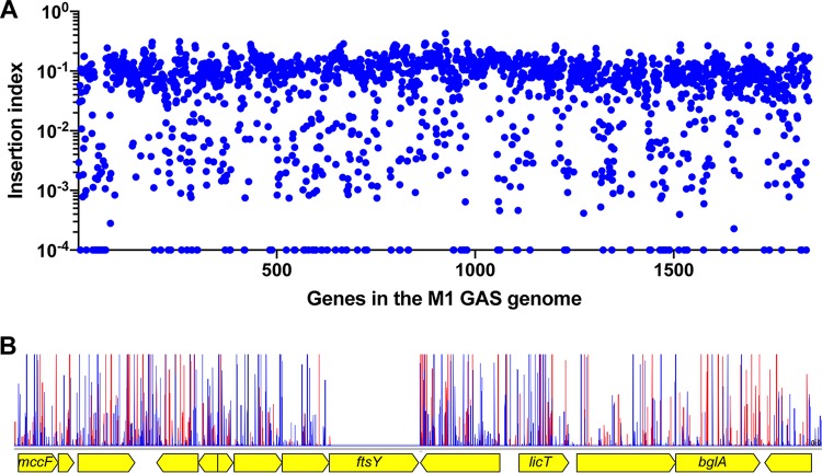
A transposon insertion mutant library was generated using serotype M1 strain MGAS2221 as the parental organism. Strain MGAS2221 was chosen for transposon mutagenesis because (i) it is genetically representative of a pandemic clone that arose in the 1980s and disseminated worldwide (30–32), (ii) MGAS2221 has wild-type alleles of major transcriptional regulators that affect virulence, such as covR and covS, ropB, mga, and rocA, and (iii) it has been used in many mouse and primate infection studies (31). Using plasmid pGh9:ISS1 (plasmid pGh9 carrying the insertion sequence S1 [26]), we successfully generated a dense transposon mutant library in strain MGAS2221 containing 140,249 unique transposon insertions. On average, the library contained one transposon insertion for every 13 nucleotides. Of the genes in the MGAS2221 genome, 1,720 out of 1,841 (93.4%) have at least one transposon insertion. The nearly random distribution of transposon insertions and the high density of transposon insertions in the S. pyogenes genome are illustrated in Fig. 1. By analyzing the mutant library using the tradis_essentiality TraDIS toolkit Script (25), we identified 432 genes (~23.5% of the genes in the genome) that were essential for the serotype M1 GAS strain MGAS2221 under our experimental conditions (40°C, in THY broth [Todd-Hewitt broth with yeast extract] supplemented with 0.5 μg/ml erythromycin; see Materials and Methods). The list of identified essential genes is presented in Table S1 in the supplemental material.
FIG 1 .
Near-random distribution (A) and the high density of transposon insertions (B) of the M1 GAS input mutant library. (A) Insertion index (number of insertion sites divided by gene length; y axis) of each gene (x axis) in the M1 GAS reference genome. (B) A representative section of the transposon insertion map. As expected, the essential gene ftsY has no insertion because it is not represented in the library. Red vertical spikes are forward reads; blue vertical spikes are reverse reads. Read orientations indicate the direction of the transposon insertion.
Essential genes identified in the genome of serotype M1 GAS strain MGAS2221 under the conditions tested. Download TABLE S1, PDF file, 0.1 MB (135.3KB, pdf) .
Copyright © 2017 Zhu et al.
This content is distributed under the terms of the Creative Commons Attribution 4.0 International license.
Genes that contribute to fitness of GAS over time in human saliva.
We exposed the transposon mutant library to pooled human saliva ex vivo to screen for genes that contribute to fitness in this fluid. TraDIS was used to identify genes with a significantly altered mutant frequency in the output mutant pools compared to the input pool at 12, 24, and 48 h after saliva inoculation. Genes with a significantly decreased mutant frequency (fold change of >1.5, q value of <0.1) in the output mutant pools were regarded as important for saliva growth and persistence, which can be referred to as fitness. To ensure that the statistical power was adequate, genes with fewer than 10 transposon insertions in any of the four input mutant pools were excluded from the analysis, as recommended by van Opijnen et al. (33).
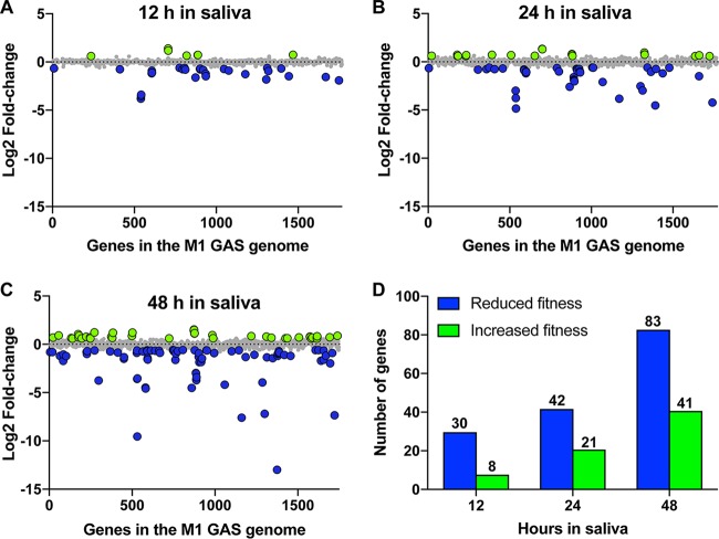
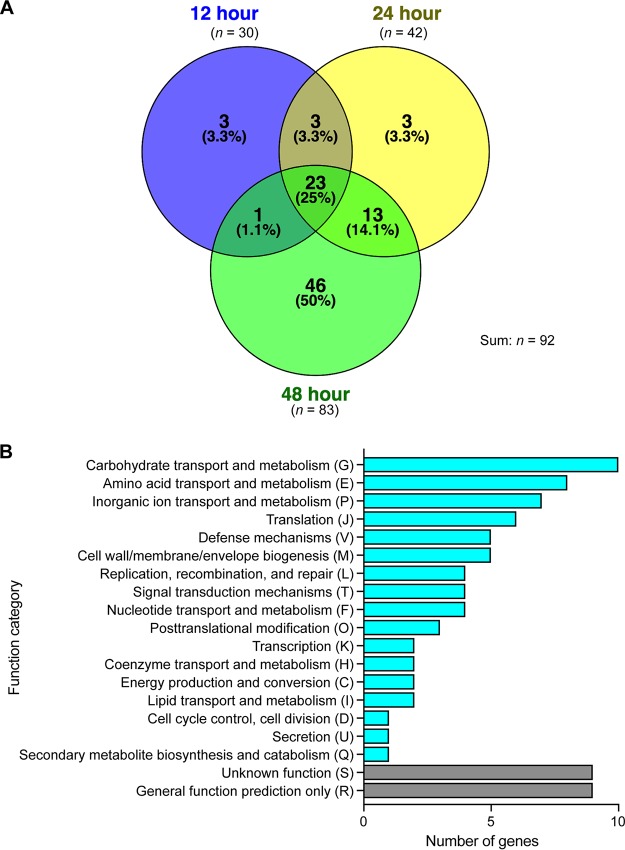
We identified 30 (12 h), 42 (24 h), and 83 (48 h) genes with significantly decreased mutant frequencies, providing evidence that these genes contribute to GAS fitness in human saliva (Fig. 2 and 3; Table S2). In total, 92 genes were identified at the three time points (Fig. 3A; Table S2). Clusters of orthologous groups (COG) classification of the 92 genes showed that numerically, the three more prevalent categories included genes involved in carbohydrate transport and metabolism (n = 10 genes), amino acid transport and metabolism (n = 8 genes), and inorganic ion transport and metabolism (n = 7 genes) (Fig. 3B). Our previously published data from an experimental pharyngitis infection study involving 20 cynomolgus macaques (15) identified genes expressed during GAS oropharyngeal infection. Of the 92 saliva fitness genes identified by TraDIS, ~74% were also expressed during GAS oropharyngeal infection (Table S2). Moreover, many of these 92 genes (e.g., nagA, pstS, oppA, and malX) are upregulated during GAS oropharyngeal infection in cynomolgus macaques (15). Together, our results suggest that many of the GAS genes that contribute to fitness in saliva ex vivo may also contribute to pathogen fitness in the oropharynx of nonhuman primates (NHPs). However, experiments will be required to directly test this hypothesis.
FIG 2 .
TraDIS analyses of GAS fitness genes during exposure to human saliva ex vivo. (A to C) Genome-scale summary of the changes in mutant abundance (y axis) for each of the genes (x axis) in the output mutant pools recovered after 12 h, 24 h, and 48 h of incubation in human saliva ex vivo. Gene mutations (insertions) conferring significantly decreased (blue circles) or increased (green circles) fitness are highlighted. Insertion mutations that lacked a significantly altered fitness phenotype (gray circles) are also indicated. (D) Summary of GAS genes identified to be important for fitness in saliva after the indicated period of saliva incubation.
FIG 3 .
(A) Venn diagram showing the number of GAS genes identified to be important for fitness in saliva after the indicated period of incubation. (B) Functional categorization of the 92 identified GAS saliva fitness genes. Note that in panel A the circle sizes are not proportional to the numbers of genes identified, for the sake of improving the visual presentation and clarity.
Gene mutations conferring decreased fitness after 12, 24, and 48 h of incubation with human saliva. Download TABLE S2, PDF file, 0.1 MB (60.6KB, pdf) .
Copyright © 2017 Zhu et al.
This content is distributed under the terms of the Creative Commons Attribution 4.0 International license.
Additionally, we determined that some genes at 12 h (n = 8 genes), 24 h (n = 21 genes), and 48 h (n = 41 genes) were significantly associated with potentially increased GAS fitness in saliva. The magnitudes of the fold changes in these genes were relatively modest compared to the results with genes that were decreased in fitness (Fig. 2A to C; Table S3).
Gene mutations conferring increased fitness after 12, 24, and 48 h of incubation with human saliva. Download TABLE S3, PDF file, 0.05 MB (48.7KB, pdf) .
Copyright © 2017 Zhu et al.
This content is distributed under the terms of the Creative Commons Attribution 4.0 International license.
The fitness scores of all genes in the M1 GAS genome (including the genes with less than 10 insertions) with a significant change in sequence read counts at the three time points (12 h, 24 h, and 48 h) are listed in Table S4, Table S5, and Table S6, respectively.
Fitness score of each gene in the M1 GAS genome after the 12-h saliva incubation. Download TABLE S4, PDF file, 0.1 MB (76.5KB, pdf) .
Copyright © 2017 Zhu et al.
This content is distributed under the terms of the Creative Commons Attribution 4.0 International license.
Fitness score of each gene in the M1 GAS genome after the 24-h saliva incubation. Download TABLE S5, PDF file, 0.1 MB (122.6KB, pdf) .
Copyright © 2017 Zhu et al.
This content is distributed under the terms of the Creative Commons Attribution 4.0 International license.
Fitness score of each gene in the M1 GAS genome after the 48-h saliva incubation. Download TABLE S6, PDF file, 0.1 MB (131.1KB, pdf) .
Copyright © 2017 Zhu et al.
This content is distributed under the terms of the Creative Commons Attribution 4.0 International license.
Validation of six genes required for wild-type GAS fitness in human saliva ex vivo.
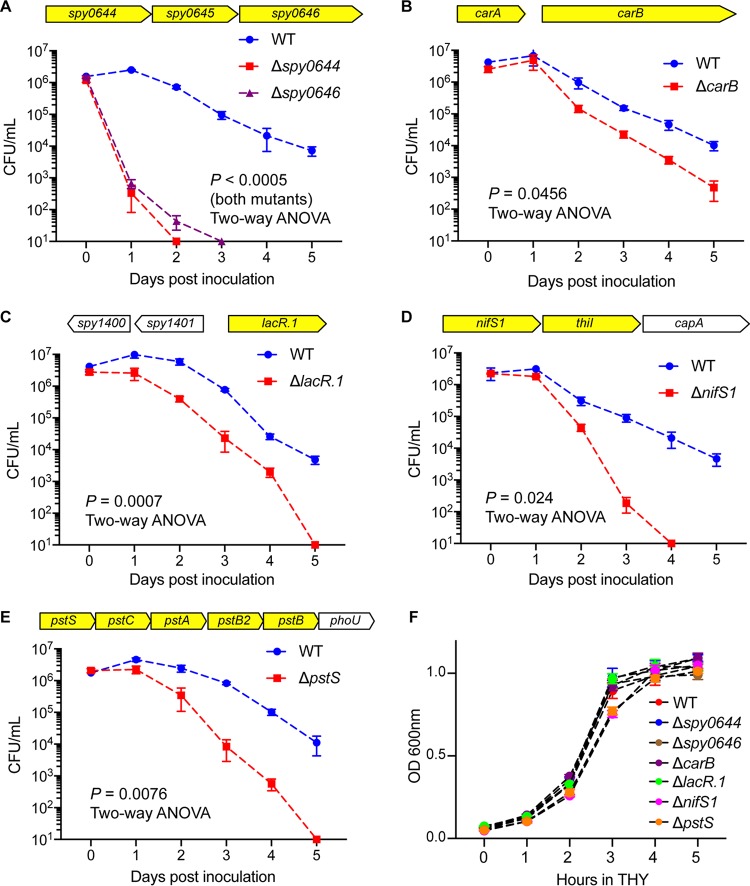
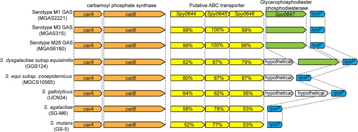
To validate the TraDIS screen findings, we analyzed the saliva fitness phenotype of each isogenic mutant strain generated from six genes (those for Spy0644, Spy0646, LacR.1, CarB, NifS1, and PstS) identified in the screen. These six genes were chosen for analysis because (i) they have not been previously shown to participate in GAS fitness in human saliva, (ii) transposon insertions into these genes represented a range of altered fitness changes, (iii) the genes are present in the core genome of all sequenced GAS genomes, (iv) the genes are known to be expressed in the oropharynx of NHPs during experimental infection (15), and (v) these gene products participate in a variety of biological pathways (Spy0644 and Spy0646 [putative ABC transporters], LacR.1 [carbohydrate metabolism], CarB [pyrimidine and arginine synthesis], NifS1 [amino acid metabolism], and PstS [phosphate transport]). To test the hypothesis that inactivating each of these six genes impaired GAS fitness in human saliva ex vivo, we used targeted insertional mutagenesis (31, 34, 35) to create isogenic mutant strains from wild-type parental strain MGAS2221. The genome of each isogenic strain was sequenced before use to ensure that no spurious mutations had been introduced during mutant construction. Consistent with our hypothesis, the results (Fig. 4) confirmed that these six isogenic mutant strains had significantly decreased fitness in human saliva compared to parental strain MGAS2221 (Fig. 4). Importantly, we discovered that mutant strains Δspy0644 and Δspy0646 had severely impaired fitness in saliva (Fig. 4A). Greater than 99% of the Δspy0644 and Δspy0646 strain inocula were not present as viable cells at the 24-h time point post-saliva inoculation (Fig. 4A). Genes for Spy0644, Spy0645, and Spy0646 likely constitute an operon that encodes an ABC transporter system (Fig. 4A; Table S2). On the basis of genome sequencing of thousands of strains of 20 evolutionarily diverse M protein serotypes that commonly cause human infections, these three genes are part of the core genome of GAS (32, 36). That is, these genes are present in all GAS genomes sequenced and, moreover, they are highly conserved in terms of genome location and context and of primary amino acid sequence. In addition, homologs of this three-gene region are present in related species of pathogenic streptococci, including Streptococcus agalactiae, Streptococcus dysgalactiae, Streptococcus equi, Streptococcus gallolyticus, Streptococcus mutans, and others, and are conserved in a location downstream of carB (Fig. 5). This suggests that a functional relationship exists between this ABC transporter and the metabolic activities of CarB. In this regard, we also identified carB to be important for wild-type GAS fitness in saliva (Table S2), and we confirmed that the ΔcarB isogenic mutant strain was significantly impaired in persistence in saliva (Fig. 4B). carB encodes the large subunit of carbamoylphosphate synthetase (37, 38). Carbamoylphosphate is a precursor for both pyrimidine and arginine synthesis (38). Interestingly, carB also was reported to be required for GAS fitness in human blood, a body fluid with a very different chemical composition than that for saliva (39). These results indicated that pyrimidine and arginine syntheses mediated in part by carB contribute to GAS fitness in multiple host niches. The ΔpstS mutant strain was also significantly attenuated for growth in human saliva (Fig. 4E). pstS encodes a putative phosphate binding protein and is part of a six-gene operon that encodes a phosphate uptake system (Fig. 4E). A genome-wide transposon mutagenesis screen found that pstS is required for the fitness of Streptococcus pneumoniae in human saliva ex vivo (40). We also noted that pst operon genes have been reported to contribute to the virulence of multiple Gram-negative pathogens, including Proteus mirabilis and Escherichia coli (41–45). These results suggest efficient phosphate uptake is important for fitness of multiple human and animal pathogens that must successfully interact with their specific host niches, including saliva in the oropharynx. Growth of the six mutants in rich medium in vitro (THY) showed that none of the mutants has severe growth defects (Fig. 4F). To summarize, the saliva growth phenotype of these six isogenic mutant strains strongly reflected the fitness data obtained from the high-throughput TraDIS screen.
FIG 4 .
Validation of the findings from the TraDIS saliva screen. (A to E) The saliva persistence phenotype was determined for each of six GAS isogenic mutant strains. Highlighted genes (yellow) are the putative saliva fitness genes identified by TraDIS. P values were determined by a repeated-measures 2-way ANOVA. (F) The growth phenotype in rich medium (THY) was also determined for each of the six GAS isogenic mutant strains.
FIG 5 .
Homologous regions encoding genes for Spy0644 to Spy0646 in GAS and other bacteria. Yellow arrows represent genes for Spy0644, Spy0645, Spy0646, and their homologs. Percentages denote amino acid identities in comparison to serotype M1 GAS strain MGAS2221.
DISCUSSION
Our results present for the first time a genome-wide view of the GAS genes that contribute to pathogen fitness in human saliva, and GAS is only the second pathogen (40) for which such a screen has been performed. The work also represents the first application of TraDIS to GAS, a human-specific pathogen responsible for greater than 700 million infections each year, including 600 million pharyngitis cases (3).
Some years ago we initiated study of GAS-human saliva molecular interactions (16) with the goal of obtaining new insight into GAS gene activity during the earliest stage of oropharyngeal infection. A common theme that has emerged from many studies (16, 17, 46, 47) is that genes involved in complex carbohydrate catabolism play important roles in growth and persistence in human saliva. Saliva in the human oropharynx contains many nutrients and diverse molecules critical to innate and acquired immunity (48–50). Knowledge gained about how GAS responds to saliva can contribute to a broader understanding of host-pathogen interactions and microbial persistence on mucosal surfaces. Expression microarray analyses, immunologic methods, and in vivo gene quantification identified a genetic program used by GAS to survive in human saliva ex vivo (14, 16, 17). A key discovery was that a two-component regulatory system (TCS) of previously unknown function played a central role in pathogen survival in saliva (17), and this revealed an intimate link between metabolism, virulence factor production, and GAS persistence in saliva. However, the strategy used was unable to directly identify the specific genes contributing to GAS persistence in saliva. Our TraDIS analysis discovered that 25 of 92 (27%) genes contributing to fitness in human saliva ex vivo are involved in carbohydrate, amino acid, and inorganic ion transport and/or metabolism. Inactivation of genes in these categories is likely to significantly impair core metabolic processes, such as nutrient acquisition and use. Our results add to the important theme of an intimate linkage between metabolism and GAS persistence in human saliva. We note that a similar spectrum of genes was abundantly represented in an ex vivo human saliva transposon mutagenesis screen conducted of S. pneumoniae (40). For example, the opp operon (oligopeptide transport) and pst operon (phosphate uptake) are required for fitness in both GAS and S. pneumoniae (40) (Table S2), suggesting that certain mechanisms contributing to bacterial fitness in saliva are shared by multiple pathogens in the oropharyngeal niche.
By mining data available from our previously conducted NHP pharyngitis study, we found a large majority (74% [Table S2]) of the 92 genes discovered here that contribute to fitness in human saliva also were expressed in vivo in the monkey oropharynx (15). Several explanations may account for the lack of evidence for expression of 24% of the genes. First, the NHP study was conducted relatively early in our understanding of the annotation of the genome of the input serotype M1 strain MGAS5005. It is possible that not all of the genome of MGAS5005 was represented on the Affymetrix gene chip used in that study. Second, it is possible that some of the genes were expressed but at levels too low to detect with the techniques available at that time. Third, it may be that some of the genes in MGAS2221 are expressed at different time points than we used in the two studies, for example, farther into the asymptomatic carriage phase (that is, later than day 7), the endpoint used in our current study. Finally, human saliva ex vivo is a different environment than the primate oropharynx, a niche that also contains, for example, host innate and acquired immune cells and epithelial cells. Therefore, it was not unexpected that we did find potential differences in evidence of gene expression between the two data sets. Despite this, the remarkable 74% gene overlap unambiguously showed that many similar mechanisms are at work on GAS regardless of whether it is exposed to human saliva ex vivo or inoculated into the primate oropharynx.
For decades, the analysis of GAS-mediated processes contributing to pharyngitis was restricted predominantly to inferences obtained by evaluating serologic responses to a relatively few extracellular molecules that participate in pathogen-host molecular interactions, such as M protein, DNase B, and streptolysin O (51–53). Although important information has been obtained from these descriptive studies, the inability to directly identify large numbers of GAS genes contributing to pharyngitis means that we have a very imprecise understanding of molecular processes contributing to this important human infection. Given the 74% overlap in genes between our TraDIS screen and NHP pharyngitis expression data, it is reasonable to conclude that our study advances understanding of molecular events occurring in the oropharynx and thereby provides a critical foundation for subsequent molecular pathogenesis studies.
Growth and persistence in human saliva contribute to the ability of GAS to be successfully transmitted by respiratory droplets. The work of Hamburger (9–11) demonstrated that individuals with higher CFU of GAS in saliva were more likely to transmit the organism to others. This observation implies that any process detrimentally affecting fitness in saliva (such as gene inactivation, as found herein) is likely to decrease the probability of successful transmission. It also suggests that mutants with substantial fitness defects are more highly likely to have decreased ability to disseminate and/or cause clinical pharyngitis. The isogenic mutants constructed by inactivation of the Spy0644 gene (here denoted sptA, for streptococcal persistence) and the Spy0646 gene (sptC) had the most pronounced growth phenotypes of the six isogenic mutant strains during saliva growth ex vivo. Of note, the transposon mutants of these two genes had the lowest fitness scores at the early time point (12 h) of any of the six mutants we tested (Table S2). In contrast, according to the screen, carB and lacR.1 transposon mutants had significantly decreased fitness only at the latest time point (48 h) (Table S2). Isogenic mutants ΔcarB and ΔlacR.1 had a moderate growth phenotype on saliva, compared to the ΔsptA and ΔsptC strains (Fig. 4B and C). These findings suggest that a relationship exists between the magnitude of the fold change at an early time point and growth of the resulting isogenic mutant strain in saliva ex vivo. However, more data generated with additional isogenic mutant strains are required to rigorously test this idea. If true, use of these data could be an important characteristic used to help identify through triage the GAS genes for which more in-depth analyses should be conducted, including translation research activities.
Our results showed that mutants with insertions in pst operon genes were significantly attenuated for growth in human saliva (Fig. 4E; Table S2). The pst operon encodes a high-affinity phosphate transporter and has been reported to contribute to bacterial virulence and fitness in a wide variety of pathogens (41, 43–45, 54–56). For example, genome-wide transposon mutagenesis screens found that the pst operon is required for the fitness of S. pneumoniae in human saliva ex vivo (40) and mouse lung infections (57). With the oral pathogen S. mutans, deletion of pstS resulted in decreased production of extracellular polysaccharide and reduced the ability to adhere to saliva-coated surfaces (56). The pst operon genes in Gram-negative pathogens have been reported to contribute to the virulence of Proteus mirabilis and Escherichia coli (41–45, 54). Together, these results suggest efficient phosphate uptake is important for the ability of multiple human and animal pathogens to survive and thrive in their specific host niches, including S. pyogenes in human saliva.
The transposon mutagenesis screen study we performed had several limitations. In the human oropharynx, saliva is constantly replenished, whereas in our experimental system a single aliquot of pooled saliva was used for the entire incubation period. A second important limitation was the lack of intact immune cells contributing to innate and acquired immunity in the saliva preparation used. These and other cells (e.g., epithelial cells) were lacking, because the saliva was filter sterilized prior to use. It is also possible that very small genes were missed in the screen because we excluded from the analysis genes with fewer than 10 inserts, a common practice in transposon mutagenesis studies (33, 58). Overcoming some of these limitations will require experimental infection of an intact animal that faithfully recapitulates all phases of human pharyngitis, such as NHPs. A third limitation is that the six isogenic mutants we used to validate the TraDIS results were generated by insertional inactivation. Although the isogenic mutants have no spurious mutations and their phenotype recapitulated the TraDIS findings, this does not rule out potential polar effects on neighboring genes, especially genes located in the same operon. To overcome this limitation in future follow-up in-depth functional studies on key genes and operons identified in this initial screen, nonpolar deletion mutants and complemented mutant strains could be used.
In summary, by identifying genes contributing to fitness in human saliva ex vivo, our work complements important information obtained in other transposon mutant screens that identified GAS genes that contribute to fitness in human blood ex vivo and mouse soft tissue disease after subcutaneous infection and also for growth in vitro (39, 59, 60). It is reasonable to suggest that the information we obtained from this genome-wide screen for genes contributing to fitness in human saliva can be successfully exploited for future pathogenesis investigations.
MATERIALS AND METHODS
Bacterial strains and growth conditions.
Strain MGAS2221 is genetically representative of a pandemic clone of serotype M1 that arose in the 1980s and has spread worldwide (30–32). Isogenic mutant strains ΔcarB, ΔlacR.1, ΔsptA, ΔsptC, ΔnifS1, ΔpstS were derived from parental strain MGAS2221, the organism used for construction of the transposon mutant library. All GAS strains were grown in Todd-Hewitt broth supplemented with 0.2% yeast extract (THY broth) at 37°C with 5% CO2.
Generation of GAS transposon mutant library.
A transposon mutant library was generated in strain MGAS2221 using transposon plasmid pGh9:ISS1 based on a recently described protocol (26). Briefly, pGh9:ISS1 was transformed into strain MGAS2221 by electroporation (26). A single colony of the transformants was picked and grown overnight at 28°C (permissive temperature) in THY broth supplemented with 0.5 μg/ml erythromycin. The resulting overnight culture was heat shocked at 40°C (nonpermissive temperature) for 3 h to permit random transposition and integration of pGh9:ISS1 into the GAS genome. The GAS cells in the culture were harvested by centrifugation, plated on THY agar supplemented with 0.5 μg/ml erythromycin, and grown overnight at 37°C. The transposon mutant library (i.e., pooled transposon mutants) was collected by washing the colonies off the agar plates with THY broth containing 25% glycerol. The bacterial cell suspension (transposon mutant library) was stored at −80°C. This process was repeated on three separate occasions, and the three libraries were pooled.
Human saliva collection and processing.
Human saliva was collected from healthy volunteers under a Houston Methodist Research Institute Institutional Review Board human subjects protocol and processed as described previously (16, 17). Briefly, saliva was collected from five healthy donors, pooled, clarified by centrifugation at 45,000 × g for 15 min, and sterilized with a 0.20-μm filter (Corning Inc.). The resulting sterile saliva was used for subsequent transposon mutant library screening and individual strain growth.
Exposure of the transposon mutant library to human saliva ex vivo.
The transposon mutant library was inoculated into 20 ml of THY and grown at 37°C to mid-exponential phase (an optical density [OD] of 0.5). The bacteria were harvested by centrifugation and washed three times with an equal volume of phosphate-buffered saline (PBS) to remove trace THY broth. A 50-µl aliquot of the cell suspension in PBS was inoculated into four tubes, each containing 40 ml of filtered saliva (Fig. S1). The four inoculated tubes constituted four biological replicates for the saliva persistence assay (Fig. S1). Immediately after inoculation, 200 µl of the inoculated saliva from each of the four replicate cultures was plated onto four THY plates and incubated at 37°C for 12 h. GAS cells growing on the plates were harvested and represented the composition of the input mutant pools (0-h mutant pools, n = 4). Mutant pools present at 12 h, 24 h, and 48 h postinoculation were recovered by plating 200 µl of the inoculated saliva onto THY agar plates at the aforementioned time points and incubating plates for 12 h at 37°C. The collected mutant pools (4 replicates, 4 time points; n = 16) were stored at −80°C for TraDIS analysis.
Experimental strategy of TraDIS mutant screens in human saliva. Download FIG S1, PDF file, 0.5 MB (523.4KB, pdf) .
Copyright © 2017 Zhu et al.
This content is distributed under the terms of the Creative Commons Attribution 4.0 International license.
DNA preparation and massive parallel sequencing.
Genomic DNA preparation and DNA sequencing were performed according to procedures described previously for TraDIS analysis, with minor modifications (26). Briefly, genomic DNA of mutant pools collected at the various time points was isolated using a DNeasy blood and tissue kit (Qiagen). Two micrograms of purified genomic DNA was fragmented by incubating with NEBNext dsDNA Fragmentase (New England Biolabs) for 25 min at 37°C to obtain DNA fragments in the range of 200 to 1,000 bp. A Y-adaptor (26) (Table S7) was ligated to 1 μg of fragmented DNA by using the NEBNext Ultra II DNA library prep kit for Illumina (New England Biolabs). The adaptor-ligated DNA fragments were then purified using AMPure XP beads (Agencourt, Beckman Coulter, Inc.) and digested with restriction enzyme BamHI for 3 h at 37°C to minimize the mapping of TraDIS reads to the transposon plasmid backbone (26). The resulting DNA was purified, and 100 ng of the purified library DNA was subjected to PCR using the specific ISS1 primer and one of the 8 indexing PCR primers per DNA library (Table S7), in order to amplify regions that spanned the 5′ end of ISS1 and the GAS genomic regions adjacent to the chromosomal location of the transposon. The PCR-amplified libraries were sequenced using a single-end 76-cycle protocol on a NextSeq550 instrument (Illumina) using a custom Read 1 primer (Table S7) and a custom Index Read sequencing primer (Table S7).
Primers used in the study. Download TABLE S7, PDF file, 0.04 MB (40.7KB, pdf) .
Copyright © 2017 Zhu et al.
This content is distributed under the terms of the Creative Commons Attribution 4.0 International license.
Processing of DNA sequencing reads and data analysis.
The raw Illumina reads obtained from the input and output pools were parsed with the FASTX Barcode Splitter program (http://hannonlab.cshl.edu/fastx_toolkit/commandline.html). After removing adaptor, low-quality reads, and index sequences, PRINSEQ Lite version 0.20.4 (http://prinseq.sourceforge.net/) was used to eliminate reads shorter than 40 nucleotides. The resulting sequencing reads were analyzed with the TraDIS toolkit (25) according to previously described methods (26, 61). Briefly, bacteria_tradis was used to trim transposon tag sequences and map the remaining reads to the serotype M1 strain MGAS5005 reference genome. The plot files generated by bacteria_tradis were analyzed by tradis_gene_insert_sites to generate spreadsheets listing the read count, insertion count, and insertion index for each gene. The output files from the tradis_gene_insert_sites analysis were transferred to tradis_comparison.R to compare the reads mapped per gene between the input pools (t0 pools) and the output pools (t12, t24, and t48 pools). Essential genes were determined by analyzing the input library using the tradis_essentiality TraDIS Script toolkit (25).
Construction of isogenic mutant strains.
Construction of isogenic mutant strains was performed by previously described methods (31, 34, 35). An internal fragment from six different genes identified as important for fitness in saliva (genes for Spy0644, Spy0646, CarB, LacR.1, NifS1, and PstS) was amplified by PCR from genomic DNA from wild-type parental M1 strain MGAS2221 with relevant primers (Table S7), digested with BamHI, and cloned into suicide vector pBBL740 (62). The resulting constructs were transformed into strain MGAS2221 to inactivate each of the six genes. pBBL740 has a cat gene which confers chloramphenicol resistance. The plasmid integrant strains (mutants) were selected using THY agar plates supplemented with 5 μg/ml chloramphenicol. The genome of each isogenic strain was sequenced before use to confirm the mutant construct and ensure that no spurious mutations had been introduced during mutant construction.
Saliva growth assay.
To compare the ability of GAS strains to grow in human saliva, we first grew GAS strains to mid-exponential phase in THY broth (OD of 0.5). GAS cells were washed three times with PBS and suspended with an equivalent volume of PBS. A 100-µl aliquot of the GAS-PBS suspension was inoculated into 10-ml aliquots of filter-sterilized human saliva. GAS strains in saliva were incubated at 37°C with 5% CO2. Samples (100 µl) of the GAS-saliva suspension were recovered over time, and GAS CFU were determined by serial dilution and growth on blood agar plates. Four biological replicates were included for each strain. Statistical significance was assessed by a repeated-measures 2-way analysis of variance (ANOVA).
ACKNOWLEDGMENTS
This work was supported by funds from the Fondren Foundation (to James M. Musser) and the Horse Trust (G4104) (to Andrew S. Waller). Amelia R. L. Charbonneau is supported by the University of Cambridge Doctoral Training Partnership scheme, which is funded by the Biotechnology and Biological Sciences Research Council, United Kingdom (1503883).
We thank Kathryn Stockbauer for critical reading of the manuscript. We thank Matthew Ojeda Saavedra for assistance with the Illumina sequencing.
Footnotes
This paper was submitted via the mSphereDirect® pathway.
Contributor Information
Sarah E. F. D'Orazio, University of Kentucky.
Robert G. Quivey, Jr., University of Rochester School of Medicine & Dentistry.
Harry L. T. Mobley, University of Michigan Medical School.
REFERENCES
- 1.Bäumler A, Fang FC. 2013. Host specificity of bacterial pathogens. Cold Spring Harb Perspect Med 3:a010041. doi: 10.1101/cshperspect.a010041. [DOI] [PMC free article] [PubMed] [Google Scholar]
- 2.Monack DM, Mueller A, Falkow S. 2004. Persistent bacterial infections: the interface of the pathogen and the host immune system. Nat Rev Microbiol 2:747–765. doi: 10.1038/nrmicro955. [DOI] [PubMed] [Google Scholar]
- 3.Carapetis JR, Steer AC, Mulholland EK, Weber M. 2005. The global burden of group A streptococcal diseases. Lancet Infect Dis 5:685–694. doi: 10.1016/S1473-3099(05)70267-X. [DOI] [PubMed] [Google Scholar]
- 4.Peter G, Smith AL. 1977. Group A streptococcal infections of the skin and pharynx (second of two parts). N Engl J Med 297:365–370. doi: 10.1056/NEJM197708182970706. [DOI] [PubMed] [Google Scholar]
- 5.Bisno AL. 2001. Acute pharyngitis. N Engl J Med 344:205–211. doi: 10.1056/NEJM200101183440308. [DOI] [PubMed] [Google Scholar]
- 6.Eisenberg MJ. 1993. Rheumatic heart disease in the developing world: prevalence, prevention, and control. Eur Heart J 14:122–128. doi: 10.1093/eurheartj/14.1.122. [DOI] [PubMed] [Google Scholar]
- 7.Walker MJ, Barnett TC, McArthur JD, Cole JN, Gillen CM, Henningham A, Sriprakash KS, Sanderson-Smith ML, Nizet V. 2014. Disease manifestations and pathogenic mechanisms of group A Streptococcus. Clin Microbiol Rev 27:264–301. doi: 10.1128/CMR.00101-13. [DOI] [PMC free article] [PubMed] [Google Scholar]
- 8.Wilkening RV, Federle MJ. 2017. Evolutionary constraints shaping Streptococcus pyogenes-host interactions. Trends Microbiol 25:562–572. doi: 10.1016/j.tim.2017.01.007. [DOI] [PMC free article] [PubMed] [Google Scholar]
- 9.Hamburger M. 1944. Studies on the transmission of hemolytic streptococcus infections. I. Cross infections in army hospital wards. J Infect Dis 75:58–70. doi: 10.1093/infdis/75.1.58. [DOI] [Google Scholar]
- 10.Hamburger M, Puck TT, Hamburger VG, Johnson MA. 1944. Studies on the transmission of hemolytic Streptococcus infections. II. Beta hemolytic streptococci in the saliva of persons with positive throat cultures. J Infect Dis 75:71–78. doi: 10.1093/infdis/75.1.71. [DOI] [Google Scholar]
- 11.Hamburger M, Robertson OH. 1948. Expulsion of group A hemolytic streptococci in droplets and droplet nuclei by sneezing, coughing, and talking. Am J Med 4:690–701. doi: 10.1016/S0002-9343(48)90392-1. [DOI] [PubMed] [Google Scholar]
- 12.Kaplan EL, Couser R, Huwe BB, McKay C, Wannamaker LW. 1979. Significance of quantitative salivary cultures for group A and non-group A and non-group A beta-hemolytic streptococci in patients with pharyngitis and in their family contacts. Pediatrics 64:904–912. [PubMed] [Google Scholar]
- 13.Sitkiewicz I, Musser JM. 2006. Expression microarray and mouse virulence analysis of four conserved two-component gene regulatory systems in group a streptococcus. Infect Immun 74:1339–1351. doi: 10.1128/IAI.74.2.1339-1351.2006. [DOI] [PMC free article] [PubMed] [Google Scholar]
- 14.Virtaneva K, Graham MR, Porcella SF, Hoe NP, Su H, Graviss EA, Gardner TJ, Allison JE, Lemon WJ, Bailey JR, Parnell MJ, Musser JM. 2003. Group A Streptococcus gene expression in humans and cynomolgus macaques with acute pharyngitis. Infect Immun 71:2199–2207. doi: 10.1128/IAI.71.4.2199-2207.2003. [DOI] [PMC free article] [PubMed] [Google Scholar]
- 15.Virtaneva K, Porcella SF, Graham MR, Ireland RM, Johnson CA, Ricklefs SM, Babar I, Parkins LD, Romero RA, Corn GJ, Gardner DJ, Bailey JR, Parnell MJ, Musser JM. 2005. Longitudinal analysis of the group A Streptococcus transcriptome in experimental pharyngitis in cynomolgus macaques. Proc Natl Acad Sci U S A 102:9014–9019. doi: 10.1073/pnas.0503671102. [DOI] [PMC free article] [PubMed] [Google Scholar]
- 16.Shelburne SA III, Granville C, Tokuyama M, Sitkiewicz I, Patel P, Musser JM. 2005. Growth characteristics of and virulence factor production by group A Streptococcus during cultivation in human saliva. Infect Immun 73:4723–4731. doi: 10.1128/IAI.73.8.4723-4731.2005. [DOI] [PMC free article] [PubMed] [Google Scholar]
- 17.Shelburne SA III, Sumby P, Sitkiewicz I, Granville C, DeLeo FR, Musser JM. 2005. Central role of a bacterial two-component gene regulatory system of previously unknown function in pathogen persistence in human saliva. Proc Natl Acad Sci U S A 102:16037–16042. doi: 10.1073/pnas.0505839102. [DOI] [PMC free article] [PubMed] [Google Scholar]
- 18.Shea PR, Virtaneva K, Kupko JJ III, Porcella SF, Barry WT, Wright FA, Kobayashi SD, Carmody A, Ireland RM, Sturdevant DE, Ricklefs SM, Babar I, Johnson CA, Graham MR, Gardner DJ, Bailey JR, Parnell MJ, Deleo FR, Musser JM. 2010. Interactome analysis of longitudinal pharyngeal infection of cynomolgus macaques by group A Streptococcus. Proc Natl Acad Sci U S A 107:4693–4698. doi: 10.1073/pnas.0906384107. [DOI] [PMC free article] [PubMed] [Google Scholar]
- 19.Langridge GC, Phan MD, Turner DJ, Perkins TT, Parts L, Haase J, Charles I, Maskell DJ, Peters SE, Dougan G, Wain J, Parkhill J, Turner AK. 2009. Simultaneous assay of every Salmonella typhi gene using one million transposon mutants. Genome Res 19:2308–2316. doi: 10.1101/gr.097097.109. [DOI] [PMC free article] [PubMed] [Google Scholar]
- 20.Chaudhuri RR, Morgan E, Peters SE, Pleasance SJ, Hudson DL, Davies HM, Wang J, van Diemen PM, Buckley AM, Bowen AJ, Pullinger GD, Turner DJ, Langridge GC, Turner AK, Parkhill J, Charles IG, Maskell DJ, Stevens MP. 2013. Comprehensive assignment of roles for Salmonella typhimurium genes in intestinal colonization of food-producing animals. PLoS Genet 9:e1003456. doi: 10.1371/journal.pgen.1003456. [DOI] [PMC free article] [PubMed] [Google Scholar]
- 21.Weerdenburg EM, Abdallah AM, Rangkuti F, Abd El Ghany M, Otto TD, Adroub SA, Molenaar D, Ummels R, Ter Veen K, van Stempvoort G, van der Sar AM, Ali S, Langridge GC, Thomson NR, Pain A, Bitter W. 2015. Genome-wide transposon mutagenesis indicates that Mycobacterium marinum customizes its virulence mechanisms for survival and replication in different hosts. Infect Immun 83:1778–1788. doi: 10.1128/IAI.03050-14. [DOI] [PMC free article] [PubMed] [Google Scholar]
- 22.Moule MG, Spink N, Willcocks S, Lim J, Guerra-Assunção JA, Cia F, Champion OL, Senior NJ, Atkins HS, Clark T, Bancroft GJ, Cuccui J, Wren BW. 2015. Characterization of new virulence factors involved in the intracellular growth and survival of Burkholderia pseudomallei. Infect Immun 84:701–710. doi: 10.1128/IAI.01102-15. [DOI] [PMC free article] [PubMed] [Google Scholar]
- 23.Grant AJ, Oshota O, Chaudhuri RR, Mayho M, Peters SE, Clare S, Maskell DJ, Mastroeni P. 2016. Genes required for the fitness of Salmonella enterica serovar Typhimurium during infection of immunodeficient gp91−/− phox mice. Infect Immun 84:989–997. doi: 10.1128/IAI.01423-15. [DOI] [PMC free article] [PubMed] [Google Scholar]
- 24.Subashchandrabose S, Smith S, DeOrnellas V, Crepin S, Kole M, Zahdeh C, Mobley HL. 2016. Acinetobacter baumannii genes required for bacterial survival during bloodstream infection. mSphere 1:e00013-15. doi: 10.1128/mSphere.00013-15. [DOI] [PMC free article] [PubMed] [Google Scholar]
- 25.Barquist L, Mayho M, Cummins C, Cain AK, Boinett CJ, Page AJ, Langridge GC, Quail MA, Keane JA, Parkhill J. 2016. The TraDIS toolkit: sequencing and analysis for dense transposon mutant libraries. Bioinformatics 32:1109–1111. doi: 10.1093/bioinformatics/btw022. [DOI] [PMC free article] [PubMed] [Google Scholar]
- 26.Charbonneau ARL, Forman OP, Cain AK, Newland G, Robinson C, Boursnell M, Parkhill J, Leigh JA, Maskell DJ, Waller AS. 2017. Defining the ABC of gene essentiality in streptococci. BMC Genomics 18:426. doi: 10.1186/s12864-017-3794-3. [DOI] [PMC free article] [PubMed] [Google Scholar]
- 27.Ruiz L, Bottacini F, Boinett CJ, Cain AK, O’Connell-Motherway M, Lawley TD, van Sinderen D. 2017. The essential genomic landscape of the commensal Bifidobacterium breve UCC2003. Sci Rep 7:5648. doi: 10.1038/s41598-017-05795-y. [DOI] [PMC free article] [PubMed] [Google Scholar]
- 28.Senior NJ, Sasidharan K, Saint RJ, Scott AE, Sarkar-Tyson M, Ireland PM, Bullifent HL, Rong Yang Z, Moore K, Oyston PCF, Atkins TP, Atkins HS, Soyer OS, Titball RW. 2017. An integrated computational-experimental approach reveals Yersinia pestis genes essential across a narrow or a broad range of environmental conditions. BMC Microbiol 17:163. doi: 10.1186/s12866-017-1073-8. [DOI] [PMC free article] [PubMed] [Google Scholar]
- 29.Musser JM, Kapur V, Szeto J, Pan X, Swanson DS, Martin DR. 1995. Genetic diversity and relationships among Streptococcus pyogenes strains expressing serotype M1 protein: recent intercontinental spread of a subclone causing episodes of invasive disease. Infect Immun 63:994–1003. [DOI] [PMC free article] [PubMed] [Google Scholar]
- 30.Sumby P, Porcella SF, Madrigal AG, Barbian KD, Virtaneva K, Ricklefs SM, Sturdevant DE, Graham MR, Vuopio-Varkila J, Hoe NP, Musser JM. 2005. Evolutionary origin and emergence of a highly successful clone of serotype M1 group a Streptococcus involved multiple horizontal gene transfer events. J Infect Dis 192:771–782. doi: 10.1086/432514. [DOI] [PubMed] [Google Scholar]
- 31.Zhu L, Olsen RJ, Nasser W, Beres SB, Vuopio J, Kristinsson KG, Gottfredsson M, Porter AR, DeLeo FR, Musser JM. 2015. A molecular trigger for intercontinental epidemics of group A Streptococcus. J Clin Invest 125:3545–3559. doi: 10.1172/JCI82478. [DOI] [PMC free article] [PubMed] [Google Scholar]
- 32.Nasser W, Beres SB, Olsen RJ, Dean MA, Rice KA, Long SW, Kristinsson KG, Gottfredsson M, Vuopio J, Raisanen K, Caugant DA, Steinbakk M, Low DE, McGeer A, Darenberg J, Henriques-Normark B, Van Beneden CA, Hoffmann S, Musser JM. 2014. Evolutionary pathway to increased virulence and epidemic group A Streptococcus disease derived from 3,615 genome sequences. Proc Natl Acad Sci U S A 111:E1768–E1776. doi: 10.1073/pnas.1403138111. [DOI] [PMC free article] [PubMed] [Google Scholar]
- 33.van Opijnen T, Bodi KL, Camilli A. 2009. Tn-seq: high-throughput parallel sequencing for fitness and genetic interaction studies in microorganisms. Nat Methods 6:767–772. doi: 10.1038/nmeth.1377. [DOI] [PMC free article] [PubMed] [Google Scholar]
- 34.Zhu L, Olsen RJ, Lee JD, Porter AR, DeLeo FR, Musser JM. 2017. Contribution of secreted NADase and streptolysin O to the pathogenesis of epidemic serotype M1 Streptococcus pyogenes infections. Am J Pathol 187:605–613. doi: 10.1016/j.ajpath.2016.11.003. [DOI] [PMC free article] [PubMed] [Google Scholar]
- 35.Zhu L, Olsen RJ, Horstmann N, Shelburne SA, Fan J, Hu Y, Musser JM. 2016. Intergenic variable-number tandem-repeat polymorphism upstream of rocA alters toxin production and enhances virulence in Streptococcus pyogenes. Infect Immun 84:2086–2093. doi: 10.1128/IAI.00258-16. [DOI] [PMC free article] [PubMed] [Google Scholar]
- 36.Beres SB, Kachroo P, Nasser W, Olsen RJ, Zhu L, Flores AR, de la Riva I, Paez-Mayorga J, Jimenez FE, Cantu C, Vuopio J, Jalava J, Kristinsson KG, Gottfredsson M, Corander J, Fittipaldi N, Di Luca MC, Petrelli D, Vitali LA, Raiford A, Jenkins L, Musser JM. 2016. Transcriptome remodeling contributes to epidemic disease caused by the human pathogen Streptococcus pyogenes. mBio 7:e00403-16. doi: 10.1128/mBio.00403-16. [DOI] [PMC free article] [PubMed] [Google Scholar]
- 37.Nyunoya H, Lusty CJ. 1983. The carB gene of Escherichia coli: a duplicated gene coding for the large subunit of carbamoyl-phosphate synthetase. Proc Natl Acad Sci U S A 80:4629–4633. doi: 10.1073/pnas.80.15.4629. [DOI] [PMC free article] [PubMed] [Google Scholar]
- 38.Arioli S, Monnet C, Guglielmetti S, Mora D. 2009. Carbamoylphosphate synthetase activity is essential for the optimal growth of Streptococcus thermophilus in milk. J Appl Microbiol 107:348–354. doi: 10.1111/j.1365-2672.2009.04213.x. [DOI] [PubMed] [Google Scholar]
- 39.Le Breton Y, Mistry P, Valdes KM, Quigley J, Kumar N, Tettelin H, McIver KS. 2013. Genome-wide identification of genes required for fitness of group A Streptococcus in human blood. Infect Immun 81:862–875. doi: 10.1128/IAI.00837-12. [DOI] [PMC free article] [PubMed] [Google Scholar]
- 40.Verhagen LM, de Jonge MI, Burghout P, Schraa K, Spagnuolo L, Mennens S, Eleveld MJ, van der Gaast-de Jongh CE, Zomer A, Hermans PW, Bootsma HJ. 2014. Genome-wide identification of genes essential for the survival of Streptococcus pneumoniae in human saliva. PLoS One 9:e89541. doi: 10.1371/journal.pone.0089541. [DOI] [PMC free article] [PubMed] [Google Scholar]
- 41.Jacobsen SM, Lane MC, Harro JM, Shirtliff ME, Mobley HL. 2008. The high-affinity phosphate transporter Pst is a virulence factor for Proteus mirabilis during complicated urinary tract infection. FEMS Immunol Med Microbiol 52:180–193. doi: 10.1111/j.1574-695X.2007.00358.x. [DOI] [PubMed] [Google Scholar]
- 42.Armbruster CE, Forsyth-DeOrnellas V, Johnson AO, Smith SN, Zhao L, Wu W, Mobley HLT. 2017. Genome-wide transposon mutagenesis of Proteus mirabilis: essential genes, fitness factors for catheter-associated urinary tract infection, and the impact of polymicrobial infection on fitness requirements. PLoS Pathog 13:e1006434. doi: 10.1371/journal.ppat.1006434. [DOI] [PMC free article] [PubMed] [Google Scholar]
- 43.Daigle F, Fairbrother JM, Harel J. 1995. Identification of a mutation in the pst-phoU operon that reduces pathogenicity of an Escherichia coli strain causing septicemia in pigs. Infect Immun 63:4924–4927. [DOI] [PMC free article] [PubMed] [Google Scholar]
- 44.Cheng C, Tennant SM, Azzopardi KI, Bennett-Wood V, Hartland EL, Robins-Browne RM, Tauschek M. 2009. Contribution of the pst-phoU operon to cell adherence by atypical enteropathogenic Escherichia coli and virulence of Citrobacter rodentium. Infect Immun 77:1936–1944. doi: 10.1128/IAI.01246-08. [DOI] [PMC free article] [PubMed] [Google Scholar]
- 45.Lamarche MG, Dozois CM, Daigle F, Caza M, Curtiss R III, Dubreuil JD, Harel J. 2005. Inactivation of the pst system reduces the virulence of an avian pathogenic Escherichia coli O78 strain. Infect Immun 73:4138–4145. doi: 10.1128/IAI.73.7.4138-4145.2005. [DOI] [PMC free article] [PubMed] [Google Scholar]
- 46.Shelburne SA III, Sumby P, Sitkiewicz I, Okorafor N, Granville C, Patel P, Voyich J, Hull R, DeLeo FR, Musser JM. 2006. Maltodextrin utilization plays a key role in the ability of group A Streptococcus to colonize the oropharynx. Infect Immun 74:4605–4614. doi: 10.1128/IAI.00477-06. [DOI] [PMC free article] [PubMed] [Google Scholar]
- 47.Shelburne SA III, Sahasrobhajane P, Suber B, Keith DB, Davenport MT, Horstmann N, Kumaraswami M, Olsen RJ, Brennan RG, Musser JM. 2011. Niche-specific contribution to streptococcal virulence of a MalR-regulated carbohydrate binding protein. Mol Microbiol 81:500–514. doi: 10.1111/j.1365-2958.2011.07708.x. [DOI] [PMC free article] [PubMed] [Google Scholar]
- 48.Wiesner J, Vilcinskas A. 2010. Antimicrobial peptides: the ancient arm of the human immune system. Virulence 1:440–464. doi: 10.4161/viru.1.5.12983. [DOI] [PubMed] [Google Scholar]
- 49.Marcotte H, Lavoie MC. 1998. Oral microbial ecology and the role of salivary immunoglobulin A. Microbiol Mol Biol Rev 62:71–109. [DOI] [PMC free article] [PubMed] [Google Scholar]
- 50.Humphrey SP, Williamson RT. 2001. A review of saliva: normal composition, flow, and function. J Prosthet Dent 85:162–169. doi: 10.1067/mpr.2001.113778. [DOI] [PubMed] [Google Scholar]
- 51.McCarty M. 1949. The inhibition of streptococcal desoxyribonuclease by rabbit and human antisera. J Exp Med 90:543–553. doi: 10.1084/jem.90.6.543. [DOI] [PMC free article] [PubMed] [Google Scholar]
- 52.Kaplan EL, Rothermel CD, Johnson DR. 1998. Antistreptolysin O and anti-deoxyribonuclease B titers: normal values for children ages 2 to 12 in the United States. Pediatrics 101:86–88. doi: 10.1542/peds.101.1.86. [DOI] [PubMed] [Google Scholar]
- 53.Hysmith ND, Kaplan EL, Cleary PP, Johnson DR, Penfound TA, Dale JB. 2017. Prospective longitudinal analysis of immune responses in pediatric subjects after pharyngeal acquisition of group A streptococci. J Pediatr Infect Dis Soc 6:187–196. doi: 10.1093/jpids/piw070. [DOI] [PMC free article] [PubMed] [Google Scholar]
- 54.O’May GA, Jacobsen SM, Longwell M, Stoodley P, Mobley HL, Shirtliff ME. 2009. The high-affinity phosphate transporter Pst in Proteus mirabilis HI4320 and its importance in biofilm formation. Microbiology 155:1523–1535. doi: 10.1099/mic.0.026500-0. [DOI] [PMC free article] [PubMed] [Google Scholar]
- 55.Lamarche MG, Wanner BL, Crépin S, Harel J. 2008. The phosphate regulon and bacterial virulence: a regulatory network connecting phosphate homeostasis and pathogenesis. FEMS Microbiol Rev 32:461–473. doi: 10.1111/j.1574-6976.2008.00101.x. [DOI] [PubMed] [Google Scholar]
- 56.Luz DE, Nepomuceno RS, Spira B, Ferreira RC. 2012. The Pst system of Streptococcus mutans is important for phosphate transport and adhesion to abiotic surfaces. Mol Oral Microbiol 27:172–181. doi: 10.1111/j.2041-1014.2012.00641.x. [DOI] [PubMed] [Google Scholar]
- 57.Hava DL, Camilli A. 2002. Large-scale identification of serotype 4 Streptococcus pneumoniae virulence factors. Mol Microbiol 45:1389–1406. doi: 10.1046/j.1365-2958.2002.t01-1-03106.x. [DOI] [PMC free article] [PubMed] [Google Scholar]
- 58.van Opijnen T, Camilli A. 2013. Transposon insertion sequencing: a new tool for systems-level analysis of microorganisms. Nat Rev Microbiol 11:435–442. doi: 10.1038/nrmicro3033. [DOI] [PMC free article] [PubMed] [Google Scholar]
- 59.Le Breton Y, Belew AT, Valdes KM, Islam E, Curry P, Tettelin H, Shirtliff ME, El-Sayed NM, McIver KS. 2015. Essential genes in the core genome of the human pathogen Streptococcus pyogenes. Sci Rep 5:9838. doi: 10.1038/srep09838. [DOI] [PMC free article] [PubMed] [Google Scholar]
- 60.Le Breton Y, Belew AT, Freiberg JA, Sundar GS, Islam E, Lieberman J, Shirtliff ME, Tettelin H, El-Sayed NM, McIver KS. 2017. Genome-wide discovery of novel M1T1 group A streptococcal determinants important for fitness and virulence during soft tissue infection. PLoS Pathog 13:e1006584. doi: 10.1371/journal.ppat.1006584. [DOI] [PMC free article] [PubMed] [Google Scholar]
- 61.Dembek M, Barquist L, Boinett CJ, Cain AK, Mayho M, Lawley TD, Fairweather NF, Fagan RP. 2015. High-throughput analysis of gene essentiality and sporulation in Clostridium difficile. mBio 6:e02383-14. doi: 10.1128/mBio.02383-14. [DOI] [PMC free article] [PubMed] [Google Scholar]
- 62.Ramirez-Peña E, Treviño J, Liu Z, Perez N, Sumby P. 2010. The group A Streptococcus small regulatory RNA FasX enhances streptokinase activity by increasing the stability of the ska mRNA transcript. Mol Microbiol 78:1332–1347. doi: 10.1111/j.1365-2958.2010.07427.x. [DOI] [PMC free article] [PubMed] [Google Scholar]
Associated Data
This section collects any data citations, data availability statements, or supplementary materials included in this article.
Supplementary Materials
Essential genes identified in the genome of serotype M1 GAS strain MGAS2221 under the conditions tested. Download TABLE S1, PDF file, 0.1 MB (135.3KB, pdf) .
Copyright © 2017 Zhu et al.
This content is distributed under the terms of the Creative Commons Attribution 4.0 International license.
Gene mutations conferring decreased fitness after 12, 24, and 48 h of incubation with human saliva. Download TABLE S2, PDF file, 0.1 MB (60.6KB, pdf) .
Copyright © 2017 Zhu et al.
This content is distributed under the terms of the Creative Commons Attribution 4.0 International license.
Gene mutations conferring increased fitness after 12, 24, and 48 h of incubation with human saliva. Download TABLE S3, PDF file, 0.05 MB (48.7KB, pdf) .
Copyright © 2017 Zhu et al.
This content is distributed under the terms of the Creative Commons Attribution 4.0 International license.
Fitness score of each gene in the M1 GAS genome after the 12-h saliva incubation. Download TABLE S4, PDF file, 0.1 MB (76.5KB, pdf) .
Copyright © 2017 Zhu et al.
This content is distributed under the terms of the Creative Commons Attribution 4.0 International license.
Fitness score of each gene in the M1 GAS genome after the 24-h saliva incubation. Download TABLE S5, PDF file, 0.1 MB (122.6KB, pdf) .
Copyright © 2017 Zhu et al.
This content is distributed under the terms of the Creative Commons Attribution 4.0 International license.
Fitness score of each gene in the M1 GAS genome after the 48-h saliva incubation. Download TABLE S6, PDF file, 0.1 MB (131.1KB, pdf) .
Copyright © 2017 Zhu et al.
This content is distributed under the terms of the Creative Commons Attribution 4.0 International license.
Experimental strategy of TraDIS mutant screens in human saliva. Download FIG S1, PDF file, 0.5 MB (523.4KB, pdf) .
Copyright © 2017 Zhu et al.
This content is distributed under the terms of the Creative Commons Attribution 4.0 International license.
Primers used in the study. Download TABLE S7, PDF file, 0.04 MB (40.7KB, pdf) .
Copyright © 2017 Zhu et al.
This content is distributed under the terms of the Creative Commons Attribution 4.0 International license.


