Abstract
Anterior–posterior (A–P) polarity of mouse embryos is established by distal visceral endoderm (DVE) at embryonic day (E) 5.5. Lefty1 is expressed first at E3.5 in a subset of epiblast progenitor cells (L1epi cells) and then in a subset of primitive endoderm cells (L1dve cells) fated to become DVE. Here we studied how prospective DVE cells are selected. Lefty1 expression in L1epi and L1dve cells depends on Nodal signaling. A cell that experiences the highest level of Nodal signaling begins to express Lefty1 and becomes an L1epi cell. Deletion of Lefty1 alone or together with Lefty2 increased the number of prospective DVE cells. Ablation of L1epi or L1dve cells triggered Lefty1 expression in a subset of remaining cells. Our results suggest that selection of prospective DVE cells is both random and regulated, and that a fixed prepattern for the A–P axis does not exist before the blastocyst stage.
In the mouse embryo, anterior-posterior polarity is established by distal visceral endoderm (DVE) at embryonic day 5.5 but how this arises is unclear. Here, the authors show that expression of Lefty1 earlier can define DVE, and that future DVE cells are selected by Nodal signalling and stochasticity.
Introduction
In Drosophila, the anterior–posterior (A–P) body axis is specified by maternal determinants that are asymmetrically distributed within the oocyte with respect to future A–P polarity1. Such maternal determinants do not appear to exist for mammals such as the mouse, however, with the mechanism by which A–P polarity is established in these animals having remained unknown. A–P polarity is established in the mouse embryo when the distal visceral endoderm (DVE) migrates toward the future anterior side at embryonic day (E) 5.5 (refs. 2–7). Concomitant with DVE migration, all visceral endoderm (VE) cells in the embryonic region undergo global movement, resulting in the localization of some VE cells at the distal tip of the embryo. These VE cells at the distal tip will become the anterior visceral endoderm (AVE) and migrate toward the future anterior side of the embryo by following the migration of DVE8. Development of the A–P axis is thus a self-organizing process that does not require maternal cues.9,10
Whereas A–P polarity of the mouse embryo is firmly established during the period from E5.5 to E6.5, its origin can be traced back to preimplantation stages of development. Lefty1 is a marker of both DVE and AVE, but its expression begins in the blastocyst. It is expressed first in a subset of epiblast progenitor cells and then in a subset of primitive endoderm (PrE) progenitors, the latter of which is fated to become DVE. Expression of Lefty1 therefore marks prospective DVE cells in peri-implantation embryos8. Although generation of Lefty1+ future DVE cells9 and Cerl1+ DVE cells10,11 occurs in an embryo-autonomous manner, generation of fully functional DVE may require interaction with the uterus12. Whereas Nodal signaling13 and expression of its target gene Eomes 14 are essential for DVE formation, it has remained unknown how Lefty1 expression is induced and how prospective DVE cells are selected in peri-implantation embryos. In this study, we have now addressed these questions by studying the regulation of Lefty1 expression and its role in specification of future DVE cells. Our results suggest that selection of prospective DVE cells in mouse peri-implantation embryo is both random and regulated.
Results
Lefty1 expression is regulated by Nodal signaling
We have previously shown that Lefty1 is expressed first (at E3.5) in a subset of epiblast progenitor cells and then (between E3.75 and E4.5) in a subset of PrE progenitors fated to become DVE8, with these Lefty1+ cell subsets being herein designated L1epi cells and L1dve cells, respectively. Some DVE cells were previously reported to be derived from epiblast (Sox2+ cells) that transmigrates into VE12. We examined this possibility by testing whether Oct3/4+ and Sox2+ epiblast contributes to DVE. We were unable to detect Oct3/4 (mTomato)+ cells (7/7 embryos at E5.5), Oct3/4+ cells (14/14 embryos at E5.5) or Sox2+ cells (4/4 embryos at E5.5, 5/5 embryos at E6.0) in the DVE region (Supplementary Fig. 1), however, suggesting that all DVE cells are derived from L1dve cells between E3.75 and E4.5, as we previously described8.
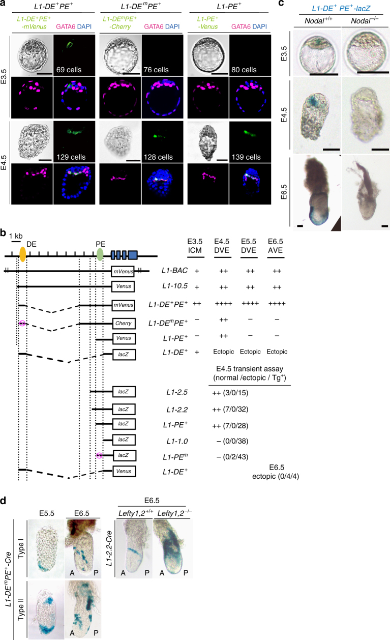
We examined how Lefty1 expression is regulated in both L1epi and L1dve cells (Fig. 1). A Lefty1(mVenus or Cherry) bacterial artificial chromosome (BAC) transgene that recapitulates Lefty1 expression in embryos8 was active in epiblast progenitor cells8 within the inner cell mass (ICM) of E3.5 embryos and in the PrE of E4.5 embryos8,9 (Supplementary Fig. 2a, b, c), representing Lefty1 expression in L1epi and L1dve cells, respectively. L1-10.5-Venus, a transgene that contains the 10.5-kb upstream region of Lefty1 and which recapitulates Lefty1 expression at E6.5 and E8.0 (refs. 9,15) (Fig. 1b), was also active at E3.5 (presumably in L1epi cells) and at E4.5 (presumably in L1dve cells) (Fig. 1b).
Fig. 1.
Lefty1 expression in L1epi and L1dve cells is regulated by Nodal-Foxh1 signaling. a Expression of three Lefty1 transgenes (L1-DE + PE + -mVenus, L1-DE m PE + -Cherry, and L1-PE + -Venus) was examined in mouse embryos at E3.5 and E4.5. Embryos were immunostained for transgene expression as well as for GATA6 (a PrE-specific marker), and they were counterstained with 4’,6-diamidino-2-phenylindole (DAPI). Bright-field images are also shown. The expression pattern of L1-DE + PE + -lacZ in wild-type embryos has been described previously8. The number of cells in each embryo is indicated. Scale bars, 50 μm. b Structures of various Lefty1 reporter transgenes and summary of their activities at the indicated stages. L1-BAC is the Lefty1(mVenus) BAC transgene generated by replacement of lacZ in the Lefty1(lacZ) BAC transgene9 with mVenus. The positions of two Foxh1-dependent enhancers, DE (distal enhancer) and PE (proximal enhancer), are indicated. The Foxh1-binding sites in DE or PE are mutated in L1-DE m PE and L1-PE m, respectively. c The expression of L1-DE + PE + -lacZ was examined by X-gal staining in Nodal +/+ and Nodal –/– embryos at the indicated stages. Scale bars, 50 µm. d L1-DE m PE + -Cre or L1-2.2-Cre transgenic mice were crossed with Rosa26R transgenic mice, and transgenic embryos recovered at E5.5 or E6.5 were stained with X-gal. Two types of embryos were observed for the L1-DE m PE + -Cre cross: type I (8/24 embryos), in which only DVE and DVE-derived cells were marked at E5.5 and E6.5, respectively; and type II (16/24 embryos), in which the extraembryonic region was positive in addition to DVE and DVE-derived cells at E5.5 and E6.5. DVE-derived cells were detected on the lateral side of E6.5 embryos produced from the L1-2.2-Cre cross (6/7 embryos). The number of DVE-derived cells was increased in E6.5 embryos produced from a cross of L1-2.2-Cre mice with Lefty1,2 –/– mice expressing Rosa26R (2/3 embryos)
Given that left–right (L–R) asymmetric expression of Lefty1 at E8.0 is regulated by Nodal-Foxh1 signaling15, we examined the possible role of such signaling in Lefty1 expression at E3.5 and E4.5. Culture of E3.2 embryos harboring a Lefty1(mVenus) BAC transgene with the Nodal signaling inhibitor SB431542 for 24 h prevented the emergence of Lefty1 expression (11/11 embryos) (Supplementary Fig. 2h). Foxh1-binding sequences that are conserved between mouse and human9 are present within the 10.5-kb upstream region of Lefty1, with two such sequences being located in the region around –1.5 kb and two in the region around –10 kb (ref. 9). Foxh1 binding has been detected at both of these regions in human embryonic stem cells by chromatin immunoprecipitation–sequencing (ChIP-seq) analysis16,17 (Supplementary Fig. 2i). The Foxh1-dependent enhancers located at –10 and –1.5 kb of Lefty1 are hereafter referred to as DE (distal enhancer) and PE (proximal enhancer), respectively.
The L1-DE + PE +-mVenus transgene, which contains DE and PE, was active in a few epiblast progenitor cells at E3.5 (L1epi cells) and in GATA6+ cells in PrE at E4.5 (L1dve cells) (Fig. 1a, b, and Supplementary Fig. 2b). L1-PE +-Venus, or L1-PE + -mVenus which contains PE but lacks DE, was inactive in the ICM at E3.5 but active in a few GATA6+ cells of PrE at E4.5 (L1dve cells) (Fig. 1a, b, and Supplementary Fig. 2c). Similarly, L1-PE +-lacZ was inactive at E3.5, active at E4.5, and inactive at E6.5 (Supplementary Fig. 2e). L1-DE + transgenes were active in the ICM at E3.5 but showed ectopic expression in the epiblast between E4.5 and E6.5 (Fig. 1b and Supplementary Fig. 2f, g). L1-DE + PE + -lacZ was not expressed at E3.5, E4.5, or E6.5 in Nodal −/− embryos (11/12 embryos at E3.5, 8/8 embryos at E4.5, 2/2 embryos at E6.5) (Fig. 1c), suggesting that the activity of both DE and PE is Nodal dependent. Consistent with this notion, L1-DE m PE + -Cherry or L1-DE m PE + -lacZ, in which the Foxh1-binding sites of DE are mutated, was inactive at E3.5 but was active at E4.5 (Fig. 1a, b, and Supplementary Fig. 2d), whereas L1-PE m -lacZ, in which the Foxh1-binding sequences of PE are mutated, was not active at E4.5 (Fig. 1b). Furthermore, SB431542 abolished expression of L1-DE + PE + -mVenus (6/6 embryos) and L1-PE + -Venus (4/4 embryos) in E3.2 embryos cultured for 24 h (Supplementary Fig. 2h).
L1-DE m PE +-Cre, a Cre transgene driven by PE, marked DVE at E5.5 and DVE-derived cells at E6.5 but failed to label epiblast at both stages (24/24 embryos) (Fig. 1d). Similarly, L1-2.2-Cre, which contains PE, specifically marked DVE-derived cells, excluding the epiblast, at E6.5 (6/7 embryos) (Fig. 1d). Together, these results suggested that the Foxh1-binding sites in DE are essential for Lefty1 expression in L1epi cells of the ICM at E3.5, whereas the Foxh1-binding sites in PE regulate Lefty1 expression in L1dve cells at E4.5. DE may also contribute to the regulation of Lefty1 expression at E4.5, given that the expression level of L1-DE m PE +-lacZ at this time (Supplementary Fig. 2d) was lower than that of L1-DE + PE +-lacZ (Fig. 1c) (note that the LacZ staining time for the former embryo was 12 h, whereas that for the latter embryo was 15 min).
Nodal signaling induces Lefty1 expression
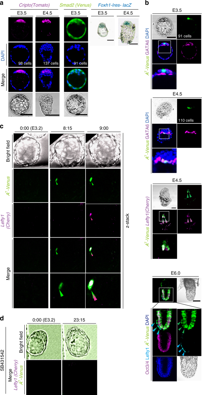
Given that our results suggested that Nodal-Foxh1 signaling regulates Lefty1 expression in L1epi and L1dve cells, we next examined expression of Nodal signaling components. Cripto (E3.5: 5/5 embryos; E4.5: 6/6 embryos), Foxh1(E3.5: 4/5 embryos; E4.5: 8/8 embryos), and Smad2(E3.5: 6/7 embryos) were all found to be expressed between E3.5 and E4.5 (Fig. 2a)9. Nodal-Foxh1 signaling activity can be monitored with a transgene driven by seven tandem repeats of a Foxh1-binding sequence18. When examined with such a transgene, A 7 -Venus, Nodal-Foxh1 signaling was found to be active in a few ICM cells at E3.5 (presumably L1epi cells, 11/11 embryos), in a few GATA6+ cells at E4.5 (L1dve cells, given that they also expressed Lefty1, 12/14 embryos), and in AVE and epiblast cells at E6.0, as expected (10/10 embryos) (Fig. 2b). Culture of E3.2 embryos for 24 h with SB431542 prevented A 7 -Venus expression (11/11 embryos) (Supplementary Fig. 3a), whereas it was maintained in the control embryos (5/5 embryos), confirming that such expression is dependent on Nodal signaling. Simultaneous monitoring of both A 7 -Venus and Lefty1 expression in live embryos from E3.2 revealed that a single cell became positive for Venus expression after culture for ~ 8 h (Fig. 2c, Supplementary Movie 1). This cell then began to express Lefty1. Lefty1+ cells in such live imaging at this time (equivalent to E3.5) were positive for Venus (15/16 embryos), which is consistent with the notion that Lefty1 expression in L1epi cells is induced by Nodal signaling. Furthermore, expression of A 7 -Venus and Lefty1(Cherry) was abolished in the presence of SB431542 (Fig. 2d, Supplementary Movie 2). Lefty1+ cells at E4.5 were also positive for Venus (7/7 embryos) (Fig. 2b), suggesting that Lefty1 expression in L1dve cells is also regulated by Nodal signaling. While Lefty1 expression was found in PrE at E4.5, Cripto was not expressed in PrE (Fig. 2a). It is most likely that PrE cells can receive Nodal siganing because Cryptic, another co-receptor for Nodal, is expressed in PrE19, and because Cripto and Cryptic can act non-cell-autonomously20,21. In support of this, Lefty1+ cells at E5.5 (DVE cells) are lost in the Cripto -/-, Cryptic -/- double mutant embryo19.
Fig. 2.
Lefty1 expression begins in cells positive for Nodal-Foxh1 signaling. a Expression of Nodal signaling components in mouse embryos at E3.5 and E4.5. Expression of Cripto(Tomato) and Smad2(Venus) BAC transgenes as well as that of GATA6 were monitored by immunofluorescence staining, whereas that of a Foxh1-Ires-lacZ transgene was monitored by X-gal staining. Scale bars, 50 μm. b Embryos harboring A 7 -Venus were examined for Venus and GATA6 immunofluorescence at E3.5 and E4.5 (top two panels). Note that Venus is expressed in a GATA6– cell at E3.5 and in GATA6+ cells at E4.5. An E4.5 embryo harboring A 7 -Venus and a Lefty1(Cherry) BAC transgene was examined for Venus and Cherry immune fluorescence (third panel). Arrowheads indicate two Lefty1+ cells that are also positive for Venus. An E6.0 embryo harboring A 7 -Venus was subjected to immunofluorescence staining for Venus, Lefty, and Oct3/4 (bottom panel). Arrowheads indicate Cherry in magenta, Venus in green and Lefty1 in light blue. Boxed regions are shown at higher magnification in the images immediately below. Scale bars, 50 μm. c An E3.2 embryo harboring both Lefty1(Cherry) and A 7 -Venus transgenes was cultured for 9 h. Fluorescence of Venus and Cherry was examined at the indicated times. A single cell positive for Venus was detected at 8.25 h (arrowhead). This cell had being positive for Cherry at 9 h. d An E3.2 embryo harboring both Lefty1(Cherry) and A 7 -Venus transgenes was cultured for 23.15 h in the presence of 10 µM SB431542 and monitored for Venus and Cherry fluorescence. Note that Venus or Cherry fluorescence is not apparent at 23.15 h
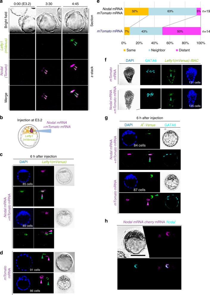
We next monitored Nodal and Lefty1 expression simultaneously in live embryos between E3.2 and E4.0 (Fig. 3a, Supplementary Movie 3). Nodal expression, as revealed with a Nodal(Tomato) BAC transgene, was dynamic, beginning in one cell, rapidly expanding to all cells, and being maintained until E4.0. Lefty1 expression (in L1epi cells) was found to begin in the same cell that had earlier initiated Nodal expression (12/24 embryos) or in a neighboring cell (11/24 embryos).
Fig. 3.
Lefty1 expression in L1epi cells is induced by Nodal. a An E3.2 embryo harboring both Lefty1(mVenus) and Nodal(Tomato) BAC transgenes was cultured for 5 h, with fluorescence of mVenus and Tomato being examined at the indicated times. Nodal expression began in two cells at 3.5 h (magenta arrowheads), with these two cells becoming positive for Lefty1 expression by 4.75 h (green arrowheads). b Schematic illustration of mRNA injection experiments in (c) and (d). mTomato mRNA (encoding a membrane-localized Tomato) was injected with or without Nodal mRNA into a single cell of E3.2 embryos harboring the Lefty1(mVenus) BAC transgene. Embryos were examined for mTomato and mVenus immunofluorescence 6 h after mRNA injection. c Embryos injected with both mTomato and Nodal mRNAs. mVenus is expressed in the injected cell (upper panel) or in a neighboring cell of the injected cell (lower panel). d Two embryos injected with mTomato mRNA alone. mTomato+ and mVenus+ cells (arrowheads) do not overlap. e Summary of the location of the mVenus+ cell relative to the injected cell for experiments similar to that in c and d. f E3.2 embryos harboring the Lefty1(mVenus) BAC transgene were injected with mTomato mRNA alone or together with Nodal mRNA, allowed to develop in utero, recovered at E4.5, and examined for Lefty1(mVenus) and GATA6 expression. Note that there is no substantial difference in the number of Lefty1-expressing cells between the two types of injected embryos (Supplementary Fig. 3b, c). g E3.2 embryos harboring A 7 -Venus were injected with mTomato mRNA alone or together with Nodal mRNA and were examined for GATA6 and Venus expression after 6 h. When mTomato mRNA alone was injected, the Venus+ cell was randomly located relative to the injected cell (lower panel). However, when Nodal mRNA was co-injected, the injected cell or a neighboring cell was positive for Venus (upper panel). h An E3.2 embryo co-injected with excess Nodal mRNA (200 ng/µl) and with mTomato mRNA was immunostained for Nodal at 6 h after mRNA injection. Scale bar, 50 μm
We also examined the effect of ectopic Nodal expression on Lefty1 expression. Injection of Nodal mRNA and mTomato mRNA into E3.2 embryos harboring the Lefty1(mVenus) BAC transgene and examination of the embryos 6 h later revealed that Lefty1 expression began either in the cell that received Nodal mRNA (6/19 embryos) or in a neighboring cell (12/19 embryos) (Fig. 3b–e). In the remaining embryo (1/19 embryos), Lefty1 expression was found in a distant cell. Injection of Nodal mRNA did not increase the number of Lefty1+ cells at E4.5 (Fig. 3f and Supplementary Fig. 3b, c). When Nodal mRNA and mTomato mRNA (encoding a membrane-localized form of Tomato) were injected into E3.2 embryos harboring the A 7 -Venus transgene, Venus was detected in the cell that received Nodal mRNA or in a neighboring cell (Fig. 3g). We confirmed that Nodal protein was produced in the excess injected cell (Fig. 3h). When mTomato mRNA alone was injected, however, Venus+ cells were located randomly relative to the mTomato+ cell (Fig. 3d, e). Collectively, these results suggested that Lefty1 expression in L1epi and L1dve cells is induced by Nodal-Foxh1 signaling.
Lefty activity restricts the number of prospective DVE cells
We next investigated whether Lefty proteins might play a role in DVE formation. In addition to Lefty1, Lefty2 was also expressed in mouse embryos between E3.5 and E4.5. Expression of Lefty2, as revealed with a Lefty2(lacZ) BAC transgene, was thus detected in a subset of ICM cells at E3.5 (Supplementary Fig. 4a, b: 16/16 embryos). At E4.5, Lefty2 was expressed in a subset of PrE and EPI cells (Supplementary Fig. 4b). Lefty2 expression at E4.5 was also dependent on Nodal-Foxh1 signaling, given that such expression was not apparent in Nodal −/− (3/3 embryos) or Foxh1 −/− (4/4 embryos) embryos (Supplementary Fig. 4a). Monitoring of Lefty2 and Lefty1 expression from E3.2 also revealed that both genes were expressed in the same ICM cells (L1epi cells) (6/10 embryos) (Supplementary Fig. 4b and Supplementary Movie 4) or in different ICM cells (4/10 embryos). Both genes also initiated their expression with similar timing (Supplementary Fig. 4c and Supplementary Movie 4). Given that Lefty2 was also found to be expressed in ICM cells at E3.5, we generated a mutant (Lefty1,2 −/−) mouse lacking both Lefty1 and Lefty2 (Supplementary Fig. 5a). Staining of Lefty1,2 −/− embryos with antibodies that recognize both Lefty1 and Lefty2 confirmed the absence of Lefty proteins (5/5 embryos) (Supplementary Fig. 5b).
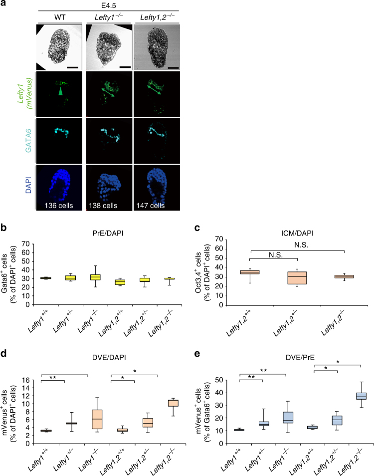
We then examined the effect of Lefty1 and Lefty2 deletion on the number of future DVE cells. Prospective DVE cells were identified and counted in Lefty1(mVenus) BAC transgenic embryos at E4.5 (Fig. 4a). The number of future DVE cells (mVenus+ cells) was increased in the absence of Lefty genes (Fig. 4a, d, e, and Supplementary Table 2), whereas the number of PrE cells (Fig. 4b, Supplementary Table 2) or ICM cells (Fig. 4c, Supplementary Fig. 5, and Supplementary Table 2) was not significantly affected. mVenus+ cells thus constituted ~ 10% of total PrE cells (GATA6+ cells) in wild-type (WT) embryos, whereas they accounted for 25 to 30% of PrE cells in some Lefty1 −/− embryos (Fig. 4e, Supplementary Table 2). This finding is consistent with our previous observation that the number of cells expressing Cerl1 (a marker for DVE) at E5.5 was increased in Lefty1 −/− embryos22. Moreover, future DVE cells accounted for 30–50% of PrE cells in Lefty1,2 −/− embryos (Fig. 4e, Supplementary Table 2). These results suggested that Lefty1 is not only a marker for future DVE cells in the blastocyst, but that it also restricts the number of prospective DVE cells together with Lefty2. Although the number of prospective DVE cells was increased in Lefty1,2 −/− embryos at E4.5, the expression patterns of Cerl1 and Hex appeared normal at E6.5 (9/9 embryos for Cerl1, 6/6 embryos for Hex) and E7.5 (5/5 embryos for Cerl1) (Supplementary Fig. 5d, e), suggesting that AVE is formed normally even though such AVE cells lack Lefty1. Since Cerl1 is required to position the primitive streak at the posterior side of the embryo23, together with Lefty1 and Lefty2, Cerl1 may have a redundant role in AVE formation.
Fig. 4.
Lefty1 and Lefty2 restrict the number of prospective DVE cells. a The number of prospective DVE cells [GATA6+ cells expressing the Lefty1(mVenus) BAC transgene] was examined in WT, Lefty1 –/–, and Lefty1,2 –/– embryos at E4.5. The embryos were subjected to immunofluorescence staining for mVenus and GATA6. Scale bars, 50 μm. Arrowheads and double-headed arrows indicate mVenus+ cells (prospective DVE cells). b–e Summary of the number of PrE cells (GATA6+ cells) as a percentage of total cells (b), the number of ICM cells positive for Oct3/4 staining as a percentage of total cells (c) (Supplementary Fig. 5c), the number of prospective DVE cells as a percentage of total cells (d), and the number of prospective DVE cells as a percentage of total PrE cells (e) in embryos of the indicated genotypes at E4.5. Data are presented as box-and-whisker plots (first and third quartile, the line represents the median; whiskers: minimum to maximum). *P < 0.05, **P < 0.01; NS, not significant (t test)
Nodal-Lefty regulatory network
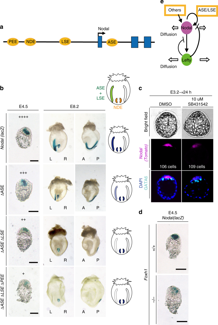
Our results indicated that Lefty1 expression is induced by Nodal signaling and that Lefty1 restricts the number of Lefty1+ cells, a scenario reminiscent of the self-enhancement and lateral inhibition (SELI) Nodal-Lefty regulatory network that operates during L–R patterning at E8.024. Given that Lefty proteins diffuse faster than Nodal25, whether a Nodal-Lefty SELI system operates in peri-implantation embryos will depend on whether Nodal expression is positively regulated by Nodal itself. We therefore investigated how Nodal expression is regulated at the blastocyst stage—in particular, whether it is positively autoregulated as it is at E8.0. Transcription of Nodal is regulated by several enhancers, including two Foxh1-dependent enhancers, ASE26,27 and LSE27, which contribute to L–R asymmetric expression of Nodal at E8.0 (Fig. 5a) and which are also active at the peri-implantation stage28. Another enhancer, PEE, whose activity depends on Wnt–β-catenin signaling, is also active in a subset of cells in the blastocyst28.
Fig. 5.
Regulation of Nodal expression in peri-implantation embryos. a Four transcriptional enhancers—ASE, LSE, NDE, and PEE—contribute to regulation of Nodal expression. b Expression of Nodal(lacZ) BAC transgenes containing all enhancers or lacking either ASE alone, ASE and LSE, or ASE, LSE, and PEE was determined in embryos at E4.5 and E8.2 by X-gal staining. Scale bars, 50 μm. In E4.5 embryos, the relative levels of Nodal expression in the different embryos are indicated by plus signs. Frontal and lateral views are shown for E8.2 embryos. X-gal+ regions are schematically summarized in diagrams on the right. Note that ASE and LSE are active in the lateral plate mesoderm, whereas NDE is active in the node40. c E3.2 embryos harboring a Nodal(Tomato) BAC transgene were cultured for 24 h with 10 µM SB431542 or dimethyl sulfoxide (DMSO) vehicle and were then examined for Tomato and GATA6 immunofluorescence. d Expression of the Nodal(lacZ) BAC transgene in Foxh1 +/+ and Foxh1 –/– embryos at E4.5. Scale bars, 50 µm. e Regulatory relation between Nodal and Lefty at the peri-implantation stage. The genes constitute a SELI system, as they do for L–R patterning at E8.0
We found that ASE, LSE, and PEE all contribute to the regulation of Nodal expression at E4.5. Deletion of ASE thus attenuated Nodal expression (14/15 embryos) (Fig. 5b). Whereas additional deletion of LSE did not have a further substantial effect on Nodal expression (17/19 embryos), which of both LSE and PEE did further reduce it (7/10 embryos) (Fig. 5b). Given that ASE and LSE are dependent on Foxh1 and that PEE is dependent on Wnt–β-catenin, these results suggested that Nodal expression in peri-implantation mouse embryos is positively regulated by Nodal signaling. Consistent with this notion, culture of E3.2 embryos with SB431542 for 24 h resulted in marked attenuation of Nodal expression, presumably in L1epi and L1dve cells (13/13 embryos) (Fig. 5c). Furthermore, Nodal expression in L1dve cells at E4.5 was greatly reduced in Foxh1 −/− embryos (4/4 embryos) (Fig. 5d). Together, these observations suggested that Nodal expression in peri-implantation embryos is positively regulated by Nodal signaling via Foxh1-dependent enhancers (Fig. 5e).
L1epi and L1dve cells are selected randomly
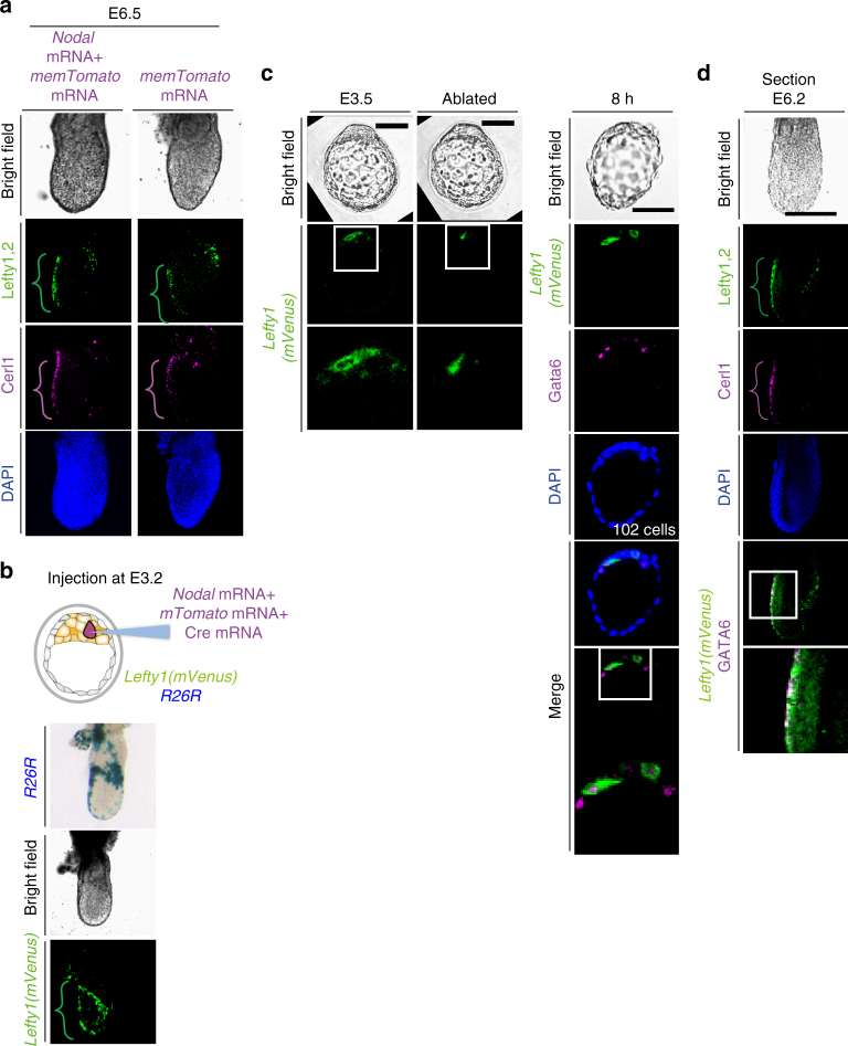
Injection of Nodal mRNA into a single cell at E3.2 induced Lefty1 expression in the same cell, generating an L1epi cell (Fig. 3c), but it did not increase the overall number of L1epi cells (Supplementary Fig. 3b, c). When such injected embryos were returned to the uterus and allowed to develop further, they developed normally at least up to E6.5 (5/5 embryos), showing normal expression patterns of Cerl1, Lefty1, and Lefty2 (Fig. 6a). Both Cerl1 and Lefty1 were thus detected at the anterior side, suggesting that the AVE was formed normally. Lefty2 was expressed at the opposite (posterior) side, suggesting that the primitive streak was formed correctly. Examination of the fate of the injected cell revealed that it survived and contributed to DVE and DVE-derived cells at E6.5 (Fig. 6b). We also removed all (usually two or three) Lefty1+ cells from Lefty1(mVenus) transgenic embryos at E3.5 by laser ablation. Cell ablation was confirmed by monitoring of the cell membrane, with successful ablation resulting in rupture of the membrane and the appearance of fluorescent cell debris (Fig. 6c). Culture of the ablated embryos revealed that Lefty1 was expressed in a different cell by 8 h after the ablation (8/8 embryos) (Fig. 6c). On return to the uterus, such ablated embryos again developed an apparently normal A–P axis by E6.5, with Lefty1 and Cerl1 expression being apparent on the anterior side of the embryo and Lefty2 expression on the posterior side (10/10 embryos) (Fig. 6d).
Fig. 6.
Ablation of L1epi cells at E3.5 results in the appearance of new L1epi cells and does not impair A–P patterning. a E3.2 embryos in which a single cell had been injected with mTomato mRNA alone or together with Nodal mRNA were transferred to a pseudopregnant mouse, allowed to develop until E6.5, and then recovered for immunofluorescence staining of A–P markers. The brackets denote AVE located on the anterior side of the embryos. Lefty1, Lefty2, and Cerl1 were detected in AVE, the primitive streak, and AVE, respectively, suggesting that a normal A–P axis was established. b An E3.2 Rosa26R (R26R) embryo harboring a Lefty1(mVenus) BAC transgene was injected with Nodal mRNA, mTomato mRNA, and Cre mRNA, transferred to a pseudopregnant mouse, allowed to develop until E6.5, and then recovered for X-gal staining and immunofluorescence staining for mVenus. Note that the injected cell survived and contributed to DVE and DVE-derived cells. c All mVenus+ cells (L1epi cells) were removed by laser ablation from an E3.5 embryo harboring a Lefty1(mVenus) BAC transgene, after which the embryo was cultured for 8 h and then subjected to immunofluorescence staining for mVenus and GATA6. Note that fluorescent membrane debris was detected immediately after ablation and that new mVenus+ cells had appeared by 8 h after the ablation. Boxed areas are shown at higher magnification in the images immediately below. Scale bar, 50 µm. d After culture, an embryo such as that in (c) was transferred to a pseudopregnant mouse, allowed to develop until E6.5, and then recovered for immunofluorescence staining of A–P markers. Lefty1, Lefty2, and Cerl1 were detected in AVE, the primitive streak, and AVE, respectively. Scale bar, 100 µm
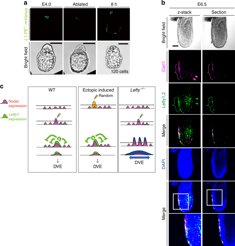
Similarly, when all (usually four or five) L1dve cells at E4.0 were ablated, a new Lefty1+ cell appeared in the PrE region by 8 h after the ablation (Fig. 7a). On return of such embryos to the uterus, they developed an apparently normal A–P axis by E6.5 (Fig. 7b).
Fig. 7.
Ablation of L1dve cells at E4.0 results in the appearance of new L1dve cells and does not impair A–P patterning. a All mVenus+ cells (L1dve cells) were removed by laser ablation from an E4.0 embryo harboring an L1-PE + -mVenus transgene (Fig. 1b), after which the embryo was cultured for 8 h and then subjected to immunofluorescence staining for mVenus. Note that a new mVenus+ cell had appeared by 8 h after the ablation. Scale bars, 50 μm. b After culture, an embryo such as that in a was transferred to a pseudopregnant mouse, allowed to develop until E6.5, and then recovered for immunofluorescence staining of A–P markers. Lefty1 and Cerl1 were detected in the AVE, suggesting that a normal A–P axis was established. Arrowheads indicate Cerl1+ definitive endoderm cells in magenta, and Lefty2+ definitive ectoderm cells in green. The boxed regions of the merged images are shown at higher magnification in the images immediately below. c Model for the spatial distribution of Nodal expression and Lefty1 expression in a peri-implantation embryo based on observations in the present study. In the WT embryo (left panel), a cell that first expresses Nodal beyond a threshold level (dotted line) begins to express Lefty1. The Lefty1 protein then produced rapidly represses Nodal signaling in nearby cells, preventing them from expressing Lefty1. Injection of Nodal mRNA into a cell (middle panel) induces expression of Lefty1. In the absence of Lefty (right panel), a larger number of cells manifest Nodal expression or activity beyond the threshold and are fated to become DVE. See text for further details
Together, these results suggested that both L1epi and L1dve cells are not predetermined but are selected in a regulated and robust manner. Furthermore, the A–P axis can be established normally even if an ectopic cell begins to express Lefty1 between E3.5 and E4.0; that is, an ectopic cell can be specified to become a prospective DVE cell.
Discussion
Lefty1 is expressed first in L1epi cells at E3.5 and subsequently in L1dve cells. Although Lefty1 expression specifically marks future DVE cells in the mouse embryo at E4.0 to E4.5, it is not absolutely required to specify DVE cells, given that Cerl1+Hex+ cells are formed at E6.5 in the absence of both Lefty1 and Lefty2. Rather, Lefty1 and Lefty2 function to restrict the number of future DVE cells. Thus, in the absence of either Lefty1 alone or both Lefty1 and Lefty2, the number of future DVE cells is increased at E4.5. The number of DVE cells at E5.5 (Cerl1-expressing cells) was previously shown to be increased in Lefty1 −/− embryos22. However, we found that the number and position of AVE cells at E6.5 (Cerl1-expressing cells) remained essentially normal in Lefty1,2 −/− embryos. Although DVE guides the anterior migration of AVE cells that arise later, the increased number of DVE cells in the mutant embryos does not appear to influence the appearance and migration of AVE cells.
Then, why does Lefty1 expression occur in two steps? In particular, what is the role of the first phase of Lefty1 expression in L1epi cells? Lefty1 expression in L1epi cells is transient, disappearing shortly after L1dve cells arise8. Lefty1 produced in L1epi cells may establish an uneven distribution of Nodal activity within the blastocyst, as revealed by the pattern of A 7 -Venus expression at E3.5 (Fig. 2c), and thereby allow only one or two cells to become L1epi cells (Fig. 7c). This would in turn restrict the number of L1dve cells and determine the position of these cells in the blastocyst. In support of this notion, L1dve cells arise in a region remote from L1epi cells8. Unfortunately, it is not technically feasible to address the role of Lefty1 in L1epi cells directly, given that a Cre transgene active specifically in E3.5 epiblasts is not currently available (the expression of zygotic Sox2-Cre 29 is not sufficiently early). Nonetheless, it is of note that the number of prospective DVE cells was increased to a markedly greater extent in Lefty1,2 −/− embryos than in Lefty1 −/− embryos. Whereas Lefty2 and Lefty1 are both expressed in epiblast-fated cells at E3.5 (Supplementary Fig. 4a–c), they are expressed in different domains at E4.5, suggesting that Lefty1 and Lefty2 in epiblast-fated cells at E3.5 regulate the number of future DVE cells.
Ablation of L1epi or L1dve cells at E3.5 and E4.0, respectively, resulted in the initiation of Lefty1 expression in remaining cells that presumably took over the role of the ablated cells. Injection of Nodal mRNA into a single cell of E3.2 embryos induced Lefty1 expression, but did not increase the number of L1epi cells. Together, these results suggest that selection of both L1epi and L1dve cells is random and regulated (Fig. 7c). Consistent with this notion, the positions of L1epi cells at E3.5 and of L1dve cells at E4.0 appear random, although L1dve cells eventually occupy the future anterior side of the PrE at E4.5. Furthermore, embryos with Nodal mRNA injected into a single cell developed a normal A–P axis at E6.5, suggesting that the A–P axis is established normally even if an ectopic cell is chosen to become an L1epi or L1dve cell. Overall, our data suggest that selection of prospective DVE cells is both random and regulated, and that there may be no fixed prepattern for the future A–P axis before the blastocyst stage, although some cellular asymmetries along the future A–P axis have been detected around E5.5 (refs. 30,31). Further understanding of the origin of the A–P axis in mammals may require investigation of the origin and regulation of Nodal activity during earlier stages.
Methods
Mice. Various Lefty1 BAC transgenes were constructed from the mouse Lefty1 BAC clone RP23-390I1. The Lefty1(lacZ) BAC and Lefty1(mVenus) BAC transgenes were described previously8,9. Lefty1(Cherry) BAC and Lefty2(mTomato) BAC transgenes were similarly constructed. A transgenic mouse line harboring L1-DE + PE + -lacZ was previously established9; this transgene recapitulates asymmetric Lefty1 expression in PrE and AVE. L1-mVenus, L1-Venus and L1-Cre transgenes were constructed by replacing lacZ of L1-DE + PE + -lacZ and related constructs with mVenus or Venus. The A 7 -Venus transgene contains seven tandem repeats of a Foxh1-binding sequence as well as an Hsp68-Venus hybrid gene. A Lefty2(lacZ) BAC transgene was constructed from the mouse Lefty2 BAC clone RP23-390I1 (the same clone as for Lefty1 above) by replacement of exon 1 with lacZ. A Smad2(Venus) BAC transgene was constructed by replacement of exon 1 of Smad2 in the mouse BAC clone RP23-90N19 with Venus. Cripto(Tomato) and Nodal(Tomato) BAC transgenes were constructed from RP23-322L8 and RP23-55A6, respectively, by replacement of exon 1 of each gene with tdTomato. Nodal(lacZ) BAC transgenes lacking either ASE alone, ASE and LSE, or ASE, LSE, and PEE were constructed from a Nodal(lacZ) BAC32. An Oct3/4(mTomato) BAC transgene was constructed by insertion of the coding sequence for mTomato after the initiation codon of the mouse Oct3/4 BAC clone RP23-38P5. Recombinant BAC clones were generated with the use of the highly efficient recombination system for Escherichia coli 33. BAC DNA was prepared by CsCl centrifigation and was linearized before microinjection34. Transgenic mice were generated as described previously15. Other mice used in this study have been reported: Lefty1 +/– (ref. 35), Foxh1 +/– (ref. 36), Nodal +/– (ref. 37), Nodal(lacZ) BAC32, and Foxh1-Ires-lacZ BAC38. Lefty1,2 +/– mice were generated as described in Supplementary Fig. 5a. All transgenic mice were generated in C57BL6 and C3H F1 hybrid mice, whereas Lefty1,2 +/– mice were under the C57BL/129 mixed background. All mouse experiments were approved by the relevant committees of Osaka University and RIKEN Center for Developmental Biology, license numbers FBS-12-019 and AH28-01.
X-gal staining
Transgenic embryos were stained with the X-gal (5-bromo-4-chloro-3-indolyl-β-D-galactopyranoside) substrate26.
Immunofluorescence analysis
Embryos were recovered in phosphate-buffered saline (PBS) and staged on the basis of their morphology. They were fixed for 15 min at room temperature in PBS containing 4% paraformaldehyde, washed twice with PBS, permeabilized for 20 min at room temperature with 0.2% Triton X-100 in PBS, and incubated first for 1 h at room temperature with TSA blocking reagent (Perkin-Elmer) and then overnight at 4 °C with primary antibodies diluted in blocking reagent. They were then washed three times with PBS before incubation with secondary antibodies diluted in blocking reagent. Nuclei were stained by incubation for 30 min at room temperature with DAPI (1/2000 dilution in PBS) (Wako). All images were acquired with the use of a laser-scanning confocal microscope system (FV1000, Olympus) and a UPLSAPO 20× objective lens (numerical aperture, 0.75; Olympus). Primary and secondary antibodies applied for immunofluorescence staining are listed in Supplementary Table 1.
Injection of mRNA
Capped synthetic mRNAs encoding Nodal, mTomato and Cre were generated by in vitro transcription from the Nodal/pSP64T, mTomato/PCS2 and Cre/PCS2 vectors with the use of an SP6 mMessage mMachine Kit (Ambion AM1340). Nodal (35 ng/μl) and mTomato (100 ng/μl) mRNAs were introduced into a single cell of E3.2 embryos with the use of a Piezo-expert microinjector (Eppendorf). Injected embryos were cultured for 1.5 h and then checked with an M205FC fluorescence stereomicroscope (Leica), with only those with a single mTomato-positive cell being studied further.
Whole-mount in situ hybridization
Whole-mount in situ hybridization was performed according to standard procedures39 with digoxigenin-labeled riboprobes specific for Cerl1 or Hex. Embryos were genotyped by polymerase chain reaction analysis of partially purified embryonic DNA.
Time-lapse microscopy and image processing
Peri-implantation embryos were recovered in modified Whitten’s medium8 and then transferred to glass-bottom culture dishes (Mat Tk, P35G-0-14-C) in fresh medium for culture in a CO2 incubator. Time-lapse images were obtained with a Cell Voyager CV1000 CSU confocal system (Yokogawa). The images were acquired from multiple positions at 15-min intervals and 3 μm apart in the z-axis for optical sectioning with a 20× objective lens (Olympus UplanApo; numerical aperture, 0.70). Confocal images were processed with IMARIS (Bitplane) for analysis of cell behavior.
Laser ablation of cells
All mVenus+ cells in E3.5 embryos harboring Lefty1(mVenus) (Fig. 6) or in E4.0 embryos harboring L1-PE + -mVenus were ablated with the use of a TCS SP5 multiphoton microscope (Leica). The embryos were immersed in Whitten’s medium containing 20 mM Hepes (pH 7.2). Membrane fluorescence of target cells was scanned in the confocal scanning mode with a 488-nm argon laser. Target cells were ablated with a pulsed TiSa 1-W 800-nm laser at full power in the two-photon mode for 1 to 2 s. The IR laser beam was restricted to the center of the cytoplasm of target cells in the region of interest (ROI) scan mode to prevent bleaching of membrane fluorescence and damage to neighboring cells. Successful ablation was confirmed by detection of changes to the cell membrane in the confocal scanning mode with the 488-nm argon laser. Ablated cells were thus identified by the presence of debris at the cell membrane (Fig. 6c). All images were acquired with a Leica HCX APO 20 × water-immersion objective lens (numerical aperture, 1.0).
Statistical analysis
The data were analyzed with t test. A P value of <0.05 was considered statistically significant and a P value of <0.01 was highly significant.
Data availability
The authors declare that all data supporting the findings of this study are available within the article and its Supplementary Information files or from the corresponding author upon reasonable request.
Electronic supplementary material
Description of Additional Supplementary Files
Acknowledgements
We thank A. Fukumoto, Y. Uegaki, K. Ohnishi-Sugimura, E. Kajikawa and K. Miyama for technical assistance. This work was also supported by a grant from CREST (Core Research for Evolutional Science and Technology) of JST as well as by a Grant-in-Aid from the Ministry of Education, Culture, Sports, Science, and Technology of Japan (no. 17H01435). K.T. was supported by KAKENHI Grants-in Aid for Scientific Research on Innovative Areas (no. 15H01511 and 24116706) from the Japan Society for the Promotion of Science and KAKENHI Grants-in Aid for Young Scientists (A) (no. 24687028).
Author contributions
The project was planned, the experiments were designed, and the manuscript was written by K.T. and H.H. Most experiments were performed and the data analyzed by K.T. Transgenes were constructed by H.N.
Competing interests
The authors declare no competing financial interests.
Footnotes
Electronic supplementary material
Supplementary Information accompanies this paper at doi:10.1038/s41467-017-01625-x.
Publisher's note: Springer Nature remains neutral with regard to jurisdictional claims in published maps and institutional affiliations.
Contributor Information
Katsuyoshi Takaoka, Email: katsuyoshi.takaoka@mpibpc.mpg.de.
Hiroshi Hamada, Email: hiroshi.hamada@riken.jp.
References
- 1.Huynh JR, St Johnston D. The origin of asymmetry: early polarisation of the Drosophila germline cyst and oocyte. Curr. Biol. 2004;14:R438–R449. doi: 10.1016/j.cub.2004.05.040. [DOI] [PubMed] [Google Scholar]
- 2.Beddington RS, Robertson EJ. Anterior patterning in mouse. Trends Genet. 1998;14:277–284. doi: 10.1016/S0168-9525(98)01499-1. [DOI] [PubMed] [Google Scholar]
- 3.Beddington RS, Robertson EJ. Axis development and early asymmetry in mammals. Cell. 1999;96:195–209. doi: 10.1016/S0092-8674(00)80560-7. [DOI] [PubMed] [Google Scholar]
- 4.Thomas P, Beddington R. Anterior primitive endoderm may be responsible for patterning the anterior neural plate in the mouse embryo. Curr. Biol. 1996;6:1487–1496. doi: 10.1016/S0960-9822(96)00753-1. [DOI] [PubMed] [Google Scholar]
- 5.Rossant J, Tam PP. Emerging asymmetry and embryonic patterning in early mouse development. Dev. Cell. 2004;7:155–164. doi: 10.1016/j.devcel.2004.07.012. [DOI] [PubMed] [Google Scholar]
- 6.Rossant J, Tam PP. Blastocyst lineage formation, early embryonic asymmetries and axis patterning in the mouse. Development. 2009;136:701–713. doi: 10.1242/dev.017178. [DOI] [PubMed] [Google Scholar]
- 7.Nowotschin S, Hadjantonakis AK. Cellular dynamics in the early mouse embryo: from axis formation to gastrulation. Curr. Opin. Genet. Dev. 2010;20:420–427. doi: 10.1016/j.gde.2010.05.008. [DOI] [PMC free article] [PubMed] [Google Scholar]
- 8.Takaoka K, Yamamoto M, Hamada H. Origin and role of distal visceral endoderm, a group of cells that determines anterior-posterior polarity of the mouse embryo. Nat. Cell Biol. 2011;13:743–752. doi: 10.1038/ncb2251. [DOI] [PubMed] [Google Scholar]
- 9.Takaoka K, et al. The mouse embryo autonomously acquires anterior-posterior polarity at implantation. Dev. Cell. 2006;10:451–459. doi: 10.1016/j.devcel.2006.02.017. [DOI] [PubMed] [Google Scholar]
- 10.Morris SA, et al. Dynamics of anterior-posterior axis formation in the developing mouse embryo. Nat. Commun. 2012;3:673. doi: 10.1038/ncomms1671. [DOI] [PMC free article] [PubMed] [Google Scholar]
- 11.Bedzhov I, et al. Development of the anterior-posterior axis is a self-organizing process in the absence of maternal cues in the mouse embryo. Cell. Res. 2015;25:1368–1371. doi: 10.1038/cr.2015.104. [DOI] [PMC free article] [PubMed] [Google Scholar]
- 12.Hiramatsu R, et al. External mechanical cues trigger the establishment of the anterior-posterior axis in early mouse embryos. Dev. Cell. 2013;27:131–144. doi: 10.1016/j.devcel.2013.09.026. [DOI] [PubMed] [Google Scholar]
- 13.Robertson EJ. Dose-dependent Nodal/Smad signals pattern the early mouse embryo. Semin. Cell Dev. Biol. 2014;32:73–79. doi: 10.1016/j.semcdb.2014.03.028. [DOI] [PubMed] [Google Scholar]
- 14.Nowotschin S, et al. The T-box transcription factor Eomesodermin is essential for AVE induction in the mouse embryo. Genes Dev. 2013;27:997–1002. doi: 10.1101/gad.215152.113. [DOI] [PMC free article] [PubMed] [Google Scholar]
- 15.Saijoh Y, et al. Distinct transcriptional regulatory mechanisms underlie left-right asymmetric expression of lefty-1 and lefty-2. Genes Dev. 1999;13:259–269. doi: 10.1101/gad.13.3.259. [DOI] [PMC free article] [PubMed] [Google Scholar]
- 16.Oki S, Maehara K, Ohkawa Y, Meno C. SraTailor: graphical user interface software for processing and visualizing ChIP-seq data. Genes. Cells. 2014;19:919–926. doi: 10.1111/gtc.12190. [DOI] [PubMed] [Google Scholar]
- 17.Kim SW, et al. Chromatin and transcriptional signatures for Nodal signaling during endoderm formation in hESCs. Dev. Biol. 2011;357:492–504. doi: 10.1016/j.ydbio.2011.06.009. [DOI] [PubMed] [Google Scholar]
- 18.Saijoh Y, et al. Left-right asymmetric expression of lefty2 and nodal is induced by a signaling pathway that includes the transcription factor FAST2. Mol. Cell. 2000;5:35–47. doi: 10.1016/S1097-2765(00)80401-3. [DOI] [PubMed] [Google Scholar]
- 19.Chu J, Shen MM. Functional redundancy of EGF-CFC genes in epiblast and extraembryonic patterning during early mouse embryogenesis. Dev. Biol. 2010;342:63–73. doi: 10.1016/j.ydbio.2010.03.009. [DOI] [PMC free article] [PubMed] [Google Scholar]
- 20.Chu J, et al. Non-cell-autonomous role for Cripto in axial midline formation during vertebrate embryogenesis. Development. 2005;132:5539–5551. doi: 10.1242/dev.02157. [DOI] [PubMed] [Google Scholar]
- 21.Lee GH, et al. A GPI processing phospholipase A2, PGAP6, modulates Nodal signaling in embryos by shedding CRIPTO. J. Cell. Biol. 2016;215:705–718. doi: 10.1083/jcb.201605121. [DOI] [PMC free article] [PubMed] [Google Scholar]
- 22.Yamamoto M, et al. Antagonism between Smad1 and Smad2 signaling determines the site of distal visceral endoderm formation in the mouse embryo. J. Cell Biol. 2009;184:323–334. doi: 10.1083/jcb.200808044. [DOI] [PMC free article] [PubMed] [Google Scholar]
- 23.Perea-Gomez A, et al. Nodal antagonists in the anterior visceral endoderm prevent the formation of multiple primitive streaks. Dev. Cell. 2002;3:745–756. doi: 10.1016/S1534-5807(02)00321-0. [DOI] [PubMed] [Google Scholar]
- 24.Nakamura T, et al. Generation of robust left-right asymmetry in the mouse embryo requires a self-enhancement and lateral-inhibition system. Dev. Cell. 2006;11:495–504. doi: 10.1016/j.devcel.2006.08.002. [DOI] [PubMed] [Google Scholar]
- 25.Wang Y, Wang X, Wohland T, Sampath K. Extracellular interactions and ligand degradation shape the nodal morphogen gradient. Elife. 2016;5:e13879. doi: 10.7554/eLife.13879. [DOI] [PMC free article] [PubMed] [Google Scholar]
- 26.Adachi H, et al. Determination of left/right asymmetric expression of nodal by a left side-specific enhancer with sequence similarity to a lefty-2 enhancer. Genes Dev. 1999;13:1589–1600. doi: 10.1101/gad.13.12.1589. [DOI] [PMC free article] [PubMed] [Google Scholar]
- 27.Norris DP, Robertson EJ. Asymmetric and node-specific nodal expression patterns are controlled by two distinct cis-acting regulatory elements. Genes Dev. 1999;13:1575–1588. doi: 10.1101/gad.13.12.1575. [DOI] [PMC free article] [PubMed] [Google Scholar]
- 28.Granier C, et al. Nodal cis-regulatory elements reveal epiblast and primitive endoderm heterogeneity in the peri-implantation mouse embryo. Dev. Biol. 2011;349:350–362. doi: 10.1016/j.ydbio.2010.10.036. [DOI] [PubMed] [Google Scholar]
- 29.Hayashi S, Lewis P, Pevny L, McMahon AP. Efficient gene modulation in mouse epiblast using a Sox2Cre transgenic mouse strain. Mech. Dev. 2002;119:S97–S101. doi: 10.1016/S0925-4773(03)00099-6. [DOI] [PubMed] [Google Scholar]
- 30.Hoshino H, Shioi G, Aizawa S. AVE protein expression and visceral endoderm cell behavior during anterior-posterior axis formation in mouse embryos: Asymmetry in OTX2 and DKK1 expression. Dev. Biol. 2015;402:175–191. doi: 10.1016/j.ydbio.2015.03.023. [DOI] [PubMed] [Google Scholar]
- 31.Yamamoto M, et al. Nodal antagonists regulate formation of the anteroposterior axis of the mouse embryo. Nature. 2004;428:387–392. doi: 10.1038/nature02418. [DOI] [PubMed] [Google Scholar]
- 32.Uehara M, Yashiro K, Takaoka K, Yamamoto M, Hamada H. Removal of maternal retinoic acid by embryonic CYP26 is required for correct Nodal expression during early embryonic patterning. Genes Dev. 2009;23:1689–1698. doi: 10.1101/gad.1776209. [DOI] [PMC free article] [PubMed] [Google Scholar]
- 33.Copeland NG, Jenkins NA, Court DL. Recombineering: a powerful new tool for mouse functional genomics. Nat. Rev. Genet. 2001;2:769–779. doi: 10.1038/35093556. [DOI] [PubMed] [Google Scholar]
- 34.Gong S, et al. A gene expression atlas of the central nervous system based on bacterial artificial chromosomes. Nature. 2003;425:917–925. doi: 10.1038/nature02033. [DOI] [PubMed] [Google Scholar]
- 35.Meno C, et al. lefty-1 is required for left-right determination as a regulator of lefty-2 and nodal. Cell. 1998;94:287–297. doi: 10.1016/S0092-8674(00)81472-5. [DOI] [PubMed] [Google Scholar]
- 36.Yamamoto M, et al. The transcription factor FoxH1 (FAST) mediates Nodal signaling during anterior-posterior patterning and node formation in the mouse. Genes Dev. 2001;15:1242–1256. doi: 10.1101/gad.883901. [DOI] [PMC free article] [PubMed] [Google Scholar]
- 37.Wu Q, et al. Nodal/activin signaling promotes male germ cell fate and suppresses female programming in somatic cells. Development. 2013;140:291–300. doi: 10.1242/dev.087882. [DOI] [PMC free article] [PubMed] [Google Scholar]
- 38.Shiratori H, Yashiro K, Shen MM, Hamada H. Conserved regulation and role of Pitx2 in situs-specific morphogenesis of visceral organs. Development. 2006;133:3015–3025. doi: 10.1242/dev.02470. [DOI] [PubMed] [Google Scholar]
- 39.Wilkinson, D. G. Whole mount in situ hybridization of vertebrate embryos. In In Situ Hybridization: A Practical Approach 2nd edn. (IRL Press, 1992).
- 40.Krebs LT, et al. Notch signaling regulates left-right asymmetry determination by inducing Nodal expression. Genes Dev. 2003;17:1207–1212. doi: 10.1101/gad.1084703. [DOI] [PMC free article] [PubMed] [Google Scholar]
Associated Data
This section collects any data citations, data availability statements, or supplementary materials included in this article.
Supplementary Materials
Description of Additional Supplementary Files
Data Availability Statement
The authors declare that all data supporting the findings of this study are available within the article and its Supplementary Information files or from the corresponding author upon reasonable request.


