Abstract
Objective
To determine the regionally variant quality of collagen alignment in human TMJ discs and its statistical correlation with viscoelastic properties.
Design
For quantitative analysis of the quality of collagen alignment, horizontal sections of human TMJ discs with Pricrosirius Red staining were imaged under circularly polarized microscopy. Mean angle and angular deviation of collagen fibers in each region were analyzed using a well-established automated image-processing for angular gradient. Instantaneous and relaxation moduli of each disc region were measured under stress-relaxation test both in tensile and compression. Then Spearman correlation analysis was performed between the angular deviation and the moduli. To understand the effect of glycosaminoglycans on the correlation, TMJ disc samples were treated by chondroitinase ABC (C-ABC).
Results
Our imaging processing analysis showed the region-variant direction of collagen alignment, consistently with previous findings. Interestingly, the quality of collagen alignment, not only the directions, was significantly different in between the regions. The angular deviation of fiber alignment in the anterior and intermediate regions were significantly smaller than the posterior region. Medial and lateral regions showed significantly bigger angular deviation than all the other regions. The regionally variant angular deviation values showed statistically significant correlation with the tensile instantaneous modulus and the relaxation modulus, partially dependent on C-ABC treatment.
Conclusion
Our findings suggest the region-variant degree of collagen fiber alignment is likely attributed to the heterogeneous viscoelastic properties of TMJ disc that may have significant implications in development of regenerative therapy for TMJ disc.
Keywords: TMJ disc, collagen fiber alignment, viscoelastic properties, automated image processing
INTRODUCTION
Temporomandibular joint disorders (TMJDs) affect over 10 million Americans with an annual cost for treatment at ~$4 billion as per National Institute of Dental and Craniofacial Research (NIDCR). Damage or displacement of TMJ discs are highly associated with TMJDs, frequently leading to surgical procedure such as discectomy (Allen & Athanasiou, 2006a; Dimitroulis, 2011). Given the controversial clinical outcome of discectomy (Dimitroulis, 2011; Hagandora & Almarza, 2012), synthetic or alloplastic TMJ disc replacements have been applied to relieve the symptoms but failed to result in satisfactory outcome (Estabrooks, Fairbanks, Collett, & Miller, 1990; Henry & Wolford, 1993; Kalpakci, Willard, Wong, & Athanasiou, 2011). More recently, TMJ disc regeneration has been attempted using stem cells, biomaterials, and biochemical signals to overcome limitations of the previous disc grafts (Ahtiainen et al., 2013; Allen & Athanasiou, 2006a; Hagandora, Gao, Wang, & Almarza, 2013; Legemate, Tarafder, Jun, & Lee, 2016; MacBarb, Chen, Hu, & Athanasiou, 2013; Tarafder et al., 2016).
The complex biochemical structure and composition of the disc represent a challenging feature for TMJ disc regeneration. Collagen is the predominant extracellular matrix component in TMJ disc that is densely aligned dependent on region and direction. Histological and SEM analyses revealed that collagen fibers have circumferential orientation in the peripheral bands and anteroposterior alignment in the intermediate zone (Allen & Athanasiou, 2006a; Kalpakci et al., 2011; Scapino, Canham, Finlay, & Mills, 1996; Willard, Kalpakci, Reimer, & Athanasiou, 2012). The anisotropic collagen fibers orientation is largely attributed to the tensile properties of TMJ discs varied by region and direction (Allen & Athanasiou, 2006a; Kalpakci et al., 2011; Scapino et al., 1996). In the intermediate zone, tensile modulus in the anteroposterior direction is a higher order in the mediolateral direction (Kalpakci et al., 2011). Similarly, tensile modulus in the peripheral band is significantly higher in the circumferential direction in parallel to the collagen alignment than in the perpendicular direction of collagen alignment (Kalpakci et al., 2011).
The anisotropic collagen structure has been applied in few recent studies for TMJ disc tissue engineering (Legemate et al., 2016; MacBarb et al., 2013; Tarafder et al., 2016). In a biconcave and TMJ-shaped molds, meniscus cells and articular chondrocytes were co-cultured to engineer anisotropic collagen structure in support with passive axial compressive loads and bioactive agents (MacBarb et al., 2013). The engineered anisotropic fibrocartilage exhibited higher tensile properties in the direction of collagen alignment, reminiscent of native TMJ discs (MacBarb et al., 2013). More recently, TMJ disc regeneration has been attempted using 3D-printed bioscaffolds that were constructed with repeats of biodegradable microfibers oriented in the anteroposterior and circumferential directions in the intermediate zone and the peripheral bands, respectively (Legemate et al., 2016; Tarafder et al., 2016). The region/direction-dependent mechanical properties of TMJ discs were successfully reconstructed in the 3D-printed scaffolds (Legemate et al., 2016) that consequently led to TMJ disc regeneration in rabbits (Tarafder et al., 2016).
Despite the meritorious findings, the previous analysis of collagen fibers alignment in TMJ disc has been limited in qualitative measurements. Histological and scanning electronic microscope evaluation used in the previous studies were only sufficient to provide the major orientation direction of collagen fibers not degree and/or quality of the fibers’ alignment (Scapino et al., 1996; Shi, Wright, Ex-Lubeskie, Bradshaw, & Yao, 2013; Stankovic et al., 2013). However, previous mechanical characterizations of TMJ discs consistently demonstrated that tensile properties in parallel to collagen orientation are different in between different regions despite the relatively homogeneous collagen contents (Allen & Athanasiou, 2006a; Kalpakci et al., 2011). Accordingly, we performed a quantitative analysis of collagen fibers alignment in different regions to test a hypothesis that qualify or degree of fibers alignment is varied in different regions of TMJ discs. Then we tested a statistical correlation between the degree of fiber alignment and the regional-variant mechanical properties of TMJ disc. Our data below suggest that the quality of collagen fiber alignment are varied in different regions and statistically correlated with viscoelastic properties of TMJ disc.
MATERIALS AND METHODS
Quantitative analysis of collagen fibers alignment in TMJ discs
Total 12 tissue samples were prepared from human TMJ discs obtained from National Disease Research Interchange (NDRI) (age 47 – 65; 55.3 ± 9 years old; male 67%; female 33%). The fresh-harvested samples were delivered overnight kept at 4 °C without fixation or freezing. Due to the nature of this study, it was granted an exemption in writing by the Columbia University IRB. For histological analysis, harvested TMJ disc samples were fixed, embedded in paraffin, and horizontally sectioned in 5-μm thickness. The tissue sections were then stained with Picrosirius Red and imaged under circularly polarized microscope (Lee et al., 2015). Randomly selected sections from anterior, posterior, medial, and lateral bands, and intermediate zones throughout different depths were imaged (n = 10 per section and region), and then collagen fiber alignment was analyzed using a digital image processing technique as established in our previous works (Lee et al., 2015; Lee et al., 2005). Sections from the superficial zone was excluded given the purpose of our study. The automated image-processing method has been to estimate local directionality and angular deviation in images of oriented textiles, as well as in biological tissues and cultured cells (Lee et al., 2005). In this method, the local orientation in images is determined by forming a pixel-by-pixel gradient vector and the spatial intensity gradient was calculated in the horizontal and vertical directions (Lee et al., 2005). The analysis of each image resulted in quantitative data of distribution of fiber orientations, ranging from −90° to 90°, where 0° was defined as the mean angle. The degree of collagen fiber alignment was quantified as the angular deviation value that represent a statistical deviation of alignment angle of a selected set of fiber segments (Lee et al., 2005). The angular deviation values was calculated using circular statistics (Lee et al., 2005) and the algorithm was implemented using MATLAB (Mathworks Inc., Natick, MA, USA).
Mechanical tests
TMJ disc samples from different regions were prepared for mechanical tests, separately from the samples for imaging. For tensile tests, tissue samples in parallel with the regional collagen orientation were prepared in a dog shape with length of 25 mm and average thickness of 1 mm. For compression tests, disc-shaped samples (5×2 mm2) were prepared from different regions of the TMJ disc samples (Supplementary Fig. 1). After preconditioning of 15 cycles of 0 – 5% strain, a 20% step strain was applied and held up to 15 mins allowing all the samples to reach their relaxation plateau, while maintaining the humidity of tissue samples. The time vs. stress curves were then fitted to a Prony series of stress relaxation model (Kim et al., 2005) as follow:
where E(t) is the time function of modulus, E(∞) is relaxation modulus (Er), τj is relaxation time. E(t) when t = 0 represents instantaneous modulus (Ei). Then Ei, Er and τj were calculated from each data curve using MATLAB curve fitting tool as per our prior method (Lee et al., 2014; Legemate et al., 2016). A Prony series for stress relaxation function is consisted of a series of constants and Maxwell elements, which has widely been utilized to evaluate viscoelastic properties of polymers and biological tissues showing better fitting to data from quasi-linear tests (e.g. creep and stress relaxation) (Chen, 2000; Palacio-Torralba et al., 2015). All the mechanical tests were performed using BioDynamics 5100 testing system (TA instruments, New Castle, DE), equipped with tensile jigs and compression plateaus designed for soft tissue mechanics. To determine effects of glycosaminoglycans (GAGs) on the mechanical properties of TMJ disc, separate tissue samples were treated by c-ABC (1 U/mL) for 3 hours (n = 5 each region). GAGs contents before and after the c-ABC treatment were measured by Blyscan™ GAG Assay kit (Biocolor Ltd, UK) following our previous methods (Lee, Cook, et al., 2010; Lee, Shah, Moioli, & Mao, 2010).
Statistical Analysis
Upon confirmation of normal data distribution, One-way ANOVA with a post-hoc Tukey test were used to compare between the groups with p value < 0.05 considered significant. Spearman correlation analysis was performed to test statistical correlation between the regional angular deviation and the instantaneous and relaxation moduli using SPSS (IBM Corporation, Armonk, NY). Following previously described methods (Andarawis-Puri, Sereysky, Sun, Jepsen, & Flatow, 2012), the Spearman correlation coefficient (R) was calculated with a significance level at p < 0.01.
RESULTS
Regionally variant collagen alignment in human TMJ discs
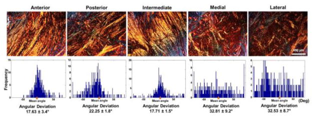
Our automated imaging processing demonstrated the regionally variant direction of collagen alignment (Fig. 1), consistently with previous findings. Collagen fibers were orientated circumferentially in the peripheral bands, whereas they were aligned in the anteroposterior direction in the intermediate zone (Fig. 1A). Mean orientation angles of collagen fibers calculated from our imaging processing algorithm were also matched with the previously described anisotropic collagen alignment (Fig. 1B). Interestingly, the degree of collagen alignment was significantly different in between the regions of TMJ discs (Fig. 2). Polarized images showed more densely and highly aligned collagen fibers in the anterior and posterior bands and the intermediate zone (Fig. 2A, B, and C) as compared to medial and lateral regions (Fig. 2D & E). Representative histograms of collagen fiber orientation angles consistently showed the varied degree of fiber alignment in the different regions (Fig. 2F – J). More collagen fibers were oriented along the mean orientation angle in the anterior band and the intermediate zone in comparison with the posterior bands and the medial and lateral regions (Fig. 2F–J). Quantitatively, the values of angular deviation were significantly smaller in the anterior bands (Fig. 2F) and the intermediate zone (Fig. 2H) than the posterior bands (Fig. 2G) and the medial and lateral zones (Fig. 2I & J) (n = 10 per group; p < 0.001). The medial and lateral regions showed significantly poorer collagen alignment than all the other regions (Fig. 2I & J) (p < 0.001).
Fig. 1.
The anisotropic collagen orientation in TMJ disc. Circularly polarized images of Picrosirius Red (PR) stained tissue sections showed circumferentially oriented collagen fibers in the peripheral zones and the anteroposterior fiber orientation in the intermediate zone (A), corresponding to the mean orientation angles of the collagen fibers from an automated digital imaging processing (B).
Fig. 2.
Quantitative analysis of collagen fiber alignment. Polarized images with PR staining (A–E) show highly aligned collagen fibers in the anterior band (A) and intermediate zone (C) as compared to the posterior band (B). Medial and lateral regions show less alignment of collagen fibers (D, E). Histograms of fiber alignment angle (F–J) consistently show more fibers aligned along with the mean angles in the anterior (F) and intermediate zone (H) in comparison with the other regions (G, I, J). Quantitatively, AD values were significantly smaller in the anterior and the intermediate zone as compared to the other zones (n = 10 per group; p < 0.001).
Instantaneous and relaxation moduli of TMJ discs and their correlation with collagen alignment
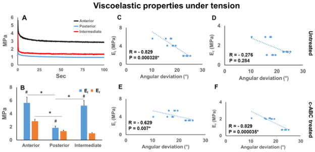
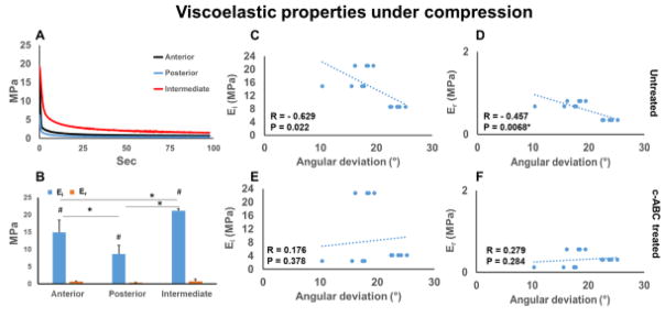
The stress relaxation curves from the TMJ disc samples showed a good-fitting to a Prony series (R2 = 0.997 ± 0.1) as compared to a standard solid stress relaxation model (R2 = 0.883 ± 0.28) (Supplementary Figure. 2). TMJ disc samples showed tensile stress relaxation curves in a region-variant manner (Fig. 3A & B). In tensile tests, the instantaneous modulus (Ei) were significantly higher in the anterior band and the intermediate zone as compared to the posterior band (Fig. 3B). The relaxation modulus (Er) was significantly higher in the anterior bands in comparison with the posterior and the intermediate zone (Fig. 3B). The Ei was statistically correlated with angular deviation both in untreated and c-ABC treated TMJ discs (Fig. 3C & D). In contrast, the Er was not statistically correlated with angular deviation in untreated TMJ discs (Fig. 3D) but the c-ABC treatment resulted in a statistically significant correlation between Er and angular deviation (Fig. 3F). Similarly, stress relaxation curves and the instantaneous and relaxation moduli under compression showed a regional variance (Fig. 4A & B). Under compression, Ei with or without c-ABC treatment failed to show a statistically significant correlation with angular deviation (Fig. 4C, E). A significant correlation with angular deviation was found in Er of untreated TMJ discs (Fig. 4D), not with c-ABC treatment (Fig. 4F). Our GAGs assay showed that the c-ABC treatment for 3 hours degraded up to 65% GAGs in TMJ discs, consistently with previous works (Lumpkins & McFetridge, 2009; Willard et al., 2012).
Fig. 3.
Correlation between the degree of fiber alignement and tensile viscoelastic properties of TMJ discs. Stress relaxation curves (A) and the instantaneous (Ei) and relaxation moduli (Er) (B) were varied in the different regions of TMJ discs. Ei was significantly correlated with the AD with or without C-ABC treatment (C, E), wheares Er was correlated with AD only when treated with C-ABC (D, F) (n = 5 per group) (*: p<0.01; #: p<0.01 comapred to Er).
Fig. 4.
Correlation between the degree of fiber alignement and compressive viscoelastic properties. Stress relaxation curves (A) and the instantaneous (Ei) and relaxation moduli (Er) (B) were varied in the different regions of TMJ discs. Under compression, Ei with our without C-ABC treatment was not correlated with AD (C, E), and Er was correlated in untreated samples (D) not with C-ABC treatment (F) (n = 5 per group) (*: p<0.01; #: p<0.01 comapred to Er).
DISCUSSION
The directions of the anisotropic orientation of collagen fibers in TMJ discs have been well-described in previous studies (Allen & Athanasiou, 2006a; Kalpakci et al., 2011; Perez del Palomar & Doblare, 2006; Shi et al., 2013; Stankovic et al., 2013). In the present study, we demonstrated that not only the direction but also degree of collagen fibers alignment are varied in the different regions of human TMJ discs. Our data also suggested that the variant degree of collagen fiber alignment is attributed to the regionally variant instantaneous modulus (Ei) and in part of relaxation modulus (Er) under stress relaxation test. These novel findings likely provide a potential explanation for the previous findings showed that tensile properties of TMJ discs, prepared along with the regional collagen orientation, are varied in different regions in spite of the relatively homogeneous collagen density throughout the TMJ discs(Kalpakci et al., 2011).
As a fibrocartilaginous tissue, TMJ discs exhibit viscoelastic properties that have been widely evaluated by stress relaxation tests, largely under compression (Allen & Athanasiou, 2006a, 2006b; Kuo, Zhang, Bacro, & Yao, 2010; Lumpkins & McFetridge, 2009; Willard et al., 2012). Previous works showed that the regional distribution of GAGs contributes to the viscoelastic properties with a significant difference in between the regions of TMJ discs (Beek, Aarnts, Koolstra, Feilzer, & van Eijden, 2001; Lumpkins & McFetridge, 2009; Willard et al., 2012). As GAGs are negatively charged playing roles in water absorbance, its mechanical functions have been predominantly examined under compression (Beek et al., 2001; Lumpkins & McFetridge, 2009; Willard et al., 2012). However, a recent study demonstrated that the GAG-containing fibrocartilaginous micro-domains play essential roles in the transmission of tensile forces as interconnected with collagen fibers network (Han et al., 2016). Similarly, our data demonstrated that the correlation between tensile relaxation modulus and angular deviation of collage fibers alignment were significantly altered by C-ABC treatment, further advocating a role of GAGs in tensile behavior of TMJ discs.
Likely due to the predominant mechanical roles of collagen fibers in tensile, we found no statistically significant correlation between the compressive instantaneous modulus and the angular deviation. However, the compressive relaxation modulus of TMJ discs showed a statistically significant correlation with the angular deviation of collagen fibers alignment in a GAGs-dependent manner. This finding suggests a potential mechanical role of collagen fibers under compressive loads. Consistently, a recent study demonstrated that not only GAG but also density and alignment of collagen fibers determine compressive modulus of TMJ discs via the mechanical reinforcement by collagen network (Fazaeli, Ghazanfari, Everts, Smit, & Koolstra, 2016). Accordingly, it is postulated that the degree of collagen alignment is associated with the collagen-GAGs network that consequently contributes to the compressive relaxation modulus.
Despite the novel finding of the correlation between the viscoelastic properties and the degree of collagen fibers alignment in TMJ discs, the present study has several limitations. First, we had a limited number of human TMJ disc samples. By preparing multiple tissue samples from each TMJ disc, we had a sufficient sample number to result in a statistically significant outcome in analysis of the viscoelastic properties and the collagen fiber alignment. However, our sample number was not sufficient to investigate any difference in the donor’s gender. In addition, all the tissue donors were over 45 years old so our data may not represent structure-mechanics characteristics of TMJ discs in young patients. Thus, follow-up studies with additional sample numbers are necessary to study age- and/or gender-differences in the degree of collagen alignment and its correlation with mechanical properties. Another limitation of this study is the incomplete characterization of biochemical structure determining the correlation between the angular deviation of collagen alignment and mechanical properties which are dependent upon interactions with GAGs and the type of loads. Multi-scale mechanical evaluation (Han et al., 2016) may be applied in a follow-up study to obtain more in-depth understanding of the complex mechanical behavior of TMJ discs.
The regionally variant viscoelastic properties of human TMJ discs in the present study show somewhat different level and tendency from some of previous reports showing higher instantaneous modulus in the posterior band than the anterior (Wright et al., 2016). However, another study showed a higher peak tensile modulus in the anterior band than the posterior in human TMJ disc (Kalpakci et al., 2011), corresponding to our findings. Similarly, dynamic modulus of human TMJ discs were higher in the anterior band than the posterior (Beek et al., 2001). Such discrepancy in the regional mechanical properties in between the different studies are likely due to various loading regime, testing conditions, sample preparation, and donor age and sex (Allen & Athanasiou, 2006a).
In conclusion, this study revealed the region-variant degree of collagen fibers alignment that is likely attributed to the heterogeneous mechanical properties of TMJ disc. One of the most important criteria for functional tissue engineering is to recapitulate native-like mechanical properties in an engineered tissue. Thus, our findings demonstrating the complex and region-variant viscoelastic properties may have significant implication in our ultimate research goal to engineer functional replacement TMJ discs.
Supplementary Material
Highlights.
Human TMJ disc shows region-variant directions of collagen alignment.
Degree of collagen alignment, not only the directions, is regionally variant.
The region-variant alignment degree is correlated with viscoelastic properties.
Acknowledgments
We thank A. Tsu for administrative assistance. This research was partially supported by NIH/NIDCR 1R03DE026794-01 grant (C.H.L).
Footnotes
Publisher's Disclaimer: This is a PDF file of an unedited manuscript that has been accepted for publication. As a service to our customers we are providing this early version of the manuscript. The manuscript will undergo copyediting, typesetting, and review of the resulting proof before it is published in its final citable form. Please note that during the production process errors may be discovered which could affect the content, and all legal disclaimers that apply to the journal pertain.
References
- Ahtiainen K, Mauno J, Ella V, Hagstrom J, Lindqvist C, Miettinen S, … Seppanen R. Autologous adipose stem cells and polylactide discs in the replacement of the rabbit temporomandibular joint disc. J R Soc Interface. 2013;10(85):20130287. doi: 10.1098/rsif.2013.0287. [DOI] [PMC free article] [PubMed] [Google Scholar]
- Allen KD, Athanasiou KA. Tissue Engineering of the TMJ disc: a review. Tissue Eng. 2006a;12(5):1183–1196. doi: 10.1089/ten.2006.12.1183. [DOI] [PubMed] [Google Scholar]
- Allen KD, Athanasiou KA. Viscoelastic characterization of the porcine temporomandibular joint disc under unconfined compression. J Biomech. 2006b;39(2):312–322. doi: 10.1016/j.jbiomech.2004.11.012. [DOI] [PubMed] [Google Scholar]
- Andarawis-Puri N, Sereysky JB, Sun HB, Jepsen KJ, Flatow EL. Molecular response of the patellar tendon to fatigue loading explained in the context of the initial induced damage and number of fatigue loading cycles. J Orthop Res. 2012;30(8):1327–1334. doi: 10.1002/jor.22059. [DOI] [PMC free article] [PubMed] [Google Scholar]
- Beek M, Aarnts MP, Koolstra JH, Feilzer AJ, van Eijden TM. Dynamic properties of the human temporomandibular joint disc. J Dent Res. 2001;80(3):876–880. doi: 10.1177/00220345010800030601. [DOI] [PubMed] [Google Scholar]
- Chen T. NASA, editor. Determining a Prony series for a viscoelastic material from time varying strain data. 2000;TM-210123 [Google Scholar]
- Dimitroulis G. A critical review of interpositional grafts following temporomandibular joint discectomy with an overview of the dermis-fat graft. Int J Oral Maxillofac Surg. 2011;40(6):561–568. doi: 10.1016/j.ijom.2010.11.020. [DOI] [PubMed] [Google Scholar]
- Estabrooks LN, Fairbanks CE, Collett RJ, Miller L. A retrospective evaluation of 301 TMJ Proplast-Teflon implants. Oral Surg Oral Med Oral Pathol. 1990;70(3):381–386. doi: 10.1016/0030-4220(90)90164-n. [DOI] [PubMed] [Google Scholar]
- Fazaeli S, Ghazanfari S, Everts V, Smit TH, Koolstra JH. The contribution of collagen fibers to the mechanical compressive properties of the temporomandibular joint disc. Osteoarthritis Cartilage. 2016;24(7):1292–1301. doi: 10.1016/j.joca.2016.01.138. [DOI] [PubMed] [Google Scholar]
- Hagandora CK, Almarza AJ. TMJ disc removal: comparison between pre-clinical studies and clinical findings. J Dent Res. 2012;91(8):745–752. doi: 10.1177/0022034512453324. [DOI] [PubMed] [Google Scholar]
- Hagandora CK, Gao J, Wang Y, Almarza AJ. Poly (glycerol sebacate): a novel scaffold material for temporomandibular joint disc engineering. Tissue Eng Part A. 2013;19(5–6):729–737. doi: 10.1089/ten.tea.2012.0304. [DOI] [PMC free article] [PubMed] [Google Scholar]
- Han WM, Heo SJ, Driscoll TP, Delucca JF, McLeod CM, Smith LJ, … Elliott DM. Microstructural heterogeneity directs micromechanics and mechanobiology in native and engineered fibrocartilage. Nat Mater. 2016;15(4):477–484. doi: 10.1038/nmat4520. [DOI] [PMC free article] [PubMed] [Google Scholar]
- Henry CH, Wolford LM. Treatment outcomes for temporomandibular joint reconstruction after Proplast-Teflon implant failure. J Oral Maxillofac Surg. 1993;51(4):352–358. doi: 10.1016/s0278-2391(10)80343-x. discussion 359–360. [DOI] [PubMed] [Google Scholar]
- Kalpakci KN, Willard VP, Wong ME, Athanasiou KA. An interspecies comparison of the temporomandibular joint disc. J Dent Res. 2011;90(2):193–198. doi: 10.1177/0022034510381501. [DOI] [PMC free article] [PubMed] [Google Scholar]
- Kim SJ, Shin JW, Lee CH, Shin HJ, Kim SH, Jeong JH, Lee JW. Biomechanical comparisons of three different tibial tunnel directions in posterior cruciate ligament reconstruction. Arthroscopy. 2005;21(3):286–293. doi: 10.1016/j.arthro.2004.11.004. [DOI] [PubMed] [Google Scholar]
- Kuo J, Zhang L, Bacro T, Yao H. The region-dependent biphasic viscoelastic properties of human temporomandibular joint discs under confined compression. J Biomech. 2010;43(7):1316–1321. doi: 10.1016/j.jbiomech.2010.01.020. [DOI] [PMC free article] [PubMed] [Google Scholar]
- Lee CH, Cook JL, Mendelson A, Moioli EK, Yao H, Mao JJ. Regeneration of the articular surface of the rabbit synovial joint by cell homing: a proof of concept study. Lancet. 2010;376(9739):440–448. doi: 10.1016/S0140-6736(10)60668-X. [DOI] [PMC free article] [PubMed] [Google Scholar]
- Lee CH, Lee FY, Tarafder S, Kao K, Jun Y, Yang G, Mao JJ. Harnessing endogenous stem/progenitor cells for tendon regeneration. J Clin Invest. 2015;125(7):2690–2701. doi: 10.1172/JCI81589. [DOI] [PMC free article] [PubMed] [Google Scholar]
- Lee CH, Rodeo SA, Fortier LA, Lu C, Erisken C, Mao JJ. Protein-releasing polymeric scaffolds induce fibrochondrocytic differentiation of endogenous cells for knee meniscus regeneration in sheep. Sci Transl Med. 2014;6(266):266ra171. doi: 10.1126/scitranslmed.3009696. [DOI] [PMC free article] [PubMed] [Google Scholar]
- Lee CH, Shah B, Moioli EK, Mao JJ. CTGF directs fibroblast differentiation from human mesenchymal stem/stromal cells and defines connective tissue healing in a rodent injury model. J Clin Invest. 2010;120(9):3340–3349. doi: 10.1172/JCI43230. [DOI] [PMC free article] [PubMed] [Google Scholar]
- Lee CH, Shin HJ, Cho IH, Kang YM, Kim IA, Park KD, Shin JW. Nanofiber alignment and direction of mechanical strain affect the ECM production of human ACL fibroblast. Biomaterials. 2005;26(11):1261–1270. doi: 10.1016/j.biomaterials.2004.04.037. [DOI] [PubMed] [Google Scholar]
- Legemate K, Tarafder S, Jun Y, Lee CH. Engineering Human TMJ Discs with Protein-Releasing 3D-Printed Scaffolds. J Dent Res. 2016;95(7):800–807. doi: 10.1177/0022034516642404. [DOI] [PubMed] [Google Scholar]
- Lumpkins SB, McFetridge PS. Regional variations in the viscoelastic compressive properties of the temporomandibular joint disc and implications toward tissue engineering. J Biomed Mater Res A. 2009;90(3):784–791. doi: 10.1002/jbm.a.32148. [DOI] [PubMed] [Google Scholar]
- MacBarb RF, Chen AL, Hu JC, Athanasiou KA. Engineering functional anisotropy in fibrocartilage neotissues. Biomaterials. 2013;34(38):9980–9989. doi: 10.1016/j.biomaterials.2013.09.026. [DOI] [PMC free article] [PubMed] [Google Scholar]
- Palacio-Torralba J, Hammer S, Good DW, Alan McNeill S, Stewart GD, Reuben RL, Chen Y. Quantitative diagnostics of soft tissue through viscoelastic characterization using time-based instrumented palpation. J Mech Behav Biomed Mater. 2015;41:149–160. doi: 10.1016/j.jmbbm.2014.09.027. [DOI] [PubMed] [Google Scholar]
- Perez del Palomar A, Doblare M. The effect of collagen reinforcement in the behaviour of the temporomandibular joint disc. J Biomech. 2006;39(6):1075–1085. doi: 10.1016/j.jbiomech.2005.02.009. [DOI] [PubMed] [Google Scholar]
- Scapino RP, Canham PB, Finlay HM, Mills DK. The behaviour of collagen fibres in stress relaxation and stress distribution in the jaw-joint disc of rabbits. Arch Oral Biol. 1996;41(11):1039–1052. doi: 10.1016/s0003-9969(96)00079-9. [DOI] [PubMed] [Google Scholar]
- Shi C, Wright GJ, Ex-Lubeskie CL, Bradshaw AD, Yao H. Relationship between anisotropic diffusion properties and tissue morphology in porcine TMJ disc. Osteoarthritis Cartilage. 2013;21(4):625–633. doi: 10.1016/j.joca.2013.01.010. [DOI] [PMC free article] [PubMed] [Google Scholar]
- Stankovic S, Vlajkovic S, Boskovic M, Radenkovic G, Antic V, Jevremovic D. Morphological and biomechanical features of the temporomandibular joint disc: an overview of recent findings. Arch Oral Biol. 2013;58(10):1475–1482. doi: 10.1016/j.archoralbio.2013.06.014. [DOI] [PubMed] [Google Scholar]
- Tarafder S, Koch A, Jun Y, Chou C, Awadallah MR, Lee CH. Micro-precise spatiotemporal delivery system embedded in 3D printing for complex tissue regeneration. Biofabrication. 2016;8(2):025003. doi: 10.1088/1758-5090/8/2/025003. [DOI] [PubMed] [Google Scholar]
- Willard VP, Kalpakci KN, Reimer AJ, Athanasiou KA. The regional contribution of glycosaminoglycans to temporomandibular joint disc compressive properties. J Biomech Eng. 2012;134(1):011011. doi: 10.1115/1.4005763. [DOI] [PMC free article] [PubMed] [Google Scholar]
- Wright GJ, Coombs MC, Hepfer RG, Damon BJ, Bacro TH, Lecholop MK, … Yao H. Tensile biomechanical properties of human temporomandibular joint disc: Effects of direction, region and sex. J Biomech. 2016;49(16):3762–3769. doi: 10.1016/j.jbiomech.2016.09.033. [DOI] [PMC free article] [PubMed] [Google Scholar]
Associated Data
This section collects any data citations, data availability statements, or supplementary materials included in this article.


