Abstract
Obstructive Sleep Apnea Syndrome (OSA) is a multifactorial disease that highly alters a persons quality of life. It is characterized by the repeated interruption of breathing during sleep, due to an obstruction or the collapse of the upper airways. Since it is a multifactorial etiological disorder, it requires a thorough diagnosis and treatment with an interdisciplinary team, which comprises several professionals such as a surgical dentist, phonoaudiologist, otorhinolaryngologist, sleep doctor, neurologist and physiotherapist. The diagnosis and the degree of severity of the syndrome is determined through a polysomnography examination. After that, the best form of treatment is devised depending on the gravity of the case. In cases of moderate to severe apnea, invasive treatment through surgical procedures such as maxillomandibular advancement remains the preferred option as it increases the posterior air space, reducing and/or eliminating the obstruction. Thus, improving the patients respiratory function and, consequently, his quality of life as it is shown in the clinical case at hand. In which the male patient, facial pattern type I, 41 years of age, diagnosed with moderate OSA (Apnea-Hypopnea Index - AHI of 23.19), decided to have a surgical treatment instead of a conservative one, resulting in the cure of apnea (AHI of 0.3).
Keywords: Orthognathic Surgery, Sleep Apnea Syndromes, Esthetics
INTRODUCTION
Obstructive Sleep Apnea Syndrome (OSA) is a disorder characterized by repeated nighttime episodes of collapsing of the upper airways, which may be partial or complete. The apnea or hypopnea, determined by the degree of the obstruction, causes progressive asphyxia so that the desaturation of oxygen makes the individual require increasingly higher respiratory efforts, until he is awoken1,2.
According to Libman et al.3, the prevalence of this syndrome in the adult population is high, although the data varies according to the sample and the type of diagnosis used in the study. OSA is multifactorial and, among the predisposing and risk factors, we have the small lumen of the upper airways, unstable respiratory control, synergist breathing muscles not performing their function, smoking, obesity, fluid retention, age and adenotonsillar hypertrophy2.
OSA brings grave consequences such as excessive daytime sleepiness, which can generate traffic accidents. Besides, it may be associated with a significant cardiovascular morbidity and connected with metabolic syndromes. In addition, many patients may describe difficulties in concentration, memory loss and mood swings, irritability and depression causing a great impact on quality of life4.
According to the American Academy of Sleep Medicine, the diagnosis of OSA is given through the analysis of clinical history, physical, radiographic and polysomnographic examinations. This last one consists of the evaluation of nighttime cardiac and respiratory parameters during sleep, aiming to detect obstructive events and alterations in the saturation of oxygen in the blood. The index used to define the severity of the syndrome is the Apnea-Hypopnea Index (AHI). Calculated as the number of obstructive events by hour of sleep. It can characterize minimal, mild, moderate or severe cases1,5.
Patients with OSA must be treated in a multidisciplinary way and several options for treatment are available nowadays. Continuous Positive Air Pressure (CPAP) is very effective, improving quality of life and reducing the clinical developments of sleep apnea. Alternative options include weight control, mandibular protrusion devices and a number of surgical approaches of the upper airways. Patients with OSA have below-average dimensions of the upper airways and the orthognathic surgery allows maxillomandibular repositioning so that, all the muscles involved are moved forward, which results in a volumetric increase of the airway6.
This article aims at performing a revision of the literature about the aspects of the Obstructive Sleep Apnea Syndrome and its surgical treatment of Bimaxillary Advancement in a Pattern I individual with the clinical case report.
REVISION OF THE LITERATURE
The Obstructive Sleep Apnea Syndrome is characterized by episodes of obstruction in the upper airways during sleep. It may be associated to clinical signs and symptoms like snoring, excessive daytime sleepiness, impaired memory and fatigue. This syndrome results in oxygen desaturation, which in turn provokes an increase in blood pressure due to the need of a greater respiratory effort and an increase in the levels of carbon dioxide in the blood. The resulting hypoxia is linked to a wide range of problems deriving from the oxidative stress as well as to mortality concerning the coronary arteries3,6.
If not diagnosed and treated promptly, this sleep disorder can bring about important medical consequences and the options for treatment become limited. Because of that, its prevention through early diagnosis and treatment, during the growing and developing phase of children, is the best way forward for it can cause important alterations in many organs, structures and systems of the craniocervical/oral region7.
Jordan et al.2 point out, as predisposing factors of OSA, problems in the anatomy of the upper airways, where the craniofacial structure or body fat has reduced the size of the Pharyngeal lumen, leading to a higher probability of collapsing. The dilator muscles, especially the genioglossus are also studied. Lung volume may be a casual factor since, when small, the airway is also small and may, therefore easily collapse. Obesity frequently reduces the residual functioning ability of the respiratory volume, particularly when in supine position. Sensorial pharyngeal compromise may also contribute to the collapsing of the upper airway. Liquid retention and fluid displacement from the legs into the neck during the night can affect the mechanics of the airways as well.
Among the available options for the treatment of OSA, the Continuous Positive Air Pressure (CPAP) device is the first choice for patients with moderate to severe forms of the disease. However, some patients are intolerant to this treatment, claiming discomfort when using the mask, while others do not accept sleeping while connected to a mechanic device8. In these cases, surgical treatment must be considered, whose objective is the removal of the cause of obstruction of the upper airways, enlarging the lumen after a precise detection of the place where the obstruction occurs.
Maxillomandibular advancement induces an anterior displacement of the soft palate and of the tongue, increasing the pharyngeal space. This is a highly efficient treatment, which requires short, medium and long term monitoring. Nevertheless, by being a very invasive procedure, it must be reserved for selected patients with craniofacial malformations or when other forms of treatment have failed1.
It is important to point out that OSA also attacks patients with facial morphology type Pattern I, that is, a straight profile. Therefore, a question arises: What is the repercussion of a Bimaxillary Advancement in facial aesthetics? With the improvements in surgical techniques and in the modification of the occlusal plane with counter-clockwise rotation during fixation, this alternative has become possible for such individuals, favoring clearance of the airway for the improvement of the respiratory function with little to no compromise in facial harmony, which was observed in this clinical case report.
CLINICAL CASE REPORT
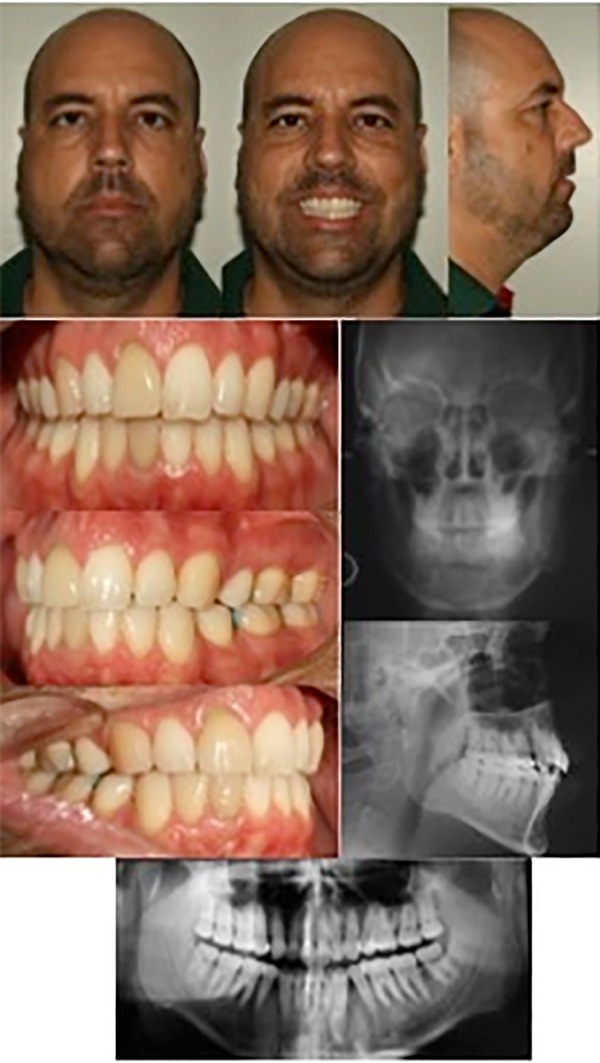
Patient H.A.L.P.S., male, 42, facial features pattern I, that is, straight profile, dolichofacial and class I Angle. This same subject had already undergone conventional orthodontic treatment for the correction of the masticatory function and for aesthetics, during which the first four premolar teeth were extracted (figure 1).
Figure 1.

Initial photographs and radiographs.
Polysomnography revealed an IAH of 23.19 and average basal oxyhemoglobin desaturation of 95% and a minimum of 84%, with obstructive events of up to 4 minutes in duration (table 1). These data associated with the physical examination, clinical history of signs and symptoms (daytime sleepiness, concentration difficulties, frequent headaches) and with the cephalometric and facial analysis (table 2), confirm the diagnosis and the moderate severity of this OSA.
Table 1.
Initial and polysomnographic control values.
| Initial | 6 months post surgical | 2,5 years post surgical | |
|---|---|---|---|
| AIH/hour | 23,19 | 4,1 | 0,3 |
| Moderate | Normal | Normal | |
| Average O2 saturation | 95% | 97% | 98% |
| Minimal O2 saturation | 84% | 93% | 95% |
Table 2.
Initial and cephalometric control values.
| Cephalometric measurements | Preoperative | Postoperative | Standard |
|---|---|---|---|
| SNA | 77° | 95° | 82°(+ -2) |
| SNB | 75° | 82° | 80°(+ -2) |
| ANB | 2° | 12° | 2°(+ -2) |
| 1.NA | 26° | 5° | 22° |
| 1-NA | 7mm | 2mm | 4mm |
| 1.NB | 22° | 32° | 25° |
| 1-NB | 9mm | 4mm | 4mm |
| AFAI | 95mm | 88mm | 67mm |
| CoA | 97mm | 107mm | 91mm |
| CoGN | 139mm | 140mm | 112mm |
| FMA | 24° | 25° | 25°(+ -5) |
| FMIA | 90° | 86° | 68° |
| IMPA | 62° | 59° | 87° |
| Upphw-PP1 | 18mm | 25mm | 26mm |
| PP2-PP2' | 15mm | 18mm | 12mm |
| PNS-P | 43mm | 37mm | |
| Mpphw-MaphW | 13mm | 22mm | 22mm |
| PAS | 8mm | 13mm | |
| LAS | 10mm | 14mm |
The treatment of choice was Bimaxillary Advancement Surgery, considering that the patient had not accepted a conservative, palliative treatment with CPAP. As orthodontic presurgical planning, a Morelli fixed metallic orthodontic appliance was installed, Edgewise prescription, passive form, rectangular steel archwires 0.019”x0.025” with individual spurs and ligatures (figure 2).
Figure 2.

Intra-oral photographs of the preoperative orthodontic preparation.
The models were mounted in semi-adjustable articulator for model surgery and confection of surgical models based on the measurements of the predictive surgical tracing (10 mm maxillary advancement with a 2 mm impaction of the anterior nasal spine, 3 mm lowering of the posterior nasal spine and 15 mm mandibular advancement with counter-clockwise rotation of the occlusal plane), which was carried out together with the surgeon.
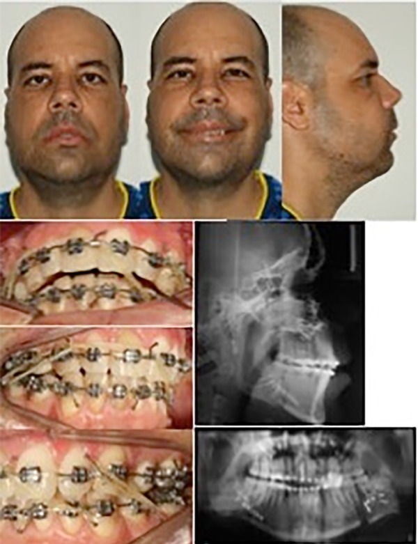
Thirty days after surgery, the patient returned to the office for occlusion examination and guidance in the use of intermaxillary elastics (figure 3). During this postoperative period, the patient was already reporting an improvement in the quality of his sleep and, therefore, a considerable gain in his general well-being.
Figure 3.

Photographs and radiographs after 30 days of orthognathic surgery.
The improvement in the flow of air through the posterior superior air column was observed in postsurgical lateral teleradiography (table 2), (figure 3) and the cure of OSA was confirmed, six months later, through control polysomnography which showed an AHI of 4.1 events/hour of sleep, minimal oxyhemoglobin saturation of 93% and an average of 97% (table 1).
Orthodontic treatment extended for another year after surgery, in order to refine occlusion. Contentions were installed and the balance between masticatory function, respiratory function and facial aesthetics was reached, considering that the subject’s profile remained straight, that is, Pattern I and quite harmonious.
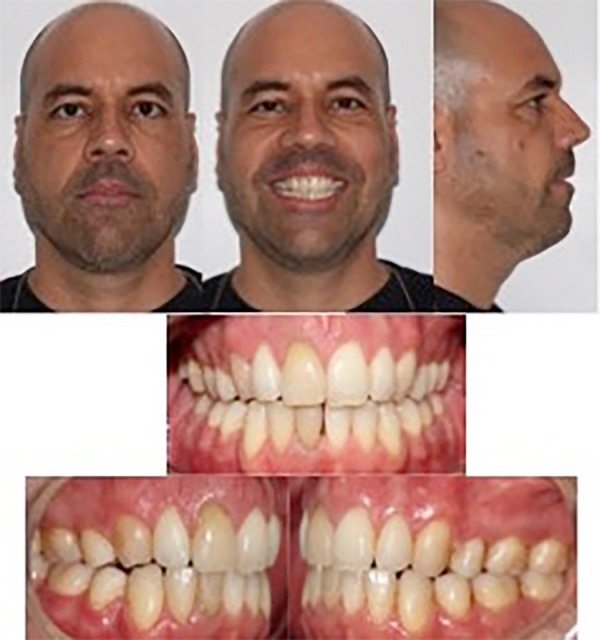
In a mid-term control, two and a half years after the orthognathic surgery, this same subject underwent a new polysomnography test and the data turned up promising about the improvement of OSA. AHI was even better than before at 0.3 events/hour, with minimal oxygen saturation of 95% and average of 98% (table 1). He seemed very pleased with his face, occlusion, breathing and sleep (figure 4).
Figure 4.

Control photographs after two and a half years of orthognathic surgery
DISCUSSION
Obstructive Sleep Apnea Syndrome (OSA) consists of periods of apnea in which breathing through the nose or the mouth ceases for longer than 10 seconds and hypopneas, when there is a 50% reduction in the breathing volume for longer than 10 seconds5.
In conformity with reports from many authors and their experiences with orthognathic surgery, we can observe, in the vast majority of patients, common symptomatology, clinic and radiographic characteristics such as: snoring, sleepiness, arrhythmia, nasal obstruction, oropharyngeal abnormalities like tonsil hyperplasia, adenoidal enlargement and macroglossia among others3,6,9,10.
In this case report, the male patient, a young adult, corroborates the prevalence registered in literature, as OSA affects, in the majority of cases, men between 40 and 60 years of age11,12. However, a study which evaluates oral health in ill patients who make use of CPAP, Tsuda et al.13 indicated that the prevalence of OSA is higher in elderly individuals than it is in middle-aged ones.
There are some options for the medical treatment of OSA, including Continuous Positive Air Pressure (CPAP) appliances, Bilevel Positive Airway Pressure (BIPAP) devices, intraoral devices, as well as weight loss. The CPAP nasal device is used for the pneumatic support of the upper airway. A non-surgical alternative which uses a mask, a tube and a flow generator but, in spite of being widely used, it does not offer a cure since a night without it makes all the quality of sleep obtained through its use disappear14,15.
Likewise, combined maxillary and mandibular advancement orthognathic surgery has been the chosen procedure in the treatment of respiratory disorders in order to make the patient free from the use of any mechanical devices. Countless studies report the benefits of orthognathic surgery for mandibular advancement in the upper airway and the association of this procedure with the counter-clockwise rotation of the occlusal plane, when properly performed, is a stable procedure in the long term and maximizes the aesthetic function after surgery even in patients who already have a straight facial profile1,14.
The cephalometric radiographies are extensively used in the diagnosis and surgical monitoring. One of its limitations resides in the fact that this is a 2D technique used to assess 3D structures. On the other hand, a Computed Tomography scan offers the potential for 3D reconstructions of such structures favoring assessments that are more precise even if, regrettably, its price makes it inaccessible for the population in general14.
In accordance with Lye et al.16 and Raffaini and Pisani17 maxillomandibular advancement results not only in the augmentation of the anteroposterior diameter of the airway but also in its medial-lateral diameter, thus expanding the airway in three dimensions whilst favoring a larger respiratory flow and a reduction in resistance, which in turn helps the treatment of OSA.
It is clear that the pattern of expansion of superior air space is different between maxilla and mandible. An expansion in the anteroposterior direction is bigger with the advancement of the maxilla and the medial-lateral diameter grows more when the mandible is moved forward due to the force vectors form the tensor veli palatini, levator veli palatini, palatoglossus and genioglossus muscles9,16.
Al-Moraissi and Wolford18, Ferraz et al.6 and Mehra and Wolford19 concluded that surgical success is attained with an AHI below 10/hour or a higher-than-50% reduction after surgery, while for the cure of the disease the AHI should be lower than 5/h. Such success depends on a maxillomandibular advancement beyond 9.9 mm. These changes were observed in the clinical case report presented, where AHI was reduced from 23.19/h to 0.3/h. This result was obtained thanks to a surgical maxillary advancement of 10mm and mandibular advancement of 15 mm with rotation of the occlusal plane. For the authors, such surgeries are safe procedures. There are no reports of any deaths or major complications other than transitory labial mental paresthesia. Present in 100% of cases with an 85% improvement rate in up to 1 year after surgery.
Most authors firmly state that maxillomandibular advancement surgery not only treats OSA but also re-establishes dental occlusion (as long as there is orthodontic treatment prior to surgery). In addition, it improves facial harmony for many of the patients who exhibit retrusion or maxillomandibular imbalance and also, it maintains a balanced facial profile for those who already exhibit a straight pattern before the surgical procedure20,21.
CONCLUSION
OSA has arisen as the most frequent and important sleep disorder while connecting to several systemic alterations. Mainly, cardiovascular ones.
It is the dental surgeon’s responsibility to know the diagnostic parameters, the established definitions and the limits of his field of work when working as part of the multidisciplinary teams that monitor and treat respiratory sleep disorders.
Several therapeutics have been proposed depending on the gravity of the diagnosis, yet, the most definite and effective one for the treatment of moderate to severe OSA cases is the orthognathic surgery.
REFERENCES
- 1.Spicuzza L, Caruso D, Maria G. Obstructive sleep apnoea syndrome and its management. Ther Adv Chronic Dis. 2015;6(5):273–285. doi: 10.1177/2040622315590318. [DOI] [PMC free article] [PubMed] [Google Scholar]
- 2.Jordan AS, McSharry DG, Malhotra A. Adult obstructive sleep apnoea. Lancet. 2014;383(9918):736–747. doi: 10.1016/S0140-6736(13)60734-5. [DOI] [PMC free article] [PubMed] [Google Scholar]
- 3.Libman E, Bailes S, Fichten CS, Rizzo D, Creti L, Baltzan M, et al. CPAP Treatment Adherence in Women with Obstructive Sleep Apnea. Sleep Disord. 2017;2017:2760650–2760650. doi: 10.1155/2017/2760650. [DOI] [PMC free article] [PubMed] [Google Scholar]
- 4.Feitoza CC, Azevedo WRS, Emery JMT, Pereira CV, Vargas Jr CS, Pizzol KED. Cirurgia ortognática no tratamento da síndrome da apneia obstrutiva do sono - relato de caso. OrthoScience. 2017;10(38):98–105. [Google Scholar]
- 5.Epstein LJ, Kristo D, Strollo PJ Jr, Friedman N, Malhotra A, Patil SP, et al. Adult Obstructive Sleep Apnea Task Force of the American Academy of Sleep Medicine Clinical guideline for the evaluation, manegement and long-term care of obstructive sleep apnea in adults. J Clin Sleep Med. 2009;5(3):263–276. [PMC free article] [PubMed] [Google Scholar]
- 6.Ferraz O, Guimarães TM, Rossi RR, Cunali PA, Fabbro CD, Chaves CM, et al. Effectiveness of Maxillomandibular advancement (MMA) surgery in sleep apnea treatment: Case report. Sleep Sci. 2016;9(3):134–139. doi: 10.1016/j.slsci.2016.07.005. [DOI] [PMC free article] [PubMed] [Google Scholar]
- 7.Caples SM, Rowley JA, Prinsell JR, Pallanch JF, Elamin MB, Katz SG, et al. Surgical modifications of the upper airway for obstructive sleep apnea in adults: a systematic review and meta-analysis. Sleep. 2010;33(10):1396–1407. doi: 10.1093/sleep/33.10.1396. [DOI] [PMC free article] [PubMed] [Google Scholar]
- 8.Feres MA, Feres R. A Ortodontia no diagnóstico e tratamento da apneia obstrutiva do sono em crianças. OrthoScience. 2013;6(23):410–414. [Google Scholar]
- 9.Phan NT, Wallwork B, Panizza B. Surgery for adult patients with obstructive sleep apnoea: A review for general practitioners. Aust Fam Physician. 2016;45(8):574–578. [PubMed] [Google Scholar]
- 10.Al-Moraissi EA, Wolford LM. Is Counterclockwise Rotation of the Mandibular Complex Stable Compared to Clockwise Rotation in the Dentofacial Deformities? A Systematic Review and Meta-Analysis. J Oral Maxillofac Surg. 2016;74(10):2066.e1–2066.e12. doi: 10.1016/j.joms.2016.06.001. [DOI] [PubMed] [Google Scholar]
- 11.Mehra P, Downie M, Pita MC, Wolford LM. Pharyngeal airway space changes after counterclockwise rotation of the maxillomandibular complex. Am J Orthod Dentofacial Orthop. 2001;120(2):154–159. doi: 10.1067/mod.2001.114647. [DOI] [PubMed] [Google Scholar]
- 12.Capelloza Filho L, Ribeiro AA, Guedes FP, Nary Filho H, Maltagliat LA, Cardoso MA. Tratamento ortodôntico cirúrgico da birretrusão associado à SAOS: relato de caso. OrthoScience. 2012;5(19):311–324. [Google Scholar]
- 13.Prado BN, Fernandes EG, Moreira TCA, Gavranich Junior J. Apneia Obstrutiva do Sono: Diagnóstico e Tratamento. Rev Odontol Univ Cid São Paulo. 2010;22(3):233–239. [Google Scholar]
- 14.Okushi T, Tonogi M, Arisaka T, Kobayashi S, Tsukamoto Y, Morishita H, et al. Effect of maxillomandibular advancement on morphology of velopharyngeal space. J Oral Maxillofac Surg. 2011;69(3):877–884. doi: 10.1016/j.joms.2010.10.012. [DOI] [PubMed] [Google Scholar]
- 15.La Piana GE, Scartabellati A, Chiesa L, Ronchi L, Raimond P, Carro MA, et al. Long-term adherence to CPAP treatment in patients with obstructive sleep apnea: importance of educational program. Patient Prefer Adherence. 2011;5:555–562. doi: 10.2147/PPA.S24018. [DOI] [PMC free article] [PubMed] [Google Scholar]
- 16.Echarri P, Pérez-Campoy MA, Coromina J, Grandi D. Papel do médico dentista no tratamento da roncopatia e do síndrome de apneia/hipoapneia obstrutiva do sono (SAHOS) J Dentistry. 2015;17:26–30. [Google Scholar]
- 17.Gharibeh T, Mehra R. Obstructive sleep apnea syndrome: natural history, diagnosis, and emerging treatment options. Nature Sci Sleep. 2010;2:233–255. doi: 10.2147/NSS.S6844. [DOI] [PMC free article] [PubMed] [Google Scholar]
- 18.Tsuda H, Moritsuchi Y, Higuchi Y, Tsuda T. Oral health under use of continuous positive airway pressure and interest in alternative therapy in patients with obstructive sleep apnoea: a questionnaire-based survey. Gerodontology. 2015;33(3):416–420. doi: 10.1111/ger.12184. [DOI] [PubMed] [Google Scholar]
- 19.Flores-Mir C. Dentistry and Obstructive Sleep Apnea. J Clin Sleep Med. 2016;12(9):1213–1214. doi: 10.5664/jcsm.6114. [DOI] [PMC free article] [PubMed] [Google Scholar]
- 20.de Sousa Miranda W, Álvares de Castro Rocha V, Lara Dos Santos Marques K, Trindade Neto AI, do Prado CJ, Zanetta-Barbosa D. Three-dimensional evaluation of superior airway space after orthognathic surgery with counterclockwise rotation and advancement of the maxillomandibular complex in Class II patients. Oral Surg Oral Med Oral Pathol Oral Radiol. 2015;120(4):453–458. doi: 10.1016/j.oooo.2015.06.037. [DOI] [PubMed] [Google Scholar]
- 21.Li KK, Guilleminault C, Riley RW, Powell NB. Obstructive sleep apnea and maxillomandibular advancement: an assessment of airway changes using radiographic and nasopharyngoscopic examinations. J Oral Maxillofac Surg. 2002;60(5):526–530. doi: 10.1053/joms.2002.31849. [DOI] [PubMed] [Google Scholar]
- 22.Raffaini M, Pisani C. Clinical and cone-beam computed tomography evaluation of the three-dimensional increase in pharyngeal airway space following maxillo-mandibular rotation-advancement for Class II-correction in patients without sleep apnoea (OSA) J Craniomaxillofac Surg. 2013;41(7):552–557. doi: 10.1016/j.jcms.2012.11.022. [DOI] [PubMed] [Google Scholar]


