Abstract
Extreme environments, such as subterranean habitats, are suspected to be responsible for morphologically inseparable cryptic or sibling species and can bias biodiversity assessment. A DNA barcode is a short, standardized DNA sequence used for taxonomic purposes and has the potential to lessen the challenges presented by a biotic inventory. Here, we investigate the diversity of the genus Leptonetela Kratochvíl, 1978 that is endemic to karst systems in Eurasia using DNA barcoding. We analyzed 624 specimens using one mitochondrial gene fragment (COI). The results show that DNA barcoding is an efficient and rapid species identification method in this genus. DNA barcoding gap and automatic barcode gap discovery (ABGD) analyses indicated the existence of 90 species, a result consistent with previous taxonomic hypotheses, and supported the existence of extreme male pedipalpal tibial spine and median apophysis polymorphism in Leptonetela species, with direct implications for the taxonomy of the group and its diversity. Based on the molecular and morphological evidence, we delimit and diagnose 90 Leptonetela species, including the type species Leptonetela kanellisi(Deeleman-Reinhold, 1971). Forty-six of them are previously undescribed. The female of Leptonetela zhai Wang & Li, 2011 is reported for the first time. Leptonetela tianxinensis (Tong & Li, 2008) comb. nov. is transferred from the genus Leptoneta Simon, 1872;the genus Guineta Lin & Li, 2010 syn. nov. is a junior synonym of Leptonetela; Leptonetela gigachela(Lin & Li, 2010) comb. nov. is transferred from Guineta. The genus Sinoneta Lin & Li, 2010 syn. nov. is a junior synonym of Leptonetela; Leptonetela notabilis(Lin & Li, 2010) comb. nov. and Leptonetela sexdigiti(Lin & Li, 2010) comb. nov. are transferred from Sinoneta; Leptonetela sanchahe Wang & Li nom. nov. is proposed as a replacement name for Sinoneta palmata(Chen et al, 2010) because Leptonetela palmata is preoccupied.
Keywords: DNA barcoding, Phylogeny, Phenotype, Species delineation
INTRODUCTION
Subterranean ecosystems, such as caves and cracks, are evident mainly in karst areas, which represent nearly 4% of the rocky outcrops of the world. These environments are marked by permanent darkness, a lack of diurnal and annual rhythms, and extremely scarce food sources (Culver & White, 2005; Howarth, 1983; Poulson & White, 1969). Many studies show that despite stressful and unfavorable conditions, the subsurface habitat harbors diverse animal communities (mainly invertebrates) (Amara-Zettler et al, 2002; Flot et al, 2010; López-García et al, 2001; Mathieu et al, 1997; Niemiller et al, 2012; Sket, 1999). Troglobionts are expected to adopt strategies that are characterized by significant geographic isolation and numerous local endemics (Convey, 1997; Waterman, 2001). Because the diversity of possible adaptive responses declines with stress intensity (Nevo, 2001), evolution in harsh environments is also expected to be influenced by convergence (Little & Vrijenhoek, 2003; Rothschild & Mancinelli, 2001; Waterman, 2001). Therefore, in subterranean, and more generally in extreme environments, diversification and speciation processes should be largely influenced by island-like habitats, such as caves, allopatric speciation and vicariant events, and could be masked by morphological convergence. For these groups of organisms, morphology alone cannot determine species boundaries, so identifying morphologically inseparable cryptic or sibling species requires an integrative approach that often includes DNA analysis.
DNA barcoding relies on the use of a standardized DNA region as a tag for accurate and rapid species identification (Hebert & Gregory, 2005) and has been used to help overcome the 'taxonomic impediment' (Herbert et al, 2003a; Tautz et al, 2003). It aids in the identification of species in applied settings, the association of morphologically distinct life-cycle forms within a species, the detection of host-specific lineages and the detection of morphologically cryptic species (Miller & Foottit, 2009). DNA barcoding has been used in a diverse range of vertebrate and invertebrate taxa (Clare et al, 2007; Ratnasingham & Hebert, 2007) and has enabled an increasing number of taxa to be identified. For example, a survey of crustacean stygofauna suggests that there could be substantial levels of subterranean biodiversity hidden in Australia's acquifer (Asmyhr & Cooper, 2012). Nevertheless, the exclusive use of single-locus molecular gene fragments is not without risks, for identical mitochondrial DNA sequences can be present in unrelated species due to introgression, or incomplete lineage sorting (Ballard & Whitlock, 2004). Additionally, the use of a divergence threshold for distinguishing intra-vs. interspecific sequence variation (Hebert et al, 2003a) can seriously compromise species identification and suffers from severe statistical problems (Vences et al, 2005). Furthermore, species misidentification has been observed when a reference database is not comprehensive; such that is does not contain all the species of the group under study (Meyer & Paulay, 2005).
The South China karst, a United Nations Educational, Scientific and Cultural Organization (UNESCO) World Heritage Site since 2007, is noted for its karst features and landscapes as well as rich biodiversity. Numerous subterraean species have been reported in this region, especially invertebrate fauna (Zhang, 1986). The spider genus Leptonetela is discontinuously distributed in the South China karst and the Balkan Peninsula, a karstic region in Europe. The genus has 54 catalogued species (World Spider Catalog, 2017), and with one exception (L. pungitia Wang & Li, 2011), nearly all Leptonetela species are endemic to either a single cave or a cave system. The spiders are cave adapted as shown by morphological features, such as vestigial eyes and highly reduced skin pigmentation. Over the past nine years, we have conducted extensive surveys of subterranean biodiversity in Eurasia. More than 1 500 caves were visited, and we ultimately sampled 122 Leptonetela populations (caves). Rapid and accurate identification within this genus is difficult due to congeneric species sharing similar morphological traits, a lack of obvious morphological differences between closely related species and some species only differ in one or a few quantitative differences, such as the location, length ratio or thickness of the male pedipalpal tibial spines and the number of teeth on the median apophysis.
In this study, we test the usefulness of DNA barcoding for species identification in the subterranean genus Leptonetela and investigate the diversity of the genus. The standard molecular barcode, cytochrome c oxidase subunit Ⅰ (COI) was used. A species discovery method, automatic barcode gap discovery (ABGD) (Puillandre et al, 2012), and a species validation method, DNA barcoding gap analysis, (Hebert et al, 2003b) were both used, depending on whether the samples were partitioned prior to analysis. The main goals of our study were: (ⅰ) to test whether the COI barcoding fragment can reliably resolve and identify subterranean Leptonetela species by comparing the COI barcode fragment results with those from morphological data; (ⅱ) to test taxonomic value of morphological characters used in traditional methods of classification.
MATERIALS AND METHODS
Taxon sampling
We sampled 624 Leptonetela individuals from 122 populations (caves) (Supplementary Table S1) in Eurasia (Insular and Peninsular Greece, and Southeast Asia; see inset in Figure 1). Nine individuals from three other genera of the family Leptonetidae were chosen as outgroups. All specimens were collected alive, fixed in absolute ethanol, and the legs were removed for subsequent DNA extraction. The remaining specimens were preserved in 80% ethanol for identification and morphological examination. Voucher specimens and all type specimens were deposited in the Institute of Zoology, Chinese Academy of Sciences (IZCAS), Beijing, China.
Figure 1.
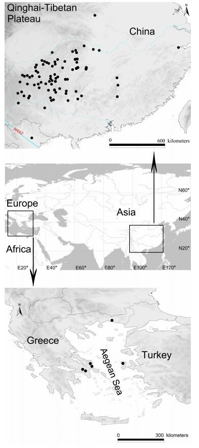
Area of endemic of Leptonetela
Molecular protocols
Total genomic DNA was extracted using the Animal Genomic DNA Isolation Kit (Dingguo, Beijing, China) following the manufacturer's protocol. We amplified the cytochrome c oxidase subunit Ⅰ (COI) barcode region using the primer pairs LCOI490/HCO2198 (Folmer et al, 1994). PCR reaction conditions were: initial denaturation at 94 ℃ for 1 min; 35 cycles of denaturation at 94 ℃ for 1 min, annealing at 45 ℃ for 45 s, and elongation at 70 ℃ for 60 s; and a final extension at 72 ℃ for 5 min. The 25 μL PCR reactions included 17.25 μL of double-distilled H2O, 2.5 μL of 10× Taq buffer (mixed with MgCl2; TianGen Biotech, Beijing, China), 2.0 μL of dNTP Mix (2.5 mmol/L), 1 μL of each forward and reverse 10 μmol/L primer, 1 μL of DNA template, and 0.25 μL Taq DNA polymerase (2.5 U/μL; TianGen Biotech, Beijing, China). Double-stranded PCR products were visualized by agarose gel electrophoresis (1% agarose). PCR products were purified and sequenced by Sunny Biotechnology Co., Ltd (Shanghai, China) using the ABI 3730XL DNA analyser. Sequences were aligned using ClustalW in Mega 6.0 (Tamura et al, 2013), with visual inspection, translation, and manual adjustment to minimize alignment error. The most appropriate phylogenetic model for the sequence alignment was selected using jModelTest2 (Darriba et al, 2012) under the Akaike Information Criterion (Posada & Crandall, 1998).
Phylogenetic analyses
Phylogenetic analyses were performed using maximum likelihood (ML) in RAXML v. 7.0.3 with the GTRCAT model (Stamatakis, 2006). One hundred replicate ML inferences were performed in the search for an optimal ML tree, each initiated with a random starting tree and employing the default rapid hill-climbing algorithm. Clade confidence was assessed with a rapid bootstrap of 1 000 replicates.
Species delineation
We analyzed the COI barcode dataset (see Supplementary Table S1) using two species delineation methods. DNA barcoding gap analyses require an a priori species designation. Therefore, we divided the 624 Leptonetela individuals of 122 populations (caves) into 90 putative species based on morphological characters and geographic information. In our DNA barcoding gap analysis, we examined the overlap between the mean intraspecific and interspecific Kimura 2-parameter (K2P) (Kimura, 1980) and uncorrected p-distance (Nei & Kumar, 2000) for each candidate species, as calculated by Mega v. 6.0 (Tamura et al, 2013).
The automatic barcode gap discovery procedure (ABGD) (<xref ref-type="bibr" rid="b31-ZoolRes-38-6-321">Puillandre et al, 2012</xref>), which does not require assigning samples to putative species, calculates all pairwise distances in the dataset, evaluates intraspecific divergences, and then sorts the samples into candidate species using the calculated distances. We performed ABGD analyses online (<a href="http://wwwabi.snv.jussieu.fr/public/abgd/" target="_blank">http://wwwabi.snv.jussieu.fr/public/abgd/</a>), using three different distance metrics: Jukes-Cantor (JC69) (<xref ref-type="bibr" rid="b18-ZoolRes-38-6-321">Jukes & Cantor, 1969</xref>), Kimura 2-parameter (K2P) (<xref ref-type="bibr" rid="b19-ZoolRes-38-6-321">Kimura, 1980</xref>), and simple distance (<italic>p</italic>-distance) (<xref ref-type="bibr" rid="b26-ZoolRes-38-6-321">Nei & Kumar, 2000</xref>). We analyzed the data using two different values for the parameters Pmin (0.0001 and 0.001), Pmax (0.1 and 0.2), and relative gap width (X=1 or 1.5), with all other parameters at default values.
Taxonomy
The terminology and the measurements in this paper generally follow Wang & Li (2011) and Ledford et al (2011). All measurements were taken in millimetres (mm). The left palpi of male spiders are illustrated, except where otherwise indicated. Abbreviations used in text include: PL: prolateral lobe; E: embolus; C: conductor; MA: median apophysis; At: atrium; SS: spermathecae stalk; SH: spermathecae.
Nomenclatural acts
This article conforms to the requirements of the amended International Code of Zoological Nomenclature. All nomenclatural acts contained within this published work have been registered in ZooBank. The ZooBank LSIDs (Life Science Identifiers) can be resolved and the associated information viewed by appending the LSID to the prefix "<a href="http://zoobank.org/" target="_blank">http://zoobank.org/</a>". The LSID for this publication is: urn:lsid:zoobank.org:pub:7ECB1BDC-8893-4D0F-8BEA-17ECE327FC47
RESULTS
In total, 624 DNA barcodes were analyzed. A full list of the analyzed specimens can be found in Supplementary Table S1. Fragment lengths of the analyzed DNA barcodes ranged from 107 (0.005%) to 617 bp (89%). For all populations, except L. kanellisi and L. robustispina, four or more DNA barcodes were generated. All nucleotides were translated into functional protein sequences in the correct reading frame, with no stop codons or indels observed. Similar to other arthropod studies, our data indicated a high AT-content for this mitochondrial gene fragment: the mean sequence compositions were A=20.5%, C=12.6%, G=24.4%, T=41.4%.
Phylogenetic inference
The ML gene tree topology suggests that Leptonetela is monophyletic, with the node highly supported (Figure 2; bootstrap value, BS=92). Our analyses revealed all Leptonetela species formed non-overlapping clusters, with bootstrap support values of 100. In contrast, relationships among putative species were largely unresolved, usually with low bootstrap support on the ML gene tree, particularly at deeper phylogenetic levels.
Figure 2.

Maximum likelihood COI gene tree for 624 terminals of Leptonetela, with the results of two different species delimitation approaches
Species delineation
DNA barcoding gap analysis: Based on our a priori species hypotheses, Interaspecific divergences ranged from zero to 5.3/5.0% (K2P/uncorrected p-distance) whereas interspecific distances were between 3.1/3% and 31.9/25% (K2P/uncorrected p-distance). Maximum intraspecific distances > 3% were found for two species, including L. reticulopecta (4.3/4.0%), and L. pentakis Lin & Li, 2010 (5.3/5%). The lowest interspecific distance were revealed for the two species pairs L.changtu Wang & Li sp. nov. with L. chuan Wang & Li sp. nov. and L. kangsa Wang & Li sp. nov. with L. shibingensis Guo, Yu & Chen, 2016 with a value of 3.1/3%. Minimum interspecific pairwise distances < 5%, and > 3% were found for two species pairs: L. shibingensis with L. shanji Wang & Li sp. nov. and L. dao Wang & Li sp. nov. with L. xiaoyan Wang & Li sp. nov. The mean interspecific distance between the 90 tentative species was 17.9/15.6% (K2P/uncorrected p-distance), and the mean intraspecific distance within each species was 0.2% (both K2P and uncorrected p-distance) in Leptonetela. A histogram of the gap and overlap between intra-and interspecies genetic distances are show in Figure 3.
Figure 3.
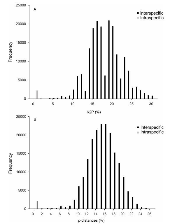
DNA barcoding for Leptonetela
ABGD analysis
The ABGD analyses of the COI dataset, using the originally specified parameter combinations and partitions resulted mostly in 90 distinct species that correspond to the 90 species observed in the previous taxonomic hypotheses based on morphology. The result was the same regardless of the model of evolution employed (Jukes-Cantor (JC), K2P, Simple Distance). The settings P min/P max=0.0001/0.2 yielded the most significant P values. However, at lower values of prior intraspecific distance (P), recursive partitioning of ABGD recognized more species (Table 1) when P min/P max=0.0001/0.2, P values=0.159, JC and K2P distance resulted in 98 species, and simple distance resulted in 95 species.
Table 1.
Results of the automatic barcode gap discovery (ABGD) analyses
| Prior intraspecific divergence (P) | |||||||||||
| Substitution mode | P min/P max | X | Partition | 0.001 | 0.0017 | 0.0028 | 0.0046 | 0.0077 | 0.0129 | 0.0215 | 0.0359 |
| JC | 0.001/0.1 | 1.5 | Initial | 90 | 90 | 90 | 90 | 90 | 90 | 90 | 90 |
| Recursive | 171 | 136 | 136 | 106 | 106 | 99 | 92 | 90 | |||
| K2P | 0.001/0.1 | 1.5 | Initial | 90 | 90 | 90 | 90 | 90 | 90 | 90 | 90 |
| Recursive | 171 | 136 | 136 | 106 | 106 | 99 | 92 | 90 | |||
| Simple | 0.001/0.1 | 1.5 | Initial | 90 | 90 | 90 | 90 | 90 | 90 | 90 | 90 |
| Recursive | 105 | 105 | 105 | 97 | 96 | 95 | 92 | 90 | |||
| JC | 0.001/0.1 | 1 | Initial | 90 | 90 | 90 | 90 | 90 | 90 | 90 | 90 |
| Recursive | 169 | 132 | 132 | 106 | 106 | 99 | 92 | 90 | |||
| K2P | 0.001/0.1 | 1 | Initial | 90 | 90 | 90 | 90 | 90 | 90 | 90 | 90 |
| Recursive | 172 | 137 | 137 | 106 | 106 | 99 | 92 | 90 | |||
| Simple | 0.001/0.1 | 1 | Initial | 90 | 90 | 90 | 90 | 90 | 90 | 90 | 90 |
| Recursive | 105 | 105 | 105 | 97 | 96 | 95 | 92 | 90 | |||
| 0.0001 | 0.0002 | 0.0005 | 0.0013 | 0.0029 | 0.0068 | 0.0159 | 0.0369 | ||||
| JC | 0.0001/0.2 | 1.5 | Initial | 90 | 90 | 90 | 90 | 90 | 90 | 90 | 90 |
| Recursive | 171 | 171 | 171 | 171 | 136 | 106 | 98 | 90 | |||
| K2P | 0.0001/0.2 | 1.5 | Initial | 90 | 90 | 90 | 90 | 90 | 90 | 90 | 90 |
| Recursive | 171 | 171 | 171 | 171 | 136 | 106 | 98 | 90 | |||
| Simple | 0.0001/0.2 | 1.5 | Initial | 90 | 90 | 90 | 90 | 90 | 90 | 90 | 90 |
| Recursive | 105 | 105 | 105 | 105 | 105 | 96 | 95 | 90 | |||
DISCUSSION
DNA barcoding is widely recognized as a useful tool for species identification across the animal kingdom (Chesters et al, 2012; Wang et al, 2011). Our research represents an important step towards the application of DNA barcodes for identification of Leptonetela taxa, and for 119 taxa (97%), our data represent the first published DNA barcodes.
Classically, geographic isolation is considered a primary feature of troglobitic taxa (Hedin, 1997; Hedin & Thomas, 2010). Our DNA barcoding result is consistent with this view and similar to other DNA barcoding studies, in which COI showed high genetic structure between populations within species (Tavares et al, 2001).
Choosing appropriate thresholds that can delimit species is one of the main challenges and concerns for DNA barcoding research (Ferguson, 2002). Our DNA barcoding gap analysis shows an overlap in the range of intra-and interspecific COI sequence divergence. The interspecific genetic divergences between L. chuan Wang & Li sp. nov. and L. changtu Wang & Li sp. nov., L. kangsa Wang & Li sp. nov. and L. shibingensis, as well as between L. shibingensis and L. shanji Wang & Li sp. nov. was 3.1/3.0% based on K2P/uncorrected and p-distance models. Compared with other species L. chuan Wang & Li sp. nov., and L. changtu Wang & Li sp. nov., L. kangsa Wang & Li sp. nov., L. shibingensis and L. shanji Wang & Li sp. nov. are more closely distributed. In morphology, L. chuan Wang & Li sp. nov. and L. changtu Wang & Li sp. nov. can be distinguished by the shape of the median apophysis and the conductor (median apophysis palm-shaped, edge with sclerotized spots, conductor semicircular in L. changtu Wang & Li sp. nov., median apophysis rectangular, with 5 larger teeth distally, conductor triangular in ventral view in L. chuan Wang & Li sp. nov.); L. kangsa Wang & Li sp. nov., L. shibingensis and L. shanji Wang & Li sp. nov. can be distinguished by the location and pattern of male pedipalpal tibial spines (Ⅰ spine located at the middle in L. shibingensis and L. shanji Wang & Li sp. nov.; Ⅰ spine asymmetrically bifurcated in L. shanji Wang & Li sp. nov., male pedipalpal tibia Ⅰ spine located at base and not bifurcated in L. kangsa Wang & Li sp. nov.). Nevertheless, we found two species with maximum pairwise distance > 3%, including L. reticulopecta (specimens from Tianshegnqiao Cave are clearly distant from the rest) with 4.3/4.0%, L. pentakis (specimens from Liaoya cave is clearly distant from the rest) with 5.3/5.0%. Then we achieved a threshold of 3.11/3.0% (K2P/uncorrected and p-distance), excluding taxa from Tianshegnqiao Cave and Liaoya Cave. This threshold was interestingly close to the 3% commonly used in barcoding literature (Hebert et al, 2003a, b).
Here, we were highly successful using ABGD for identification. In ABGD analysis, the taxa from Tianshegnqiao Cave and Liaoya Cave were identified as L. reticulopecta and L. pentakis, respectivelly. Given that all specimens of L. reticulopecta and L. pentakis are morphologically very similar, we are currently unable to ascertain if the observed genetic distances simply represent a high level of intraspecific variation or reflect cryptic species between the taxa of L. reticulopecta and L. pentakis. To answer this question, more specimens need to be collected and analyzed, using both morphological characters and nuclear sequence data.
In conclusion, our study demonstrates the power of an integrative approach, in which both classical and DNA barcoding taxonomy complements each other and both contribute to a more accurate taxonomic classification.
Taxonomy
Key to species of Leptonetela
(Mostly referring to characters of the male pedipalp)
1 Spermathecae thin and loosely twisted…………………………L. strinatii (Brignoli, 1976) (male unknown)
- Not as above……………………………………………………………2
2 Male pedipalp with median apophysis………………………3
- Male pedipalp without median apophysis…………………9
3 Median apophysis like pine needles, sclerotized………4
- Not as above……………………………………………………………33
4 Median apophysis appears as 4 pine needle-like appendages…………………………………………………………………………5
- Median apophysis divided into more or less than 4 pine needle-shaped appendages…………………………………………6
5 Tibia Ⅰ spine strong, conspicuous, with bifurcated tip……………………………………………………………………L. chakou sp.nov.
- Tibia Ⅰ spine strong, located at the middle of tibia prolaterally……………………………………………L. grandispina Lin & Li, 2010
6 Cymbium roughly double the length of bulb………………7
- Cymbium roughly the same length as bulb…………………8
7 Median apophysis divided into 15 pine needle-like appendages………………………L. liuzhai Wang & Li sp. nov.
- Median apophysis divided into 2 pine needle-like appendages………………………L. shuilian Wang & Li sp. nov.
8 Cymbium constricted medially, median apophysis divided into 5 pine needle-like appendages…………………………………………………………………………………L. pentakis Lin & Li, 2010
- Cymbium not constricted medially, median apophysis divided into 2 pine needle-like appendages………………………………………………………………………………L. dao Wang & Li sp. nov.
9 Male pedipalp with 5 tibial spines prolaterally…………10
- Male pedipalp with more than 5 tibial spines prolaterally…29
10 Cymbium constricted and wrinkled medially……………11
- Cymbium not constricted or wrinkled medially…………22
11 Tibial spines slender and without bifurcated tip…………12
- Tibial spines strong or with bifurcated tip…………………16
12 Prolateral lobe tongue-shaped…………………………………13
- Prolateral lobe absent………L. sanyan Wang & Li sp. nov.
13 Pedipalpal tibia with one spine significantly longer than other spines………………………………………………………………………14
- Pedipalp tibia Ⅰ, Ⅱ spines nearly the same length…………………………………………………………………L. meitan Lin & Li, 2010
14 Conductor bamboo leaf-shaped in ventral view…………15
- Conductor C-shaped in ventral view………………………………………………………………………L. liangfeng Wang & Li sp. nov.
15 Embolus and conductor long, intersecting……………………………………………………………………………L. suae Lin & Li, 2010
- Embolus and conductor short, not intersecting………………………………………………………………………L. tongzi Lin & Li, 2010
16 Pedipalpal tibia Ⅰ spine with bifurcated tip…………………17
- Pedipalpal tibia Ⅰ spine without bifurcated tip……………19
17 Pedipalpal tibia Ⅰ spine strong, asymmetrically bifurcate…18
- Pedipalpal tibia Ⅰ spine slender, symmetrically bifurcate………………………………………………………L. danxia Lin & Li, 2010
18 Pedipalpal tibia Ⅰ spine located proximally at tibia, thin spines Ⅱ, Ⅴ and Ⅵ arranged in a triangle, conductor bamboo leaf-shaped in ventral view………L. andreevi Deltshev, 1985
- Pedipalpal tibia Ⅰ spine located at distal 1/3 of tibia, conductor C shaped in ventral view………………………………………………………………………………L. furcaspina Lin & Li, 2010
19 Pedipalpal tibia Ⅰ spine longest…………………………………20
- Pedipalpal tibia Ⅱ spine longest………………………………21
20 Pedipalpal tibia Ⅰ spine bent distally, conductor reduced……………………………………………L. langdong Wang & Li sp. nov.
- Pedipalpal tibia Ⅰ spine not bent distally, conductor semicircular in ventral view……………………………L. yaoi Wang & Li, 2011
21 Eyes absent, pedipalpal tibia Ⅲ, Ⅴ and Ⅵ spines more slender than Ⅰ, Ⅱ spines……………L. lineata Wang & Li, 2011
- Six eyes, pedipalpal tibial spines equally strong………………………………………………………………L. caucasica Dunin, 1990
22 Conductor developed………………………………………………23
- Conductor reduced…………………………………………………26
23 Pedipalpal tibia Ⅰ spine without bifurcated tip……………24
- Pedipalpal tibia Ⅰ spine with bifurcated tip, other spines concentrated distally, tip of conductor bifurcated…………………………………………………………………L. anshun Lin & Li, 2010
24 Conductor bamboo leaf-shaped in ventral view…………25
- Conductor C-shaped in ventral view, pedipalpal tibia Ⅰ spine longest………………………………L. dashui Wang & Li sp. nov.
25 Pedipalpal tibia Ⅰ spine strong, prolateral bulbal lobe reduced………………………………………L. qiangdao Wang & Li sp. nov.
- Pedipalpal tibia Ⅰ spine slender, prolateral bulbal lobe tongue-shaped……………L. nuda (Chen, Jia & Wang, 2010)
26 Cymbium with a distal and proximal spine prolaterally, pedipalpal tibial spines equidistant……………………………………………………………………………L. curvispinosa Lin & Li, 2010
- Not as above……………………………………………………………27
27 Pedipalpal tibia Ⅰ spine slender, asymmetrically bifurcated…………………………………………L. wangjia Wang & Li sp. nov.
- Not as above……………………………………………………………28
28 Pedipalpal tibia Ⅰ, Ⅱ, and Ⅲ spines concentrated in the mid of tibia, 2 additional spines located distally, prolateral bulbal lobe reduced……………………L. maxillacostata Lin & Li, 2010
- Pedipalpal tibia Ⅰ spine longest, located far from others, prolateral lobe small, tongue shaped…………………………………………………………………………L. chenjia Wang & Li sp. nov.
29 Male pedipalp with 6 tibial spines retrolaterally…………30
- Male pedipalp with 7 tibial spines retrolaterally…………32
30 Pedipalpal tibia Ⅰ, Ⅱ spines strong, equally length, Ⅱ spine asymmetrically bifurcated, conductor reduced………………………………………………………………L. gang Wang & Li sp. nov.
- Pedipalpal tibial spines slender, not bifurcated, conductor developed………………………………………………………………31
31 Pedipalpal tibia with 2 large spines prolaterally, cymbium not constricted medially, earlobe-shaped process absent, and cymbium long, twice the length of bulb…………………………………………………………………L. gigachela (Lin & Li, 2010)
- Pedipalpal tibia without prolateral spines, cymbium constricted medially, retrolaterally attaching an earlobe-shaped process, cymbium less than twice the length of bulb…………………………………………………………………L. wenzhu Wang & Li sp. nov.
32 Cymbium with 1 horn-shaped spine on the earlobe-shaped process, conductor thin, triangular in ventral view………………………………………………………L. rudong Wang & Li sp. nov.
- Earlobe-shaped process of cymbium without spine, conductor broad, C shaped in ventral view…L. la Wang & Li sp. nov.
33 Median apophysis like a pointed process or lamelliform..34
- Median apophysis finger-shaped or harrow-like………50
34 Cymbium not constricted medially, earlobe-shaped process reduced……………………………………………………………………35
- Cymbium constricted medially, earlobe-shaped process developed…………………………………………………………………38
35 Male pedipalpal tibia with 6 spines retrolaterally……………36
- Male pedipalpal tibia only with 5 spines retrolaterally……37
36 Pedipalpal tibia with 4 long spines prolaterally, the retrolateral Ⅰ spine longest, Ⅱ Ⅲ spines short and strong, median apophysis pointed, conductor bamboo leaf-shaped………………………………………………………………………………L. bama Lin & Li, 2010
- Pedipalpal tibia with 3 long spines prolaterally, the retrolateral Ⅰ spine longest and strongest, median apophysis "M"-shaped, conductor reduced………………………L. yangi Lin & Li, 2010
37 Pedipalpal tibia with 1 long spine prolaterally, the retrolateral Ⅰ spine longest and strongest, median apophysis pointed, conductor bamboo leaf-shaped………L. liping Lin & Li, 2010
- Pedipalpal tibia with 3 long spines prolaterally, the retrolateral spines Ⅰ slender, and longest, median apophysis obtuse triangle shaped, conductor narrow, triangular……………………………………………………………L. mayang Wang & Li sp. nov.
38 Cymbium with 1 strong spine on the earlobe-shaped process…………………………………………………………………………………39
- No spine on the earlobe-shaped process…………………43
39 Male pedipalp tibia with 5 spines retrolaterally…………40
- Male pedipalp tibia with more than 5 spines retrolaterally…41
40 Cymbium with 1 curved spine retrolaterally, median apophysis pointed, with 3 sclerotized apices distally, conductor C shaped…………………………………L. jiahe Wang & Li sp. nov.
- Cymbium without curved spine retrolaterally, median apophysis punctate in ventral view, conductor vestigial………………………………………………L. panbao Wang & Li sp. nov.
41 Pedipalpal tibia Ⅰ spine strong, Ⅱ spine asymmetrically bifurcated, median apophysis lamelliform, conductor triangular…………………………………………………L. jiulong Lin & Li, 2010
- Pedipalpal tibia Ⅰ spine slender, not bifurcated…………42
42 Pedipalpal tibia with 3 long spines prolaterally, 6 spines retrolaterally, median apophysis semicircular………………………………………………………………L. parlonga Wang & Li, 2011
- Pedipalpal tibia with 5 long spines prolaterally, 7 spines retrolaterally, median apophysis mita-shaped, embolus with 1 tooth distally……………………………L. mita Wang & Li, 2011
43 Pedipalpal tibia with 5 spines retrolaterally………………44
- Pedipalpal tibia with more than 5 spines retrolaterally……49
44 Pedipalpal tibia with 3 long spines prolaterally………………45
- Pedipalpal tibia with 1 or 2 long spines prolaterally………47
45 Conductor C shaped in ventral view……………………………46
- Conductor bamboo leaf-shaped in ventral view, retrolateral spines Ⅰ longest, median apophysis triangular………………………………………………………………L. xianren Wang & Li sp. nov.
46 Pedipalpal tibia Ⅰ spine longest, the rest concentrated at distal end of tibia, median apophysis spatula-shaped in ventral view…………………………L. rudicula Wang & Li, 2011
- Pedipalpal tibia Ⅰ spine longest, Ⅰ, Ⅱ, and Ⅲ spines equally strong, median apophysis single quote shaped, " ′ " in ventral view…………………………L. longli Wang & Li sp. nov.
47 Pedipalpal tibia with 1 long spine prolaterally, median apophysis tongue-shaped, conductor triangular……………………………………………………………L. pungitia Wang & Li, 2011
- Pedipalpal tibia with 2 long spines prolaterally………………48
48 Pedipalpal tibia Ⅰ spine strongest, Ⅲ-Ⅴ spines in a triangular arrangement, median apophysis punctate, conductor triangular…………………………L. chiosensis Wang & Li, 2011
- Pedipalpal tibia Ⅰ spine longest, spine Ⅰ-Ⅲ equally strong, Ⅳ-Ⅴ situated distally median apophysis "m"-shaped, conductor triangular…………L. feilong Wang & Li sp. nov.
49 Pedipalpal tibia with 6 spines retrolaterally, tibia Ⅰ spine close to others, median apophysis flake-like, sclerotized distally, conductor broad, undulate distally………………………………………………………………L. tiankeng Wang & Li sp. nov.
- Pedipalpal tibia with 7 spines retrolaterally, tibia Ⅰ spine distant from others, median apophysis small worm-shaped, conductor thin, triangular…………………………………………………………………L. lophacantha (Chen, Jia & Wang, 2010)
50 Median apophysis index finger like…………………………51
- Median apophysis harrow-like…………………………………72
51 Embolus bifurcated……………L. xinhua Wang & Li sp. nov.
- Embolus not bifurcated……………………………………………52
52 Base of median apophysis swollen…………………………53
- Base of median apophysis not swollen……………………56
53 Male pedipalpal tibia with 5 spines retrolaterally………54
- Male pedipalpal tibia with 6 slender spines retrolaterally, spines Ⅰ longest, conductor smooth, semicircular……………………………………………L. quinquespinata (Chen & Zhu, 2008)
54 Pedipalpal tibia Ⅰ spine much stronger than Ⅱ, asymmetrically bifurcated……………………………………L. jinsha Lin & Li, 2010
- Pedipalpal tibia Ⅰ spine similarly strong as Ⅱ, not bifurcated…………………………………………………………………………………55
55. Cymbium constricted medially, earlobe-shaped process with 2 long, curved spines retrolaterally, base of median apophysis distinctly swollen, conductor smooth, broad, semicircular…………………………L. gubin Wang & Li sp. nov.
- Cymbium not constricted medially, earlobe-shaped process small, base of median apophysis slightly swollen, conductor rugose, thin, triangular……………L. lujia Wang & Li sp. nov.
56 Median apophysis bifurcated distally…………………………………………………………………………L. wuming Wang & Li sp. nov.
- Median apophysis not bifurcated distally…………………57
57 Pedipalpal tibia Ⅰ spine located at the base of tibia……58
- Pedipalpal tibia Ⅰ spine located medially……………………59
58 Pedipalpal tibia Ⅰ spine asymmetrically bifurcated, tibia with 4 long spines prolaterally………………………………………………………………………………L. shibingensis Guo, Yu & Chen, 2016
- Pedipalpal tibia Ⅰ spine not bifurcated…………………………………………………………………………L. kangsa Wang & Li sp. nov.
59 Male pedipalp tibia with 6 spines retrolaterally…………60
- Male pedipalp tibia with 5 spines retrolaterally…………62
60 Pedipalpal tibia with 4 spines prolaterally, cymbium with 1 curved spine at the base of retrolateral surface, median apophysis weakly sclerotized…………………………………………………………………………………L. xiaoyan Wang & Li sp. nov.
- Not as above……………………………………………………………61
61 Male pedipalp tibia with 2 spines prolaterally, conductor short, broad and rugose………L. oktocantha Lin & Li, 2010
- Male Pedipalp tibia without spine prolaterally, conductor smooth, semicircular……………L. hexacantha Lin & Li, 2010
62 Median apophysis curved distally……………………………63
- Median apophysis not curved distally………………………65
63 Cymbium with 1 horn-shaped spine on the earlobe-shaped process retrolaterally, tibia spines gradually shorted, conductor smooth, C shaped……………………………………………………………………L. mengzongensis Wang & Li, 2011
- Cymbium without spine on the earlobe-shaped process retrolaterally………………………………………………………………64
64 Male pedipalp tibia with 2 long setae prolaterally, tibia Ⅰ Ⅱ and Ⅲ spines equally in length, conductor broad, semicircular…………………………………………………L. hamata Lin & Li, 2010
- Male pedipalp tibia with 4 long spines prolaterally, tibia Ⅰ Ⅱ spines equally in length, conductor long, curved distally……………………………………………L. tetracantha Lin & Li, 2010
65 Pedipalpal tibia Ⅰ spur strong……………………………………66
- Pedipalpal tibia Ⅰ spine slender…………………………………70
66 Pedipalpal tibia Ⅰ spine asymmetrically bifurcated………67
- Pedipalpal tibia Ⅰ spine not bifurcated, conductor broad, C shaped, median apophysis distinctly sclerotized…………………………………………………………L. reticulopecta Lin & Li, 2010
67 Median apophysis tapering……………………………………68
- Median apophysis blunt…………………………………………69
68 Pedipalpal tibia Ⅰ spine located at the middle of tibia………………………………………………………L. shanji Wang & Li sp. nov.
- Pedipalpal tibia Ⅰ spine located at the basal of tibia………………………………………………………………L. digitata Lin & Li, 2010
69 Pedipalpal tibia Ⅱ-Ⅴ spines slender flexible, Ⅰ and Ⅱ spines equally length, conductor shorter than median apophysis………………………………………L. tianxinensis (Tong & Li, 2008)
- Pedipalpal tibia Ⅱ spine slender, Ⅲ spine strong, conductor longer than median apophysis……………………………………………………………………………………L. nanmu Wang & Li sp. nov.
70 Pedipalpal tibia Ⅰ spine located at the base of tibia, other spines concentrated distally on tibia, conductor smooth, semicircular………………………L. huoyan Wang & Li sp. nov.
- Not as above……………………………………………………………71
71 Pedipalpal tibia Ⅰ Ⅱ spines adjacent, the rest short, concentrated distally, outermost plumose, tibia with 2 spines prolaterally, conductor bifurcate………………………………………………………………………L. geminispina Lin & Li, 2010
- Pedipalpal tibia Ⅰ-Ⅳ spines spaced at regular intervals, Ⅳ and Ⅴ adjacent, tibia Ⅰ-Ⅲ equal in length, conductor short, C shaped…………………………L. tianxingensis Wang & Li, 2011
72 Median apophysis harrow-like, horrow pin reduced to sclerotized spots………………………………………………………73
- Median apophysis harrow-like, horrow pin not reduced…75
73 Pedipalpal tibial spines slender, equally strong, median apophysis long, half the length of bulb………………………………………………………………………L. liuguan Wang & Li sp. nov.
- Pedipalpal tibial spines not equally strong, median apophysis short, 1/5 the length of bulb…………………………74
74 Pedipalpal tibia Ⅰ, Ⅱ spines equally strong, stronger than other spines, Ⅲ-Ⅴ in triangular arrangement, cymbium constricted medially, with one curved spine at the base of constriction retrolaterally…………L. penevi Wang & Li, 2016
- Pedipalpal tibia Ⅰ Ⅱ Ⅲ spines equally strong, stronger than other spines, Ⅲ-Ⅴ not triangular arrangement, cymbium not constricted medially…………L. changtu Wang & Li sp. nov.
75 Median apophysisi harrow-like, the horrow pin not constant in size………………………………………………………………………76
- Median apophysisi harrow-like, the horrow pin constant in size…………………………………………………………………………80
76 Pedipalpal tibia Ⅰ spine not bifurcated…………………………77
- Pedipalpal tibia Ⅰ spine strong, asymmetrically bifurcated, other 4 spines slender, median apophysis with 5 small teeth and 1 large, horn-shaped tooth…………………………………………………………………………L. lianhua Wang & Li sp. nov.
77 Pedipalpal tibia Ⅰ spine longest…………………………………78
- Pedipalpal tibia Ⅱ spine longest………………………………79
78 Median apophysis palmate, with six teeth distally…………………………………………L. megaloda (Chen, Jia & Wang, 2010)
- Median apophysis antler-like, with 4 small teeth and 1 large tooth, which bears 2 small teeth…………………………………………………………………………………L. niubizi Wang & Li sp. nov.
79 Two large teeth on the periphery of median apophysis, 2 small teeth in the middle……………………………………………………………………L. hangzhouensis (Chen, Shen & Gao, 1984)
- Two large teeth on the periphery of median apophysis, 5 small teeth in the middle.. L. microdonta (Xu & Song, 1983)
80 Median apophysis short and broad…………………………81
- Median apophysis long and thin……………………………87
81 Pedipalpal tibia Ⅰ spine strongest……………………………82
- Pedipalpal tibia Ⅰ spine not strongest………………………83
82 Pedipalpal tibia Ⅰ, Ⅱ spines equally strong, stronger than other 3 spines, median apophysis with 6 small teeth apically………………………………L. identica (Chen, Jia & Wang, 2010)
- Pedipalpal tibia Ⅱ, Ⅲ spines equally strong, spine Ⅱ longest, median apophysis with 5 sharp teeth apically……………………………………………………………………………L. meiwang sp.nov.
83 Pedipalpal tibia Ⅰ spine bifurcated……………………………84
- Pedipalpal tibia Ⅰ spine not bifurcated………………………89
84 Distal edge of median apophysis with 6 teeth……………85
- Distal edge of median apophysis with 5 or 10 teeth…86
85 Teeth of median apophysis needle-shaped, earlobe-shaped process of cymbium absent; in the female, anterior margin of atrium with one pointed process medially…………………………………………………………………L. zakou Wang & Li sp. nov.
- Teeth of median apophysis normal, cymbium with earlobe-shaped process, female anterior margin of atrium without pointed process………………L. sexdentata Wang & Li, 2011
86 Distal edge of median apophysis with 5 teeth, conductor C shaped, tip of conductor undulate in the female, anterior margin of atrium with one pointed process medially…………………………………L. longyu Wang & Li sp. nov.
- Distal edge of median apophysis with 10 teeth, conductor C shaped, distal edge of conductor smooth; in the female, anterior margin of atrium without pointed process…………………………………………………L. shicheng Wang & Li sp. nov.
87 Pedipalpal tibia Ⅱ spine tapering………………………………88
- Pedipalpal tibia Ⅰ spine blunt…L. flabellaris Wang & Li, 2011
88 Distal edge of median apophysis with 5 teeth, conductor short, C shaped………………………L. palmata Lin & Li, 2010
- Distal edge of median apophysis with 7 teeth, conductor long, triangular shaped………………………………………………………………………………L. kanellisi (Deeleman-Reinhold, 1971)
89 Pedipalpal tibia with clusters of short spines dorsally……90
- Pedipalpal tibia without clusters of short spines dorsally………………………………………………………………………………………91
90 Distal edge of median apophysis linear, with 8 teeth……………………………………………………L. encun Wang & Li sp. nov.
- Distal edge of median apophysis semicircular, with 12 teeth………………………L. robustispina (Chen, Jia & Wang, 2010)
91 Base of pedipalpal tibia swollen………………………………92
- Base of pedipalpal tibia not swollen…………………………96
92 Pedipalpal tibia Ⅰ spine bifurcate………………………………93
- Pedipalpal tibia Ⅰ spine trifurcate…………………………………………………………………………………L. notabilis (Lin & Li, 2010)
93 Conductor triangular, longer than median apophysis, median apophysis with 7 teeth………………………………………………94
- Conductor C shaped, shorter than median apophysis, median apophysis with 6 teeth……………………………………95
94 Spermathecae not twisted distally………………………………………………………………………………L. shuang Wang & Li sp. nov.
- Spermathecae twisted distally……………………………………………………………………………L. sanchahe Wang & Li nom. nov.
95 Spermathecae weakly twisted……………………………………………………………………………L. sexdigiti (Lin & Li, 2011)
- Spermathecae strongly twisted………………………………………………………………………………………L. lihu Wang & Li sp. nov.
96 Pedipalpal tibia Ⅰ spine strongset……………………………97
- Pedipalpal tibia Ⅰ, Ⅱ, Ⅲ spines equally strong, stronger than other spines……………………………………………………………103
97 Pedipalpal tibia Ⅱ spine bifurcate………………………………98
- Pedipalpal tibia Ⅰ spine not bifurcate………………………101
98 Pedipalpal tibia Ⅱ-Ⅴ spine slender, curved, equally strong…………………………………………………………………………………99
- Pedipalpal tibia Ⅱ-Ⅴ spine not equally strong…………100
99 Distal edge of median apophysis with 6 teeth……………………………………………………L. arvanitidisi Wang & Li, 2016
- Distal edge of median apophysis with 5 teeth………………………………………………………L. erlong Wang & Li sp. nov.
100 Distal edge of median apophysis with 4 teeth, tibia Ⅱ, Ⅲ spines equally strong, stronger than other 2 spines………………………………………………L. tawo Wang & Li sp. nov.
- Distal edge of median apophysis with 3 teeth, tibia Ⅲ-Ⅴ spines equally strong, slender than spine Ⅱ……………………………………………………………L. paragamiani Wang & Li, 2016
101 Pedipalpal tibia Ⅱ-Ⅴ spines equally strong………………102
- Pedipalpal tibia Ⅲ-Ⅴ spines equally strong, slender than spine Ⅱ, distal edge of median apophysis with 4 teeth……………………………………………………L. deltshevi (Brignoli, 1979)
102 Distal edge of median apophysis with 5 teeth, conductor C shaped…………………………L. gittenbergeri Wang & Li, 2011
- Distal edge of median apophysis with 6 teeth, conductor semicircular………………………………L. zhai Wang & Li, 2011
103 Pedipalpal tibia Ⅰ Ⅱ spines equally strong……………………………………………………………………………L. thracia Gasparo, 2005
- Pedipalpal tibia Ⅱ and Ⅲ spines equally strong………104
104 Distal edge of median apophysis with 3 teeth, tibia with 3 large spines prolaterally………L. dabian Wang & Li sp. nov.
- Distal edge of median apophysis with 6 teeth, tibia with 6 long setae prolaterally…………L. chuan Wang & Li sp. nov.
Family Leptonetidae Simon, 1890
Genus Leptonetela Kratochvíl, 1978
Type species: Leptonetela kanellisi (Deeleman-Reinhold, 1971) from Greece.
Diagnosis. The genus Leptonetela can be distinguished from other leptonetid genera by the following combination of male pedipalpal characters: femur lacking spines and tibia with a longitudinal row of spines on the retrolateral surface.
Redescription. Carapace yellowish or white. Sternum shield-shaped. Opisthosoma gray, ovoid, covered with short hairs. Male pedipalpal patella with one short spine dorso-distally; tibia with trichobothria dorsally; cymbium with strong, thorny spine distally; bulb yellowish, ovoid, with two appendages inserted ventrally, median apophysis chitinous, conductor membranous, median apophysis and conductor absent in some species, embolus transparent, membranous. Female genital area covered with short hairs. Vulva with a pair of spermathecae and sperm ducts, spermathecae twisted and weakly sclerotized.
Distribution. Greece, Turkey, Georgia, Azerbaijan, Vietnam and China.
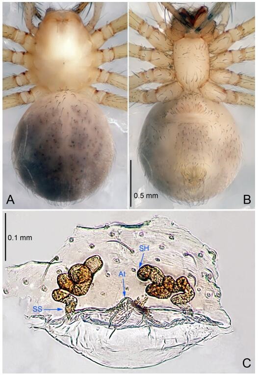
Leptonetela chakou Wang & Li sp. nov. Figures 4-5, 97
Figure 4.
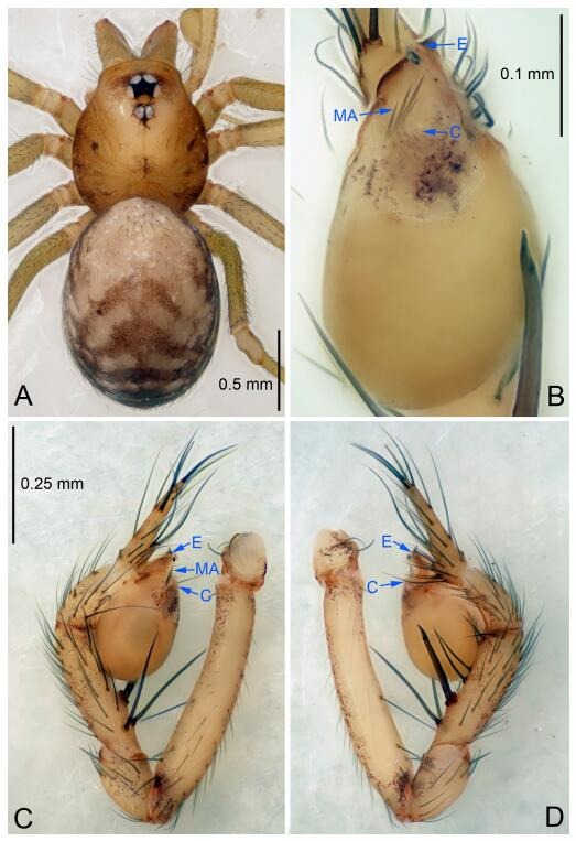
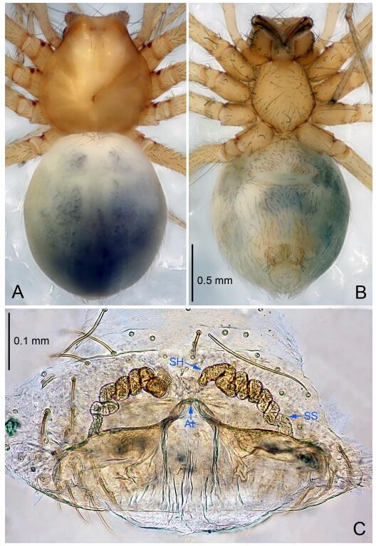
Leptonetela chakou sp. nov., holotype male
Figure 5.
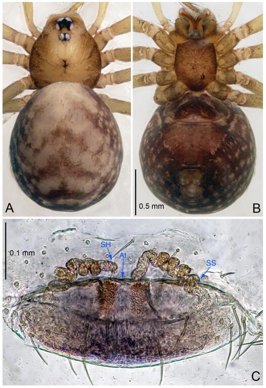
Leptonetela chakou sp. nov., one of the paratype females
Figure 97.
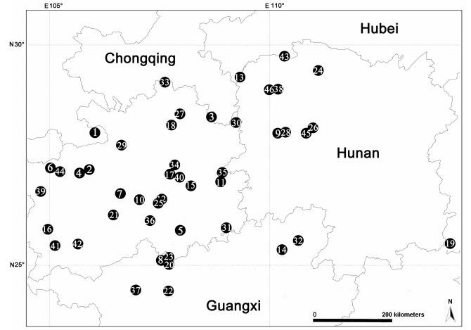
Locality records for forty-six new species of Leptonetela in China
Type material. Holotype: male (IZCAS), Chakou Cave, N27.93°, E106.14°, Shalang, Shibao Town, Gulin County, Luzhou City, Sichuan Province, China, 20 April 2014, Y. Li, H. Zhao & Y. Lin leg. Paratypes: 1 male and 3 females, same data as holotype.
Etymology. The specific name refers to the type locality; noun.
Diagnosis. This new species is similar to L. dao Wang & Li sp. nov., L. grandispina Lin & Li, 2010, L. liuzhai Wang & Li sp. nov., L. pentakis Lin & Li, 2010, and L. shuilian Wang & Li sp. nov., but can be distinguished by the male pedipalpal tibia with 5 spines retrolaterally, the basal spine strong, conspicuous and with a bifurcate tip (Figure 4D) (6 short spines, with spine Ⅱ largest in L. grandispina, 5 slender spines in L. dao Wang & Li sp. nov., L. liuzhai Wang & Li sp. nov., L. pentakis and L. shuilian Wang & Li sp. nov.); the median apophysis divided into 4 pine needle like structures (Figure 4B) (median apophysis divided into 2 pine needlelike structures a in L. dao Wang & Li sp. nov. and L. shuilian Wang & Li sp. nov., 15 pine needlelike structures in L. liuzhai Wang & Li sp. nov., and 5 pine needle like structures in L. pentakis); from L. dao Wang & Li sp. nov., L. grandispina, L. pentakis by the conductor reduced in this new species (Figure 4B); from L. liuzhai Wang & Li sp. nov. by the cymbium 1.3 times longer than bulb (Figure 4C-D) (cymbium 2 times longer than bulb in L. liuzhai Wang & Li sp. nov. and L. shuilian Wang & Li sp. nov.).
Description. Male (holotype). Total length 2.25 (Figure 4A). Carapace 0.87 long, 0.87 wide. Opisthosoma 1.50 long, 1.00 wide. Carapace brown. Eyes six. Median groove, cervical grooves and radial furrows distinct. Clypeus 0.13 high. Opisthosoma gray, ovoid, with pigmented stripe. Leg measurements: Ⅰ 7.63 (2.05, 0.35, 2.35, 1.75, 1.13); Ⅱ 5.71 (1.63, 0.30, 1.60, 1.30, 0.88); Ⅲ 4.73 (1.25, 0.30, 1.13, 1.20, 0.85); Ⅳ 6.30 (1.75, 0.35, 1.75, 1.45, 1.00). Male pedipalp (Figure 4C-D): tibia with 2 large spines prolaterally, and 5 spines retrolaterally, Ⅰ spine strong, conspicuous, tip bifurcated. Cymbium constricted medially, attaching to an earlobe-shaped process. Embolus triangular, bearing a basal tooth. Median apophysis sclerotized, divided into 4 pine needle like structures. Conductor membranous, reduced (Figure 4B).
Female (one of the paratypes). Similar to male in color and general features, but larger and with shorter legs. Total length 2.27 (Figure 5A-B). Carapace 0.88 long, 0.80 wide. Opisthosoma 1.50 long, 1.25 wide. Clypeus 0.12 high. Leg measurements: Ⅰ 5.83 (1.50, 0.35, 1.55, 1.38, 1.05); Ⅱ 4.43 (1.13, 0.30, 1.25, 1.00, 0.75); Ⅲ 3.62 (1.00, 0.25, 1.00, 0.75, 0.62); Ⅳ 4.96 (1.38, 0.35, 1.35, 1.13, 0.75). Vulva (Figure 5C): spermathecae coiled, atrium fusiform.
Distribution. China (Sichuan).
Leptonetela dao Wang & Li sp. nov. Figures 6-7, 97
Figure 6.
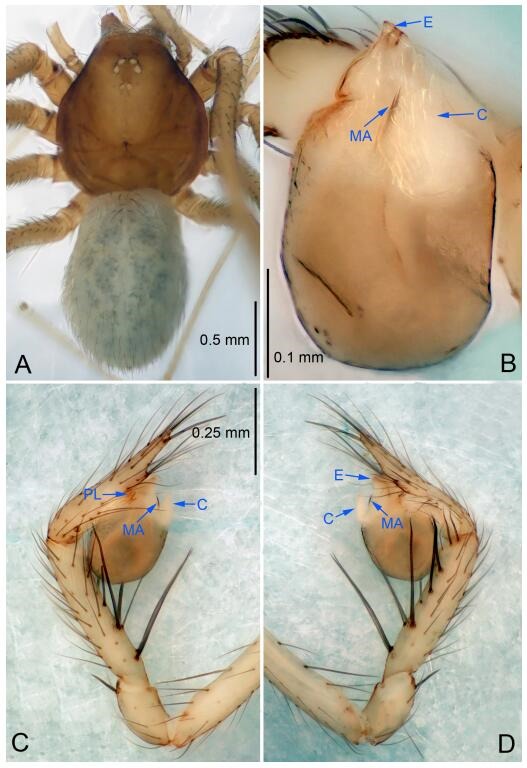
Leptonetela dao sp. nov., holotype male
Figure 7.
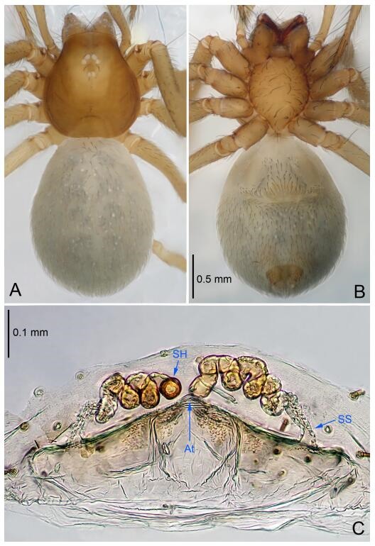
Leptonetela dao sp. nov., one of the paratype females
Type material. Holotype: male (IZCAS), Dao Cave, N27.19°, E105.06°, Shuanglong, Salaxi County, Bijie City, Guizhou Province, China, 18 November 2011, H. Chen & Z. Zha leg. Paratypes: 1 male and 20 females, same data as holotype; 5 males and 7 females, Shanlanqiao Cave, N26.28°, E106.04°, Shanlanqiao, Qianyanqiao Town, Anshun City, Guizhou Province, China, 4 November 2011, H. Chen & Z. Zha leg.
Etymology. The specific name refers to the type locality; noun.
Diagnosis. This new species is similar to L. chakou Wang & Li sp. nov., L. grandispina Lin & Li, 2010, L. liuzhai Wang & Li sp. nov. L. pentakis Lin & Li, 2010, and L. shuilian Wang & Li sp. nov., but can be separated from L. chakou Wang & Li sp. nov., L. grandispina, L. liuzhai Wang & Li sp. nov. and L. pentakis by median apophysis divided into 2 pine needlelike (Figure 6B) (median apophysis divided into 4 pine needle like structures in L. chakou Wang & Li sp. nov. and L. grandispina, 15 pine needlelike structures in L. liuzhai Wang & Li sp. nov., and 5 pine needlelike structures in L. pentakis); from L. chakou Wang & Li sp. nov., L. grandispina by the tibial spines slender (Figure 6D) (the tibia Ⅰ spine in L. chakou Wang & Li sp. nov. and Ⅱ spines in L. grandispina strong); from L. chakou Wang & Li sp. nov., L. pentakis by the cymbium not constricted medially (Figure 6C); from L. liuzhai Wang & Li sp. nov. and L. shuilian Wang & Li sp. nov. by the cymbium 1.2 times longer than bulb (Figure 6C-D) (cymbium 2 times longer than bulb in L. liuzhai Wang & Li sp. nov. and L. shuilian Wang & Li sp. nov.).
Description. Male (holotype). Total length 2.28 (Figure 6A). Carapace 1.15 long, 1.03 wide. Opisthosoma 1.28 long, 0.93 wide. Carapace brown. Eyes six, reduced to white vestiges. Median groove, cervical grooves and radial furrows distinct. Clypeus 0.15 high. Opisthosoma gray, ovoid. Leg measurements: Ⅰ 10.36 (2.76, 0.40, 3.24, 2.40, 1.56); Ⅱ 8.72 (2.44, 0.36, 2.60, 1.72, 1.60); Ⅲ 6.20 (2.04, 0.32, 1.52, 1.40, 0.92); Ⅳ 8.80 (2.56, 0.40, 2.60, 2.04, 1.20). Male pedipalp (Figure 6C-D): tibia with 5 slender spines prolaterally and 5 slender spines retrolaterally, with Ⅰ spine longest. Cymbium not wrinkled, earlobe-shaped process small. Embolus triangular, prolateral lobe small, oval. Median apophysis sclerotized, divided into 2 pine needle like structures. Conductor broad, C shaped in ventral view (Figure 6B).
Female (one of the paratypes). Similar to male in color and general features, but larger and with longer legs. Total length 2.76 (Figure 7A-B). Carapace 1.13 long, 1.10 wide. Opisthosoma 1.65 long, 1.40 wide. Clypeus 0.13 high. Leg measurements: Ⅰ 11.36 (3.00, 0.40, 3.60, 2.60, 1.76); Ⅱ 9.08 (2.64, 0.36, 2.80, 1.88, 1.40); Ⅲ 7.44 (2.24, 0.36, 1.96, 1.64, 1.24); Ⅳ 9.68 (2.80, 0.40, 3.00, 2.08, 1.40). Vulva (Figure 7C): spermathecae coiled, atrium triangular, anterior margin of atrium with short hairs.
Distribution. China (Guizhou).
Leptonetela liuzhai Wang & Li sp. nov. Figures 8-9, 97
Figure 8.
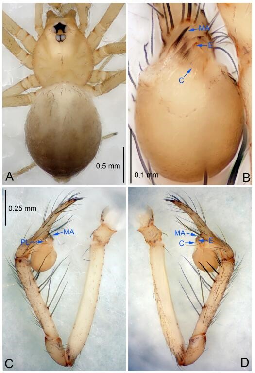
Leptonetela liuzhai sp. nov., holotype male
Figure 9.
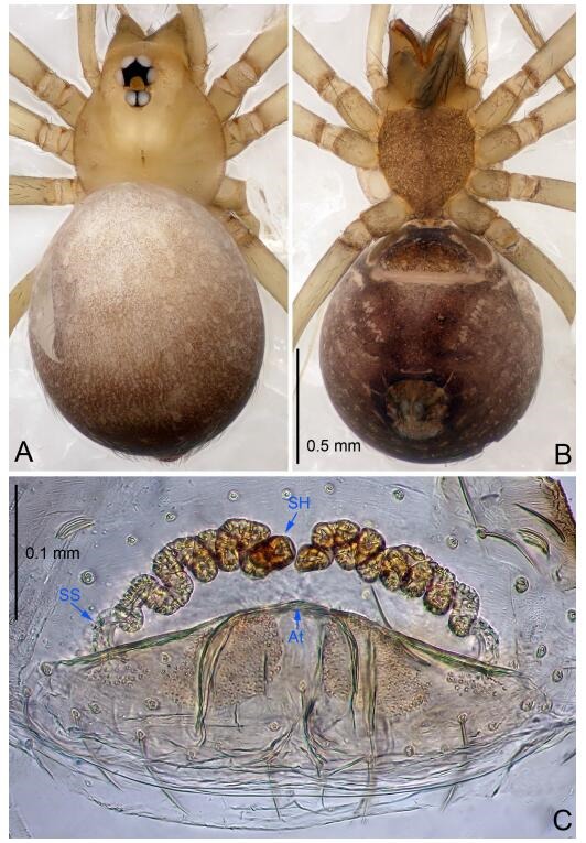
Leptonetela liuzhai sp. nov., one of the paratype females
Type material. Holotype: male (IZCAS), nameless Cave, N25.27°, E107.43°, Longli, Liuzhai Town, Nandan County, Hechi City, Guangxi Zhuang Autonomous Region, China, 29 January 2015, Y. Li & Z. Chen leg. Paratypes: 2 males and 6 females, same data as holotype.
Etymology. The specific name refers to the type locality; noun.
Diagnosis. This new species is similar to L. chakou Wang & Li sp. nov., L. dao Wang & Li sp. nov., L. grandispina Lin & Li, 2010, L. pentakis Lin & Li, 2010, and L. shuilian Wang & Li sp. nov. but can be separated from L. chakou Wang & Li sp. nov., L. dao Wang & Li sp. nov., L. grandispina, and L. pentakis by the male pedipalpal cymbium double the length of bulb, median apophysis divided into 15 pine needle-like structures (Figure 8B) (cymbium not double the length of bulb in L. chakou Wang & Li sp. nov., L. dao Wang & Li sp. nov., L. grandispina, and L. pentakis; median apophysis with 4 pine needlelike structures in L. chakou Wang & Li sp. nov. and L. grandispina, 2 pine needlelike structures in L. dao Wang & Li sp. nov. and L. shuilian Wang & Li sp. nov., and 5 pine needlelike structures in L. pentakis); from L. chakou Wang & Li sp. nov., and L. grandispina by the tibial spines slender (Figure 8D) (Ⅰ tibial spine in L. chakou Wang & Li sp. nov. and Ⅱ spines in L. grandispina strong); from L. chakou Wang & Li sp. nov., and L. pentakis by the cymbium not constricted medially in this new species (Figure 8C-D).
Description. Male (holotype). Total length 2.25 (Figure 8A). Carapace 1.00 long, 0.88 wide. Opisthosoma 1.35 long, 1.10 wide. Carapace yellowish. Ocular area with a pair of setae, eyes six. Median groove needle-shaped, cervical grooves and radial furrows distinct. Clypeus 0.15 high. Opisthosoma gray, ovoid. Leg measurements: Ⅰ 8.30 (2.25, 0.25, 2.35, 1.95, 1.50); Ⅱ 6.68 (1.88, 0.25, 2.00, 1.55, 1.00); Ⅲ 5.70 (1.63, 0.20, 1.62, 1.35, 0.90); Ⅳ 7.49 (2.13, 0.25, 2.13, 1.85, 1.13). Male pedipalp (Figure 8C-D): tibia with 5 long spines prolaterally and 5 spines retrolaterally, with tibia Ⅰ spine longest. Cymbium not wrinkled, earlobe-shaped process small, cymbium double the length of bulb. Embolus triangular, prolateral lobe reduced. Median apophysis sclerotized, divided into 15 pine needlelike structures. Conductor reduced (Figure 8B).
Female (one of the paratypes). Similar to male in color and general features, but larger and with shorter legs. Total length 2.50 (Figure 9A-B). Carapace 1.50 long, 0.88 wide. Opisthosoma 1.13 long, 1.38 wide. Clypeus 0.13 high. Leg measurements: Ⅰ 7.30 (2.00, 0.25, 2.25, 1.75, 1.05); Ⅱ 5.51 (1.63, 0.20, 1.55, 1.25, 0.88); Ⅲ 4.76 (1.38, 0.25, 1.25, 1.13, 0.75); Ⅳ 6.50 (1.87, 0.25, 1.88, 1.50, 1.00). Vulva (Figure 9C): spermathecae coiled, atrium fusiform.
Distribution. China (Guangxi).
Leptonetela shuilian Wang & Li sp. nov. Figures 10-11, 97
Figure 10.
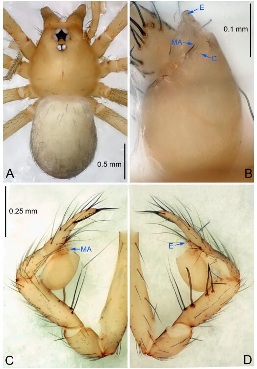
Leptonetela shuilian sp. nov., holotype male
Figure 11.
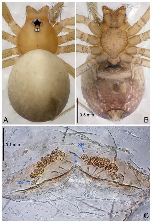
Leptonetela shuilian sp. nov., one of the paratype females
Type material. Holotype: male (IZCAS), Shuilian Cave, N24.43°, E106.97°, Pingle, Fengshan County, Hechi City, Guangxi Zhuang Autonomous Region, China, 22 March 2015, Y. Li & Z. Chen leg. Paratypes: 6 males and 4 females, same data as holotype.
Etymology. The specific name refers to the type locality; noun.
Diagnosis. This new species is similar to L. chakou Wang & Li sp. nov., L. dao Wang & Li sp. nov., L. grandispina Lin & Li, 2010, L. pentakis Lin & Li, 2010, and L. liuzhai Wang & Li sp. nov. but can be separated from L. chakou Wang & Li sp. nov., L. dao Wang & Li sp. nov., L. grandispina Lin & Li, 2010, L. pentakis Lin & Li, 2010 by the male pedipalpal cymbium double the length of bulb; from L. chakou Wang & Li sp. nov., L. grandispina, L. liuzhai Wang & Li sp. nov. and L. pentakis by the median apophysis divided into 2 pine needlelike structures in L. chakou Wang & Li sp. nov. (Figure 10B) (median apophysis divided into 4 pine needlelike structures in L. chakou Wang & Li sp. nov. and L. grandispina, 15 pine needlelike structures in L. liuzhai Wang & Li sp. nov., and 5 pine needlelike structures in L. pentakis); from L. chakou Wang & Li sp. nov. and L. grandispina by the tibial spines slender (Figure 10D) (Ⅰ tibial spine in L. chakou Wang & Li sp. nov. and Ⅱ spines in L. grandispina strong); from L. chakou Wang & Li sp. nov. and L. pentakis by the cymbium not constricted medially in this new species.
Description. Male (holotype). Total length 2.25 (Figure 10A). Carapace 1.13 long, 1.00 wide. Opisthosoma 1.25 long, 0.90 wide. Carapace yellow. Ocular area with a pair of setae, eyes six. Median groove needle-shaped, cervical grooves and radial furrows indistinct. Clypeus 0.12 high. Opisthosoma gray, ovoid. Leg measurements: Ⅰ -(2.63, -, 2.88, 2.35, 1.60); Ⅱ -(2.13, -, 2.25, 2.00, 1.10); Ⅲ -(1.88, -, 1.75, 1.50, 0.95); Ⅳ -(2.38, -, 2.38, 2.10, 1.25). Male pedipalp (Figure 10C-D): tibia with 3 long spines prolaterally, and 5 spines retrolaterally, with Ⅰ spine longest, tip bifurcated. Cymbium not wrinkled, earlobe-shaped process absent, cymbium double the length of bulb. Embolus spoon-shaped; prolateral lobe reduced. Median apophysis sclerotized, divided into 2 sharp pine needlelike structures. Conductor reduced (Figure 10B).
Female (one of the paratypes). Similar to male in color and general features, but smaller and with shorter legs. Total length 2.10 (Figure 11A-B). Carapace 1.00 long, 0.85 wide. Opisthosoma 1.50 long, 1.13 wide. Clypeus 0.10 high. Leg measurements: Ⅰ 7.30 (1.75, 0.35, 2.25, 1.70, 1.25); Ⅱ 5.33 (1.40, 0.30, 1.63, 1.00, 1.00); Ⅲ 5.01 (1.25, 0.25, 1.38, 1.25, 0.88); Ⅳ 6.48 (1.60, 0.30, 1.88, 1.60, 1.10). Vulva (Figure 11C): spermathecae coiled, apical part free, atrium semicircular, anterior margin of the atrium with short hairs.
Distribution. China (Guangxi).
Leptonetela chenjia Wang & Li sp. nov. Figures 12-13, 97
Figure 12.
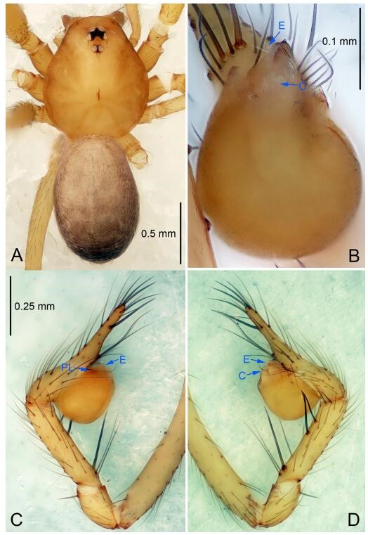
Leptonetela chenjia sp. nov., holotype male
Figure 13.
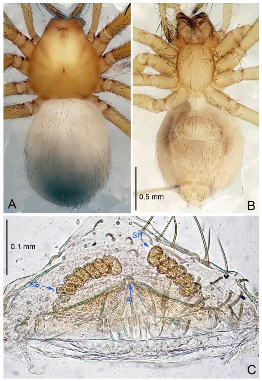
Leptonetela chenjia sp. nov., one of the paratype females
Type material. Holotype: male (IZCAS), Chenjia Cave, N28.38°, E108.67°, Tianba, Songtao County, Tongren City, Guizhou Prvince, China, 9 March 2013, H. Zhao & J. Liu leg. Paratypes: 1 male and 2 females, same data as holotype.
Etymology. The specific name refers to the type locality; noun.
Diagnosis. This new species is similar to L. anshun Lin & Li, 2010, L. suae Lin & Li, 2010, L. tongzi Lin & Li, 2010, L. meitan Lin & Li, 2010, L. liangfeng Wang & Li sp. nov., and L. sanyan Wang & Li sp. nov., but can be distinguished by the male pedipalal tibia Ⅰ spine far apart from the other 4 spines (Figure 12D), conductor reduced (Figure 12B) (tibial spines Ⅰ bifurcated symmetrically in L. anshun; conductor tip bifurcated in L. anshun, bamboo leaf-shaped in L. sanyan Wang & Li sp. nov., and L. tongzi; thin, triangular in L. suae and L. meitan, and C shaped in L. liangfeng Wang & Li sp. nov.); is also similar to L. huoyan Wang & Li sp. nov., but can be distinguished by the absent median apophysis, reduced conductor (Figure 12B) (median apophysis present, slightly sclerotized, index finger like, conductor broad, semicircular in L. huoyan Wang & Li sp. nov.).
Description. Male (holotype). Total length 2.50 (Figure 12A). Carapace 1.25 long, 0.95 wide. Opisthosoma 1.25 long, 0.88 wide. Carapace yellow. Ocular area with a pair of setae, six eyes. Median groove needle-shaped, cervical grooves and radial furrows distinct. Clypeus 0.15 high. Opisthosoma pale brown, ovoid, with pigmented stripe. Leg measurements: Ⅰ 10.44 (2.60, 0.37, 3.05, 2.50, 1.62); Ⅱ 7.84 (2.25, 0.35, 2.25, 1.87, 1.12); Ⅲ 6.41 (1.50, 0.32, 1.87, 1.62, 1.10); Ⅳ 8.59 (2.50, 0.35, 2.37, 2.12, 1.25). Male pedipalp (Figure 12C-D): tibia with 3 long spines prolaterally, 5 spines retrolaterally, with Ⅰ spine longest, far apart from others. Cymbium not wrinkled. Embolus triangular, prolateral lobe oval. Median apophysis absent. Conductor reduced (Figure 12B).
Female (one of the paratypes). Similar to male in color and general features, but smaller and with shorter legs. Total length 2.25 (Figure 13A-B). Carapace 0.87 long, 0.80 wide. Opisthosoma 1.37 long, 1.12 wide. Clypeus 0.12 high. Leg measurements: Ⅰ 7.89 (2.12, 0.37, 2.25, 1.85, 1.30); Ⅱ 6.19 (1.62, 0.32, 1.75, 1.40, 1.10); Ⅲ 5.10 (1.45, 0.30, 1.25, 1.20, 0.90); Ⅳ 6.76 (1.80, 0.35, 1.87, 1.62, 1.12). Vulva (Figure 13C): spermathecae coiled, apical part coiled, atrium triangular.
Distribution. China (Guizhou).
Leptonetela liangfeng Wang & Li sp. nov. Figures 14-15, 97
Figure 14.
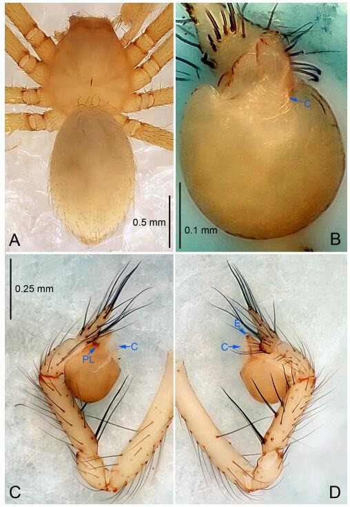
Leptonetela liangfeng sp. nov., holotype male
Figure 15.
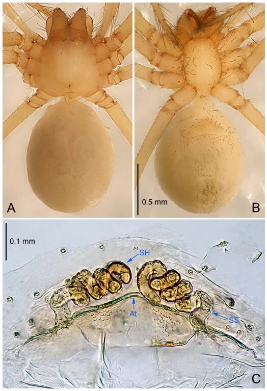
Leptonetela liangfeng sp. nov., one of the paratype females
Type material. Holotype: male (IZCAS), Liangfeng Cave, N28.32°, E107.84°, Tian, Fengle Town, Wuchuan County, Zunyi City, Guizhou Province, China, 7 August 2012, H. Zhao leg. Paratypes : 1 male and 2 females, same data as holotype.
Etymology. The specific name refers to the type locality; noun.
Diagnosis. This new species is similar to L. anshun Lin & Li, 2010, L. suae Lin & Li, 2010, L. tongzi Lin & Li, 2010, L. meitan Lin & Li, 2010, L. chenjia Wang & Li sp. nov., and L. sanyan Wang & Li sp. nov., but can be distinguished by the male pedipalpal bulb conductor C shaped (Figure 14B) (conductor tip bifurcated in L. anshun, bamboo leaf-shaped in L. sanyan Wang & Li sp. nov., and L. tongzi; thin, triangular in L. suae and L. meitan, reduced in L. chenjia Wang & Li sp. nov.); from L. anshun by the tibia Ⅰ spine slender not bifurcated (Figure 14D) (tibia Ⅰ spine symmetrically bifurcated in L. anshun).
Description. Male (holotype). Total length 2.28 (Figure 14A). Carapace 0.93 long, 0.88 wide. Opisthosoma 1.35 long, 0.88 wide. Carapace yellow. Eyes absent. Median groove needle-shaped, cervical grooves and radial furrows indistinct. Clypeus 0.13 high. Opisthosoma yellowish, ovoid. Leg measurements: Ⅰ 9.47 (2.59, 0.43, 2.88, 2.25, 1.32); Ⅱ 8.61 (2.23, 0.32, 2.60, 2.05, 1.41); Ⅲ 7.38 (2.05, 0.43, 2.08, 1.55, 1.27); Ⅳ 8.81 (2.51, 0.38, 2.17, 2.28, 1.47). Male pedipalp (Figure 14C-D): tibia with 4 long setae prolaterally and 5 spines retrolaterally, tibia Ⅰ spine longest. Cymbium constricted medially, attached to an earlobe-shaped process. Embolus triangular, prolateral lobe oval. Median apophysis absent. Conductor C shaped in ventral view (Figure 14B).
Female (one of the paratypes). Similar to male in color and general features, but smaller and with shorter legs. Total length 2.14 (Figure 15A-B). Carapace 0.88 long, 0.73 wide. Opisthosoma 1.36 long, 0.95 wide. Clypeus 0.13 high. Leg measurements: Ⅰ 7.44 (1.98, 0.38, 2.03, 1.77, 1.28); Ⅱ 7.01 (1.88, 0.37, 1.98, 1.55, 1.23); Ⅲ 5.78 (1.33, 0.25, 1.75, 1.42, 1.03); Ⅳ 7.59 (2.03, 0.33, 2.18, 1.79, 1.26). Vulva (Figure 15C): spermathecae coiled, atrium triangular.
Distribution. China (Guizhou).
Leptonetela sanyan Wang & Li sp. nov. Figures 16-17, 97
Figure 16.
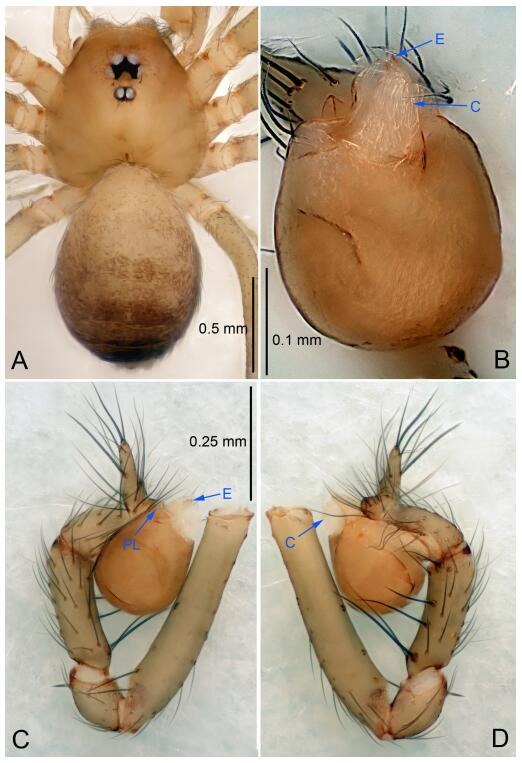
Leptonetela sanyan sp. nov., holotype male
Figure 17.
Leptonetela sanyan sp. nov., one of the paratype females
Type material. Holotype: male (IZCAS), Sanyan Cave, N29.15°, E107.60°, Heyi, Yangxi Town, Daozhen County, Guizhou Province, China, 30 May 2011, Z. Zha leg. Paratypes: 1 male and 2 females, same data as holotype.
Etymology. The specific name refers to the type locality; noun.
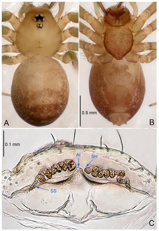
Diagnosis. This new species is similar to L. anshun Lin & Li, 2010, L. suae Lin & Li, 2010, L. tongzi Lin & Li, 2010, L. meitan Lin & Li, 2010, L. chenjia Wang & Li sp. nov., and L. liangfeng Wang & Li sp. nov., but can be separated from all above except L. tongzi by in the male conductor C shaped in this new species (Figure 16B) (conductor tip bifurcated in L. anshun, C shaped in L. liangfeng Wang & Li sp. nov., thin, triangular in L. suae and L. meitan, reduced in L. chenjia Wang & Li sp. nov.); from L. tongzi by in the female atrium triangular, anterior margin of the atrium undulate (Figure 17C) (atrium fusiform, anterior margin of the atrium with pointed process medially in L. tongzi).
Description. Male (holotype). Total length 1.78 (Figure 16A). Carapace 0.83 long, 0.83 wide. Opisthosoma 1.00 long, 0.75 wide. Carapace yellowish. Ocular area with a pair of setae, six eyes. Median groove needle-shaped, pale brown. Cervical grooves and radial furrows indistinct. Clypeus 0.13 high, slightly sloped anteriorly. Opisthosoma yellow, ovoid, with pigmented stripe. Leg measurements: Ⅰ 7.08 (2.00, 0.33, 2.15, 1.75, 1.15); Ⅱ 6.09 (1.68, 0.30, 1.73, 1.38, 1.00); Ⅲ 4.84 (1.38, 0.28, 1.25, 1.13, 0.80); Ⅳ 6.28 (1.75, 0.30, 1.78, 1.50, 0.95). Male pedipalp (Figure 16C-D): tibia with 1 long spine prolaterally, 5 spines retrolaterally, with the basal spine longest. Cymbium constricted medially, attaching an earlobe-shaped process. Embolus triangular, prolateral lobe absent. Median apophysis absent. Conductor bamboo leaf-shaped in ventral view (Figure 16B).
Female (one of the paratypes). Similar to male in color and general features, but larger and with shorter legs. Total length 2.03 (Figure 17A-B). Carapace 0.80 long, 0.75 wide. Opisthosoma 1.25 long, 0.93 wide. Clypeus 0.13 high. Leg measurements: Ⅰ 6.92 (1.88, 0.33, 2.00, 1.58, 1.13); Ⅱ 5.36 (1.50, 0.30, 1.53, 1.15, 0.88); Ⅲ 4.44 (1.20, 0.28, 1.13, 1.05, 0.78); Ⅳ 5.94 (1.73, 0.30, 1.58, 1.38, 0.95). Vulva (Figure 17C): spermathecae coiled, atrium triangular, anterior margin of the atrium undulate. Short hairs modified spermathecae, sperm ducts, and anterior margin of atrium.
Distribution. China (Guizhou).
Leptonetela wangjia Wang & Li sp. nov. Figures 18-19, 97
Figure 18.
Leptonetela wangjia sp. nov., holotype male
Figure 19.
Leptonetela wangjia sp. nov., paratype female
Type material. Holotype: male (IZCAS), Wangjia Cave, N26.98°, E107.94°, Gaoqi, Nongchang Town, Huangpin County, Guizhou Province, China, 4 March 2012, H. Zhao & J. Liu leg. Paratype: 1 female, same data as holotype.
Etymology. The specific name refers to the type locality; noun.
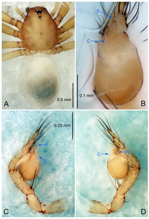
Diagnosis. This new species is similar to L. danxia Lin & Li, 2010, and L. yaoi wang & Li, 2011, but can be distinguished by the reduced pedipalpal bulb conductor (Figure 18B) (conductor C shaped in L. danxia, bamboo leaf-shaped in L. yaoi), from L. yaoi by the tibia Ⅰ spine slender, asymmetrically bifurcated (Figure 18C) (tibia Ⅰ spine strong in L. yaoi); from L. danxia by the unwrinkled cymbium (Figure 18C-D) (cymbium constricted and wrinkled at 1/3 in L. danxia).
Description. Male (holotype). Total length 2.13 (Figure 18A). Carapace 0.88 long, 0.80 wide. Opisthosoma 1.00 long, 0.88 wide. Carapace yellow. Ocular area with a pair of setae, six eyes. Median groove needle-shaped, cervical grooves and radial furrows distinct. Clypeus 0.13 high. Opisthosoma gray, ovoid. Leg measurements: Ⅰ -(1.88, 0.25, -, -, -); Ⅱ 5.66 (1.63, 0.25, 1.63, 1.25, 0.90); Ⅲ 4.71 (1.25, 0.23, 1.25, 1.13, 0.85); Ⅳ 6.25 (1.75, 0.25, 1.75, 1.50, 1.00). Male pedipalp (Figure 18C-D): tibia with 7 long setae prolaterallly and 5 spines retrolaterally, Ⅰ spine slender, longest, asymmetrically bifurcated. Cymbium not wrinkled, earlobe-shaped process absent. Embolus triangular, prolateral lobe oval. Median apophysis absent. Conductor reduced (Figure 18B).
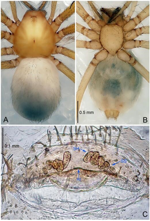
Female. Similar to male in color and general features, but larger and with longer legs. Total length 2.50 (Figure 19A-B). Carapace 1.00 long, 0.90 wide. Opisthosoma 1.50 long, 1.15 wide. Clypeus 0.20 high. Leg measurements: Ⅰ 9.26 (2.55, 0.38, 2.60, 2.10, 1.63); Ⅱ 8.25 (2.37, 0.38, 2.25, 1.90, 1.35); Ⅲ 6.90 (2.05, 0.35, 1.75, 1.65, 1.10); Ⅳ 7.86 (2.30, 0.38, 2.13, 1.75, 1.30). Vulva (Figure 19C): spermathecae coiled, atrium fusiform.
Distribution. China (Guizhou).
Leptonetela qiangdao Wang & Li sp. nov. Figures 20-21, 97
Figure 20.
Leptonetela qiangdao sp. nov., holotype male
Figure 21.
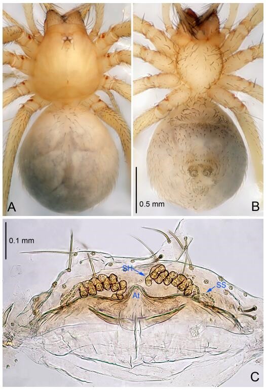
Leptonetela qiangdao sp. nov., one of the paratype females
Type material. Holotype: male (IZCAS), Qiangdao Cave, N25.83º, E109.04º, Guandong Town, Congjiang County, Qiandongnan Prefecture, Guizhou, China, 16 March 2013, H. Zhao & J. Liu leg. Paratypes: 3 females, same data as holotype.
Etymology. The specific name refers to the type locality; noun.
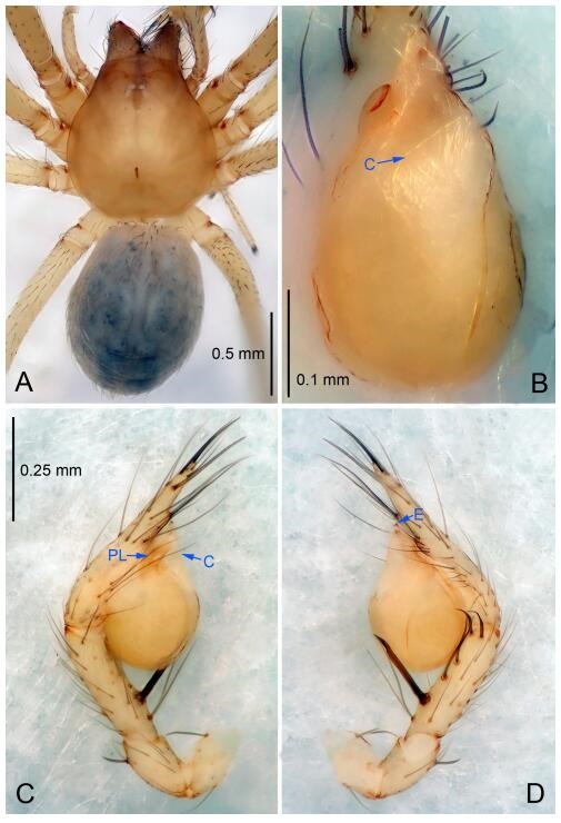
Diagnosis. This new species is similar to L. furcaspina Lin & Li, 2010, L. langdong Wang & Li sp. nov. and L. dashui Wang & Li sp. nov., but can be distinguished by the male pedipalpal tibia Ⅰ spine strong (Figure 20D), conductor bamboo leaf-shaped in ventral view (Figure 20B) (tibia Ⅰ spine strong, asymmetrically bifurcated, conductor C shaped in L. furcaspina, tibia Ⅰ spine strong, tip curved, conductor reduced in L. langdong Wang & Li sp. nov., tibia Ⅰ spine slender, Ⅱ Ⅲ spines curved basally, conductor C shaped in L. dashui Wang & Li sp. nov.).
Description. Male (holotype). Total length 1.80 (Figure 20A). Carapace 0.87 long, 0.90 wide. Opisthosoma 0.90 long, 0.75 wide. Carapace yellow. Four eyes, PME absent, ALE and PLE reduced to white points. Median groove needle-shaped, pale brown, cervical grooves and radial furrows indistinct. Clypeus 0.13 high. Opisthosoma whitish gray, ovoid, lacking distinctive pattern. Leg measurements: Ⅰ 7.88 (2.13, 0.33, 2.20, 1.92, 1.30); Ⅱ 6.41 (1.63, 0.30, 1.75, 1.58, 1.15); Ⅲ 5.40 (1.50, 0.30, 1.45, 1.23, 0.92); Ⅳ 7.43 (1.80, 0.33, 2.10, 1.72, 1.48). Male pedipalp (Figure 20C-D): tibia with 3 long setae prolaterally, 5 spines retrolaterally, with Ⅰ spine strong, longest, tip curved. Cymbium with no wrinkle medially, earlobe-shaped process small. Bulb with spoon-shaped embolus, prolateral lobe small. Median apophysis absent. Conductor bamboo leaf-shaped in ventral view (Figure 20B).
Female (one of the paratypes). Similar to male in color and general features, but larger and with shorter legs. Total length 2.25 (Figure 21A-B). Carapace 0.88 long, 0.80 wide. Opisthosoma 1.37 long, 1.13 wide. Clypeus 0.13 high. Leg measurements: Ⅰ 6.91 (2.10, 0.38, 1.88, 1.47, 1.08); Ⅱ 6.20 (1.70, 0.35, 1.75, 1.40, 1.00); Ⅲ 4.97 (1.37, 0.30, 1.25, 1.22, 0.83); Ⅳ 6.98 (1.88, 0.38, 1.92, 1.67, 1.13). Vulva (Figure 21C): spermathecae coiled, atrium trapezoidal, anterior margin of atrium undulate, covered with short hairs.
Distribution. China (Guizhou).
Leptonetela langdong Wang & Li sp. nov. Figures 22-23, 97
Figure 22.
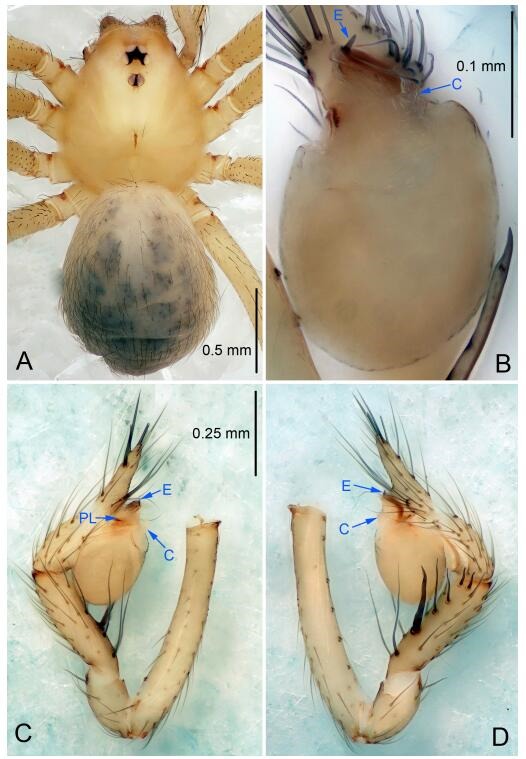
Leptonetela langdong sp. nov., holotype male
Figure 23.
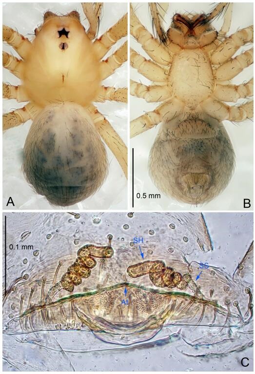
Leptonetela langdong sp. nov., one of the paratype females
Type material. Holotype: male (IZCAS), Menglonggong Cave, N27.07°, E107.76°, Langdong Village, Huangping County, Qiandongnan Prefecture, Guizhou Province, China, 3 March 2013, H. Zhao & J. Liu leg. Paratypes: 1 male and 2 females, same data as holotype.
Etymology. The specific name refers to the type locality; noun.
Diagnosis. This new species is similar to L. furcaspina Lin & Li, 2010, L. qiangdao Wang & Li sp. nov., and L. dashui Wang & Li sp. nov., but can be distinguished by the male pedipalpal tibia Ⅰ spine strong, tip curved (Figure 22D). conductor reduced (Figure 22B) (tibia Ⅰ spine strong, asymmetrically bifurcated, conductor C shaped in L. furcaspina, tibia Ⅰ spine strong, conductor bamboo leaf-shaped in L. qiangdao Wang & Li sp. nov., tibia Ⅰ spine slender, Ⅱ Ⅲ spines curved basally, conductor C shaped in L. dashui Wang & Li sp. nov.).
Description. Male (holotype): total length 2.25 (Figure 22A). Carapace 1.25 long, 0.87 wide. Opisthosoma 1.13 long, 0.88 wide. Carapace yellowish. Six eyes. Median groove needle-shaped, cervical grooves and radial furrows indistinct. Clypeus 0.13 high. Opisthosoma gray, ovoid. Leg measurements: Ⅰ 8.91 (2.38, 0.35, 2.55, 2.13, 1.50); Ⅱ 7.23 (2.00, 0.35, 1.88, 1.75, 1.25); Ⅲ 6.22 (1.75, 0.34, 1.63, 1.50, 1.00); Ⅳ 8.05 (2.25, 0.35, 2.25, 1.90, 1.30). Male pedipalp (Figure 22C-D): femur with 6 spines ventrally, tibia with 3 long spines prolaterally, 1 long seta and 5 spines retrolaterally, with Ⅰ spine strong, tip curved. Cymbium constricted medially, attached to an earlobe-shaped process. Embolus triangular, bearing a tooth basally, prolateral lobe reduced. Median apophysis absent. Conductor reduced (Figure 22B).
Female (one of the paratypes). Similar to male in color and general features, but larger and with shorter legs. Total length 2.25 (Figure 23A-B). Carapace 1.10 long, 0.60 wide. Opisthosoma 1.25 long, 0.88 wide. Clypeus 0.15 high. Leg measurements: Ⅰ 7.48 (2.10, 0.38, 2.13, 1.62, 1.25); Ⅱ 6.14 (1.75, 0.38, 1.63, 1.38, 1.00); Ⅲ 5.18 (1.50, 0.35, 1.25, 1.20, 0.88); Ⅳ 6.86 (2.00, 0.38, 1.88, 1.50, 1.10). Vulva (Figure 23C): spermathecae coiled, atrium triangular.
Distribution. China (Guizhou).
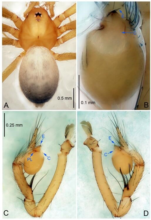
Leptonetela dashui Wang & Li sp. nov. Figures 24-25, 97
Figure 24.
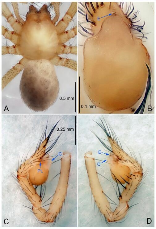
Leptonetela dashui sp. nov., holotype male
Figure 25.
Leptonetela dashui sp. nov., one of the paratype females
Type material. Holotype: male (IZCAS), Dashui Cave, N26.61°, E106.61°, Shijicheng, Jinyang New Urban Area, Guiyang City, Guizhou Province, China, 18 June 2011, Z. Zha leg. Paratypes: 1 male and 2 females, same data as holotype.
Etymology. The specific name refers to the type locality; noun.
Diagnosis. This new species is similar to L. furcaspina Lin & Li, 2010, L. qiangdao Wang & Li sp. nov., and L. langdong Wang & Li sp. nov., but can be distinguished by the slender male pedipalpal tibia Ⅰ spine, Ⅱ Ⅲ spines curved basally (Figure 24D), conductor C shaped (Figure 24B), (tibia Ⅰ spine strong, tip curved, conductor reduced in L. langdong Wang & Li sp. nov., tibia Ⅰ spine strong, asymmetrically bifurcated, conductor narrow and bifurcated in L. furcaspina, tibia Ⅰ spine strong, conductor bamboo leaf-shaped in L. qiangdao Wang & Li sp. nov.).
Description. Male (holotype). Total length 1.88 (Figure 24A). Carapace 0.88 long, 0.75 wide. Opisthosoma 1.00 long, 0.75 wide. Carapace yellowish. Eyes absent. Median groove, cervical grooves and radial furrows indistinct. Clypeus 0.13 high. Opisthosoma gray, ovoid. Leg measurements: Ⅰ 8.24 (2.25, 0.38, 2.43, 1.88, 1.30); Ⅱ 7.16 (1.98, 0.35, 2.00, 1.63, 1.20); Ⅲ 6.06 (1.75, 0.33, 1.60, 1.50, 0.88); Ⅳ 7.69 (2.13, 0.38, 2.08, 1.85, 1.25). Male pedipalp (Figure 24C-D): femur with 4 spines ventrally, tibia with 2 long setae prolaterally, 2 long setae and 5 slender spines retrolaterally, the spines equally strong, spines Ⅰ longest. Cymbium not wrinkled, earlobe-shaped process small. Embolus triangular, prolateral lobe reduced. Median apophysis absent. Conductor C shaped in ventral view (Figure 24B).
Female (one of the paratypes). Similar to male in color and general features, but larger and with shorter legs. Total length 1.93 (Figure 25A-B). Carapace 0.78 long, 0.75 wide. Opisthosoma 1.18 long, 1.00 wide. Clypeus 0.15 high. Leg measurements: Ⅰ 6.02 (1.68, 0.28, 1.73, 1.30, 1.03); Ⅱ 5.29 (1.45, 0.28, 1.43, 1.25, 0.88); Ⅲ 4.34 (1.25, 0.25, 1.13, 1.03, 0.68); Ⅳ 5.67 (1.63, 0.28, 1.58, 1.28, 0.90). Vulva (Figure 25C): spermathecae coiled, atrium fusiform, anterior margin with pointed process medially.
Distribution. China (Guizhou).
Leptonetela gang Wang & Li sp. nov. Figures 26-27, 97
Figure 26.
Leptonetela gang sp. nov., holotype male
Figure 27.
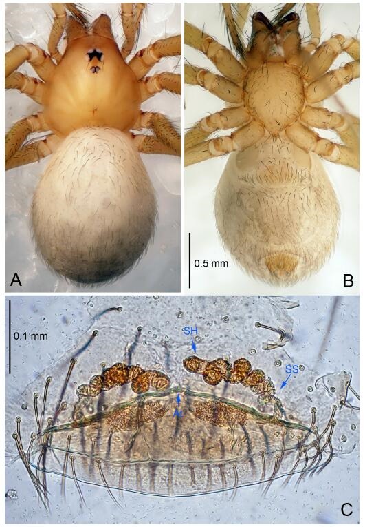
Leptonetela gang sp. nov., one of the paratype females
Type material. Holotype: male (IZCAS), Gang Cave, N26.87°, E108.91°, Tunhou, Nanming Town, Jianhe County, Kaili City, Guizhou Province, China, 15 December 2011, Z. Zha leg. Paratypes: 15 males and 6 females, same data as holotype; 4 males and 5 females, Long Cave, N26.85°, E108.79°, Longtang, Liangshang Town, Sansui County, Kaili City, Guizhou Province, China, 18 December 2011, Z. Zha leg; 5 males and 5 females, Shenxian Cave, N26.87°, E108.89°, Shixing, Xiaolan Country, Nanming Town, Jianhe County, Kaili City, Guizhou Province, China, 16 December 2011, Z. Zha leg; 5 females, Niu Cave, N26.86°, E108.93°, Cenge, Nanming Town, Jianhe County, Kaili City, Guizhou Province, China, 14 December 2011, Z. Zha leg.
Etymology. The specific name refers to the type locality; noun.
Diagnosis. This new species is similar to L. jiulong Lin & Li, 2010, but can be distinguished by the male pedipalpal tibia with 6 spines retrolaterally, tibia Ⅱ spine thickest, Ⅰ, Ⅱ spines equally length, tibia Ⅱ spine asymmetrically bifurcated (Figure 26D), median apophysis absent, conductor reduced (Figure 26B) (Ⅰ, Ⅱ spines equally strong, tibia Ⅱ spine longest and bifurcate, tibia Ⅰ spine half the length of Ⅱ, median apophysis broad and smooth, conductor rugose, triangular in L. jiulong).
Description. Male (holotype). Total length 2.63 (Figure 26A). Carapace 1.25 long, 1.00 wide. Opisthosoma 1.50 long, 1.13 wide. Carapace yellow. Ocular area with a pair of setae, six eyes. Median groove needle-shaped, cervical grooves and radial furrows distinct. Clypeus 0.13 high. Opisthosoma gray, ovoid. Leg measurements: Ⅰ 10.66 (3.00, 0.38, 3.13, 2.55, 1.60); Ⅱ 8.96 (2.50, 0.38, 2.55, 2.13, 1.40); Ⅲ 7.55 (2.25, 0.35, 2.00, 1.75, 1.20); Ⅳ 9.68 (2.75, 0.38, 2.75, 2.35, 1.45). Male pedipalp (Figure 26C-D): tibia with 5 long spines prolaterally, 6 spines retrolaterally, tibia Ⅱ spine thickest asymmetrically bifurcated, Ⅰ Ⅱ spines equally length. Cymbium constricted medially, attached to an earlobe-shaped process. Embolus triangular, median apophysis absent, conductor reduced (Figure 26B).
Female (one of the paratypes). Similar to male in color and general features, but smaller and with shorter legs. Total length 2.38 (Figure 27A-B). Carapace 1.00 long, 0.88 wide. Opisthosoma 1.50 long, 1.20 wide. Clypeus 0.13 high. Leg measurements: Ⅰ 9.26 (2.50, 0.38, 2.75, 2.13, 1.50); Ⅱ 7.51 (2.00, 0.38, 2.13, 1.75, 1.25); Ⅲ 6.29 (1.88, 0.38, 1.63, 1.40, 1.00); Ⅳ 8.11 (2.25, 0.38, 2.25, 1.88, 1.35). Vulva (Figure 27C): spermathecae coiled, atrium fusiform.
Distribution. China (Guizhou).
Leptonetela la Wang & Li sp. nov. Figure 28-29, 97
Figure 28.
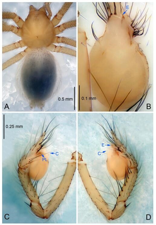
Leptonetela la sp. nov., holotype male
Figure 29.
Leptonetela la sp. nov., one of the paratype females
Type material. Holotype: male (IZCAS), La Cave in Xiaoyakou, N25.80°, E104.95°, Puan County, Qianxinan Prefecture, Guizhou Province, China, 14 July 2012, H. Zhao leg. Paratypes: 3 males and 5 females, same data as holotype.
Etymology. The specific name refers to the type locality; noun.
Diagnosis. This new species is similar to L. rudong Wang & Li sp. nov. and L. wenzhu Wang & Li sp. nov. but can be distinguished from L. wenzhu Wang & Li sp. nov. by the male pedipalpal tibia with 7 spines retrolaterally (tibia with 6 spines retrolaterally in L. wenzhu Wang & Li sp. nov.); from L. rudong Wang & Li sp. nov. by the tibia with 4 long setae prolaterally (Figure 28D) (tibia with 2 long setae, 2 spines prolaterally, cymbium with 1 horn-shaped spine on the earlobe-shaped process in L. rudong Wang & Li sp. nov.); from L. rudong Wang & Li sp. nov., and L. wenzhu Wang & Li sp. nov. by the conductor broad, C shaped (conductor thin, triangular in L. rudong Wang & Li sp. nov., bamboo leaf-shaped in L. wenzhu Wang & Li sp. nov.).
Description. Male (holotype). Total length 2.97 (Figure 28A). Carapace 1.25 long, 1.09 wide. Opisthosoma 1.71 long, 1.40 wide. Carapace yellow. Ocular area with a pair of setae, eyes absent. Median groove needle-shaped, cervical grooves and radial furrows distinct. Clypeus 0.17 high. Opisthosoma gray, ovoid. Leg measurements: Ⅰ 10.49 (2.60, 0.40, 3.16, 2.48, 1.85); Ⅱ 9.70 (2.66, 0.41, 2.81, 2.26, 1.56); Ⅲ 8.83 (2.34, 0.40, 2.81, 1.97, 1.31); Ⅳ 10.17 (2.81, 0.41, 2.88, 2.51, 1.56). Male pedipalp (Figure 28C-D): tibia with 4 long setae prolaterally and 7 slender spines retrolaterally, tibia Ⅰ Ⅱ spines of equal length, longer than other spines. Cymbium constricted medially, attaching an earlobe-shaped process. Embolus triangular, prolateral lobe oval. Median apophysis absent. Conductor C shaped in ventral view (Figure 28B).
Female (one of the paratypes). Similar to male in color and general features, but smaller and with shorter legs. Total length 2.81 (Figure 29A-B). Carapace 1.09 long, 1.01 wide. Opisthosoma 1.69 long, 1.47 wide. Clypeus 0.17 high. Leg measurements: Ⅰ 9.68 (2.56, 0.40, 2.56, 2.13, 1.62); Ⅱ 8.23 (2.34, 0.34, 2.43, 1.81, 1.31); Ⅲ 7.03 (2.19, 0.34, 2.03, 1.38, 1.09); Ⅳ 9.13 (2.51, 0.34, 2.59, 2.38, 1.31). Vulva (Figure 29C): spermathecae coiled, atrium fusiform, anterior margin of atrium with one large pointed process medially, and covered with short hairs.
Distribution. China (Guizhou).
Leptonetela rudong Wang & Li sp. nov. Figures 30-31, 97
Figure 30.
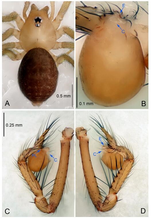
Leptonetela rudong sp. nov., holotype male
Figure 31.
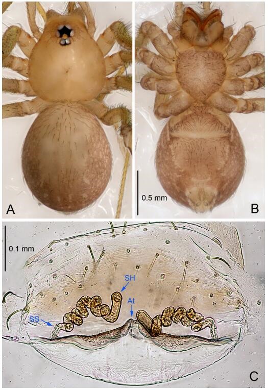
Leptonetela rudong sp. nov., one of the paratype females
Type material. Holotype: male (IZCAS), Rudong Cave, N25.57°, E110.62°, Longpan Mountain, Dongtian, Xing'an County, Guilin City, Guangxi Zhuang Autonomous Region, China, 11 July 2009, C. Wang & Z. Yao leg. Paratypes: 1 male and 3 females, same data as holotype; 3 females, Gouya Cave, N25.46°, E110.11°, Hufeng, Guanyang County, Guilin City, Guangxi Zhuang Autonomous Region, China, 30 August 2009, C. Wang & Z. Yao leg; 2 females, Jiulong Cave, N25.46°, E110.09°, Shifeng, Guanyang County, Guilin City, Guangxi Zhuang Autonomous Region, China, 30 August 2009, C. Wang & Z. Yao leg.
Etymology. The specific name refers to the type locality; noun.
Diagnosis. This new species is similar to L. la Wang & Li sp. nov., and L. wenzhu Wang & Li sp. nov. but can be distinguished by the male pedipalpal tibia with 2 long setae, 2 spines prolaterally, 1 long seta, and 6 spines retrolaterally, cymbium with 1 horn-shaped spine on the earlobe-shaped process (Figure 30C-D), conductor thin, triangular in ventral view (Figure 30B) (tibia with 4 long setae prolaterally, 7 slender spines retrolaterally, conductor broad, C shaped in L. la Wang & Li sp. nov.; tibia with 2 long setae prolaterally, 6 spines retrolaterally, with tibia Ⅰ spine strongest, conductor bamboo leaf-shaped in L. wenzhu Wang & Li sp. nov.).
Description. Male (holotype). Total length 2.12 (Figure 30A). Carapace 0.88 long, 0.85 wide. Opisthosoma 1.25 long, 1.05 wide. Carapace yellowish. Ocular area with a pair of setae, eyes six. Median groove needle-shaped, cervical grooves and radial furrows indistinct. Clypeus 0.13 high. Opisthosoma brown, ovoid. Leg measurements: Ⅰ 10.15 (2.84, 0.38, 3.00, 2.38, 1.55); Ⅱ 7.84 (2.08, 0.38, 2.23, 1.88, 1.27); Ⅲ 6.55 (1.83, 0.35, 1.75, 1.62, 1.00); Ⅳ 8.31 (2.25, 0.38, 2.38, 2.05, 1.25). Male pedipalp (Figure 30C-D): tibia with 2 long setae, 2 spines prolaterally, 1 long seta and 6 slender spines retrolaterally, with Ⅰ spine longest. Cymbium constricted medially, with 1 horn-shaped spine on the earlobe-shaped process. Embolus triangular, pedipalpal bulb oval. Median apophysis absent. Conductor thin, triangular in ventral view (Figure 30B).
Female (one of the paratypes). Similar to male in color and general features, but larger and with shorter legs. Total length 2.15 (Figure 31A-B). Carapace 0.90 long, 0.88 wide. Opisthosoma 1.33 long, 1.02 wide. Clypeus 0.13 high. Leg measurements: Ⅰ 8.64 (2.35, 0.33, 2.55, 1.88, 1.53); Ⅱ 6.87 (2.03, 0.33, 1.88, 1.50, 1.13); Ⅲ 6.06 (1.90, 0.33, 1.50, 1.35, 0.98); Ⅳ 7.39 (1.98, 0.33, 2.13, 1.78, 1.17). Vulva (Figure 31C): spermathecae coiled, atrium semicircular, anterior margin of atrium with one pointed process medially, and covered with short hairs.
Leptonetela wenzhu Wang & Li sp. nov. Figures 32-33, 97
Figure 32.
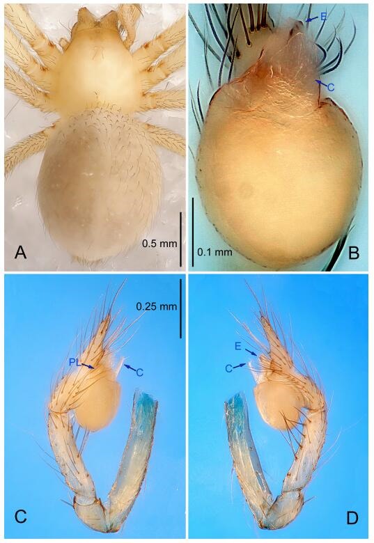
Leptonetela wenzhu sp. nov., holotype male
Figure 33.
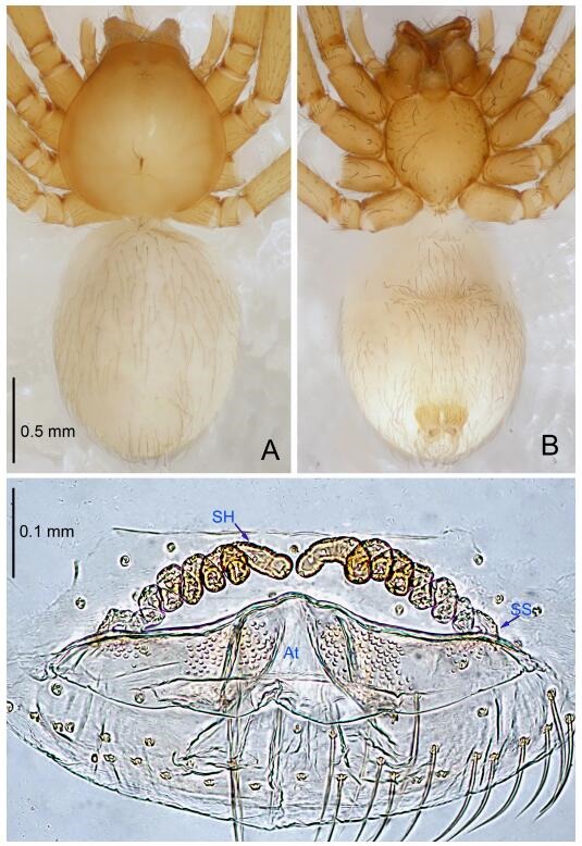
Leptonetela wenzhu sp. nov., one of the paratype females
Type material. Holotype: male (IZCAS), Wenzhu Cave, N25.44°, E105.13°, Longchang Town, Xingren City, Guizhou Province, China, 16 July 2012, H. Zhao leg. Paratypes: 1 male and 2 females, same data as holotype; 4 females, Xiaoya Cave, N25.44°, E105.13°, Yaqiao Town, Xingren City, Guizhou Province, China, 16 July 2012, H. Zhao leg.
Etymology. The specific name refers to the type locality; noun.
Diagnosis. This new species is similar to L. rudong Wang & Li sp. nov., and L. la Wang & Li sp. nov. but can be distinguished by the male pedipalpal tibia with 6 spines retrolaterally (Figure 32D), conductor bamboo leaf-shaped in ventral view (Figure 32B) (tibia with 1 long seta, 6 spines retrolaterally in L. rudong Wang & Li sp. nov., tibia with 7 spines retrolaterally in L. la Wang & Li sp. nov., conductor broad, C shaped in L. la Wang & Li sp. nov.; thin, triangular in L. rudong Wang & Li sp. nov.).
Description. Male (holotype). Total length 2.63 (Figure 32A). Carapace 1.28 long, 1.03 wide. Opisthosoma 1.34 long, 1.09 wide. Carapace yellowish. Ocular area with a pair of setae, PME and PLE absent, ALE reduced to white points. Median groove, cervical groove and radial furrows indistinct. Clypeus 0.25 high. Opisthosoma gray, ovoid. Leg measurements: Ⅰ 10.17 (2.78, 0.37, 3.12, .34, 1.56); Ⅱ 7.34 (2.53, 0.37, 1.94, 1.72, 0.78); Ⅲ 7.70 (2.19, 0.37, 2.03, 1.88, 1.22); Ⅳ 9.28 (2.60, 0.37, 2.56, 2.28, 1.47). Male pedipalp (Figure 32C-D): tibia with 6 spines retrolaterally, arranged equidistantly. Embolus triangular, prolateral lobe absent. Median apophysis absent. Conductor bamboo leaf-shaped in ventral view (Figure 32B).
Female (one of the paratypes). Similar to male in color and general features, but larger and with shorter legs. Total length 2.88 (Figure 33A-B). Carapace 1.20 long, 1.00 wide. Opisthosoma 1.60 long, 1.20 wide. Clypeus 0.18 high. Leg measurements: Ⅰ 8.62 (2.53, 0.37, 2.51, 1.90, 1.31); Ⅱ 7.36 (2.09, 0.34, 1.94, 1.65, 1.34); Ⅲ 6.18 (1.55, 0.31, 1.69, 1.47, 1.16); Ⅳ 7.98 (2.44, 0.31, 2.01, 1.88, 1.34). Vulva (Figure 33C): spermathecae coiled, atrium triangular.
Distribution. China (Guizhou).
Leptonetela longli Wang & Li sp. nov. Figures 34-35, 97
Figure 34.
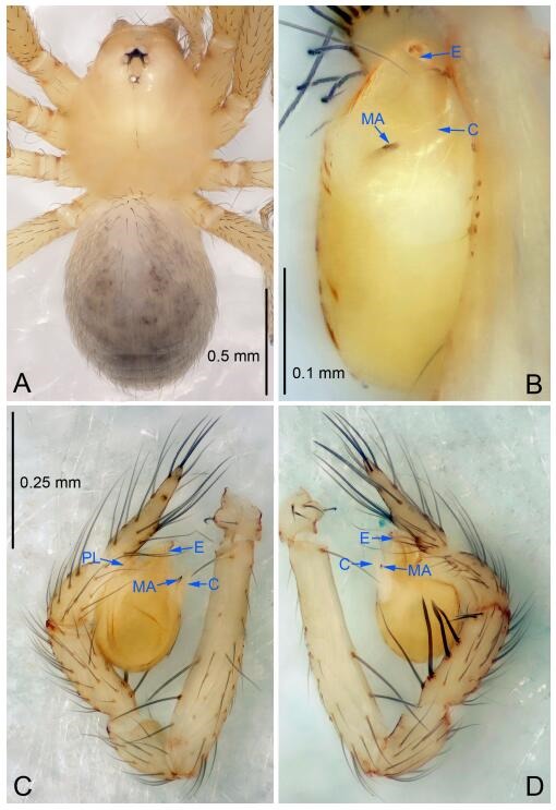
Leptonetela longli sp. nov., holotype male
Figure 35.
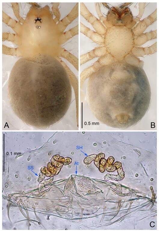
Leptonetela longli sp. nov., one of the paratype females
Type material. Holotype: male (IZCAS), Underground River, N25.27°, E107.44°, Longli, Liuzhai Town, Nandan County, Hechi City, Guangxi Zhuang Autonomous Region, China, 29 January 2015, Y. Li & Z. Chen leg. Paratypes: 3 males and 4 females, same data as holotype.
Etymology. The specific name refers to the type locality; noun.
Diagnosis. This new species is similar to L. chiosensis Wang & Li, 2011 and L. panbao Wang & Li sp. nov., but can be distinguished by the male pedipalpal tibia Ⅰ, Ⅱ and Ⅲ spines equally strong (Figure 34D), conductor C shaped (Figure 34B) (tibia Ⅰ spine strong, conductor triangular in L. chiosensis; tibia spines slender, cymbium with 1 strong spine on the earlobe-shaped process, conductor reduced, embolus with 1 tooth basally in L. panbao Wang & Li sp. nov.).
Description. Male (holotype). Total length 1.88 (Figure 34A). Carapace 0.87 long, 0.75 wide. Opisthosoma 1.00 long, 0.80 wide. Carapace yellowish. Ocular area with a pair of setae, eyes six. Median groove, cervical grooves and radial furrows indistinct. Clypeus 0.10 high. Opisthosoma gray, ovoid. Leg measurements: Ⅰ 6.38 (1.75, 0.25, 1.88, 1.50, 1.00); Ⅱ 5.03 (1.40, 0.25, 1.38, 1.25, 0.75); Ⅲ 4.25 (1.37, 0.20, 1.13, 1.00, 0.55); Ⅳ 5.71 (1.63, 0.25, 1.60, 1.35, 0.88). Male pedipalp (Figure 34C-D): tibia with 3 long spines prolaterally, 5 spines retrolaterally, tibia Ⅰ spine longest, Ⅰ, Ⅱ Ⅲ spines equally strong, stronger than other spines. Embolus triangular, prolateral lobe reduced. Median apophysis"丿"shaped in ventral view. Conductor "C" shaped in ventral view (Figure 34B).
Female (one of the paratypes). Similar to male in color and general features, but larger and with shorter legs. Total length 1.95 (Figure 35A-B). Carapace 0.88 long, 0.88 wide. Opisthosoma 1.25 long, 1.00 wide. Clypeus 0.10 high. Leg measurements: Ⅰ 5.03 (1.38, 0.25, 1.50, 1.15, 0.75); Ⅱ 4.58 (1.25, 0.25, 1.13, 1.10, 0.85); Ⅲ 3.55 (1.00, 0.20, 0.88, 0.87, 0.60); Ⅳ 4.76 (1.30, 0.25, 1.38, 1.13, 0.70). Vulva (Figure 35C): spermathecae coiled, atrium fusiform.
Distribution. China (Guangxi).
Leptonetela panbao Wang & Li sp. nov. Figures 36-37, 97
Figure 36.
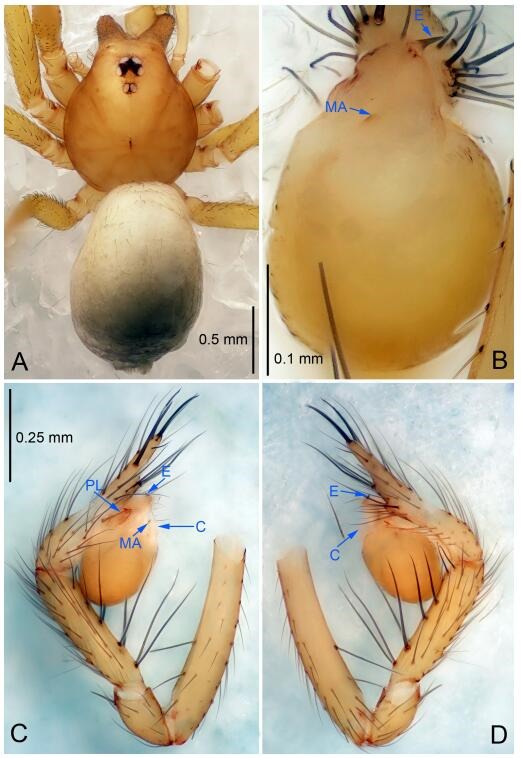
Leptonetela panbao sp. nov., holotype male
Figure 37.
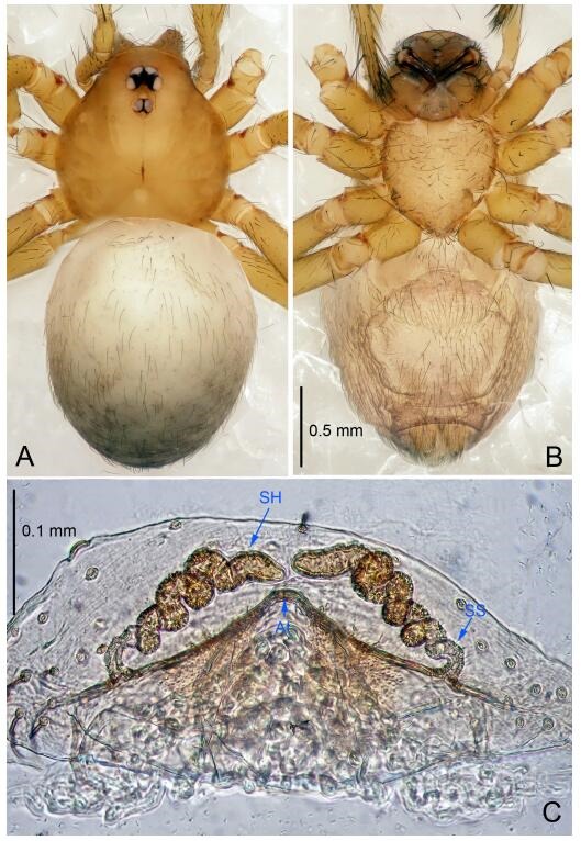
Leptonetela panbao sp. nov., one of the paratype females
Type material. Holotype: male (IZCAS), Panbao Cave, N28.38°, E108.67°, Panbao, Shichang Town, Songtao County, Tongren City, Guizhou Province, China, 8 March 2013, H. Zhao & J. Liu leg. Paratypes: 2 male and 4 females, same data as holotype.
Etymology. The specific name refers to the type locality; noun.
Diagnosis. This new species is similar to L. chiosensis Wang & Li, 2011 and L. Longli Wang & Li sp. nov., but can be distinguished by the male pedipalpal tibial spines slender, equally strong, cymbium with 1 strong spine on the earlobe-shaped process (Figure 36D), conductor reduced, embolus with 1 tooth basally (Figure 36B) (tibia Ⅰ spine stronger than other spines, conductor triangular in L. chiosensis; tibia Ⅰ, Ⅱ and Ⅲ spines equally strong, stronger than other spines, conductor C shaped in L. panbao Wang & Li sp. nov.).
Description. Male (holotype). Total length 2.38 (Figure 36A). Carapace 1.15 long, 1.00 wide. Opisthosoma 1.25 long, 0.90 wide. Carapace yellow. Six eyes. Median groove needle-shaped, cervical grooves and radial furrows distinct. Clypeus 0.13 high. Opisthosoma gray, ovoid. Leg measurements: Ⅰ 10.50 (2.75, 0.35, 3.50, 2.40, 1.50); Ⅱ 7.86 (2.13, 0.35, 2.35, 1.90, 1.13); Ⅲ 6.22 (1.75, 0.34, 1.75, 1.38, 1.00); Ⅳ 8.43 (2.38, 0.35, 2.40, 2.05, 1.25). Male pedipalp (Figure 36C-D): tibia with 4 long spines prolaterally, 5 slender spines retrolaterally, the spines equally strong, tibia Ⅰ spine longest. Cymbium not wrinkled, earlobe-shaped process small, with 1 spine. Embolus triangular, bearing a basal tooth, prolateral lobe oval. Median apophysis single quote shaped, " ′ " in ventral view. Conductor reduced (Figure 36B).
Female (one of the paratypes). Similar to male in color and general features, but larger and with shorter legs. Total length 2.50 (Figure 37A-B). Carapace 1.13 long, 1.00 wide. Opisthosoma 1.62 long, 1.25 wide. Clypeus 0.13 high. Leg measurements: Ⅰ 8.85 (2.30, 0.30, 2.62, 2.13, 1.50); Ⅱ 6.81 (1.88, 0.30, 2.00, 1.50, 1.13); Ⅲ 5.76 (1.63, 0.25, 1.63, 1.25, 1.00); Ⅳ 7.58 (2.25, 0.30, 2.13, 1.75, 1.15). Vulva (Figure 37C): spermathecae coiled, atrium triangular, anterior margin of atrium covered with short hairs.
Distribution. China (Guizhou).
Leptonetela feilong Wang & Li sp. nov. Figures 38-39, 97
Figure 38.
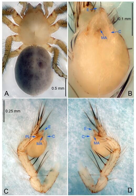
Leptonetela feilong sp. nov., holotype male
Figure 39.
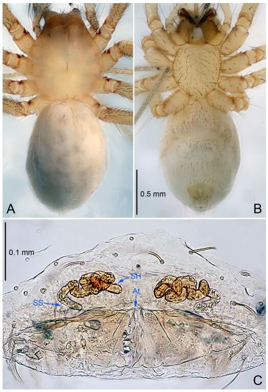
Leptonetela feilong sp. nov., one of the paratype females
Type material. Holotype: male (IZCAS), Feilong Cave, N26.44°, E107.02°, Longli Town, Qiannan Prefecture, Guizhou Province, China, 27 July 2012, H. Zhao leg. Paratypes: 9 females, same data as holotype; 1 female, Lianhua Cave, N26.43°, E106.95°, Lianhua Town, Qiannan Prefecture, Guizhou Province, China, 27 July 2012, H. Zhao leg.
Etymology. The specific name refers to the type locality; noun.
Diagnosis. This new species is similar to L. yangi Lin & Li, 2010 and L. jiahe Wang & Li sp. nov., but can be distinguished from L. yangi by the male pedipalpal cymbium constricted medially, attaching an earlobe-shaped process (Figure 38D), conductor triangular (Figure 38B) (cymbium not constricted, earlobe-shaped process absent, conductor reduced in L. yangi), from L. jiahe Wang & Li sp. nov. by the median apophysis "m"-shaped, conductor triangular (cymbium with 1 spine on the earlobe-shaped process, and 1 curved long spine medially, median apophysis like pointedprocess, with 3 sclerotized spots distally, conductor C shaped in L. jiahe Wang & Li sp. nov.).
Description. Male (holotype). Total length 2.31 (Figure 38A). Carapace 1.02 long, 1.30 wide. Opisthosoma 1.30 long, 1.14 wide. Carapace yellowish. Ocular area with a pair of setae, eyes absent. Median groove needle-shaped, cervical grooves and radial furrows indistinct. Clypeus 0.15 high. Opisthosoma gray, ovoid. Leg measurements: Ⅰ 9.53 (2.66, 0.40, 2.81, 2.19, 1.47); Ⅱ 8.09 (2.41, 0.40, 2.41, 1.93, 0.94); Ⅲ 7.17 (2.03, 0.40, 1.94, 1.72, 1.08); Ⅳ 8.82 (2.71, 0.41, 2.51, 1.94, 1.25). Male pedipalp (Figure 38C-D): tibia with 2 long setae prolaterally, 5 spines retrolaterally, spines Ⅰ, Ⅱ and Ⅲ equally strong, spines Ⅰ longest. Cymbium constricted medially, attaching an earlobe-shaped process retrolaterally. Embolus triangular, prolateral lobe nearly absent. Median apophysis "m"-shaped. Conductor triangular (Figure 8B).
Female (one of the paratypes). Similar to male in color and general features, but smaller and with shorter legs. Total length 2.13 (Figure 39A-B). Carapace 0.88 long, 0.88 wide. Opisthosoma 1.25 long, 0.87 wide. Clypeus 0.08 high. Leg measurements: Ⅰ 8.44 (2.38, 0.30, 2.51, 1.87, 1.38); Ⅱ 7.48 (2.05, 0.31, 2.25, 1.62, 1.25); Ⅲ 6.20 (1.75, 0.31, 1.75, 1.38, 1.01); Ⅳ 7.73 (2.25, 0.32, 2.25, 1.78, 1.13). Vulva (Figure 39C): spermathecae coiled, atrium fusiform.
Distribution. China (Guizhou).
Leptonetela jiahe Wang & Li sp. nov Figures 40-41, 97
Figure 40.
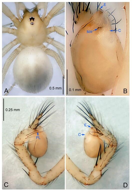
Leptonetela jiahe sp. nov., holotype male
Figure 41.
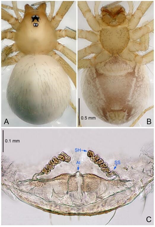
Leptonetela jiahe sp. nov., one of the paratype females
Type material. Holotype: male (IZCAS), Jiahe Cave, N25.25°, E110.20°, Lingui Town, Lingui County, Guilin City, Guangxi Zhuang Autonomous Region, China, 20 December 2013, H. Zhao leg. Paratypes: 3 males and 5 females, same data as holotype; 6 males and 5 females, Flytiger Cave, N25.25°, E110.20°, Lingui Town, Lingui County, Guilin City, Guangxi Zhuang Autonomous Region, China, 20 December 2013, H. Zhao leg.
Etymology. The specific name refers to the type locality; noun.
Diagnosis. This new species is similar to L. yangi Lin & Li, 2010, and L. feilong Wang & Li sp. nov., but can be distinguished by the male pedipalpal cymbium with 1 short spine on the earlobe-shaped process, and 1 curved, long spine retrolaterally (Figure 40D), median apophysis shaped like a pointed process, tip with 3 sclerotized spots, conductor C shaped (Figure 40B) (median apophysis "m"-shaped in L. yangi and L. feilong Wang & Li sp. nov., conductor reduced in L. yangi; triangular in L. feilong Wang & Li sp. nov.).
Description. Male (holotype). Total length 2.43 (Figure 40A). Carapace 1.03 long, 0.90 wide. Opisthosoma 1.43 long, 1.22 wide. Carapace yellowish. Ocular area with a pair of setae, six eyes. Median groove, cervical grooves and radial furrows indistinct. Clypeus 0.13 high. Opisthosoma white, ovoid. Leg measurements: Ⅰ 9.22 (2.48, 0.38, 2.66, 2.20, 1.50); Ⅱ 7.58 (2.13, 0.35, 2.20, 1.75, 1.15); Ⅲ 6.21 (1.75, 0.30, 1.63, 1.53, 1.00); Ⅳ 8.11 (2.25, 0.35, 2.13, 2.00, 1.38). Male pedipalp (Figure 40C-D): tibia with 5 slender spines retrolaterally, spines Ⅰ, Ⅱ and Ⅲ equally strong, stronger than other spines, spines Ⅰ longest. Cymbium constricted medially, retrolaterally attaching to 1 curved spine and an earlobe-shaped process, with 1 short spine. Embolus triangular, and prolateral lobe absent. Median apophysis shaped like pointed process, with 3 sclerotized spots distally. Conductor C shaped (Figure 40B).
Female (one of the paratypes). Similar to male in color and general features, but smaller and with shorter legs. Total length 2.30 (Figure 41A-B). Carapace 0.93 long, 0.80 wide. Opisthosoma 1.45 long, 1.28 wide. Clypeus 0.13 high. Leg measurements: Ⅰ -(1.95, 0.38, -, -, -); Ⅱ 5.91 (1.63, 0.35, 1.70, 1.25, 0.98); Ⅲ 4.96 (1.38, 0.33, 1.30, 1.12, 0.83); Ⅳ 6.53 (1.85, 0.35, 1.78, 1.50, 1.05). Vulva (Figure 41C): spermathecae coiled, atrium fusiform.
Distribution. China (Guangxi).
Leptonetela xianren Wang & Li sp. nov. Figures 42-43, 97
Figure 42.
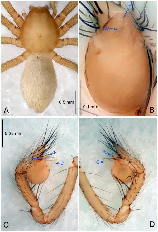
Leptonetela xianren sp. nov., holotype male
Figure 43.
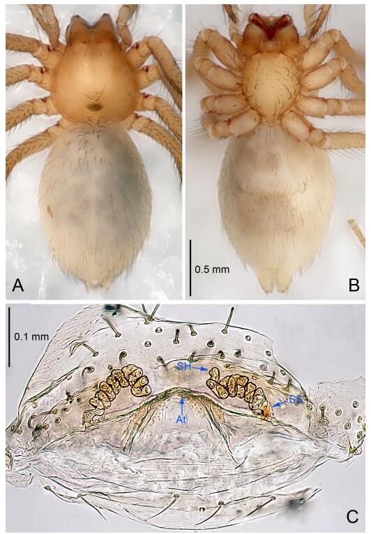
Leptonetela xianren sp. nov., one of the paratype females
Type material. Holotype: male (IZCAS), Xianren Cave, N29.73°, E110.31°, Yvpingxini, Zouma Town, Hefeng County, Enshi Tujia and Miao Autonomous Prefecture, Hubei Province, China, 27 January 2011, Y. Li & J. Liu leg. Paratypes: 2 males and 3 females, same data as holotype.
Etymology. The specific name refers to the type locality; noun.
Diagnosis. This new species is similar to L. liping Lin & Li, 2010, and L. parlonga Wang & Li, 2011 but can be distinguished by the male pedipalpal tibia with 5 slender spines retrolaterally, with spines Ⅰ longest (Figure 42D) (tibia with 5 spines retrolaterally, spines Ⅰ strong and longest in L. liping, 6 slender spines in L. parlonga); median apophysis triangular (Figure 42B) (median apophysis like pointed process in L. liping; ligulate in L. parlonga); from L. parlonga by the cymbium retrolaterally with 1 horn-shaped spine on the earlobe-shaped process in L. parlonga.
Description. Male (holotype). Total length 2.23 (Figure 42A). Carapace 0.95 long, 0.93 wide. Opisthosoma 1.25 long, 0.88 wide. Carapace yellow. Ocular zone with a pair of setae, eyes absent. Median groove, cervical groove and radial furrows indistinct. Clypeus 0.13 high. Opisthosoma gray, ovoid, lacking distinct pattern. Leg measurements: Ⅰ 8.99 (2.50, 0.38, 2.48, 2.00, 1.63); Ⅱ 8.48 (2.38, 0.37, 2.28, 1.90, 1.55); Ⅲ 7.12 (2.03, 0.33, 1.88, 1.63, 1.25); Ⅳ 7.88 (2.48, 0.38, 2.07, 1.60, 1.35). Leg formula: Ⅰ-Ⅱ-Ⅳ-Ⅲ. Male pedipalp (Figure 42C-D): femur with 5 spines ventrally, tibia with 3 long setae prolaterally, 2 long setae and 5 slender spines retrolaterally, with spines Ⅰ longest. Cymbium constricted medially, attaching an earlobe-shaped process retrolaterally. Embolus triangular, prolateral lobe indistinct. Median apophysis triangular. Conductor bamboo leaf-shaped in ventral view (Figure 42B).
Female (one of the paratypes). Similar to male in color and general features but larger and with shorter legs. Total length 2.38 (Figure 43A-B). Carapace 0.85 long, 0.83 wide. Opisthosoma 1.55 long, 1.03 wide. Clypeus 0.15 high. Leg measurements: Ⅰ 8.11 (2.25, 0.38, 2.20, 1.88, 1.40); Ⅱ 7.71 (2.18, 0.35, 2.13, 1.70, 1.35); Ⅲ 6.76 (2.00, 0.35, 1.78, 1.50, 1.13); Ⅳ 7.79 (2.45, 0.40, 2.03, 1.58, 1.33). Vulva (Figure 43C): spermathecae coiled, atrium fusiformed.
Distribution. China (Hubei).
Leptonetela tiankeng Wang & Li sp. nov. Figures 44-45, 97
Figure 44.
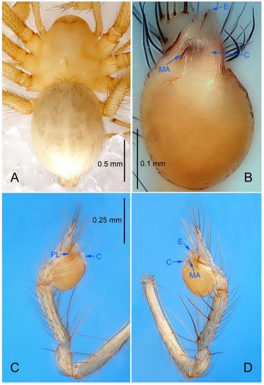
Leptonetela tiankeng sp. nov., holotype male
Figure 45.
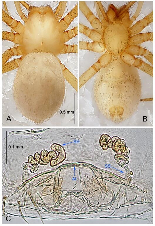
Leptonetela tiankeng sp. nov., one of the paratype females
Type material. Holotype: male (IZCAS), Tiankeng Cave, N26.64°, E104.80°, Hegou, Dewu Town, Zhongshan County, Liupanshui City, Guizhou Province, China, 9 November 2011, H. Chen & Z. Zha leg. Paratypes: 4 males and 5 females, same data as holotype; 2 females, Luoshui Cave, N26.64°, E104.80°, Hegou, Dewu Town, Zhongshan County, Liupanshui City, Guizhou Province, China, 9 November 2011, H. Chen & Z. Zha leg.
Etymology. The specific name refers to the type locality; noun.
Diagnosis. This new species is similar to L. rudicula Wang & Li, 2011, but can be distinguished by the male pedipalpal tibia with 6 spines retrolaterally (Figure 44D), prolateral lobe indistinct (Figure 44C), conductor broad and long, distal edge undulate (Figure 44B) (5 spines retrolaterally, prolateral lobe oval, conductor short, C shaped in L. rudicula).
Description. Male (holotype). Total length 2.03 (Figure 44A). Carapace 1.00 long, 0.85 wide. Opisthosoma 1.10 long, 0.88 wide. Carapace yellow. Ocular area with a pair of setae, eyes absent. Median groove needle-shaped, cervical grooves and radial furrows indistinct. Clypeus 0.13 high. Opisthosoma yellowish, ovoid. Leg measurements: Ⅰ 9.30 (2.48, 0.35, 2.81, 2.23, 1.43); Ⅱ 8.46 (2.35, 0.35, 2.30, 2.18, 1.28); Ⅲ 7.11 (2.05, 0.30, 1.98, 1.75, 1.03); Ⅳ 8.51 (2.43, 0.35, 2.33, 2.15, 1.25). Male pedipalp (Figure 44C-D): tibia with 5 long setae prolaterally, 6 slender spines retrolaterally, with spines Ⅰ longest. Cymbium slightly constricted medially, attached to an earlobe-shaped process retrolaterally. Embolus triangular, prolateral lobe indistinct. Median apophysis flake-like, sclerotized distally. Conductor broad, distal edge undulate (Figure 44B).
Female (one of the paratypes). Similar to male in color and general features, but smaller and with shorter legs. Total length 1.93 (Figure 45A-B). Carapace 0.83 long, 0.73 wide. Opisthosoma 1.13 long, 0.98 wide. Clypeus 0.13 high. Leg measurements: Ⅰ 7.68 (2.23, 0.34, 2.23, 1.63, 1.25); Ⅱ 6.41 (1.90, 0.35, 1.78, 1.38, 1.00); Ⅲ 5.69 (1.68, 0.28, 1.58, 1.38, 0.77); Ⅳ 7.19 (2.00, 0.33, 2.03, 1.70, 1.13). Vulva (Figure 45C): spermathecae coiled, atrium triangular.
Distribution. China (Guizhou).
Leptonetela mayang Wang & Li sp. nov. Figures 46-47, 97
Figure 46.
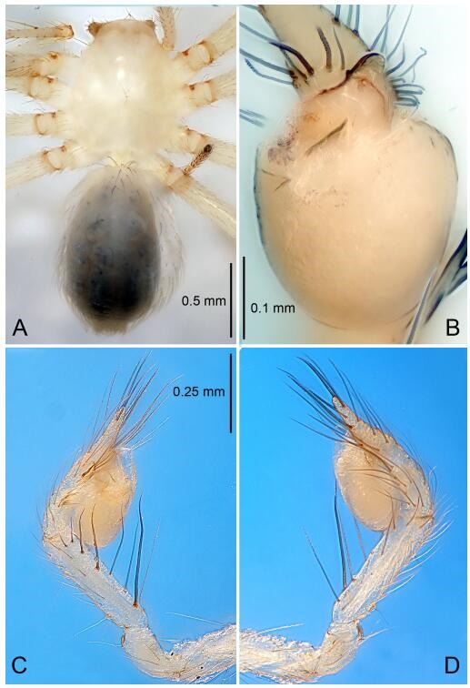
Leptonetela mayang sp. nov., holotype male
Figure 47.
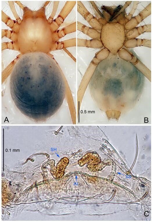
Leptonetela mayang sp. nov., paratype female
Type material. Holotype: male (IZCAS), Mayang Cave, N28.55°, E108.06°, Quankou, Dejiang County, Tongren City, Guizhou Province, China, 10 August 2012, H. Zhao leg. Paratype: 1 female, same data as holotype.
Etymology. The specific name refers to the type locality; noun.
Diagnosis. This new species can be distinguished from all other species of the genus by the male pedipalpal cymbium with one curved, short spine medially in retrolateral view, median apophysis triangular, spermathecae not tightly twisted, just spiraled in the female.
Description. Male (holotype). Total length 2.13 (Figure 46A). Carapace 1.10 long, 0.75 wide. Opisthosoma 0.90 long, 1.00 wide. Carapace white. Eye absent. Median groove, cervical grooves and radial furrows indistinct. Clypeus 0.13 high. Opisthosoma white, ovoid. Leg measurements: Ⅰ 8.98 (2.50, 0.30, 2.63, 2.05, 1.50); Ⅱ 7.68 (2.13, 0.30, 2.25, 1.75, 1.25); Ⅲ 6.76 (2.00, 0.25, 1.88, 1.63, 1.00); Ⅳ 8.21 (2.25, 0.30, 2.38, 1.88, 1.40). Male pedipalp (Figure 46C-D): tibia with 3 long setae prolaterally, and 5 slender spines retrolaterally, spines Ⅰ longest. Cymbium not wrinkled, earlobe-shaped process indistinct, and with 1 curved, short spine retrolaterally. Bulb with spoon-shaped embolus, prolateral lobe indistinct. Median apophysis triangular in ventral view. Conductor thin, triangular in ventral view (Figure 46B).
Female. Similar to male in color and general features, but larger and with shorter legs. Total length 2.37 (Figure 47A-B). Carapace 0.88 long, 0.88 wide. Opisthosoma 1.65 long, 1.00 wide. Clypeus 0.13 high. Leg measurements: Ⅰ -(2.25, 0.30, -, -, -); Ⅱ 6.81 (2.00, 0.30, 1.88, 1.50, 1.13); Ⅲ 6.16 (1.88, 0.25, 1.75, 1.38, 0.90); Ⅳ 7.28 (2.13, 0.30, 2.00, 1.60, 1.25). Vulva (Figure 47C): spermathecae spiraled, atrium triangular.
Distribution. China (Guizhou).
Leptonetela gubin Wang & Li sp. nov. Figures 48-49, 97
Figure 48.
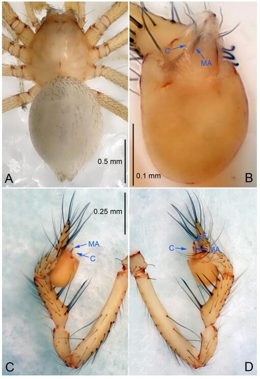
Leptonetela gubin sp. nov., holotype male
Figure 49.
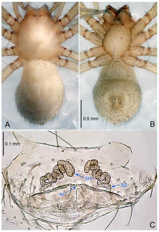
Leptonetela gubin sp. nov., one of the paratype females
Type material. Holotype: male (IZCAS), Gubin River, N26.50°, E107.52°, Gubin, Xingshan Town, Majiang County, Shengkaili City, Guizhou Province, China, 28 November 2011, H. Chen & Z. Zha leg. Paratypes: 22 males and 14 females, same data as holotype; 4 males and 5 females, nameless Cave, N26.50°, E107.52°, Gubin, Xingshan Town, Majiang County, Shengkaili City, Guizhou Province, China, 28 November 2011, H. Chen & Z. Zha leg.
Etymology. The specific name refers to the type locality; noun.
Diagnosis. This new species is similar to L. jinsha Lin & Li, 2010, L. quinquespinata (Chen & Zhu, 2008), L. xinhua Wang & Li sp. nov., L. lujia Wang & Li sp. nov. and L. xinhua Wang & Li sp. nov. but can be distinguished by the male pedipalpal tibia with 4 slender spines prolaterally, 5 slender spines retrolaterally, with spines Ⅰ, Ⅱ equal length, cymbium with 2 long curved spines on earlobe-shaped process retrolaterally (Figure 48D) (tibia with 3 long setae prolaterally, 1 long setae and 5 spines retrolaterally, with spines Ⅰ strongest, tip asymmetrically bifurcated in L. jinsha; tibia with 3 long setae prolaterally, 6 large spines retrolaterally, with spines Ⅰ longest in L. quinquespinata; tibia with 4 long setae prolaterally, 5 slender spines retrolaterally, with spines Ⅰ longest, spines Ⅱ Ⅲ equal length in L. lujia Wang & Li sp. nov.; embolus bifurcated, tibia with 5 slender spines prolaterally, 5 slender spines retrolaterally, conductor triangular in L. xinhua Wang & Li sp. nov.); from L. jinsha, L. lujia Wang & Li sp. nov. and L. xinhua Wang & Li sp. nov. by the semicircular conductor, base of median apophysis distinctly swollen, 4 times wider than the tip (Figure 48B) (conductor broad, tip undulate in L. jinsha; conductor thin, triangular in L. lujia Wang & Li sp. nov. and L. xinhua Wang & Li sp. nov.; base of median apophysis slightly swollen in L. jinsha, L. lujia Wang & Li sp. nov.).
Description. Male (holotype). Total length 1.88 (Figure 48A). Carapace 0.80 long, 0.78 wide. Opisthosoma 1.13 long, 0.93 wide. Carapace yellowish. Ocular area with a pair of setae, eyes absent. Median groove needle-shaped, cervical grooves and radial furrows distinct. Clypeus 0.13 high. Opisthosoma gray, ovoid, lacking distinctive pattern. Leg measurements: Ⅰ 7.51 (2.00, 0.38, 2.13, 1.75, 1.25); Ⅱ 6.48 (1.75, 0.35, 1.80, 1.43, 1.15); Ⅲ 5.51 (1.63, 0.35, 1.40, 1.25, 0.88); Ⅳ 6.82 (1.88, 0.35, 1.88, 1.58, 1.13). Male pedipalp (Figure 48C-D): tibia with 4 long spines prolaterally and 5 spines retrolaterally, with spines Ⅰ, Ⅱ equally length. Cymbium constricted medially, earlobe-shaped process with 2 long curved spines retrolaterally. Embolus triangular, prolateral lobe absent. Median apophysis finger-shaped, base distinctly swollen. Conductor smooth, semicircular in ventral view (Figure 48B).
Female (one of the paratypes). Similar to male in color and general features, but larger and with longer legs. Total length 2.30 (Figure 49A-B). Carapace 0.88 long, 0.85 wide. Opisthosoma 1.40 long, 0.95 wide. Clypeus 0.15 high. Leg measurements: Ⅰ 7.69 (2.13, 0.38, 2.25, 1.65, 1.28); Ⅱ 6.71 (1.90, 0.38, 1.95, 1.40, 1.08); Ⅲ 5.85 (1.75, 0.35, 1.50, 1.35, 0.90); Ⅳ 7.02 (2.00, 0.38, 1.93, 1.58, 1.13). Vulva (Figure 49C): spermathecae coiled, atrium triangular.
Distribution. China (Guizhou).
Leptonetela lujia Wang & Li sp. nov. Figures 50-51, 97
Figure 50.
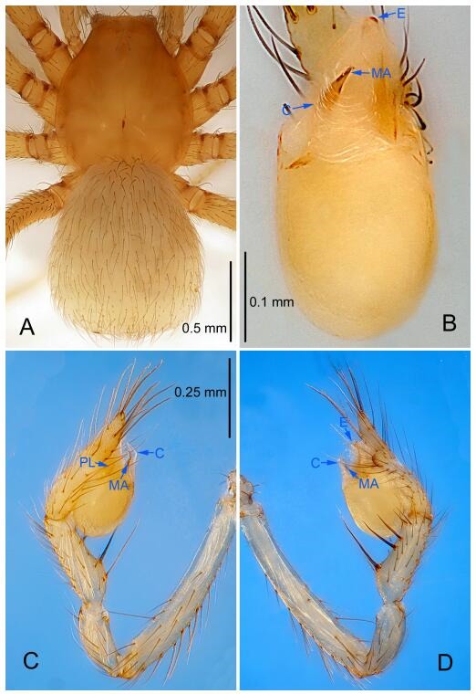
Leptonetela lujia sp. nov., holotype male
Figure 51.
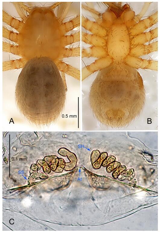
Leptonetela lujia sp. nov., one of the paratype females
Type material. Holotype: male (IZCAS), Wuming Cave, N26.48º, E107.54º, Lujia Bridge, Gubin, Xingshan Town, Majiang County, Kaili City, Guizhou Province, China, 29 November 2011, H. Chen & Z. Zha leg. Paratypes: 1 male and 2 females, same data as holotype.
Etymology. The specific name refers to the type locality; noun.
Diagnosis. This new species is similar to L. jinsha Lin et Li, 2010, L. quinquespinata (Chen & Zhu, 2008), L. xinhua Wang & Li sp. nov. and L. gubin Wang & Li sp. nov. but can be distinguished by the male pedipalpal tibia with 4 long setae prolaterally, 5 slender spines retrolaterally, with spines Ⅰ longest, spines Ⅱ Ⅲ equal length (Figure 50D), conductor thin, triangular (Figure 50B), (tibia with 3 long setae prolaterally, 1 long setae and 5 spines retrolaterally, with spines Ⅰ strongest, tip asymmetrically bifurcated, conductor broad, distal edge undulate in L. jinsha; tibia with 3 long setae prolaterally, 6 slender spines retrolaterally, with spines Ⅰ longest, conductor semicircular in L. quinquespinata; embolus bifurcated, tibia with 5 slender spines prolaterally, 5 slender spines retrolaterally, conductor triangular in L. xinhua Wang & Li sp. nov.; tibia with 4 slender spines prolaterally, 5 slender spines retrolaterally, spines Ⅰ, Ⅱ equal length, cymbium with 2 long, curved spines on earlobe-shaped process retrolaterally, conductor semicircular in L. gubin Wang & Li sp. nov.); from L. gubin and L. quinquespinata by the slightly swollen base of the median apophysis (Figure 50B) (base of median apophysis distinctly swollen, 4 times the width of tip in L. gubin Wang & Li sp. nov.; 3 times in L. quinquespinata).
Description. Male (holotype). Total length 1.72 (Figure 50A). Carapace 0.90 long, 0.85 wide. Opisthosoma 0.87 long, 0.88 wide. Carapace yellow. Ocular area with a pair of setae, eye absent. Median groove needle-shaped, cervical grooves and radial furrows indistinct. Clypeus 0.12 high. Opisthosoma yellowish, ovoid. Leg measurements: Ⅰ 7.75 (2.10, 0.37, 2.23, 1.78, 1.27); Ⅱ 6.97 (1.96, 0.37, 1.86, 1.62, 1.16); Ⅲ 5.73 (1.62, 0.32, 1.50, 1.32, 0.97); Ⅳ 7.15 (2.02, 0.30, 1.92, 1.80, 1.11). Male pedipalp (Figure 50C-D): tibia with 4 long spines prolaterally, 5 large spines retrolaterally, with spines Ⅰ longest, spines Ⅱ Ⅲ equal length. Cymbium not constricted medially, earlobe-shaped process distinct. Embolus triangular, prolateral lobe indistinct. Median apophysis index finger like. Conductor thin, triangular in ventral view (Figure 50B).
Female (one of the paratypes). Similar to male in color and general features, but larger and with shorter legs. Total length 1.70 (Figure 51A-B). Carapace 0.85 long, 0.75 wide. Opisthosoma 0.87 long, 0.83 wide. Clypeus 0.12 high. Leg measurements: Ⅰ 6.89 (1.85, 0.37, 1.97, 1.50, 1.20); Ⅱ 5.94 (1.67, 0.35, 1.62, 1.25, 1.05); Ⅲ 5.30 (1.48, 0.35, 1.38, 1.22, 0.87); Ⅳ 6.47 (1.86, 0.37, 1.70, 1.46, 1.08). Vulva (Figure 51C): spermathecae coiled, atrium triangular.
Distribution. China (Guizhou).
Leptonetela xinhua Wang & Li sp. nov. Figures 52-53, 97
Figure 52.
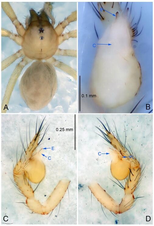
Leptonetela xinhua sp. nov., holotype male
Figure 53.
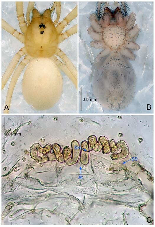
Leptonetela xinhua sp. nov., one of the paratype females
Type material. Holotype: male (IZCAS), nameless Cave, N27.85º, E111.31º, Caojia Town, Xinhua County, Loudi City, Hunan Province, China, 24 March 2016, Y. Li & Z. Chen leg. Paratypes: 3 males and 2 females, same data as holotype.
Etymology. The specific name refers to the type locality; noun.
Diagnosis. This new species is similar to L. jinsha Lin & Li, 2010, L. quinquespinata (Chen & Zhu, 2008), L. lujia Wang & Li sp. nov., and L. gubin Wang & Li sp. nov., but can be distinguished by the bifurcated embolus, male pedipalpal tibia with 5 slender spines prolaterally, 5 slender spines retrolaterally, conductor triangular (Figure 52D), (tibia with 4 long setae prolaterally, 5 slender spines retrolaterally, with spines Ⅰ longest, spines Ⅱ Ⅲ equal length, conductor thin, triangular in L. lujia Wang & Li sp. nov.; tibia with 3 long setae prolaterally, 1 long seta and 5 spines retrolaterally, with spines Ⅰ strongest, tip asymmetrically bifurcated, conductor broad, distal edge undulate in L. jinsha; tibia with 3 long setae prolaterally, 6 slender spines retrolaterally, with spines Ⅰ longest, conductor semicircular in L. quinquespinata; tibia with 4 slender spines prolaterally, 5 slender spines retrolaterally, with spines Ⅰ, Ⅱ equal length, cymbium with 2 long, curved spines on earlobe-shaped process retrolaterally, conductor semicircular in L. gubin Wang & Li sp. nov.); from L. gubin and L. quinquespinata by the base of median apophysis slightly swollen (Figure 52B) (base of median apophysis distinctly swollen, 4 times the width of tip in L. gubin Wang & Li sp. nov.; 3 times in L. quinquespinata).
Description. Male (holotype): total length 1.78 (Figure 52A). Prosoma 0.85 long, 0.71 wide. Opisthosoma 0.94 long, 0.73 wide. Prosoma yellowish. Ocular area with a pair of setae, six eyes. Median groove needle-shaped, brown. Cervical grooves and radial furrows indistinct. Clypeus 0.14 high, slightly sloped anteriorly. Opisthosoma pale brown, ovoid, covered with short hairs, lacking distinct pattern. Sternum and legs yellowish. Leg measurements: Ⅰ 5.39 (1.52, 0.28, 1.58, 1.20, 0.81); Ⅱ 4.37 (1.28, 0.29, 1.28, 1.01, 0.51); Ⅲ 3.84 (1.03, 0.25, 0.98, 0.95, 0.63); Ⅳ5.15 (1.36, 0.27, 1.50, 1.23, 0.79). Male pedipalp (Figure 52C-D): tibia with 5 slender spines prolaterally, 5 slender spines retrolaterally. Cymbium with an earlobe-shaped process retrolaterally. Embolus bifurcated, prolateral lobe triangular. Median apophysis tongue shaped in prolateral view. Conductor triangular in ventral view (Figure 52B).
Female (one of the paratypes): similar to male in color and general features, but with a larger body size and shorter legs. Total length 1.95 (Figure 53A-B). Prosoma 0.66 long, 0.53 wide. Opisthosoma 1.06 long, 0.86 wide. Clypeus 0.20 high. Leg measurements: Ⅰ 4.60 (1.30, 0.27, 1.33, 0.99, 0.71); Ⅱ 3.86 (1.11, 0.25, 1.01, 0.85, 0.64); Ⅲ 3.34 (0.95, 0.24, 0.83, 0.76, 0.56); Ⅳ 4.54 (1.33, 0.26, 1.25, 1.03, 0.67). Vulva (Figure 53C): spermathecae coiled, atrium fusiform.
Distribution. China (Hunan).
Leptonetela kangsa Wang & Li sp. nov. Figures 54-55, 97
Figure 54.
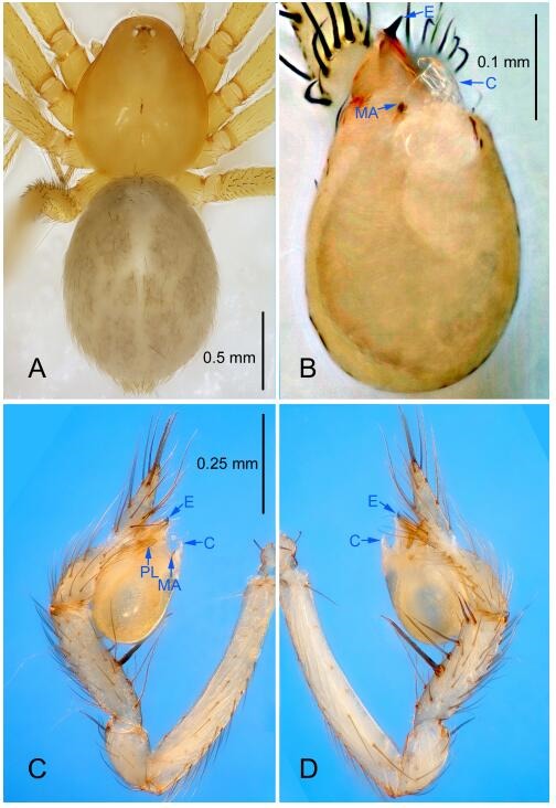
Leptonetela kangsa sp. nov., holotype male
Figure 55.
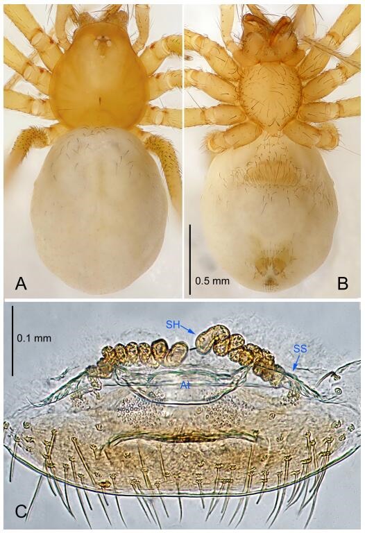
Leptonetela kangsa sp. nov., one of the paratype females
Type material. Holotype: male (IZCAS), Kangsagulie Cave, N26.79º, E108.21º, Datang, Geyi Town, Taijiang County, Kaili City, Guizhou Province, China, 5 December 2011, H. Chen & Z. Zha leg. Paratypes: 7 males and 6 females, same data as holotype.
Etymology. The specific name refers to the type locality; noun.
Diagnosis. This new species is similar to L. shibingensis and L. wuming Wang & Li sp. nov., but can be distinguished by the median apophysis index finger-like in prolaterally view, tip bifurcated (Fig. 54D) (median apophysis small, triangular in ventral view in L. shibingensis; median apophysis like victory gesture, "Ⅴ" shaped in L. wuming Wang & Li sp. nov.); from L. wuming Wang & Li sp. nov., by the tibia Ⅰ spine located at the middle of tibia, embolus with 1 basal tooth (tibia Ⅰ spine located at the base of tibia, embolus without tooth in L. wuming Wang & Li sp. nov.).
Description. Male (holotype). Total length 2.07 (Figure 54A). Carapace 0.85 long, 0.87 wide. Opisthosoma 1.25 long, 0.92 wide. Carapace yellow. Eyes six, PME reduced to white spots. Median groove needle-shaped, cervical grooves and radial furrows indistinct. Clypeus 0.12 high. Opisthosoma gray, ovoid. Leg measurements: Ⅰ 9.04 (2.50, 0.37, 2.65, 2.10, 1.42); Ⅱ 7.43 (2.10, 0.36, 2.05, 1.70, 1.22); Ⅲ 6.23 (1.77, 0.37, 1.60, 1.47, 1.02); Ⅳ 8.09 (2.22, 0.35, 2.27, 2.00, 1.25). Male pedipalp (Figure 54C-D): tibia with 4 long setae prolaterally, 5 large spines retrolaterally, with Ⅰ spine strong, located medially. Cymbium constricted medially, attaching an earlobe-shaped process retrolaterally. Embolus triangular, bearing a basal tooth, prolateral lobe oval. Median apophysis index finger-like in prolateral view, tip bifurcated. Conductor bamboo leaf-shaped in ventral view (Figure 54B).
Female (one of the paratypes). Similar to male in color and general features, but smaller and with shorter legs. Total length 2.02 (Figure 55A-B). Carapace 0.72 long, 0.72 wide. Opisthosoma 1.27 long, 1.02 wide. Clypeus 0.15 high. Leg measurements: Ⅰ 7.61 (2.07, 0.35, 2.25, 1.72, 1.22); Ⅱ 6.23 (1.72, 0.35, 1.72, 1.37, 1.07); Ⅲ 5.41 (1.62, 0.32, 1.35, 1.22, 0.90); Ⅳ 6.92 (1.80, 0.35, 2.00, 1.65, 1.12). Vulva (Figure 55C): spermathecae coiled, atrium fusiform.
Distribution. China (Guizhou).
Leptonetela wuming Wang & Li sp. nov. Figures 56-57, 97
Figure 56.
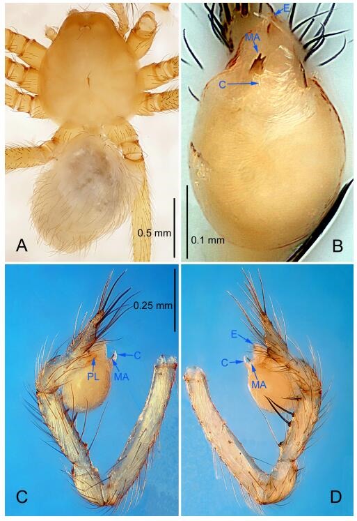
Leptonetela wuming sp. nov., holotype male
Figure 57.
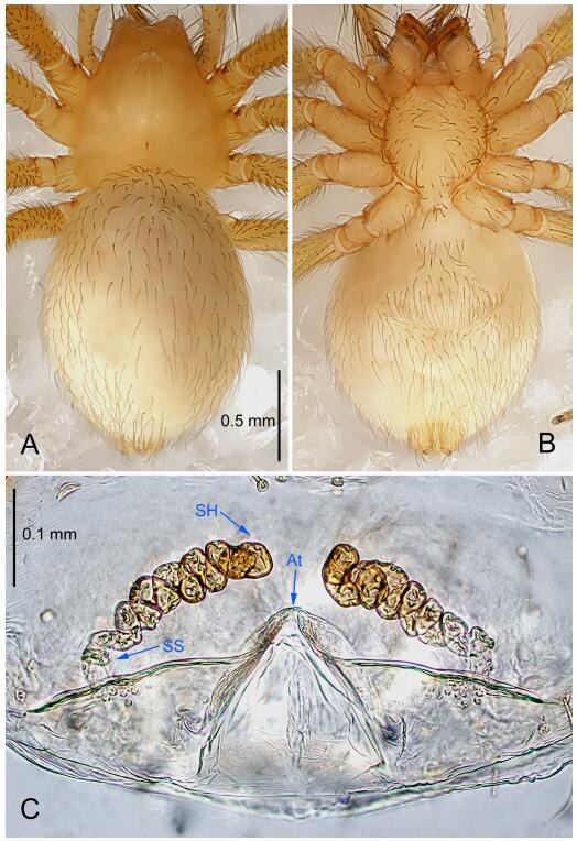
Leptonetela wuming sp. nov., one of the paratype females
Type material. Holotype: male (IZCAS), Wuming Cave, N25.43°, E105.62°, Dabei Town, Zhenfeng County, Guizhou Province, China, 18 July 2012, H. Zhao leg. Paratypes: 2 males and 5 females, same data as holotype.
Etymology. The specific name refers to the type locality; noun.
Diagnosis. This new species is similar to L. kangsa Wang & Li sp. nov., and L. shibingensis but can be distinguished by the male pedipalpal bulb embolus without basal tooth (Figure 56B), (embolus with basal tooth in the two species mentioned above); from L. kangsa Wang & Li sp. nov. and L. shibingensis by the tibia spines Ⅰ located at the base of the tibia (Figure 56D) (tibia spines Ⅰ located medially in L. kangsa Wang & Li sp. nov. and L. shibingensis).
Description. Male (holotype). Total length 1.50 (Figure 56A). Carapace 0.60 long, 0.45 wide. Opisthosoma 1.10 long, 0.60 wide. Carapace yellowish. Ocular area with a pair of setae, PLE, PME absent, ALE reduced to white spots. Median groove needle-shaped, cervical grooves and radial furrows indistinct. Clypeus 0.09 high. Opisthosoma yellowish, ovoid. Leg measurements: Ⅰ 11.75 (3.12, 0.35, 3.40, 2.88, 2.00); Ⅱ 9.35 (2.75, 0.35, 2.48, 2.49, 1.28); Ⅲ 8.47 (2.80, 0.32, 2.30, 1.96, 1.09); Ⅳ 9.81 (2.81, 0.35, 2.72, 2.56, 1.37). Male pedipalp (Figure 56C-D): tibia with 3 long setae prolaterally, 5 large spines retrolaterally, with spines Ⅰ strong, longest. Cymbium constricted medially, attached to an earlobe-shaped process retrolaterally. Embolus triangular, prolateral lobe oval. Median apophysis like victory gesture, "Ⅴ" shaped. Conductor bamboo leaf-shaped in ventral view (Figure 56B).
Female (one of the paratypes). Similar to male in color and general features, but larger and with shorter legs. Total length 3.02 (Figure 57A-B). Carapace 1.25 long, 0.90 wide. Opisthosoma 2.45 long, 1.38 wide. Clypeus 0.09 high. Leg measurements: Ⅰ 8.67 (2.44, 0.33, 2.50, 2.01, 1.39); Ⅱ 7.29 (2.10, 0.33, 2.08, 1.65, 1.13); Ⅲ 7.22 (2.08, 0.35, 2.03, 1.63, 1.13); Ⅳ 7.30 (2.51, 0.33, 1.85, 1.56, 1.05). Vulva (Figure 57C): spermathecae coiled, atrium triangular.
Distribution. China (Guizhou).
Leptonetela shanji Wang & Li Wang & Li sp. nov. Figures 58-59, 97
Figure 58.
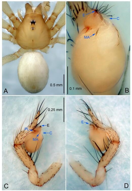
Leptonetela shanji sp. nov., holotype male
Figure 59.
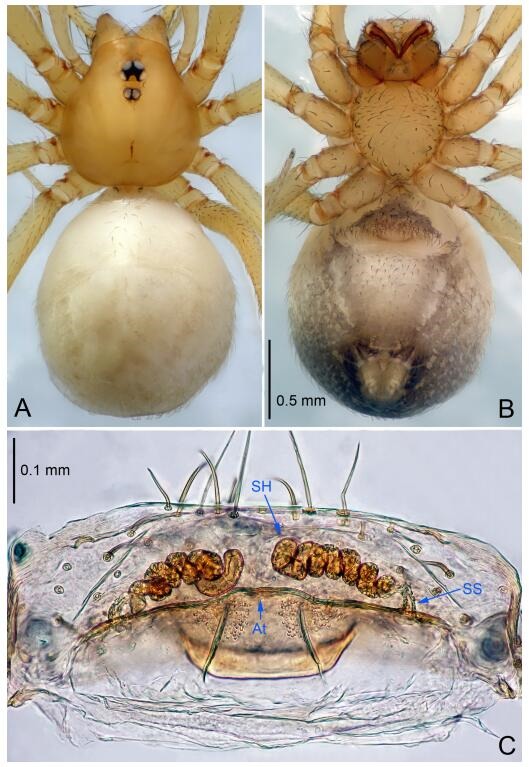
Leptonetela shanji sp. nov., one of the paratype females
Type material. Holotype: male (IZCAS), Shanji Cave, N27.28°, E107.82°, Xiaguihua, Xiaosai Town, Yuqing County, Zunyi City, Guizhou Province, China, 15 August 2012, H. Zhao leg. Paratypes: 3 males and 3 females, same data as holotype; 2 females, Guanyin Cave, N27.32°, E107.71°, Hongjun, Longxi Town, Yuqing County, Zunyi City, Guizhou Province, China, 15 August 2012, H. Zhao leg; 3 females, Liangfeng Cave, N27.27°, E107.76°, Xiaosai Town, Yuqing County, Zunyi City, Guizhou Province, China, 14 August 2012, H. Zhao leg.
Etymology. The specific name refers to the type locality; noun.
Diagnosis. This new species is similar to L. digitata Lin & Li, 2010, L. hamata Lin & Li, 2010 and L. tetracantha Lin & Li, 2010, but can be distinguished by the male pedipalpal tibia spines Ⅰ strong, located medially (Figure 58D) (tibia spines Ⅰ slender, located at the base of tibia in all above); from L. hamata and L. tetracantha by the male pediapal tibia spines Ⅰ asymmetrically bifurcated (Figure 58D) (tibia spines Ⅰ not bifurcated in L. hamata, and L. tetracantha); from L. digitata by themedian apophysis not curved (median apophysis curved in L. digitata).
Description. Male (holotype). Total length 2.08 (Figure 58A). Carapace 0.90 long, 0.95 wide. Opisthosoma 1.10 long, 0.83 wide. Carapace yellow. Ocular area with a pair of setae, six eyes. Median groove needle-shaped, cervical grooves and radial furrows indistinct. Clypeus 0.13 high. Opisthosoma gray, ovoid. Leg measurements: Ⅰ 8.54 (2.25, 0.38, 2.50, 2.03, 1.38); Ⅱ 6.89 (1.88, 0.38, 1.88, 1.60, 1.15); Ⅲ 5.70 (1.55, 0.35, 1.47, 1.35, 0.98); Ⅳ 7.59 (2.03, 0.38, 2.13, 1.88, 1.17). Male pedipalp (Figure 58C-D): tibia with 4 long spines prolaterally, 5 spines retrolaterally, with Ⅰ spine strong, asymmetrically bifurcated and located at the base of tibia. Cymbium constricted medially, attached to an earlobe-shaped process retrolaterally. Embolus triangular, bearing a small basal tooth, prolateral lobe oval. Median apophysis index finger like in prolateral view, tapering. Conductor bamboo leaf-shaped in ventral view (Figure 58B).
Female (one of the paratypes). Similar to male in color and general features, but larger and with shorter legs. Total length 2.40 (Figure 59A-B). Carapace 0.95 long, 0.88 wide. Opisthosoma 1.38 long, 1.25 wide. Clypeus 0.13 high. Leg measurements: Ⅰ 7.97 (2.13, 0.38, 2.38, 1.75, 1.33); Ⅱ 6.36 (1.75, 0.35, 1.75, 1.38, 1.13); Ⅲ 5.31 (1.45, 0.35, 1.38, 1.25, 0.88); Ⅳ 7.18 (1.95, 0.38, 2.00, 1.70, 1.15). Vulva (Figure 59C): spermathecae coiled, atrium fusiform.
Distribution. China (Guizhou).
Leptonetela xiaoyan Wang & Li sp. nov. Figures 60-61, 97
Figure 60.
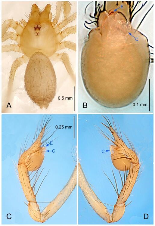
Leptonetela xiaoyan sp. nov., holotype male
Figure 61.
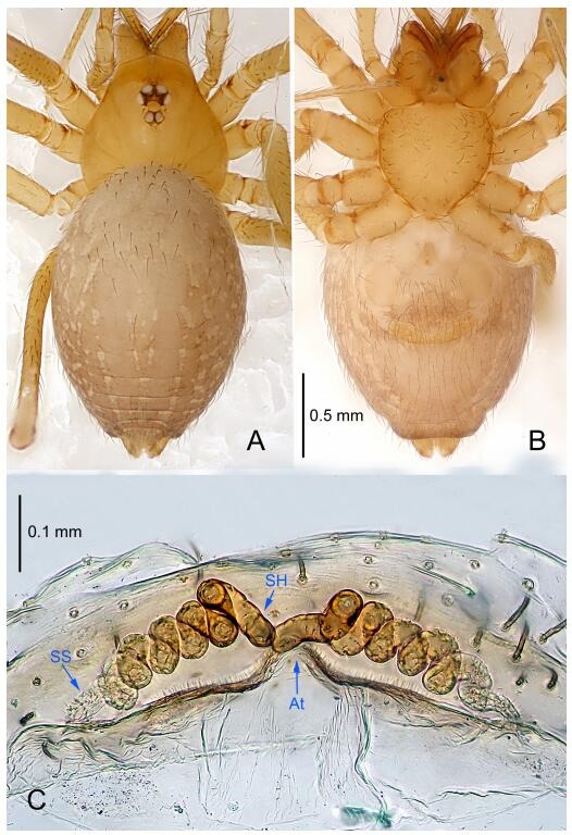
Leptonetela xiaoyan sp. nov., one of the paratype females
Type material. Holotype: male (IZCAS), Gejiaxiaoyan Cave, N27.11°, E105.24°, Shanjiao, Zhuchang Town, Bijie City, Guizhou Province, China, 27 January 2011, H. Chen & Z. Zha leg. Paratypes: 2 males and 6 females, same data as holotype.
Etymology. The specific name refers to the type locality; noun.
Diagnosis. This new species is similar to L. curvispinosa Lin & Li, 2010, but can be distinguished by the male pedipalpal tibia with 4 large spines prolaterally, 6 large spines retrolaterally (Figure 60D), median apophysis not sclerotized, little finger-shaped in prolateral view, conductor broad C tile-shaped (Figure 60B) (tibia with 3 large spines prolaterally, 5 large spines retrolaterally, median apophysis absent, conductor reduced in L. curvispinosa).
Description. Male (holotype). Total length 1.67 (Figure 60A). Carapace 0.88 long, 0.80 wide. Opisthosoma 1.15 long, 0.75 wide. Carapace yellowish. Six eyes. Median groove, cervical grooves and radial furrows indistinct. Clypeus 0.10 high. Opisthosoma yellowish, ovoid. Leg measurements: Ⅰ 9.96 (2.84, 0.35, 2.80, 2.40, 1.57); Ⅱ 7.09 (2.02, 0.32, 2.00, 1.60, 1.15); Ⅲ 6.38 (1.77, 0.32, 1.82, 1.47, 1.00); Ⅳ 7.89 (2.25, 0.35, 2.27, 1.85, 1.17). Male pedipalp (Figure 60C-D): tibia with 4 spines prolaterally and 6 spines retrolaterally, with Ⅰ spine longest. Cymbium not constricted, prolaterally with one curved spine on the base. Embolus triangular, prolateral lobe oval. Median apophysis slightly sclerotized, fingerlike in prolateral view. Conductor broad, C tile-shaped in ventral view (Figure 60B).
Female (one of the paratypes). Similar to male in color and general features, but larger and with shorter legs. Total length 2.12 (Figure 61A-B). Carapace 0.82 long, 0.82 wide. Opisthosoma 1.47 long, 1.12 wide. Clypeus 0.15 high. Leg measurements: Ⅰ 9.79 (2.62, 0.35, 3.03, 2.22, 1.57); Ⅱ 6.84 (1.90, 0.30, 1.97, 1.47, 1.20); Ⅲ 5.53 (1.52, 0.32, 1.52, 1.27, 0.90); Ⅳ 7.36 (2.07, 0.35, 1.97, 1.72, 1.25). Vulva (Figure 61C): spermathecae coiled, atrium triangular, anterior margin of atrium covered with short hairs.
Distribution. China (Guizhou).
Leptonetela huoyan Wang & Li sp. nov. Figures 62-63, 97
Figure 62.
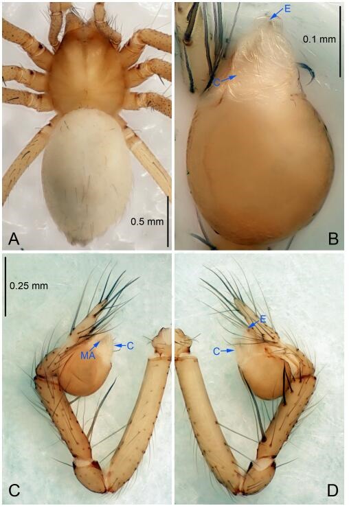
Leptonetela huoyan sp. nov., holotype male
Figure 63.
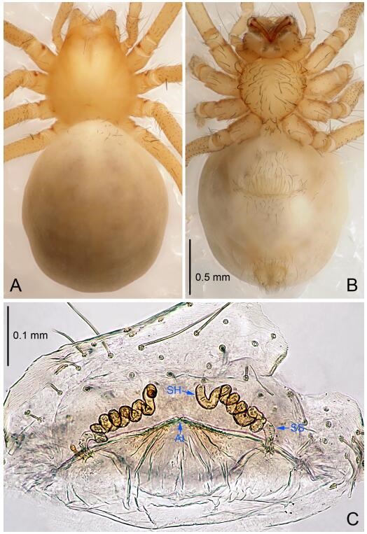
Leptonetela huoyan sp. nov., one of the paratype females
Type material. Holotype: male (IZCAS), Heyuantou nameless Cave, N29.25°, E109.35°, Huoyan Street, Guitang Dam Town, Longshan County, Hubei Province, China, 15 January 2014, Y. Li & Y. Lin leg. Paratypes: 1 male and 2 females, same data as holotype; 1 male and 4 females, nameless Cave, N29.61°, E109.17° Jieping, Xianfeng County, Enshi Tujia and Miao Autonomous Prefecture, Hubei Province, China, 17 January 2014, Y. Li & Y. Lin leg.
Etymology. The specific name refers to the type locality; noun.
Diagnosis. This new species is similar to L. anshun Lin & Li, 2010 and L. chenjia Wang & Li sp. nov., but can be distinguished by on the male pedipalpal bulb median apophysis slightly sclerotized, index finger like, conductor broad, semicircular (Fig 62B) (median apophysis absent in L. anshun, and L. chenjia Wang & Li sp. nov.; tip of conductor bifurcated in L. anshun, conductor reduced in L. chenjia Wang & Li sp. nov.); from L. anshun by the tibia spines Ⅰ slender (Figure 62D) (tibia spines Ⅰ strong, tip bifurcated in L. anshun).
Description. Male (holotype). Total length 2.25 (Figure 62A). Carapace 0.88 long, 0.83 wide. Opisthosoma 1.50 long, 0.92 wide. Carapace yellow. Eye absent. Median groove, cervical groove and radial furrows indistinct. Clypeus 0.13 high. Opisthosoma gray, ovoid. Leg measurements: Ⅰ 8.90 (2.53, 0.40, 2.57, 2.00, 1.40); Ⅱ 7.69 (2.38, 0.40, 2.13, 1.65, 1.13); Ⅲ 6.51 (2.00, 0.38, 1.75, 1.50, 0.88); Ⅳ 7.73 (2.25, 0.40, 2.15, 1.78, 1.15). Male pedipalp (Figure 62C-D): tibia with 4 long setae prolaterally, 1 long seta and 5 spines retrolaterally, with spines Ⅰ longest, distant from others, the rest of the spines concentrated distally. Cymbium constricted medially, attached to an earlobe-shaped process retrolaterally. Embolus triangular, prolateral lobe absent. Median apophysis slightly sclerotized, index finger like. Conductor broad, semicircular in ventral view (Figure 62B).
Female (one of the paratypes). Similar to male in color and general features, but larger and with shorter legs. Total length 2.53 (Figure 63A-B). Carapace 0.93 long, 0.82 wide. Opisthosoma 1.63 long, 1.38 wide. Clypeus 0.15 high. Leg measurements: Ⅰ 8.26 (2.38, 0.38, 2.32, 1.88, 1.30); Ⅱ 7.28 (2.05, 0.35, 2.00, 1.63, 1.25); Ⅲ 6.47 (1.93, 0.35, 1.62, 1.47, 1.10); Ⅳ 7.54 (2.20, 0.38, 2.08, 1.75, 1.13). Vulva (Figure 63C): spermathecae coiled, atrium fusiform.
Distribution. China (Hubei).
Leptonetela liuguan Wang & Li sp. nov. Figures 64-65, 97
Figure 64.
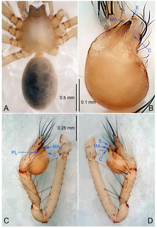
Leptonetela liuguan sp. nov., holotype male
Figure 65.
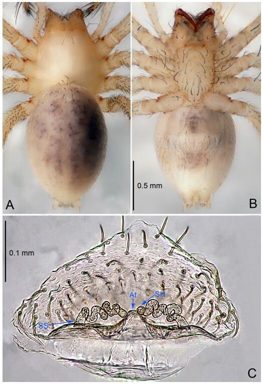
Leptonetela liuguan sp. nov., one of the paratype females
Type material. Holotype: male (IZCAS), Liuguan Cave, N26.15°, E106.46°, Mengqiu, Baiyunshan Town, Changshun County, Guizhou Province, China, 23 December 2010, Z. Zha & Z. Chen leg. Paratypes: 2 female, same data as holotype; 1 male, Fenghuang Cave, N26.09°, E106.39°, Shenglian, Zhonghuo Town, Changshun County, Guizhou Province, China, 23 December 2010, Z. Zha & Z. Chen leg.
Etymology. The specific name refers to the type locality; noun.
Diagnosis. This new species is similar to L. penevi Wang & Li, 2016 and L. changtu Wang & Li sp. nov. but can be distinguished by on the male pedipalpal bulb median apophysis long, and half the length of bulb (Figure 64B) (median apophysis short, 1/5 the length of bulb in L. palmate, and L. changtu Wang & Li sp. nov.); male pedipalpal tibia spines slender, equally strong (Figure 64D) (tibia spines Ⅰ Ⅱ equally strong, stronger than other spines in L. penevi, tibia spines Ⅰ Ⅱ Ⅲ equally strong, stronger than other spines in L. changtu Wang & Li sp. nov.).
Description. Male (holotype). Total length 1.88 (Figure 64A). Carapace 0.73 long, 0.75 wide. Opisthosoma 1.10 long, 0.88 wide. Carapace yellowish. Eyes absent. Median groove, cervical grooves and radial furrows indistinct. Clypeus 0.13 high. Opisthosoma gray, ovoid. Leg measurements: Ⅰ 8.93 (2.38, 0.40, 2.64, 2.13, 1.38); Ⅱ 7.84 (2.13, 0.40, 2.23, 1.78, 1.30); Ⅲ 6.56 (1.75, 0.38, 1.80, 1.63, 1.00); Ⅳ 8.04 (2.25, 0.40, 2.18, 1.88, 1.33). Male pedipalp (Figure 64C-D): tibia with 3 long setae prolaterally, 5 slender spines retrolaterally, the spines slim equally strong. Embolus triangular, prolateral lobe absent. Teeth of median apophysis reduced to sclerotized spots, conductor and median apophysis long, equal length and half the length of bulb (Figure 64C).
Female (one of the paratypes). Similar to male in color and general features, but larger and with shorter legs. Total length 2.08 (Figure 65A-B). Carapace 0.75 long, 0.75 wide. Opisthosoma 1.50 long, 0.88 wide. Clypeus 0.15 high. Leg measurements: Ⅰ 7.81 (2.13, 0.38, 2.30, 1.75, 1.25); Ⅱ 6.89 (1.88, 0.38, 2.00, 1.50, 1.13); Ⅲ 5.97 (1.70, 0.38, 1.63, 1.38, 0.88); Ⅳ 7.27 (2.03, 0.38, 2.05, 1.63, 1.18). Vulva (Figure 65C): spermathecae coiled, atrium fusiform, anterior margin of atrium undulate.
Distribution. China (Guizhou).
Leptonetela nanmu Wang & Li sp. nov. Figures 66-67, 97
Figure 66.
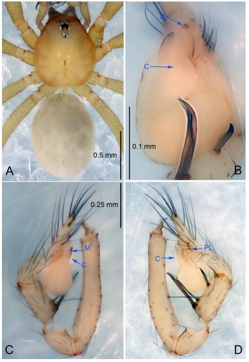
Leptonetela nanmu sp. nov., holotype male
Figure 67.
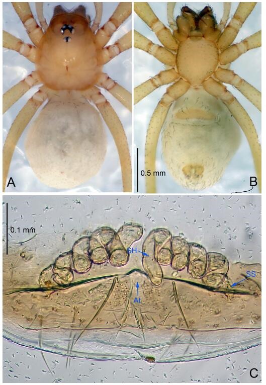
Leptonetela nanmu sp. nov., one of the paratype females
Type material. Holotype: male (IZCAS), Nanmu Cave, N28.10°, E110.08°, Pushi Town, Luxi County, Hunan Province, China, 5 April 2016, Y. Li & Z. Chen leg. Paratypes: 3 males and 2 females, same data as holotype.
Etymology. The specific name refers to the type locality; noun.
Diagnosis. This new species is similar to L. tianxingensis, but can be distinguished by on the male pedipalpal bulb conductor longer than median apophysis (Figure 66B) (conductor shorter than median apophysis in L. tianxingensis); male pedipalpal tibia Ⅲ spine strong (Figure 66D) (tibia Ⅲ spine slender in L. tianxingensis).
Description. Male (holotype): total length 1.70 (Figure 66A). Prosoma 0.81 long, 0.63 wide. Opisthosoma 0.94 long, 0.70 wide. Prosoma yellow. Six eyes, with a pair of setae on ocular area. Median groove needle-shaped, brown. Cervical grooves and radial furrows indistinct. Clypeus 0.14 high, slightly sloped anteriorly. Opisthosoma pale brown, ovoid, covered with short hairs, lacking distinctive pattern. Sternum and legs yellowish. Leg measurements: Ⅰ 5.45 (1.42, 0.26, 1.62, 1.27, 0.88); Ⅱ 4.76 (1.24, 0.25, 1.20, 1.27, 0.80); Ⅲ 4.12 (1.03, 0.23, 1.02, 0.95, 0.89); Ⅳ 5.60 (1.36, 0.22, 1.48, 1.27, 1.27). Male pedipalp (Figure 66C-D): tibia with 5 spines retrolaterally, with Ⅰ spine strongest, tip bifurcated, spines Ⅱ slender, spines Ⅲ strong. Embolus triangular, prolateral lobe oval. Median apophysis slightly sclerotized, thumb-shaped in ventral view. Conductor triangular, longer than median apophysis (Figure 66B).
Female (one of the paratypes): similar to male in color and general features, but with a larger body size and longer legs. Total length 1.98 (Figure 67A-B). Prosoma 0.88 long, 0.79 wide. Opisthosoma 1.12 long, 1.03 wide. Clypeus 0.20 high. Leg measurements: Ⅰ 5.63 (1.52, 0.28, 1.68, 1.30, 0.85); Ⅱ 4.86 (1.44, 0.27, 1.33, 0.86, 0.96); Ⅲ 4.21 (1.31, 0.21, 1.06, 0.99, 0.64); Ⅳ 5.23 (1.47, 0.25, 1.50, 1.20, 0.81). Vulva (Figure 67C): spermathecae coiled, atrium triangular.
Distribution. China (Hunan).
Leptonetela changtu Wang & Li sp. nov. Figure 68-69, 97
Figure 68.
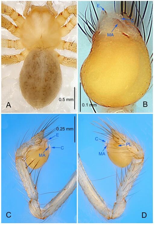
Leptonetela changtu sp. nov., holotype male
Figure 69.
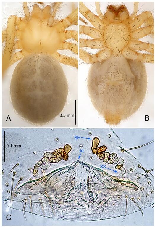
Leptonetela changtu sp. nov., one of the paratype females
Type material. Holotype: male (IZCAS), Changtu Cave, N27.14°, E105.43°, Honglin, Qianxi Town, Bijie County, Guizhou Province, China, 23 November 2011, Z. Zha & Z. Zha leg. Paratypes: 1 male and 10 females, same data as holotype.
Etymology. The specific name refers to the type locality; noun.
Diagnosis. This new species is similar to L. penevi Wang & Li, 2016 and L. liuguan Wang & Li sp. nov. but can be distinguished by the male pedipalpal tibia spines Ⅰ, Ⅱ, Ⅲ equally strong, stronger than other two spines (Figure 68C) (tibia spines Ⅰ and Ⅱ equally strong, stronger than other spines in L. penevi, tibial spines slender, equally strong in L. liuguan Wang & Li sp. nov.); from L. liuguan Wang & Li sp. nov. by median apophysis short, 1/5 the length of bulb (Figure 68B) (median apophysis long, half the length of bulb in L. liuguan Wang & Li sp. nov.); from L. penevi by the cymbium not constricted (cymbium constricted medially in L. penevi).
Description. Male (holotype). Total length 2.33 (Figure 68A). Carapace 1.06 long, 1.03 wide. Opisthosoma 1.38 long, 1.08 wide. Carapace yellowish. Ocular area with a pair of setae, eyes absent. Median groove needle-shaped, cervical grooves and radial furrows indistinct. Clypeus 0.14 high. Opisthosoma pale yellow, ovoid, with brown spots. Leg measurements: Ⅰ 10.02 (2.69, 0.39, 2.91, 2.38, 1.65); Ⅱ 8.75 (2.37, 0.38, 2.49, 2.08, 1.43); Ⅲ 7.53 (2.15, 0.38, 1.98, 1.77, 1.25); Ⅳ 9.20 (2.56, 0.38, 2.50, 2.26, 1.50). Male pedipalp (Figure 68C-D): tibia with 5 large spines retrolaterally, tibia spines Ⅰ longest, spines Ⅰ and Ⅱ equally strong, stronger than others. Cymbium not constricted. Embolus triangular, prolateral lobe oval. Median apophysis palm-shaped, teeth of median apophysis reduced to sclerotized spots. Conductor semicircular (Figure 68B).
Female (one of the paratypes). Similar to male in color and general features, but larger and with shorter legs. Total length 2.70 (Figure 69A-B). Carapace 1.10 long, 0.88 wide. Opisthosoma 1.72 long, 1.48 wide. Clypeus 0.20 high. Leg measurements: Ⅰ 9.10 (2.54, 0.43, 2.66, 1.98, 1.49); Ⅱ 7.73 (2.21, 0.41, 2.21, 1.67, 1.23); Ⅲ 6.85 (1.99, 0.40, 1.88, 1.50, 1.08); Ⅳ 8.22 (2.38, 0.41, 2.28, 1.88, 1.27). Vulva (Figure 69C): spermathecae coiled, atrium triangular.
Distribution. China (Guizhou).
Leptonetela lianhua Wang & Li sp. nov. Figures 70-71, 97
Figure 70.
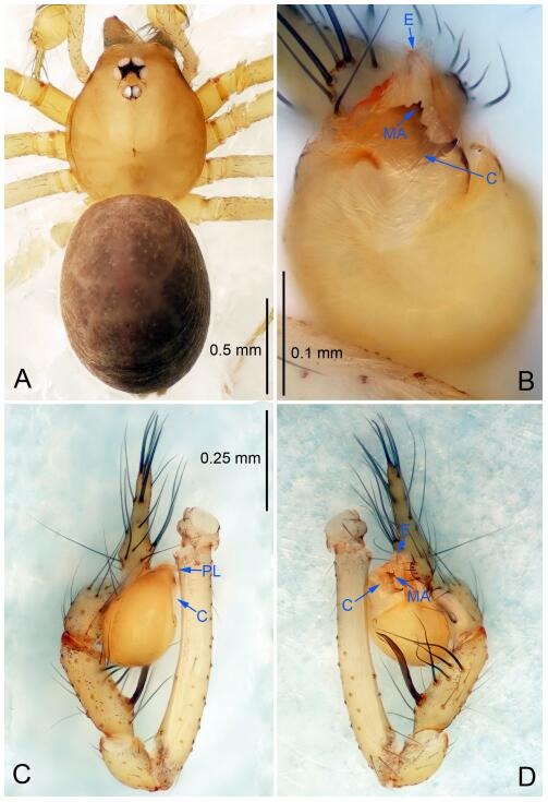
Leptonetela lianhua sp. nov., holotype male
Figure 71.
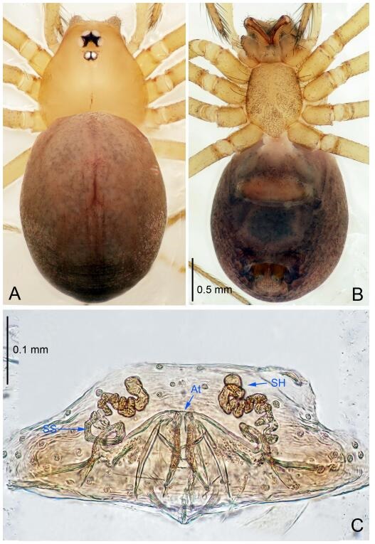
Leptonetela lianhua sp. nov., one of the paratype females
Type material. Holotype: male (IZCAS), Lianhua Cave, N25.48°, E114.09°, Niedou Town, Chongyi County, Jiangxi Province, China, 24 April 2013, Y. Luo & J. Liu leg. Paratypes: 3 males and 10 females, same data as holotype.
Etymology. The specific name refers to the type locality; noun.
Diagnosis. This new species is similar to L. niubizi Wang & Li sp. nov. but can be distinguished by the male pedipalpal tibia with 5 spines retrolaterally, with Ⅰ spine strongest, tip bifurcated, the other 4 spines slender, 2 of them longer than Ⅰ spine (Figure 70D); tip of median apophysis with 5 small teeth, and 1 ox horn-shaped large teeth (Figure 70B) (tibia with 5 slender spines retrolaterally, spines Ⅰ longest, not bifurcated, median apophysis antler-like, tip with 7 small teeth in L. niubizi Wang & Li sp. nov.).
Description. Male (holotype). Total length 2.00 (Figure 70A). Carapace 0.87 long, 0.70 wide. Opisthosoma 1.00 long, 0.87 wide. Carapace yellow. Ocular area with a pair of setae, six eyes. Median groove needle-shaped, cervical grooves and radial furrows indistinct. Clypeus 0.13 high. Opisthosoma brown, ovoid. Leg measurements: Ⅰ 9.69 (2.62, 0.25, 3.20, 2.37, 1.25); Ⅱ 7.05 (2.00, 0.25, 2.10, 1.70, 1.00); Ⅲ 5.70 (1.62, 0.22, 1.62, 1.37, 0.87); Ⅳ 7.45 (2.10, 0.25, 2.25, 1.75, 1.10). Male pedipalp (Figure 70C-D): tibia with 3 long setae prolaterally, and 5 spines retrolaterally, with spines Ⅰ strongest, tip bifurcated, and the other 4 spines slender, 2 of them longer than spines Ⅰ. Cymbium constricted medially, attached to an earlobe-shaped process retrolaterally. Embolus triangular, prolateral lobe absent. Tip of median apophysis with 5 small teeth, and 1 horn-shaped large teeth. Conductor broad C tile-shaped in ventral view (Figure 70B).
Female (one of the paratypes). Similar to male in color and general features, but larger and with shorter legs. Total length 2.25 (Figure 71A-B). Carapace 1.25 long, 0.95 wide. Opisthosoma 1.25 long, 0.75 wide. Clypeus 0.15 high. Leg measurements: Ⅰ 8.02 (2.12, 0.30, 2.50, 1.75, 1.35); Ⅱ 6.02 (1.65, 0.25, 1.75, 1.37, 1.00); Ⅲ 5.07 (1.35, 0.27, 1.50, 1.20, 0.75); Ⅳ 6.85 (2.00, 0.30, 1.95, 1.50, 1.10). Vulva (Figure 71C): spermathecae slender, coiled and atrium triangular.
Distribution. China (Jiangxi).
Leptonetela niubizi Wang & Li sp. nov. Figures 72-73, 97
Figure 72.
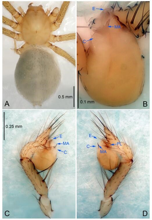
Leptonetela niubizi sp. nov., holotype male
Figure 73.
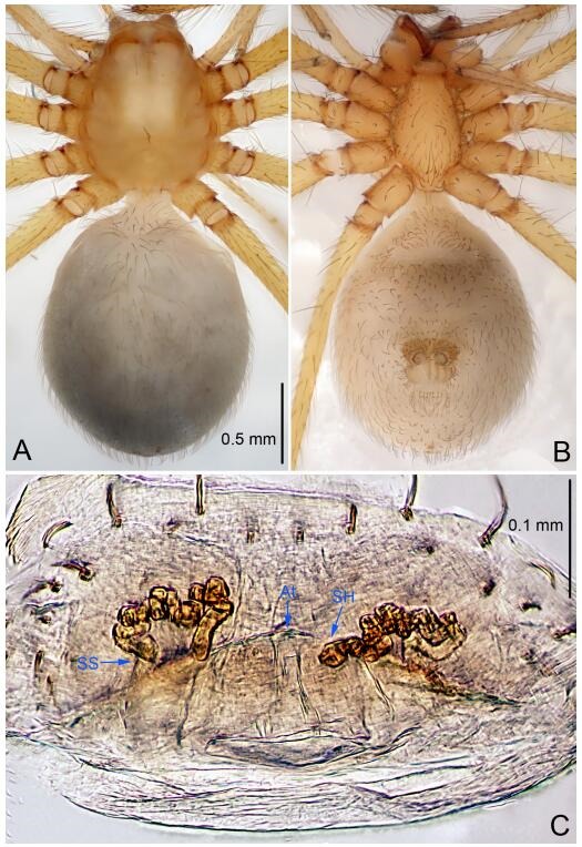
Leptonetela niubizi sp. nov., one of the paratype females
Type material. Holotype: male (IZCAS), Niubizi Cave, N27.62°, E106.67°, Leshan Town, Zunyi County, Zunyi City, Guizhou Province, China, 1 August 2012, H. Zhao leg. Paratypes: 7 females, same data as holotype.
Etymology. The specific name refers to the type locality; noun.
Diagnosis. This new species is similar to L. lianhua Wang & Li sp. nov. but can be distinguished by the male pedipalp tibia with 5 slender spines retrolaterally, spines Ⅰ longest, not bifurcated (Figure 72C), median apophysis antler-like, distal edge with 7 small teeth (Figure 72B) (tibia with 5 spines retrolateral, spines Ⅰ strongest, tip bifurcated, the other 4 spines slender, 2 of them longer than spines Ⅰ; tip of median apophysis decorated with 5 small teeth, and 1 horn-shaped large teeth in L. lianhua Wang & Li sp. nov.).
Description. Male (holotype). Total length 2.53 (Figure 72A). Carapace 0.95 long, 0.83 wide. Opisthosoma 1.58 long, 1.13 wide. Carapace yellowish. Ocular area with a pair of setae, eyes absent. Median groove needle-shaped, cervical grooves and radial furrows indistinct. Clypeus 0.15 high. Opisthosoma gray, ovoid. Leg measurements: Ⅰ 9.29 (2.56, 0.38, 2.63, 2.19, 1.53); Ⅱ 8.63 (2.50, 0.38, 2.34, 2.03, 1.38); Ⅲ 6.94 (2.03, 0.31, 1.47, 1.75, 1.38); Ⅳ -(2.55, 0.38, -, -, -). Male pedipalp (Figure 72C-D): tibia with 4 long setae prolaterally, 2 long setae and 5 slender spines retrolaterally, with spines Ⅰ longest. Cymbium constricted medially, attaching a small earlobe-shaped process retrolaterally. Embolus triangular, prolateral lobe oval. Median apophysis antler-like, distal edge decorated with 7 small teeth. Conductor short, C tile-shaped (Figure 72B).
Female (one of the paratypes). Similar to male in color and general features, but with a larger body size and shorter legs. Total length 2.60 (Figure 73A-B). Carapace 0.96 long, 0.95 wide. Opisthosoma 1.60 long, 1.25 wide. Clypeus 0.19 high. Leg measurements: Ⅰ 8.20 (2.34, 0.34, 2.44, 1.75, 1.33); Ⅱ 7.46 (2.25, 0.38, 1.88, 1.65, 1.30); Ⅲ 6.08 (2.05, 0.30, 1.08, 1.55, 1.10); Ⅳ 8.27 (2.38, 0.38, 2.25, 1.88, 1.38). Vulva (Figure 73C): spermathecae coiled, atrium triangular.
Distribution. China (Guizhou).
Leptonetela longyu Wang & Li sp. nov. Figures 74-75, 97
Figure 74.
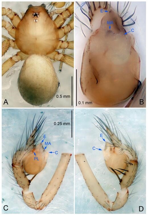
Leptonetela longyu sp. nov., holotype male
Figure 75.
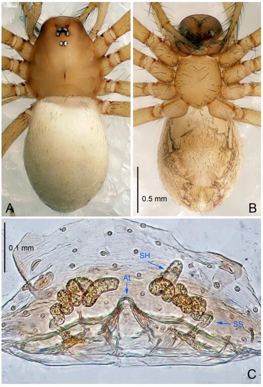
Leptonetela longyu sp. nov., one of the paratype females
Type material. Holotype: male (IZCAS), Longyu Cave, N29.40°, E110.09°, Cili County, Hunan Province, China, 5 June 2011, Z. Zha leg. Paratypes: 4 males and 5 females, same data as holotype, 5 males and 6 females, Niuerduo Cave, N29.404°, E110.73°, Cili County, Hunan Province, China, 9 April 2016, Y. Li & Z. Chen leg.
Etymology. The specific name refers to the type locality; noun.
Diagnosis. This new species is similar to L. sexdentata Wang & Li, 2011, L. shicheng Wang & Li sp. nov., L. zakou Wang & Li sp. nov. and L. meiwang Wang & Li sp. nov. but can be distinguished by median apophysis harrow-like, tip with 5 small teeth (Figure 74B) (tip of median apophysis with 6 small teeth in L. sexdentata and L. zakou Wang & Li sp. nov., 5 sharp teeth in L. meiwang Wang & Li sp. nov. and 10 in L. shicheng Wang & Li sp. nov.); from L. shicheng Wang & Li sp. nov. by the tip of conductor undulate (Figure 74B) (tip of conductor smooth in L. shicheng Wang & Li sp. nov.); from L. zakou Wang & Li sp. nov. by the teeth of median apophysis needle-shaped in L. zakou Wang & Li sp. nov.; from L. meiwang Wang & Li sp. nov. by the tibia spines Ⅰ strongest, tip asymmetrically bifurcated (tibia spines Ⅱ strongest in L. meiwang Wang & Li sp. nov.).
Description. Male (holotype). Total length 1.63 (Figure 74A). Carapace 1.05 long, 0.75 wide. Opisthosoma 0.88 long, 0.63 wide. Carapace yellow. Eyes six. Median groove needle-shaped, cervical grooves and radial furrows distinct. Clypeus 0.13 high. Opisthosoma gray, ovoid. Leg measurements: Ⅰ 6.28 (1.63, 0.25, 1.85, 1.55, 1.00); Ⅱ 4.89 (1.25, 0.25, 1.38, 1.13, 0.88); Ⅲ 4.13 (1.10, 0.23, 1.05, 1.00, 0.75); Ⅳ 5.63 (1.55, 0.25, 1.50, 1.38, 0.95). Male pedipalp (Figure 74C-D): tibia with 2 spines prolaterally and 5 spines retrolaterally, with spines Ⅰ strongest, tip asymmetrically bifurcated. Cymbium constricted medially, attaching a small earlobe-shaped process retrolaterally. Embolus triangular, prolateral lobe oval. Median apophysis short, palm-shaped, distal edge with 5 small teeth. Conductor C tile-shaped in ventral view, tip of conductor undulate (Figure 74B).
Female (one of the paratypes). Similar to male in color and general features, but larger and with longer legs. Total length 2.05 (Figure 75A-B). Carapace 0.90 long, 0.75 wide. Opisthosoma 1.13 long, 0.88 wide. Clypeus 0.12 high. Leg measurements: Ⅰ 6.28 (1.75, 0.25, 1.88, 1.40, 1.00); Ⅱ 4.94 (1.30, 0.25, 1.38, 1.13, 0.88); Ⅲ 4.41 (1.25, 0.23, 1.13, 1.05, 0.75); Ⅳ 5.58 (1.60, 0.25, 1.50, 1.35, 0.88). Vulva (Figure 75C): spermathecae coiled, atrium triangular.
Distribution. China (Hunan).
Leptonetela shicheng Wang & Li sp. nov. Figures 76-77, 97
Figure 76.
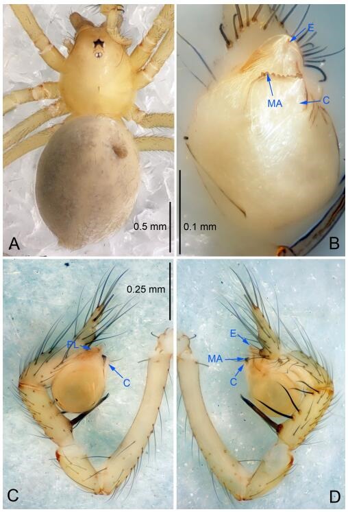
Leptonetela shicheng sp. nov., holotype male
Figure 77.
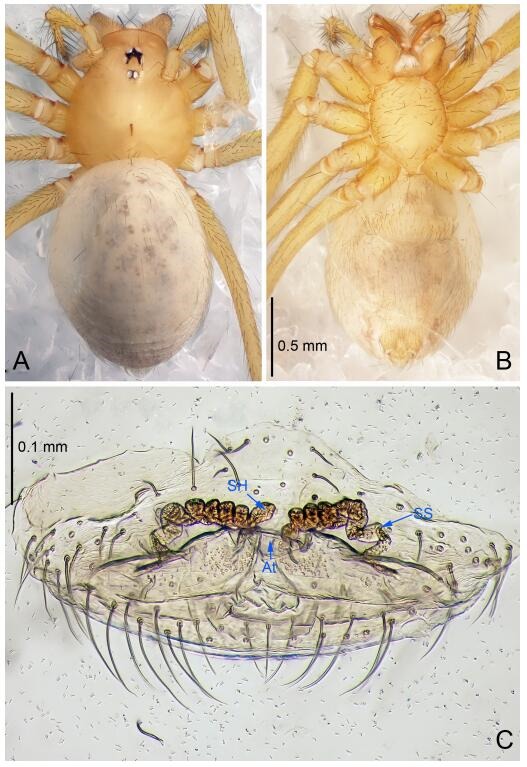
Leptonetela shicheng sp. nov., one of the paratype females
Type material. Holotype: male (IZCAS), Shicheng Cave, N27.31°, E109.07°, Jiangwu, Shanshi Town, Lianhua County, Pingxiang City, Jiangxi Province, China, 14 November 2015, Z. Chen & G. Zhou leg. Paratypes: 2 males and 5 females, same data as holotype.
Etymology. The specific name refers to the type locality; noun.
Diagnosis. This new species is similar to L. sexdentata Wang & Li, 2011, L. longyu Wang & Li sp. nov., L. zakou Wang & Li sp. nov. and L. meiwang Wang & Li sp. nov. but can be distinguished by the harrow-like median apophysis, with 10 small teeth distally (Figure 76B) (median apophysis with 6 small teeth distally in L. sexdentata and L. zakou Wang & Li sp. nov., 5 in L. longyu Wang & Li sp. nov., and L. meiwang Wang & Li sp. nov.); conductor smooth (Figure 76B) (conductor undulate distally in L. sexdentata, L. longyu Wang & Li sp. nov., and L. zakou Wang & Li sp. nov.); from L. zakou Wang & Li sp. nov. by the teeth of median apophysis needle-shaped in L. zakou Wang & Li sp. nov.; from L. meiwang Wang & Li sp. nov. by the tibia spines Ⅰ strongest, tip asymmetrically bifurcated (Figure 76D) (tibia spines Ⅱ strongest in L. meiwang Wang & Li sp. nov.).
Description. Male (holotype). Total length 2.40 (Figure 76A). Carapace 1.00 long, 0.73 wide. Opisthosoma 1.12 long, 0.87 wide. Carapace yellowish. Ocular area with a pair of setae, six eyes. Median groove needle-shaped, cervical grooves and radial furrows indistinct. Clypeus 0.08 high. Opisthosoma gray, ovoid. Leg measurements: Ⅰ 9.60 (2.60, 0.37, 2.50, 2.48, 1.65); Ⅱ 7.90 (2.25, 0.30, 2.25, 2.00, 1.40); Ⅲ 6.87 (1.75, 0.25, 1.87, 1.75, 1.25); Ⅳ 8.92 (2.37, 0.30, 2.50, 2.25, 1.50). Male pedipalp (Figure 76C-D): tibia with 2 long setae prolaterally, and 5 spines retrolaterally, with spines Ⅰ strongest, tip asymmetrically bifurcated. Cymbium constricted medially, attaching a small earlobe-shaped process retrolaterally. Embolus triangular, prolateral lobe indistinct. Median apophysis harrow-like, with 10 small teeth distally. Conductor smooth, C tile-shape in ventral view (Figure 76B).
Female (one of the paratypes). Similar to male in color and general features, but larger and with shorter legs. Total length 2.60 (Figure 77A-B). Carapace 0.87 long, 0.85 wide. Opisthosoma 1.75 long, 1.25 wide. Clypeus 0.15 high. Leg measurements: Ⅰ 8.94 (2.60, 0.37, 2.50, 2.10, 1.37); Ⅱ 7.10 (2.00, 0.30, 2.00, 1.65, 1.15); Ⅲ 5.97 (1.75, 0.25, 1.60, 1.50, 0.87); Ⅳ 7.97 (2.12, 0.35, 2.25, 2.00, 1.25). Vulva (Figure 77C): spermathecae coiled, atrium fusiform.
Distribution. China (Jiangxi).
Leptonetela zakou Wang & Li sp. nov. Figures 78-79, 97
Figure 78.
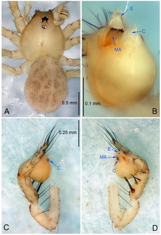
Leptonetela zakou sp. nov., holotype male
Figure 79.
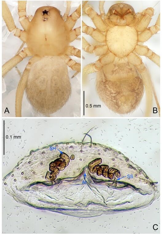
Leptonetela zakou sp. nov., one of the paratype females
Type material. Holotype: male (IZCAS), Zakou Cave, N29.35°, E109.58°, Hongyanxi Town, longshan City, Hunan Province, China, 10 January 2016, Z. Chen & Z. Wang leg. Paratypes: 3 males and 5 females, same data as holotype.
Etymology. The specific name refers to the type locality; noun.
Diagnosis. This new species is similar to L. sexdentata Wang & Li, 2011, L. longyu Wang & Li sp. nov., L. shicheng Wang & Li sp. nov., and L. meiwang Wang & Li sp. nov. but can be distinguished by on the male pedipalpal bulb median apophysis with 6 teeth, needle-shaped (Figure 78B) (median apophysis with 5 small teeth distally in L. longyu Wang & Li sp. nov., 5 sharp teeth in L. meiwang Wang & Li sp. nov., and 10 in L. shicheng Wang & Li sp. nov., ); from L. shicheng Wang & Li sp. nov. by the distally undulate conductor (Figure 78B) (conductor smooth in L. shicheng Wang & Li sp. nov.); from L. meiwang Wang & Li sp. nov. by the tibia Ⅰ spine strongest, tip asymmetrically bifurcated (Figure 78D) (tibia Ⅱ spine strongest in L. meiwang Wang & Li sp. nov.).
Description. Male (holotype). Total length 1.75 (Figure 78A). Carapace 0.87 long, 0.87 wide. Opisthosoma 1.00 long, 0.87 wide. Carapace yellowish. Ocular area with a pair of setae, six eyes. Median groove needle-shaped, cervical grooves and radial furrows indistinct. Clypeus 0.08 high. Opisthosoma gray, ovoid. Leg measurements: Ⅰ 7.62 (2.00, 0.25, 2.37, 1.75, 1.25); Ⅱ 5.62 (1.50, 0.25, 1.62, 1.25, 1.00); Ⅲ 4.57 (1.25, 0.20, 1.30, 1.12, 0.70); Ⅳ 6.47 (1.87, 0.25, 1.75, 1.50, 1.10). Male pedipalp (Figure 78C-D): tibia with 3 long setae prolaterally, 5 large spines retrolaterally, with spines Ⅰ strongest, tip asymmetrically bifurcated. Cymbium not constricted, earlobe-shaped process absent. Embolus triangular, prolateral lobe absent. Median apophysis with 6 needle-shaped teeth distally. Conductor C tile-shape in ventral view (Figure 78B).
Female (one of the paratypes). Similar to male in color and general features, but larger and with shorter legs. Total length 1.70 (Figure 79A-B). Carapace 0.87 long, 0.80 wide. Opisthosoma 1.27 long, 0.75 wide. Clypeus 0.15 high. Leg measurements: Ⅰ 6.49 (1.75, 0.25, 1.72, 1.50, 1.27); Ⅱ 4.69 (1.37, 0.25, 1.27, 1.00, 0.80); Ⅲ 3.74 (1.12, 0.20, 1.00, 0.80, 0.62); Ⅳ 5.30 (1.50, 0.25, 1.50, 1.30, 0.75). Vulva (Figure 79C): spermathecae coiled, atrium fusiform, anterior margin of atrium with one mastoid process medially.
Distribution. China (Guizhou).
Leptonetela meiwang Wang & Li sp. nov. Figures 80-81, 97
Figure 80.
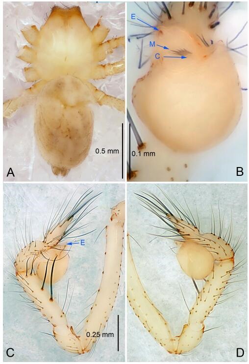
Leptonetela meiwang sp. nov., holotype male
Figure 81.
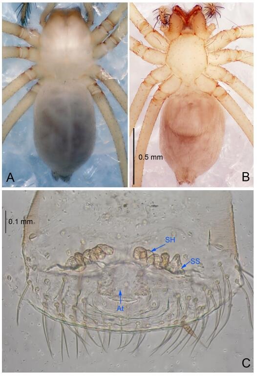
Leptonetela meiwang sp. nov., paratype female
Type material. Holotype: male (IZCAS), Meiwang Cave, N28.09°, E111.43°, Nanhua, Zhenshang Town, Lodi County, HuNan Province, China, 27 March 2016, Y. Li & Z. Chen leg. Paratypes: 1 male and 1 female, same data as holotype.
Etymology. The specific name refers to the type locality; noun.
Diagnosis. This new species is similar to L. sexdentata Wang & Li, 2011, L. longyu Wang & Li sp. nov., L. shicheng Wang & Li sp. nov. and L. zakou Wang & Li sp. nov. but can be distinguished by the harrow-like median apophysis, with 5 sharp teeth distally (Figure 80B), tibia Ⅱ spine strongest (Figure 80C) (tibia spines Ⅰ strongest, tip asymmetrically bifurcated, median apophysis with 6 small teeth distally in L. sexdentata and L. zakou Wang & Li sp. nov., 5 in L. longyu Wang & Li sp. nov., and 10 in L. shicheng Wang & Li sp. nov., ); conductor short, reduced (Figure 80B) (conductor broad, C shape in L. sexdentata, L. longyu Wang & Li sp. nov., L. shicheng Wang & Li sp. nov. and L. zakou Wang & Li sp. nov.); from L. zakou Wang & Li sp. nov. by the teeth of median apophysis needle-shaped in L. zakou Wang & Li sp. nov.
Description. Male (holotype): total length 1.75 (Figure 80A). Prosoma 0.70 long, 0.62 wide. Opisthosoma 1.20 long, 0.70 wide. Carapace yellowish. Ocular area with a pair of setae, eyes absent. Median groove needle shaped, cervical grooves and radial furrows indistinct. Clypeus 0.08 high. Opisthosoma gray, ovoid. Leg measurements: Ⅰ 9.8 (2.50, 0.37, 2.81, 2.50, 1.62); Ⅱ 8.44 (2.30, 0.35, 2.30, 2.12, 1.37); Ⅲ 7.77 (2.25, 0.30, 2.12, 2.10, 1.00); Ⅳ 9.61 (2.50, 0.37, 2.50, 2.37, 1.87). Male pedipalp (Figure 80C-D): tibia with 5 spines retrolaterally, with spines Ⅱ strongest. Cymbium constricted at middle, earlobe-shaped process absent. Embolus triangular, prolateral lobe indistinct. Median apophysis with 5 sharp teeth distally. Conductor short, reduced (Figure 80B).
Female. Similar to male in color and general features, but larger and with shorter legs. Total length 1.75 (Figure 81A-B). Prosoma 0.75 long, 0.62 wide. Opisthosoma 1.00 long, 0.75 wide. Clypeus 0.20 high. Leg measurements: Ⅰ 8.13 (2.37, 0.34, 2.30, 1.75, 1.37); Ⅱ 7.54 (2.25, 0.34, 2.00, 1.70, 1.25); Ⅲ 6.62 (1.87, 0.30, 1.75, 1.50, 1.20); Ⅳ -(2.30, 0.34, -, -, -). Vulva (Figure 81C): spermathecae coiled, atrium triangular.
Distribution. China (Hunan).
Leptonetela tawo Wang & Li sp. nov. Figure 82-83, 97
Figure 82.
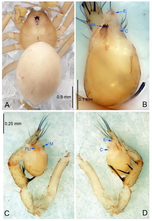
Leptonetela tawo sp. nov., holotype male
Figure 83.
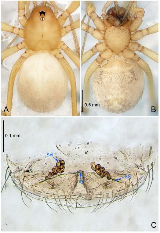
Leptonetela tawo sp. nov., one of the paratype females
Type material. Holotype: male (IZCAS), Xianren Cave, N29.18°, E109.95°, Xianren, Tawo Town, Yongshun County, Hunan Province, China, 14 January 2016, Z. Chen & Z. Wang leg. Paratypes: 2 males and 2 females, same data as holotype.
Etymology. The specific name refers to the type locality; noun.
Diagnosis. This new species is similar to L. arvanitidisi Wang & Li, 2016, L. paragamiani Wang & Li, 2016 and L. erlong Wang & Li sp. nov. but can be distinguished by on the male pedipalpal bulb median apophysis with 4 teeth distally (Figure 82B) (median apophysis with 6 teeth distally in L. arvanitidisi, 3 teeth in L. paragamiani and 5 teeth in L. erlong Wang & Li sp. nov.); tibia spines Ⅰ strongest, tip asymmetrically bifurcated, spines Ⅱ, Ⅲ equally strong, stronger than other 2 (Figure 82C) (tibia Ⅱ -Ⅴ spines slender, curved, and equally strong in L. arvanitidisi and L. erlong Wang & Li sp. nov., tibia Ⅲ -Ⅴ spines equally strong, slender than Ⅱ spine in L. paragamiani); from L. arvanitidisi by the conductor C tile-shaped (Figure 82B) (conductor triangular in L. arvanitidisi).
Description. Male (holotype). Total length 1.90 (Figure 82A). Carapace 0.87 long, 0.75 wide. Opisthosoma 1.25 long, 0.87 wide. Carapace yellowish. Ocular area with a pair of setae, six eyes. Median groove needle-shaped, cervical grooves and radial furrows indistinct. Clypeus 0.08 high. Opisthosoma gray, ovoid. Leg measurements: Ⅰ 7.69 (2.00, 0.35, 2.37, 1.72, 1.25); Ⅱ 5.95 (1.75, 0.30, 1.55, 1.35, 1.00); Ⅲ 4.60 (1.25, 0.25, 1.15, 1.10, 0.85); Ⅳ 7.15 (1.85, 0.30, 2.25, 1.60, 1.15). Male pedipalp (Figure 82C-D): tibia with 4 long setae prolaterally, 5 large spines retrolaterally, with spines Ⅰ strongest, tip asymmetrically bifurcated, tibia spines Ⅱ, Ⅲ equally strong, stronger than other 2. Cymbium not constricted, earlobe-shaped process absent. Embolus triangular, prolateral lobe indistinct. Median apophysis with 4 teeth distally. Conductor C tile-shaped in ventral view (Figure 82B).
Female (one of the paratypes). Similar to male in color and general features, but larger and with shorter legs. Total length 2.00 (Figure 83A-B). Carapace 0.87 long, 0.75 wide. Opisthosoma 1.12 long, 1.00 wide. Clypeus 0.15 high. Leg measurements: Ⅰ 5.84 (1.62, 0.35, 1.62, 1.25, 1.00); Ⅱ 4.55 (1.25, 0.35, 1.20, 1.00, 0.75); Ⅲ 3.62 (1.00, 0.30, 0.87, 0.85, 0.60); Ⅳ 5.42 (1.75, 0.35, 1.37, 1.10, 0.85). Vulva (Figure 83C): spermathecae coiled, atrium triangular.
Distribution. China (Guizhou).
Leptonetela erlong Wang & Li sp. nov. Figures 84-85, 97
Figure 84.
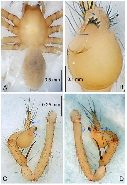
Leptonetela erlong sp. nov., holotype male
Figure 85.
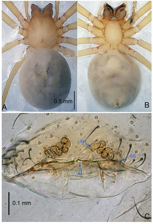
Leptonetela erlong sp. nov., one of the paratype females
Type material. Holotype: male (IZCAS), Erlong Cave, N27.82°, E110.23°, Siqian Town, Chenxi County, Huaihua City, Hunan Province, China, 19 March 2016, Y. Li & Z. Chen leg. Paratypes: 4 males and 2 females, same data as holotype.
Etymology. The specific name refers to the type locality; noun.
Diagnosis. This new species is similar to L. arvanitidisi Wang & Li, 2016, L. paragamiani Wang & Li, 2016 and L. tawo Wang & Li sp. nov. but can be distinguished by on the male pedipalpal bulb median apophysis with 5 teeth distally (Figure 84B) (median apophysis with 6 teeth in L. arvanitidisi, 4 teeth in L. tawo Wang & Li sp. nov. and 3 teeth L. paragamiani); from L. paragamiani and L. tawo Wang & Li sp. nov. by the tibia spines Ⅱ -Ⅴ slender, curved, and equally strong (Figure 84D) (tibia spines Ⅱ, Ⅲ equally strong, stronger than other 2 in L. tawo Wang & Li sp. nov., spines Ⅲ -Ⅴ equally strong, more slender than spines Ⅱ in L. paragamiani); from L. arvanitidisi by the conductor C tile-shaped (Figure 84B) (conductor triangular in L. arvanitidisi).
Description. Male (holotype): total length 1.95 (Figure 84A). Prosoma 0.50 long, 0.80 wide. Opisthosoma 1.45 long, 1.00 wide. Prosoma yellowish. Eyes absent. Median groove needle-shaped, brown. Cervical grooves and radial furrows indistinct. Clypeus 0.13 high, slightly sloped anteriorly. Opisthosoma pale brown, ovoid, covered with short hairs, lacking distinct pattern. Sternum and legs yellowish. Leg measurements: Ⅰ 6.81 (2.35, 0.35, 1.87, 1.37, 0.87); Ⅱ 5.82 (2.25, 0.35, 1.35, 1.07, 0.80); Ⅲ 5.22 (2.20, 0.30, 1.00, 0.97, 0.75); Ⅳ 6.30 (2.30, 0.35, 1.50, 1.30, 0.85). Male pedipalp (Figure 84C-D): tibia with 5 spines retrolaterally, with spines Ⅰ strongest, tip asymmetrically bifurcated, tibia spines Ⅱ -Ⅴ slender, curved, and equally strong. Cymbium constricted medially, earlobe-shaped process small. Embolus triangular, prolateral lobe indistinct. Median apophysis with 5 teeth distally. Conductor C tile-shaped in ventral view (Figure 84B).
Female (one of the paratypes). Similar to male in color and general features, but larger and with shorter legs. Total length 2.30 (Figure 85A-B). Prosoma 0.85 long, 0.95 wide. Opisthosoma 0.87 long, 0.70 wide. Clypeus 0.20 high. Leg measurements: Ⅰ 8.60 (2.50, 0.35, 2.20, 1.80, 1.75); Ⅱ 7.65 (2.35, 0.30, 1.95, 1.70, 1.35); Ⅲ 6.30 (2.15, 0.25, 2.05, 1.15, 0.70); Ⅳ 7.85 (2.25, 0.30, 2.05, 1.75, 1.50). Vulva (Figure 85C): spermathecae coiled, atrium fusiform.
Distribution. China (Hunan).
Leptonetela dabian Wang & Li sp. nov. Figures 86-87, 97
Figure 86.
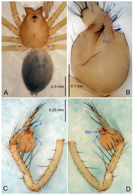
Leptonetela dabian sp. nov., holotype male
Figure 87.
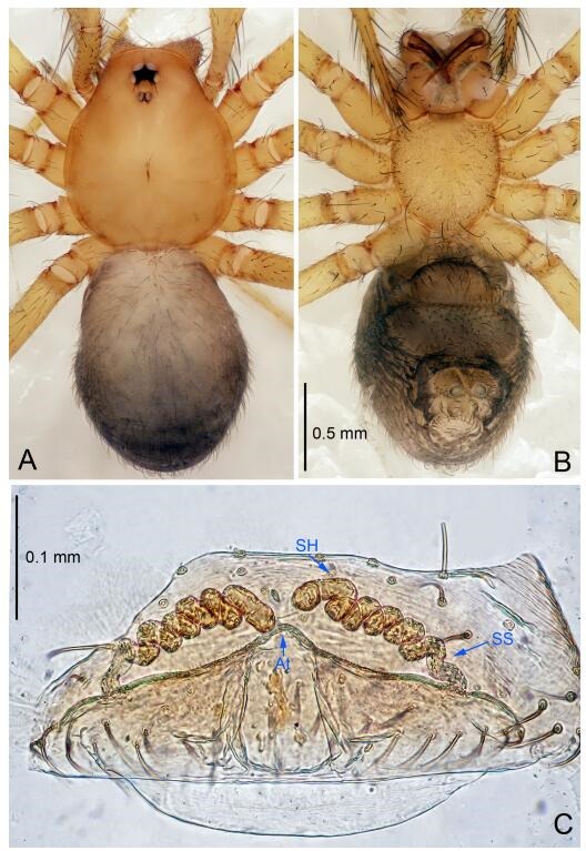
Leptonetela dabian sp. nov., one of the paratype females
Type material. Holotype: male (IZCAS), Wuming Cave, N25.75°, E107.92°, Dabian, Sandong Town, Sandu County, Qiannan Prefecture, Guizhou, China, 22 March 2013, H. Zhao & J. Liu leg. Paratypes: 2 females, same data as holotype.
Etymology. The specific name refers to the type locality; noun.
Diagnosis. This new species is similar to L. thracia Wang & Li, 2011 and L. chuan Wang & Li sp. nov., but can be distinguished by the male pedipalal tibia with 3 spines prolaterally, 5 slender spines, retrolaterally (Figure 86C-D) (tibia with 4 long setae prolaterally, 5 large spines retrolaterally, spines Ⅰ, Ⅱ equally strong, stronger than others in L. thracia; tibia with 7 long setae prolaterally, 5 large spines retrolaterally, spines Ⅰ, Ⅱ, Ⅲ equally strong, stronger than others in L. chuan Wang & Li sp. nov.); tip of median apophysis bent upwards, with 3 larger teeth distally (Figure 86B) (tip of median apophysis bent downwards, with 5 larger teeth distally in L. chuan Wang & Li sp. nov.; tip of median apophysis not bent, with 4 teeth distally in L. thracia); conductor thin, tongue shaped (Figure 86B) (conductor triangular in L. thracia and L. chuan Wang & Li sp. nov.).
Description. Male (holotype). Total length 2.38 (Figure 86A). Carapace 1.00 long, 0.80 wide. Opisthosoma 1.25 long, 0.90 wide. Carapace yellow. Six eyes. Median groove needle-shaped, cervical grooves and radial furrows indistinct. Clypeus 0.13 high. Opisthosoma gray, ovoid, with pigmented spots. Leg measurements: Ⅰ -(2.60, 0.38, 2.35, -, -); Ⅱ 7.78 (2.15, 0.38, 2.25, 1.75, 1.25); Ⅲ -(1.88, 0.35, 1.75, -, -); Ⅳ 8.26 (2.25, 0.38, 2.38, 2.00, 1.25). Male pedipalp (Figure 86C-D): tibia with 3 slender spines prolaterally, 5 large retrolateral spines equally strong. Cymbium constricted medially, attaching a small earlobe-shaped process retrolaterally. Embolus triangular, prolateral lobe oval. Tip of median apophysis bent upward, distal edge decorated with three small teeth. Conductor thin, tongue-shaped (Figure 86B).
Female (one of the paratypes). Similar to male in color and general features, but larger and with shorter legs. Total length 2.40 (Figure 87A-B). Carapace 1.00 long, 0.88 wide. Opisthosoma 1.25 long, 1.00 wide. Clypeus 0.13 high. Leg measurements: Ⅰ 9.51 (2.50, 0.38, 3.00, 2.13, 1.50); Ⅱ 7.38 (2.00, 0.38, 2.13, 1.62, 1.25); Ⅲ 6.35 (1.75, 0.35, 1.75, 1.50, 1.00); Ⅳ 8.06 (2.25, 0.38, 2.30, 1.88, 1.25). Vulva (Figure 87C): spermathecae coiled, atrium triangular, and anterior margin of atrium with short hairs.
Distribution. China (Guizhou).
Leptonetela chuan Wang & Li sp. nov. Figures 88-89, 97
Figure 88.
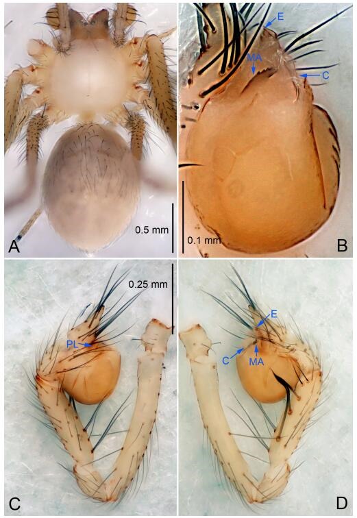
Leptonetela chuan sp. nov., holotype male
Figure 89.
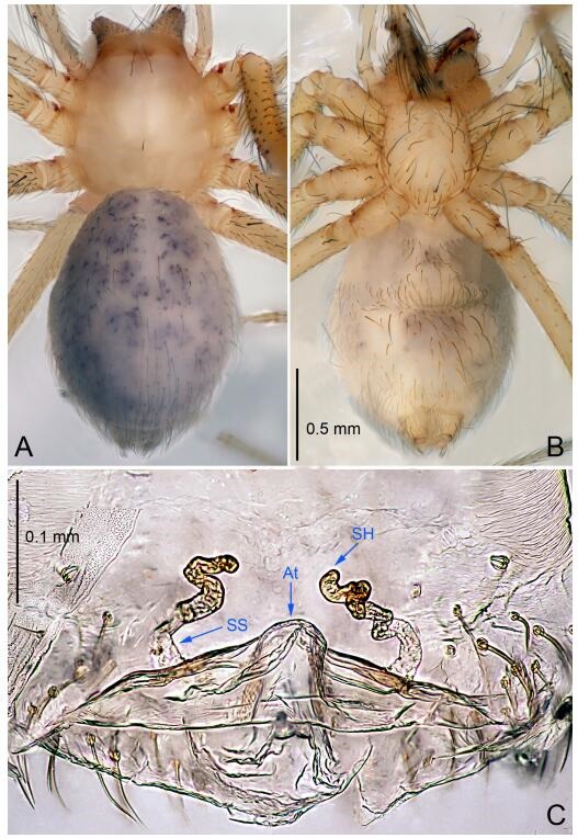
Leptonetela chuan sp. nov., one of the paratype females
Type material. Holotype: male (IZCAS), Chuan Cave, N27.08°, E105.67°, Yangchangba Town, Dafang County, Guizhou Province, China, 13 March 2011, H. Chen & Z. Zha leg. Paratype: 1 female, same data as holotype.
Etymology. The specific name refers to the type locality; noun.
Diagnosis. This new species is similar to L. thracia Wang & Li, 2011 and L. dabian Wang & Li sp. nov., but can be distinguished by the male pedipalpal tibia with 7 long setae prolaterally, 5 slender spines retrolaterally, with spines Ⅰ, Ⅱ, Ⅲ equally strong, stronger than others (Figure 88D) (tibia with 4 long setae prolaterally, 5 slender spines retrolaterally, with spines Ⅰ, Ⅱ equally strong, stronger than others in L. thracia; 3 slender spines prolaterally, 5 slender retrolaterally spines equally strong in L. dabian Wang & Li sp. nov.); tip of median apophysis bent downwards, with 5 larger teeth distally (Figure 88B) (tip of median apophysis not bent, with 4 teeth distally in L. thracia; tip of median apophysis bent upwards, with 3 larger teeth distally in L. dabian Wang & Li sp. nov.); from L. dabian Wang & Li sp. nov. by the triangular conductor (Figure 88B) (conductor thin, tongue-shaped in L. dabian Wang & Li sp. nov.).
Description. Male (holotype). Total length 2.10 (Figure 88A). Carapace 0.83 long, 0.90 wide. Opisthosoma 1.18 long, 1.05 wide. Carapace whitish. Ocular area with a pair of setae, eyes absent. Median groove, cervical grooves and radial furrows indistinct. Clypeus 0.13 high. Opisthosoma gray, ovoid. Leg measurements: Ⅰ 8.79 (2.38, 0.38, 2.50, 2.13, 1.40); Ⅱ 7.77 (2.13, 0.38, 2.18, 1.78, 1.30); Ⅲ 7.51 (1.78, 0.35, 2.10, 1.73, 1.55); Ⅳ 8.36 (2.25, 0.38, 2.38, 2.00, 1.35). Male pedipalp (Figure 88C-D): tibia with 7 long setae prolaterally, 5 large spines retrolaterally, with spines Ⅰ, Ⅱ, Ⅲ equally strong, stronger than others. Cymbium constricted medially, attached to a small earlobe-shaped process retrolaterally. Embolus triangular, prolateral lobe oval. Median apophysis bent downwards, with 5 larger teeth distally. Conductor triangular in ventral view (Figure 88B).
Female: Similar to male in color and general features, but smaller and with shorter legs. Total length 2.08 (Figure 89A-B). Carapace 0.78 long, 0.88 wide. Opisthosoma 1.33 long, 1.03 wide. Clypeus 0.15 high. Leg measurements: Ⅰ 8.31 (2.33, 0.38, 2.40, 1.85, 1.35); Ⅱ 7.21 (2.10, 0.35, 2.08, 1.58, 1.10); Ⅲ 6.92 (1.93, 0.35, 1.88, 1.63, 1.13); Ⅳ 8.14 (2.30, 0.38, 2.38, 1.83, 1.25). Vulva (Figure 89C): spermathecae loosely coiled, atrium triangular.
Distribution. China (Guizhou).
Leptonetela lihu Wang & Li sp. nov. Figures 90-91, 97
Figure 90.
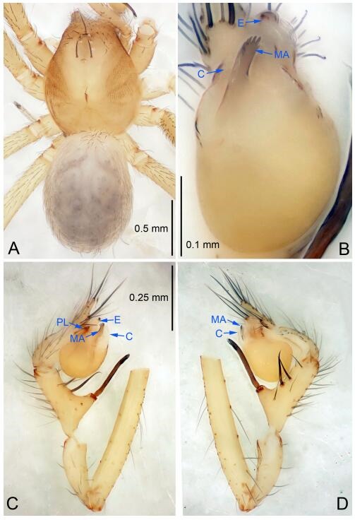
Leptonetela lihu sp. nov., holotype male
Figure 91.
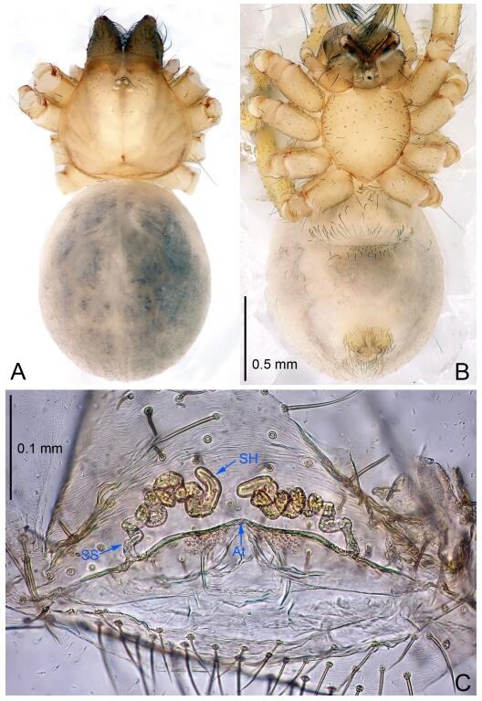
Leptonetela lihu sp. nov., one of the paratype females
Type material. Holotype: male (IZCAS), nameless Cave, N25.10°, E107.65°, Lihu Town, Nandan County, Hechi City, Guangxi Zhuang Autonomous Region, China, 31 January 2015, Y. Li & Z. Chen leg. Paratypes: 2 males and 5 females, same data as holotype.
Etymology. The specific name refers to the type locality; noun.
Diagnosis. This new species is similar to L. notabilis (Lin & Li, 2010), L. sexdigiti (Lin & Li, 2010); and L. shuang Wang & Li sp. nov., but can be separated from L. notabilis by the male pedipalpal tibia spines Ⅰ bifurcate (Figure 90D) (tibia spines Ⅰ trifurcate in L. notabilis); from L. shuang Wang & Li sp. nov. by the conductor C tile-shaped, distal edge of median apophysis with 6 teeth (Figure 90B) (conductor triangular, distal edge of median apophysis with 7 teeth in L. shuang Wang & Li sp. nov.); from L. sexdigiti by the strongly twisted spermathecae (spermathecae loosely twisted in L. sexdigiti).
Description. Male (holotype). Total length 2.13 (Figure 90A). Carapace 1.00 long, 0.88 wide. Opisthosoma 1.12 long, 0.75 wide. Carapace yellowish. Ocular area with 3 long setae, six eyes, reduced to white spots. Median groove needle-shaped, cervical grooves and radial furrows distinct. Clypeus 0.13 high. Opisthosoma gray, ovoid. Leg measurements: Ⅰ 8.23 (2.25, 0.25, 2.38, 1.95, 1.40); Ⅱ 7.00 (2.00, 0.25, 2.00, 1.63, 1.12); Ⅲ 6.10 (1.75, 0.20, 1.75, 1.45, 0.95); Ⅳ 7.75 (2.10, 0.25, 2.25, 1.90, 1.25). Male pedipalp (Figure 90C-D): basal part of tibia swollen, tibia with 5 spines retrolaterally, with spines Ⅰ strongest, longest, bifurcate and located at the base of tibia. Cymbium constricted medially, attached to a small earlobe-shaped process retrolaterally. Embolus triangular, prolateral lobe oval. Median apophysis long and thin, with 6 small teeth distally. Conductor broad, C tile-shaped in ventral view (Figure 90B).
Female (one of the paratypes). Similar to male in color and general features, but larger and with shorter legs. Total length 2.50 (Figure 91A-B). Carapace 1.25 long, 0.75 wide. Opisthosoma 1.40 long, 1.00 wide. Clypeus 0.12 high. Leg measurements: Ⅰ 8.00 (2.25, 0.25, 2.38, 1.75, 1.37); Ⅱ 7.00 (2.00, 0.25, 2.00, 1.50, 1.25); Ⅲ 5.65 (1.75, 0.20, 1.65, 1.15, 0.90); Ⅳ 7.70 (2.10, 0.25, 2.25, 1.90, 1.20). Vulva (Figure 91C): spermathecae coiled, atrium fusiform.
Distribution. China (Guangxi).
Leptonetela shuang Wang & Li sp. nov. Figures 92-93, 97
Figure 92.
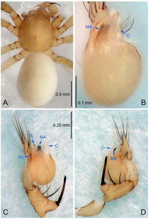
Leptonetela shuang sp. nov., holotype male
Figure 93.
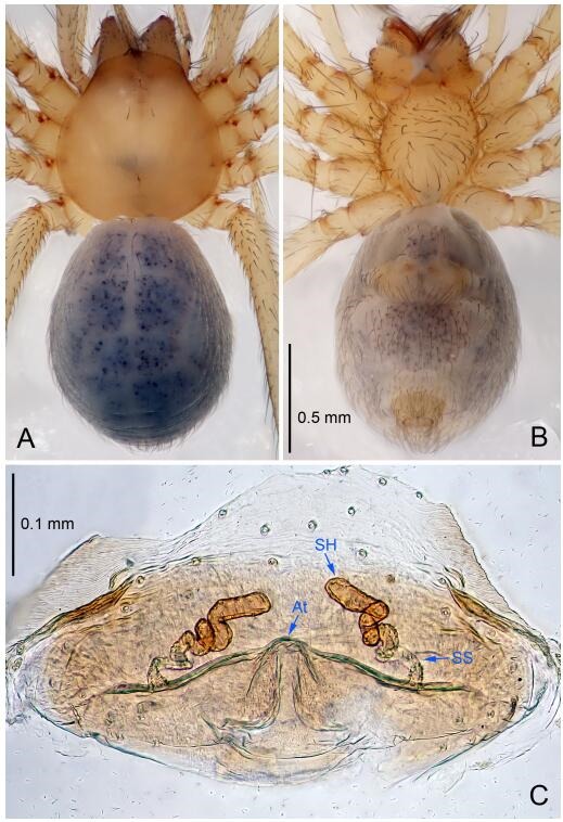
Leptonetela shuang sp. nov., one of the paratype females
Type material. Holotype: male (IZCAS), Shuang Cave, N25.93°, E107.26°, Bailong Town, Pingtang County, Qiannan Prefecture, Guizhou Province, China, 24 July 2012, H. Zhao leg. Paratypes: 2 females, same data as holotype; 2 males and 6 females, Dongkou Cave, N25.93°, E107.25°, Longxiang, Bailong Town, Pingtang County, Qiannan Prefecture, Guizhou Province, China, 25 July 2012, H. Zhao leg.
Etymology. The specific name refers to the type locality; noun.
Diagnosis. This new species is similar to L. notabilis (Lin & Li, 2010), L. sexdigiti (Lin & Li, 2010), and L. lihu Wang & Li sp. nov., but can be separated from L. notabilis by the male pedipalp tibia spines Ⅰ bifurcate (Figure 92D) (tibia spines Ⅰ trifurcate in L. notabilis); from L. sexdigiti and L. lihu Wang & Li sp. nov. by the conductor triangular, distal edge of median apophysis with 7 teeth (Figure 92B) (conductor C tile-shaped, distal edge of median apophysis with 6 teeth in L. sexdigiti and L. lihu Wang & Li sp. nov.); from L. sexdigiti by in the female spermathecae strongly twisted (Figure 93C) (spermathecae loosely twisted in L. sexdigiti).
Description. Male (holotype). Total length 2.00 (Figure 92A). Carapace 0.83 long, 0.75 wide. Opisthosoma 1.25 long, 0.80 wide. Carapace yellow. Ocular area with a pair of setae, eyes absent. Median groove needle-shaped, cervical grooves and radial furrows indistinct. Clypeus 0.13 high. Opisthosoma whitish, ovoid. Leg measurements: Ⅰ 7.74 (2.03, 0.38, 2.25, 1.80, 1.28); Ⅱ 6.65 (1.77, 0.35, 1.83, 1.50, 1.20); Ⅲ 5.68 (1.57, 0.35, 1.50, 1.28, 0.98); Ⅳ 7.38 (1.92, 0.38, 2.05, 1.78, 1.25). Male pedipalp (Figure 92C-D): basal of tibia swollen, tibia with 3 long setae prolaterally, 1 long setae and 5 spines retrolaterally, with spines Ⅰ strongest, longest, bifurcate and located at the base of the tibia. Cymbium constricted medially, attached to a small earlobe-shaped process retrolaterally. Embolus triangular, prolateral lobe oval. Median apophysis long and thin, with 7 small teeth distally. Conductor triangular (Figure 92B).
Female (one of the paratypes). Similar to male in color and general features, but smaller and with shorter legs. Total length 1.98 (Figure 93A-B). Carapace 0.88 long, 0.75 wide. Opisthosoma 1.13 long, 0.88 wide. Clypeus 0.13 high. Leg measurements: Ⅰ 7.34 (1.93, 0.38, 2.13, 1.65, 1.25); Ⅱ 6.41 (1.75, 0.35, 1.78, 1.50, 1.03); Ⅲ 5.50 (1.52, 0.35, 1.45, 1.30, 0.88); Ⅳ 7.03 (1.87, 0.38, 2.05, 1.63, 1.10). Vulva (Figure 93C): spermathecae coiled, atrium triangular.
Distribution. China (Guizhou).
Leptonetela encun Wang & Li sp. nov. Figures 94-95, 97
Figure 94.
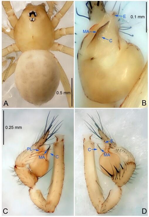
Leptonetela encun sp. nov., holotype male
Figure 95.
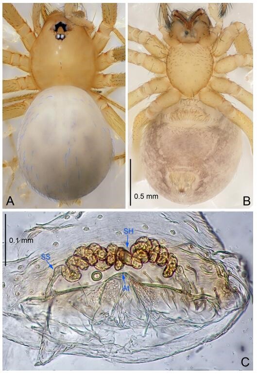
Leptonetela encun sp. nov., paratype female
Type material. Holotype: male (IZCAS), Encun Cave, N25.08°, E107.59°, En, Chengguan Town, Nandan County, Hechi City, Guangxi Zhuang Autonomous Region, China, 30 January 2015, Y. Li & Z. Chen leg. Paratypes: 1 male and 1 female, same data as holotype.
Etymology. The specific name refers to the type locality; noun.
Diagnosis. This new species is similar to L. robustispina (Chen et al, 2010) but can be distinguished by the male pedipalpal tibia with 5 spines retrolaterally, with spines Ⅰ longest, spines Ⅰ, Ⅱ, Ⅲ equally strong, stronger than others (Figure 94D), distal edge of median apophysis linear, with 8 teeth (Figure 94B) (tibia with 5 spines retrolaterally, with spines Ⅰ longest, distal edge of median apophysis semicircular, with 12 teeth in L. robustispina).
Description. Male (holotype). Total length 2.00 (Figure 94A). Carapace 0.90 long, 0.75 wide. Opisthosoma 1.25 long, 0.88 wide. Carapace yellowish, with one seta on the median part. Six eyes. Median groove needle-shaped, cervical grooves and radial furrows indistinct. Clypeus 0.10 high. Opisthosoma gray, ovoid. Leg measurements: Ⅰ -(1.88, -, -, -, -); Ⅱ 6.25 (1.75, 0.25, 1.87, 1.38, 1.00); Ⅲ 4.96 (1.38, 0.20, 1.38, 1.20, 0.80); Ⅳ 6.86 (2.00, 0.25, 1.88, 1.63, 1.10). Male pedipalp (Figure 94C-D): tibia with a few clusters of short spines dorsally, 8 long setae retrolaterally, and 5 spines retrolaterally, spines Ⅰ longest. Cymbium constricted medially, attached to a small earlobe-shaped process retrolaterally. Embolus triangular, prolateral lobe indistinct. Median apophysis harrow-like, distal edge round, with 8 small teeth. Conductor triangular in ventral view (Figure 94B).
Female: Similar to male in color and general features, but larger and with shorter legs. Total length 2.13 (Figure 95A-B). Carapace 0.88 long, 0.75 wide. Opisthosoma 1.00 long, 1.05 wide. Clypeus 0.11 high. Leg measurements: Ⅰ 6.50 (1.75, 0.25, 2.00, 1.50, 1.00); Ⅱ 5.01 (1.38, 0.25, 1.50, 1.13, 0.75); Ⅲ 4.45 (1.25, 0.20, 1.25, 1.00, 0.75); Ⅳ 5.52 (1.50, 0.25, 1.62, 1.25, 0.90). Vulva (Figure 95C): spermathecae coiled, atrium fusiform.
Distribution. China (Guangxi).
Leptonetela zhai Wang & Li, 2011 Figures 96-97
Figure 96.
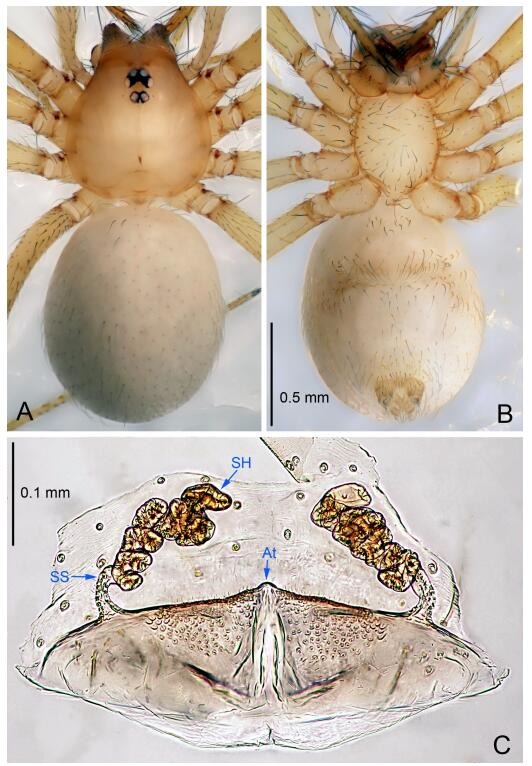
Leptonetela zhai Wang & Li, 2011, one female from the type locality
Leptonetela zhai Wang & Li, 2011: 17, Figures 69A-D, 70A-B, 71C-D.
Material examined. 4 females (IZCAS), Rudong Cave, N25.57°, E110.62°, Longpan Mountain, Dongtian, Hucheng Town, Xing'an County, Guilin City, Guangxi Zhuang Autonomous Region, China, 08 November 2012, Z. Chen & Z. Zhao leg.
Description. Male. See Wang & Li (2011).
Female. Total length 2.12 (Figure 96A-B). Carapace 0.80 long, 0.73 wide. Opisthosoma 1.27 long, 0.85 wide. Clypeus 0.12 high. Leg measurements: Ⅰ 6.61 (1.62, 0.37, 1.65, 1.87, 1.10); Ⅱ 5.39 (1.77, 0.30, 1.25, 1.12, 0.95); Ⅲ 4.16 (1.12, 0.27, 1.02, 1.00, 0.75); Ⅳ 5.72 (1.50, 0.30, 1.55, 1.35, 1.02). Vulva (Figure 96C): spermathecae coiled, atrium fusiform, anterior margin of atrium with short hairs.
Distribution. China (Guangxi).
Remarks. The female of the species is described for the first time. Females of Leptonetela zhai were collected from the same cave where the male holotype of L. zhai Wang & Li, 2011 was found.
Leptonetela tianxinensis (Tong & Li, 2008) comb. nov.
Leptoneta tianxinensis Tong & Li, 2008: 382, Figures 5A-G (♂♀).
Type material examined. Paratypes: 12 males, 6 females (IZCAS), Tianxin Cave, N33.35°, E111.88°, Sandaohe, Qilipo Town, Neixiang County, Henan Province, China, 24 June 2005, Q. Wang & Y. Tong leg.
Remarks. Our research showed that this species should be transferred to the genus Leptonetela, based on the result of DNA barcoding and morphological characters such as the pedipalpal femur lacking spines and the tibia with one strong spine retrolaterally.
Leptonetela gigachela (Lin & Li, 2010) comb. nov.
Guineta gigachela Lin & Li, 2010: 6, Figures 1A, 2A- E, 3A- B (♂♀).
Type material examined. Holotype: male (IZCAS), Qingzi Cave, N26.51°, E107.99°, Mianxi, Sankeshu Town, Kaili City, Guizhou Province, China, 26 May 2007, Y. Li & J. Liu leg. Paratypes: 2 males and 12 females, same data as holotype.
Remarks. Our studies showed that that Guineta Lin & Li, 2010 syn. nov. should be a junior synonym of Leptonetela Kratochvíl, 1978.
Leptonetela notabilis (Lin & Li, 2010) comb. nov.
Sinoneta notabilis Lin & Li, 2010: 83, Figures 55A-B, 56A-C, 57A-C (♂♀).
Type material examined. Holotype: male (IZCAS), Hebiandong Cave, Kaikou Town, Duyun City, N26.00°, E107.20°, Guizhou Province, China, 8 May 2006, Y. Li leg. Paratypes: 1 male and 1 female, same data as holotype.
Remarks. Our studies showed that Sinoneta Lin & Li, 2010 syn. nov. should be a junior synonym of Leptonetela Kratochvíl, 1978.
Leptonetela sexdigiti (Lin & Li, 2010) comb. nov.
Sinoneta sexdigiti Lin & Li, 2010: 87, Figures 58A-B, 59A-B, 60A-B (♂♀).
Type material examined. Holotype: male (IZCAS), Qiaotou Cave, Dashan, Shuangliu Town, Kaiyang County, N26.05°, E107.85°, Guizhou Province, China, 11 May 2006, Y. Li & Z. Yang leg. Paratypes: 5 males and 29 females, same data as holotype.
Leptonetela sanchahe Wang & Li nom. nov.
Qianleptoneta palmata Chen, Jia & Wang, 2010: 2902, Figures 19A-G, 20A-F, 25G (♂♀).
Sinoneta palmata Wang & Li, 2011: 4 (Transfer from Qianleptoneta).
Material examined. 1 male and 1 female (IZCAS), Sanchahe Cave, N26.53°, E107.70°, Sanchahe, Jialiang Town, Libo County, Guizhou Province, China, 16 May 2011, C. Wang & L. Lin leg.
Etymology. The specific name refers to the type locality; noun.
Remarks. Qianleptoneta palmata was collected from Sanchahe Cave in Guizhou, China and published by Chen et al. in December 2010. Wang & Li (2011) transfered Qianleptoneta palmata Chen et al, 2010 to the genus Sinoneta Lin & Li, 2010. Nevertheless, in this study our results confirmed that Qianleptoneta palmata belonged to the genus Leptonetela.
Leptonetela palmata is a preoccupied name (secondary homonym) for a species collected from Dixian Cave in Guizhou, China and published by Lin & Li, in August 2010. Subsequently, Leptonetela sanchahe Wang & Li nom. nov. is proposed for the taxon from Sanchahe Cave, in Guizhou, China.
ACKNOWLEDGEMENTS
Yi Wu and Guo Zheng helped to prepare photos of the manuscript. Sarah C. Crews kindly checked the English of the manuscript. Peter Jäger, Yanfeng Tong and Yucheng Lin provided valuable comments on an early version of the manuscript.
Funding Statement
This study was financially supported by the National Natural Sciences Foundation of China to Chunxia Wang (NSFC-31471977) and Shuqiang Li (NSFC-31530067, 31471960).Part of the lab work was supported by the Southeast Asia Biodiversity Research Institute, Chinese Academy of Sciences (2015CASEABRI005, Y4ZK111B01)
REFERENCES
- 1. Amara-Zettler LA, Gómez F, Zettler E, Keenan BG, Amils R, Sogin ML. 2002. Microbiology: eukaryotic diversity in Spain's River of Fire. Nature, 417 (6885): 137 [DOI] [PubMed] [Google Scholar]
- 2. Asmyhr MG, Cooper SJB. 2012. Difficulties barcoding in the dark: the case of crustacean stygofauna from eastern Australia. Invertebrate Systematics, 26 (6): 583- 591. [Google Scholar]
- 3. Ballard JWO, Whitlock MC. 2004. The incomplete natural history of mitochondria. Molecular Ecology, 13 (4): 729- 744. [DOI] [PubMed] [Google Scholar]
- 4. Chesters D, Wang Y, Yu F, Bai M, Zhang TX, Hu HY, Zhu CD, Li CD, Zhang YZ. 2012. The integrative taxonomic approach reveals host specific species in an encyrtid parasitoid species complex. PLoS One, 7 (5): e37655 [DOI] [PMC free article] [PubMed] [Google Scholar]
- 5. Clare EL, Lim BK, Engstrom MD, Eger JL, Hebert PDN. 2007. DNA barcoding of Neotropical bats: species identification and discovery within Guyana. Molecular Ecology Notes, 7 (2): 184- 190. [Google Scholar]
- 6. Convey P. 1997. How are the life history strategies of Antarctic terrestrial invertebrates influenced by extreme environmental conditions?. Journal of Thermal Biology, 22 (6): 429- 440. [Google Scholar]
- 7. Culver DC, White WB. 2005. Encyclopedia of Caves, Amsterdam: Elsevier Academic Press, 654 pp [Google Scholar]
- 8. Darriba D, Taboada GL, Doallo R, Posada D. 2012. jModelTest 2: more models, new heuristics and parallel computing. Nature Methods, 9 (8): 772 [DOI] [PMC free article] [PubMed] [Google Scholar]
- 9. Ferguson JWH. 2002. On the use of genetic divergence for identifying species. Biological Journal of the Linnean Society, 75 (4): 509- 516. [Google Scholar]
- 10. Flot JF, Wörheide G, Dattagupta S. 2010. Unsuspected diversity of Niphargus amphipods in the chemoautotrophic cave ecosystem of Frasassi, central Italy. BMC Evolutionary Biology, 10 171 [DOI] [PMC free article] [PubMed] [Google Scholar]
- 11. Folmer O, Black M, Hoeh W, Lutz R, Vrijenhoek R. 1994. DNA primers for amplification of mitochondrial cytochrome c oxidase subunit Ⅰ from diverse metazoan invertebrates. Molecular Marine Biology and Biotechnology, 3 (5): 294- 299. [PubMed] [Google Scholar]
- 12. Hebert PDN, Cywinska A, Ball SL, de Waard JR. 2003a. Biological identifications through DNA barcodes. Proceedings of the Royal Society B: Biological Sciences, 270 (1512): 313- 321. [DOI] [PMC free article] [PubMed] [Google Scholar]
- 13. Hebert PDN, Ratnasingham S, de Waard JR. 2003b. Barcoding animal life: cytochrome c oxidase subunit 1 divergences among closely related species. Proceedings of the Royal Society B: Biological Sciences, 270 (S1): S96- S99. [DOI] [PMC free article] [PubMed] [Google Scholar]
- 14. Hebert PDN, Gregory TR. 2005. The promise of DNA barcoding for taxonomy. Systematic Biology, 54 (5): 852- 859. [DOI] [PubMed] [Google Scholar]
- 15. Hedin M, Thomas SM. 2010. Molecular systematics of eastern North American phalangodidae (Arachnida: Opiliones: Laniatores), demonstrating convergent morphological evolution in caves. Molecular Phylogenetics and Evolution, 54 (1): 107- 121. [DOI] [PubMed] [Google Scholar]
- 16. Hedin MC. 1997. Molecular phylogenetics at the population/species interface in cave spiders of the southern Appalachians (Araneae: Nesticidae: Nesticus). Molecular Biology and Evolution, 14 (3): 309- 324. [DOI] [PubMed] [Google Scholar]
- 17. Howarth FG. 1983. Ecology of cave arthropods. Annual Review of Entomology, 28 (1): 365- 389. [Google Scholar]
- 18. Jukes TH, Cantor CR. 1969. Evolution of protein molecules. In: Munro HN. Mammalian Protein Metabolism, New York: Academic Press, 21- 32. [Google Scholar]
- 19. Kimura M. 1980. A simple method for estimating evolutionary rates of base substitutions through comparative studies of nucleotide sequences. Journal of Molecular Evolution, 16 (2): 111- 120. [DOI] [PubMed] [Google Scholar]
- 20. Ledford J, Paquin P, Cokendolpher J, Campbell J, Griswold C. 2011. Systematics of the spider genus Neoleptoneta Brignoli, 1972 (Araneae: Leptonetidae) with a discussion of the morphology and relationships for the North American Leptonetidae. Invertebrate Systematics, 25 (4): 334- 388. [Google Scholar]
- 21. Little CTS, Vrijenhoek RC. 2003. Are hydrothermal vent animals living fossils?. Trends in Ecology & Evolution, 18 (11): 582- 588. [Google Scholar]
- 22. López-García P, Rodríguez-Valera F, Pedrós-Alió C, Moreira D. 2001. Unexpected diversity of small eukaryotes in deep-sea Antarctic plankton. Nature, 409 (6820): 603- 607. [DOI] [PubMed] [Google Scholar]
- 23. Mathieu J, Jeannerod F, Hervant F, Kane TC. 1997. Genetic differentiation of Niphargus rhenorhodanensis (Amphipoda) from interstitial and karst environments. Aquatic Sciences, 59 (1): 39- 47. [Google Scholar]
- 24. Meyer CP, Paulay G. 2005. DNA barcoding: error rates based on comprehensive sampling. PLoS Biology, 3 (12): e422. [DOI] [PMC free article] [PubMed] [Google Scholar]
- 25. Miller GL, Foottit RG. 2009. The taxonomy of crop pests: the aphids. In: Foottit RG, Adler PH. Insect Biodiversity: Science and Society, Oxford, UK: Wiley-Blackwell, 463- 473. [Google Scholar]
- 26. Nei M, Kumar S. 2000. Molecular Evolution and Phylogenetics, Oxford: Oxford University Press, [Google Scholar]
- 27. Nevo E. 2001. Evolution of genome-phenome diversity under environmental stress. Proceedings of the National Academy of Sciences of the United States of America, 98 (11): 6233- 6240. [DOI] [PMC free article] [PubMed] [Google Scholar]
- 28. Niemiller ML, Near TJ, Fitzpatrick BM. 2012. Delimiting species using multilocus data: diagnosing cryptic diversity in the southern cavefish, Typhlichthys subterraneus (Teleostei: Amblyopsidae). Evolution, 66 (3): 846- 866. [DOI] [PubMed] [Google Scholar]
- 29. Posada D, Crandall KA. 1998. Modeltest: testing the model of DNA substitution. Bioinformatics, 14 (9): 817- 818. [DOI] [PubMed] [Google Scholar]
- 30. Poulson TL, White WB. 1969. The cave environment. Science, 165 (3897): 971- 981. [DOI] [PubMed] [Google Scholar]
- 31. Puillandre N, Lambert A, Brouillet S, Achaz G. 2012. ABGD, automatic barcode gap discovery for primary species delimitation. Molecular Ecology, 21 (8): 1864- 1877. [DOI] [PubMed] [Google Scholar]
- 32. Ratnasingham S, Hebert PDN. 2007. BOLD: the barcode of life data system (<a href="http://www.barcodinglife.org" target="_blank">http://www.barcodinglife.org</a>). Molecular Ecology Notes, 7 (3): 355- 364. [DOI] [PMC free article] [PubMed] [Google Scholar]
- 33. Rothschild LJ, Mancinelli RL. 2001. Life in extreme environments. Nature, 409 (6823): 1092- 1101. [DOI] [PubMed] [Google Scholar]
- 34. Sket B. 1999. The nature of biodiversity in hypogean waters and how it is endangered. Biodiversity & Conservation, 8 (10): 1319- 1338. [Google Scholar]
- 35. Stamatakis A. 2006. RAxML-Ⅵ-HPC: maximum likelihood-based phylogenetic analyses with thousands of taxa and mixed models. Bioinformatics, 22 (21): 2688- 2690. [DOI] [PubMed] [Google Scholar]
- 36. Tamura K, Stecher G, Peterson D, Filipski A, Kumar S. 2013. MEGA6: molecular evolutionary genetics analysis version 6.0. Molecular Biology and Evolution, 30 (12): 2725- 2729. [DOI] [PMC free article] [PubMed] [Google Scholar]
- 37. Tautz D, Arctander P, Minelli A, Thomas RH, Vogler AP. 2003. A plea for DNA taxonomy. Trends in Ecology & Evolution, 18 (2): 70- 74. [Google Scholar]
- 38. Tavares ES, Goncalves P, Miyaki CY, Baker AJ. 2011. DNA barcode detects high genetic structure within Neotropical bird species. PLoS One, 6 (12): e28543 [DOI] [PMC free article] [PubMed] [Google Scholar]
- 39. Vences M, Thomas M, Bonett RM, Vieites DR. 2005. Deciphering amphibian diversity through DNA barcoding: chances and challenges. Philosophical Transactions of the Royal Society B: Biological Sciences, 360 (1462): 1859- 1868. [DOI] [PMC free article] [PubMed] [Google Scholar]
- 40. Wang CX, Li SQ. 2011. A further study on the species of the spider genus Leptonetela (Araneae: Leptonetidae). Zootaxa, 2841 1- 90. [Google Scholar]
- 41. Wang JF, Jiang LY, Qiao GX. 2011. Use of a mitochondrial COI sequence to identify species of the subtribe Aphidina (Hemiptera, Aphididae). Zookeys, 122 1- 17. [DOI] [PMC free article] [PubMed] [Google Scholar]
- 42. Waterman TH. 2001. Evolutionary challenges of extreme environments (part 2). Journal of Experimental Zoology, 291 (2): 130- 168. [DOI] [PubMed] [Google Scholar]
- 43. World Spider Catalog. 2017. World spider catalog version 18.5. Natural History Museum Bern, [2017-10-04]. <a href="http://wsc.nmbe.ch" target="_blank">http://wsc.nmbe.ch</a>. [Google Scholar]
- 44. Zhang YG. 1986. The regional difference of karst cave development in China. Geographical Research, 5 (4): 36- 46. [Google Scholar]


