Abstract
This paper reports a simple, biogenic and green approach to obtain narrow band gap and visible light-active TiO2 nanoparticles. Commercial white TiO2 (w-TiO2) was treated in the cathode chamber of a Microbial Fuel Cell (MFC), which produced modified light gray TiO2 (g-TiO2) nanoparticles. The DRS, PL, XRD, EPR, HR-TEM, and XPS were performed to understand the band gap decline of g-TiO2. The optical study revealed a significant decrease in the band gap of the g-TiO2 (Eg = 2.80 eV) compared to the w-TiO2 (Eg = 3.10 eV). The XPS revealed variations in the surface states, composition, Ti4+ to Ti3+ ratio, and oxygen vacancies in the g-TiO2. The Ti3+ and oxygen vacancy-induced enhanced visible light photocatalytic activity of g-TiO2 was confirmed by degrading different model dyes. The enhanced photoelectrochemical response under visible light irradiation further supported the improved performance of the g-TiO2 owing to a decrease in the electron transfer resistance and an increase in charge transfer rate. During the TiO2 treatment process, electricity generation in MFC was also observed, which was ~0.3979 V corresponding to a power density of 70.39 mW/m2. This study confirms narrow band gap TiO2 can be easily obtained and used effectively as photocatalysts and photoelectrode material.
Introduction
Since 1972, titanium dioxide (TiO2) has been recognized as a potential photocatalyst by researcher’s worldwide1.TiO2 nanocrystals are well-known semiconductors that can catalyze solar powered reactions, such as water splitting, catalysis, photocatalysis, and environmental remediation2–5. On the other hand, TiO2 mainly absorbs light in the UV part of the solar spectrum; therefore, researchers hope that lowering its band gap energy will also enable it to absorb visible and infrared light4–9.
Previously studies have reduced the band gap of TiO2 by doping it with metals or non-metals, wrapping it with graphene, or introducing intrinsic defects into the TiO2 crystals10–22. These methods somehow increase the amount of partial visible light absorption but complete visible light and infrared light absorption is not achieved. Researchers used hydrogenation and other techniques to produce a black, red, blue form of TiO2 by introducing disorder, which absorbs light in the UV, visible and near infrared regions of the spectrum6–9. This increases the amount of solar light absorption of the black, red, and blue TiO2, which can be used to generate hydrogen gas and be applied in other visible light-induced applications, such as environmental remediation6–9,23,24. Researchers have also found that the hydrogenation process produces disorder in the surface layer of the TiO2 nanocrystals. Based on these studies, researchers have suggested that the hydrogen also ‘mops up’ broken titanium and oxygen bonds, forming new bonds that lower the band gap to the near infrared region23–28.
Significant developments in TiO2 photocatalysis and hydrogenation as well as other approaches that are novel and unique, such as surface doping or modification of TiO2, have been made to increase the photocatalytic activity under visible light irradiation25,26. Black TiO2 was reported to catalyze the photo-decomposition of organic molecules much better than normal nanophase TiO226. They also found that its ability to catalyze water splitting into hydrogen and oxygen under sunlight was improved greatly. Compared to conventional TiO2 and other metal oxide materials, it exhibits significantly higher efficiency under the same conditions6.
We previously reported band gap engineered TiO2 nanoparticles using electron beam irradiation which was simple and the reproducibility was quite high. Electron beam irradiated TiO2 nanoparticles showed notable decrease in the band gap as well as enhanced visible light-induced photo-degradation response towards methylene blue (MB) and brilliant blue G (BB) degradation, which was not possible for untreated TiO2 nanoparticles under similar conditions21. In the present study, white commercial TiO2 (w-TiO2) was modified (by forming defects) to a light gray colour (g-TiO2) using the cathode chamber of a Microbial Fuel Cell (MFC), which is a green, biogenic, novel and energy efficient process as compared to electron beam irradiation approach. The g-TiO2 showed an absorbance in the visible and near infrared region of the solar spectrum as well as improved visible light-induced photocatalytic and photoelectrochemical performances. The enhanced visible light activities of g-TiO2 might lead to various novel and efficient applications, which will open new horizons for metal oxide nanostructures with different types of defects and narrow band gap energies. Surprisingly, the treatment of TiO2 in the MFC cathode also generated electricity, which is energy efficient. The main difference between electron beam irradiation approach21 and MFC treatment is that, this approach is environmentally friendly approach which does not involve any external energy, chemicals or doping agents which make this modification method highly economical, simple, green, biogenic, useful, and efficient in the field of band gap engineering of metal oxides and has great potential for real applications in the photodegradation of several toxic dyes.
Results and Discussion
This paper reports a novel and alternative methodology to improve the visible light absorption of w-TiO2 by engineering it and forming different types of disorder. The modification process was achieved in MFC, which can produce an array of defects or disorder in the TiO2 nanoparticles, by this means, imparting novel characteristics, such as a reduced band gap, rapid charge carrier movements, and visible light-induced photocatalytic activities. This easy synthesis procedure does not include any expensive, toxic and hazardous chemicals, which make this modification method highly economical, simple, green, useful, and efficient in the field of band gap engineering of metal oxides. The complete modification process takes place in water at room temperature under atmospheric pressure. Fig. S1 presents the schematic diagram aimed at the modification of commercially obtainable TiO2 nanoparticles in a MFC. The electrons and protons generated in the anode of the MFC can either interact with some of the Ti4+ ions and reduce them to Ti3+ or interact with the TiO2 surface, which can alter the TiO2 composition21,29. The in-situ created species, such as electrons, protons, and Ti3+ ions, modified the TiO2 surface, which may impart enhanced visible light induced photocatalytic activities to g-TiO221,30,31.
X-ray diffraction studies
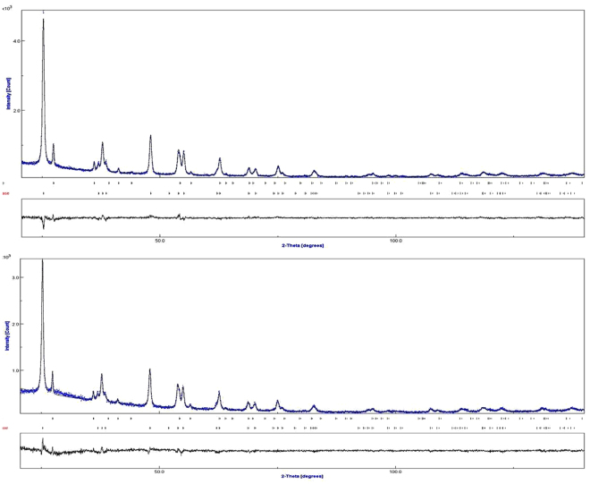
The structures of the w-TiO2 and g-TiO2 nanoparticles were examined by XRD in the 10–140° 2θ range. The strong XRD peaks (Fig. 1) indicate that the TiO2 nanoparticles were highly crystalline. The crystalline phase had an anatase and a rutile structure (Table 1) with a mean crystal size of approximately 25 nm, which is in agreement with the HRTEM observations.
Figure 1.
XRD patterns of the (a) w-TiO2, and (b) g-TiO2 nanoparticles obtained after Rietveld refinement of w-TiO2 and g-TiO2.
Table 1.
Relevant crystallographic data for w-TiO2 and g-TiO2 nanoparticles established from the powder X-ray diffraction data after Rietveld refinements.
| Sample | Phase | Phase fraction | Cell parameter(Å) | V(Å3) | ||
|---|---|---|---|---|---|---|
| a | b | c | ||||
| w-TiO2 | Anatase | 0.908 | 3.7890 | 3.7890 | 9.5131 | 136.58 |
| Rutile | 0.092 | 4.5989 | 4.5989 | 2.9583 | 62.586 | |
| g-TiO2 | Anatase | 0.901 | 3.7896 | 3.7896 | 9.5127 | 136.61 |
| Rutile | 0.099 | 4.5985 | 4.5985 | 2.9620 | 62.635 | |
The Rietveld refinement was used as a simple tool to verify the precise structure of the w-TiO2 and g-TiO2 nanoparticles and analyse the phase transformation from anatase to rutile. Using this method, the structural compositions of each samples were analysed qualitatively by fitting the experimental powder XRD profiles with respect to corresponding structural parameters (i.e. lattice parameters, atomic coordinates) and instrumental parameters (i.e. zero-point and profile parameters), as shown in Table 1. The refinement was converted to final residual factors (Table S2) of Rwp = 7.59 ± 0.3%, Rexp = 6.83 ± 0.2% and χ2 = 1.234 ± 0.2 for w-TiO2 nanoparticles, and Rwp = 7.70 ± 0.2%, Rexp = 6.69 ± 0.1% and χ2 = 1.324 ± 0.3 for g-TiO2 nanoparticles. Rwp is the weighted-profile R value, Rexp is the statistically expected R value, and χ2 is the goodness of fit (GoF), which is the square of the ratio between Rwp and Rexp. The χ2 is the goodness of fit values was calculated using given below equation (1). These calculated values of Rietveld refinement further confirmed the changes/modification of the w-TiO2 to g-TiO2 nanoparticles32.
| 1 |
where, N is the number of points and P is the number of parameters.
The Rietveld refinement of the XRD data of the w-TiO2 and g-TiO2 nanoparticles showed that after modification, the phase fraction of anatase of w-TiO2 decreased 1.234 ± 0.2 compared to g-TiO2 nanoparticles, whereas the phase fraction of rutile of w-TiO2 was increased to 1.324 ± 0.3 compared to g-TiO2 nanoparticles. The phase fraction decrease (anatase) and increase (rutile) of g-TiO2 compared to w-TiO2 was attributed to the oxidation state changes from Ti4+ to Ti3+ which was confirmed from these values (1.234 ± 0.2) and 1.324 ± 0.3). It addition, the phase fraction is also indicated by the unit cell volume of g-TiO2 nanoparticles for the anatase and rutile phase increased ~0.02% and ~0.106% compared to w-TiO2 (Table 1). The increase in the unit cell volume was attributed to the formation of some Ti3+ and defects in the sample, which is expected to be responsible for the increase in unit cell volume21,33. This XRD technique is incompatible to distinguish the oxidation state changes of two samples. The formation of Ti3+ can be easily detected by using EPR analysis (Fig. 2(b)) which is the confirmation of Ti3+formation in gray titania (Fig. 1).
Figure 2.
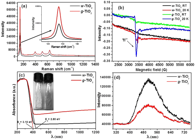
(a) Raman spectra, (b) EPR spectra at room temperature and 20 K, (c) UV-vis diffuse absorbance spectra, and (d) PL spectra of the w-TiO2 and g-TiO2 nanoparticles.
Raman spectroscopy
Raman spectroscopy was performed to observe the variations in the structure and disorder in the g-TiO2 nanoparticles after the introduction of disorder using MFC. The two polymorphs of TiO2 belong to different space groups: D4h19 (I41/amd) for anatase and D4h14 (P42/mnm) for rutile, which has distinct characteristics in Raman spectra6. For anatase TiO2, there are six Raman active modes with frequencies at 144, 197, 399, 515, 519 (superimposed with the 515 cm–1 band), and 639 cm–1 6. The modified g-TiO2 nanoparticles display the typical anatase Raman bands similar to the w-TiO2 nanoparticles, in addition to the increased intensity peak of the anatase Raman peak (Fig. 2a). The characteristic anatase mode at 144 cm–1 was present in both samples (w-TiO2 and g-TiO2) which indicate dominance of the anatase type TiO2 presence in the samples. Das et al.33 and Park et al.34 reported that the intensity of the peak increases with increasing number of non-stoichiometry defects, which increase the light absorption capacity of the TiO2 nanoparticles. Therefore, in the present case the increased intensity of the Raman bands clearly indicates that structural changes have occurred after a treatment in MFC, resulting in formation of some disorder or defects, which were confirmed further by EPR and DRS analysis21,33–36.
Electron paramagnetic resonance
The EPR spectra of the w-TiO2 and g-TiO2 nanoparticles were recorded (Fig. 2b) at room temperature (RT) and 20 K. The w-TiO2 and g-TiO2 nanoparticles at RT did not show any EPR signals, while the EPR signals for w-TiO2 and g-TiO2 nanoparticles were clear at 20 K. The EPR signal for g-TiO2 at 20 K was much stronger than that of w-TiO2 with a g value of 2.0428. The detected g value (Fig. S2) matched the characteristics of the paramagnetic Ti3+ ion centre in a distorted rhombic oxygen ligand field. The w-TiO2 showed another EPR signal at 20 K, which corresponds to oxygen vacancies, which was very small, whereas in the case of g-TiO2, this EPR signal appears to overlap with the Ti3+ signal, resulting in dramatic enhancement of the peak intensity8,29,30. Therefore, EPR showed that g-TiO2 has paramagnetic characteristics and oxygen vacancies, which enhances the visible light-induced photocatalytic activity8,31,37,38. Note that EPR is insensitive to Ti4+ species; hence, no signal is expected from it. This observation is very useful for identifying the Ti3+-related defects and oxygen vacancies related with Ti3+ lattice and F+ centres20 (Fig. 2).
UV-vis spectroscopic studies
Figure 2c shows the UV-vis diffuse absorbance spectra, i.e., optical response of the w-TiO2 and g-TiO2 nanoparticles, in which g-TiO2 nanoparticles show higher visible-light absorption than the w-TiO2 nanoparticles. The commercial w-TiO2nanoparticles only respond to ultraviolet light due to its intrinsic wide band gap. The improvement in the visible-light absorption of g-TiO2 nanoparticles was attributed to two factors: the formation of oxygen vacancies8,17, and surface disorder8,16. Mao et al. reported the surface disorder of anatase TiO2 nanoparticles following a hydrogen treatment, which shifted the valence band position by 2.18 eV16. In this case, the band gap of w-TiO2 nanoparticles shifted from 3.15 eV to 2.80 eV (Fig. 2c) for g-TiO2 nanoparticles. As a result, the energy gap between the valence band and the conduction band was narrowed dramatically to the point that was small enough for visible-light absorption. Hence, it was attributed to the absorption of visible light because of the formation of oxygen vacancies and other related defects in g-TiO2. The energy levels of the oxygen vacancies are approximately 0.75–1.18 eV below the conduction band of hydrogen-reduced TiO28,20. The visible-light absorption is associated with the transitions from the TiO2 valence band to the oxygen vacancy levels or from the oxygen vacancies to the TiO2 conduction band20–22. For the g-TiO2 nanoparticles, which appears to be self-doped Ti3+ (Fig. 2(c), red line), the considerably large absorption tail in the visible and NIR regions was observed, which is consistent with the change in colour of the powders from white to gray (Fig. 2c)8,9,21. The high absorption tail in the visible and NIR regions also provides clear evidence that the g-TiO2 nanoparticles contain a large number of oxygen vacancies8,9. The high absorption tail in the visible and NIR regions also provides clear evidence that the g-TiO2 nanoparticles contain a large number of defects/oxygen vacancies6,24.
These results also suggest that Ti3+ induced visible-light absorption that would have formed an isolated states between the forbidden gap in TiO2, as reported previously, rather than a shift in the position of either band edge, which usually takes place as a result of doping with metals or non-metals2,12–16,39. Theoretical studies also confirmed that formation of oxygen vacancies and Ti3+ could result in an electronic state vacancy band below the conduction band8,21.
PL studies
PL was performed to understand the migration, and transfer of charge carriers, efficiency of charge carrier trapping in semiconductor nanostructures. It is well identified that the PL signals of semiconductor materials result from the recombination rate of photo-induced charge carriers. In general, the lower the PL intensity, the lower the recombination rate of photo-induced electron–hole pairs, and the higher the photocatalytic activity of semiconductor photocatalysts39. In photoluminescence signals and their intensity are closely related to the improved photocatalytic activity. Further addition or modification in metal NPs reduces the PL intensities, due to shorter distance of inter band metal ions, which result in an energy transfer between nearby ions. The technique is useful because PL emission occurs mainly through the recombination of free carriers. The PL spectra of semiconductors are related to the transfer behaviour of the photo-induced electrons and holes, and can be used to estimate the recombination rate of charge carriers and understand the fate of electron–hole pairs in semiconductor nanostructures40,41. In other words, the PL emission intensity is generally associated with the recombination rate of the photo-induced electrons and holes, in which higher emission intensity reflects the fast recombination rate, whereas a lower intensity reflects the relatively slow recombination rates. This lower recombination may provide large number of charge carriers, which is actively participating in a range of oxidative and reductive photocatalytic degradation reactions of various dyes. As shown in Fig. 2d, the PL emission intensity of w-TiO2 was reduced significantly after inducing defects by MFC, which suggests that the charge carriers are trapped by the defects present in the g-TiO2, which enhances the charge separation efficiency. Different types of defects were reported to greatly affect the PL emission intensity of the metal oxides. For example, Jing et al.39, and Chetri et al.41, reported that the presence of defects quenched the PL signals of metal oxides significantly. Based on the above discussion, it could be concluded that the presence of defects in the g-TiO2 acts as trapping centres, which reduces the emission intensity of the PL signal40.
Microstructure analysis of w-TiO2 and g-TiO2 nanoparticles
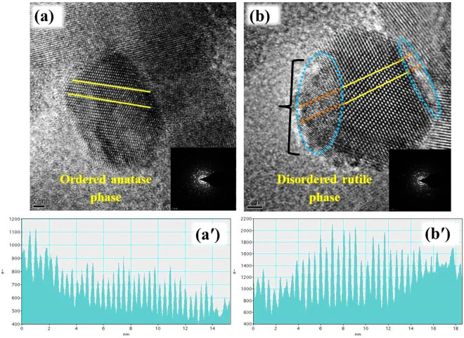
Figure 3 shows HRTEM images, SAED, and structural analysis of the w-TiO2 and g-TiO2 nanoparticles, which are in the range of 15 to 30 nm and in accordance with XRD analysis. w-TiO2 was completely crystalline, showing clearly-resolved and well-defined lattice fringes, even at the surface of the nanocrystals (Fig. 3a). The distance between the adjacent lattice planes was 0.37 nm, which is typical for anatase, and uniform throughout the whole nanocrystals (Fig. 3a). The measured lattice spacing of 0.37 nm coordinated the distance between the {101} planes of the anatase TiO2 crystal. The reflection from the similar {101} plane was prominent in the XRD patterns (Fig. 1) of the w-TiO2 and g-TiO2 nanoparticles. The spot SAED pattern [inset of Fig. 3(a),(b)] and continuous lattice confirmed the crystalline nature of the w-TiO2 and g-TiO2 nanoparticles. On the other hand, the outer edge of g-TiO2 nanoparticles gives the blurry impression, indicating an amorphous or disorder phase on the nanoparticles surface. The g-TiO2nanoparticles has a crystalline-disordered structure (Fig. 3b), and the outer layer can be seen readily, which is a structural deviation from the standard crystalline anatase. In contrast, the yellow colour encircled lattice line is disrupted and unclear at the edge of the nanoparticles. The core of the nanoparticles shows a well resolved {101} lattice plane with typical anatase plane distance on the disordered outer layer; the distances between the adjacent lattices planes are no longer uniform (Fig. 3b). This structural difference clearly verifies the disordered crystalline nature of the surface layer of the g-TiO2. Moreover, HRTEM of the w-TiO2 and g-TiO2 nanoparticles did not show any distinct changes in crystallinity (Fig. 3).
Figure 3.
HRTEM image and structural analysis of (a) w-TiO2 and (b) g-TiO2. The insets in (a) and (b) show the corresponding selected area electron diffraction pattern. (a′) shows line analysis of w-TiO2 and (b′) shows line analysis of g-TiO2. The zeros of the axis in (a′) and (b′) correspond to the left ends of the lines in (a) and (b).
Figure. 3(a′) shows the distance between the adjacent lattice planes, in the nm range, which is characteristic of anatase and uniform throughout the entire nanocrystals spectrum of w-TiO2 and (b′) displays a crystalline-disordered core-shell structure for g-TiO2 nanoparticles. Fig. S3(a), (b) as well as Fig. S3(a′), (b′) shows the HAADF-STEM images and EDX data of the w-TiO2 and g-TiO2 nanoparticles. In contrast, the difference between Fig. S3a,b suggests the surface modification of w-TiO2 in MFC. This finding was confirmed by SAED (the inset of Fig. 4a,b) and EDX analysis (Fig. S3a′,b′) of different regions of the nanoparticles.
Figure 4.
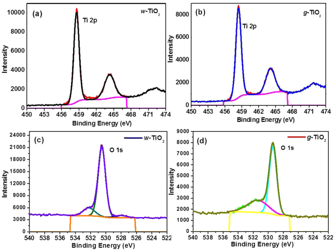
XPS (a and b) Ti 2p, and (c and d) O 1 s of the w-TiO2 and g-TiO2 nanoparticles.
XPS Analysis
XPS was performed on w-TiO2 and g-TiO2 nanoparticles for the surface characterization, oxidation states and other observations. C, O, and Ti were noticed in the survey scan spectra (Fig. S4(a) ESI). The C 1 s photoelectron peak (Fig. S4(b) ESI) at a binding energy (BE) of 284.5 eV was stronger for w-TiO2 than the g-TiO2 nanoparticles, which was ascribed to the elimination of surface carbon impurities from the g-TiO2 nanoparticles. Figure 4(a,b) shows the XP spectra of w-TiO2 and g-TiO2, respectively, in the Ti 2p BE region. The XPS Ti 2p peak was deconvoluted into two Ti 2p peaks at 458.57 and 464.27 eV for w-TiO2, whereas the peaks were observed at 458.42 and 464.10 eV for g-TiO2 nanoparticles, which were attributed to the splitting of Ti into Ti 2p3/2 and Ti 2p1/2 7,41,42. Both Ti 2p3/2 and Ti 2p1/2 peaks shifted towards a lower binding energy in the case of g-TiO2, which confirms the modification and formation of Ti3+ in the TiO2nanostructurein MFC setup. Similarly, Zhao et al.43, also reported that lower shift of Ti 2p BE is due to the formation of Ti3+ in TiO2 lattices. The amount of Ti3+ on the TiO2 surface plays a significant role, as described in the case of TiO2 doped with metal atoms. The photogenerated electrons can be confined in Ti3+, thus preventing the recombination rate of majority and minority carriers43. To control the binding states of oxygen in w-TiO2 and g-TiO2, the O 1 s XPS peak was fitted to three peaks (Fig. 4(c),(d)) cantered at 530.52, 532.22, and 528.06 eV for w-TiO2 and 530.62, 531.92, and 529.68 eV for g-TiO26,42–44. The shift in the O 1 s BE of g-TiO2 compared to w-TiO2 specifies a modification in the form of oxygen bonding, which is associated to the creation of Ti3+ 8. The photoelectron peak sat around 530.62 and 529.68 eV were allocated to the lattice oxygen in TiO2 and Ti2O3, correspondingly, however the peak at 531.92 eV was allotted to the water adsorbed on the TiO2 surface (Fig. 4).
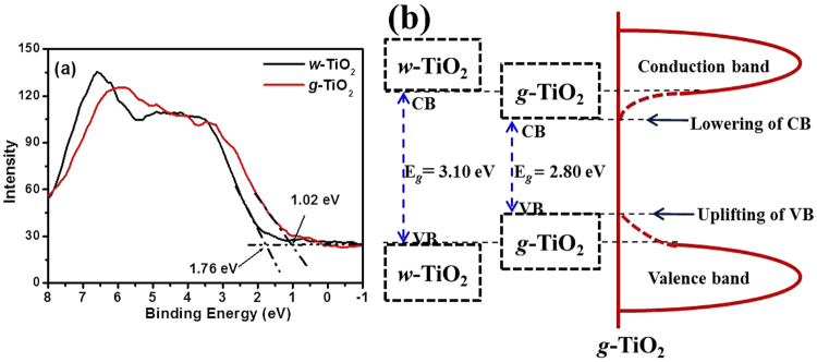
The reduction in the band gap may take place through the development of mid-gap band states whichever below the conduction band (CB) or above the valence band (VB) overlying with the respective bands. Therefore, VB XPS of the w-TiO2 and g-TiO2 nanoparticles was achieved to observe the band gap reduction phenomenon (Fig. 5(a)). The VB maximum of w-TiO2 was detected at 1.76 eV, whereas the VB maximum of the g-TiO2 was observed at 1.02 eV, showing a 0.74 eV shifts to a lower binding energy6,18,45. This shift was assigned to surface oxygen vacancies, Ti3+ formation and/or disorderliness in accordance with the TEM result and several other recent reports6,18,21,28. In particular, the band gap reduction caused by a lowering of CB was reported due to defects, such as oxygen vacancies and Ti3+ formation, which is related mainly to oxygen vacancies2,16,18,28,45. Chen et al. reported such an increase in the VB, which was due mainly to the existence of a disorder shell in the hydrogenated black TiO2 nanoparticles6. The reduction of the band gap in the g-TiO2 case was attributed to both the dropping of CB (due to oxygen vacancies and Ti3+ defect centres) and the increase in the VB (due to surface disorderliness)2,6,16,18,28.
Figure 5.
(a) VB of the w-TiO2 and g-TiO2 nanoparticles, and, (b) Proposed DOS for the g-TiO2 nanoparticles.
Based on the VB XPS results (Fig. 5(a),(b) presents a schematic illustration of the density of states (DOS) of disorder-engineered g-TiO2 nanoparticles compared to those of unmodified w-TiO2 nanoparticles. A measured band gap of 3.10 eV indicates a negligible change in the band edges of w-TiO2. The w-TiO2 displayed the typical VB DOS characteristics of TiO2, with the edge of the maximum energy at approximately 1.76 eV. Therefore, the CB minimum would occur at ~1.50 eV. For g-TiO2, the VB maximum energy showed a blue-shift toward the vacuum level at ~1.02 eV. A lower band gap from the DRS measurement for the g-TiO2 and VB XPS shift was due to the surface disorder produced after the MFC treatment. In addition, there may be CB tail states arising from the defects (Ti3+) that extend below the conduction band minimum2,6,16,18,28. The optical transitions from the blue-shifted VB edge to the band tail states are apparently responsible for optical absorption in g-TiO2. This assumption was supported by DRS observations. An additional potential advantage of this engineered and disordered g-TiO2 is that such defected and disordered metal oxides provide trapping sites (such as Ti3+) for photogenerated carriers and inhibit them from rapid recombination, thereby enhancing electron transfer and photocatalytic reactions21,29,30 (Fig. 5).
Photoelectrochemical studies
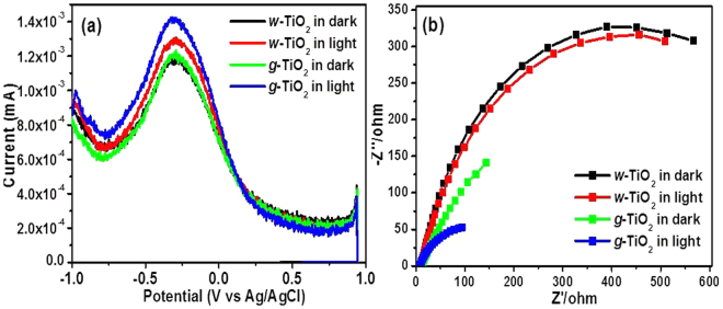
DPV is generally used to determine the charge storage capability of nanomaterials and is used frequently as a complementary technique to cyclic voltammetry. DPV was performed to understand the charge storage capability and the quantized behaviours of the w-TiO2 and g-TiO2 nanoparticles46–49. Figure 6a shows the well-defined quantized capacitance charging peaks for the w-TiO2 and g-TiO2 nanoparticles in the dark and under visible light irradiation. The peak current for w-TiO2 and g-TiO2 nanoparticles in the dark was observed at 1.170 mA and 1.218 mA, whereas, it was observed at 1.290 mA and 1.416 mA under visible light, respectively. An increase in the peak current of 0.126 mA was observed in the case of g-TiO2 nanoparticles under visible light irradiation. For the peak potential of w-TiO2 and g-TiO2 nanoparticles, no shift was observed. The increase in the peak current (0.126 mA) clearly shows the enhancement in the photoelectrochemical activity of g-TiO2 nanoparticles compared to the w-TiO2 nanoparticles, which may be due to the different types of defects formed in g-TiO2 nanoparticles. The g-TiO2 nanoparticles under visible light irradiation exhibited excellent and enhanced charge storing properties compared to w-TiO2 nanoparticles. The electrons stored on the g-TiO2 nanoparticles could be used to form different oxidative species (O2• and •OH) under visible light irradiation. These highly oxidative species might be responsible for degradation and mineralization of the organic colored dyes21,29,30. In addition, these stored electrons can be used for different photoactive devices. Overall, the g-TiO2 nanoparticles could be a good photoelectrocatalyst for electron transfer reactions such as photocatalysis and optoelectronic devices (Fig. 6).
Figure 6.
(a) DPV and (b) EIS of the w-TiO2 and g-TiO2 nanoparticles.
Generally, EIS is used to examine the electrochemical properties of the materials; hence, it was performed to understand the charge transfer resistance and charge separation efficiency between the photogenerated electrons and holes in the w-TiO2 and g-TiO2 nanoparticles. The charge separation efficiency of photogenerated electrons and holes is a critical factor for the photoelectrode and photocatalytic activities50–52. Figure 6b shows EIS Nyquist plots of the w-TiO2 and g-TiO2 nanoparticles in the dark and under visible light irradiation. The arc radius of the EIS spectra reflects the interface layer resistance arising at the electrode surface50. A smaller arc radius indicates higher charge transfer efficiency21,50–52. The arc radius of the g-TiO2 nanoparticles was smaller than that of w-TiO2 nanoparticles in the dark and under visible light irradiation. This suggests that the g-TiO2 nanoparticles have a lower resistance than w-TiO2 nanoparticles, which can accelerate the interfacial charge-transfer processes. These observations are supported by the PL results. EIS further support the important role of Ti3+ and different types of defects and oxygen vacancies, which help improve the charge separation (electrons and holes) and transfer efficiency of photogenerated electrons and holes on the surface of the g-TiO2 nanoparticles compared to the w-TiO2 nanoparticles in the dark and under visible light irradiations.
Visible light induced photocatalytic studies
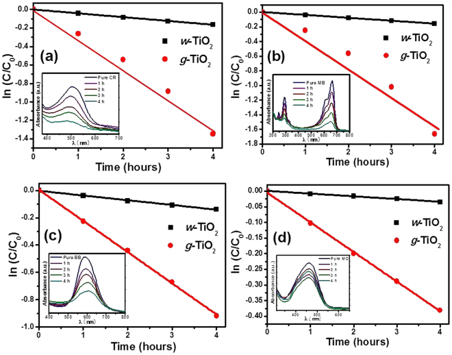
The visible light-induced photocatalytic activities of the w-TiO2 and g-TiO2 nanoparticles were estimated by degrading CR, MB, BB and MO under the visible light (λ > 500 nm) as reported earlier13,21,29,36,50. The g-TiO2 nanoparticles showed better photocatalytic degradation of CR, MB, BB, and MO than w-TiO2 (Fig. 7). The visible light-induced photocatalytic degradation was estimated from the decrease in the absorption intensity of CR, MB, BB and MO at a fixed wavelength, λmax = 492 nm, 665 nm, 588 nm, and 465 nm, respectively, during the course of the visible light-induced photocatalytic degradation experiment. The degradation was calculated using the relationship, ln C/C0 vs time (h) where C0 is the initial concentration and C is the concentration after visible light irradiation and degradation (Fig. 7). Inset of Fig. 7a,b,c, and d shows the respective decrease in absorbance after degradation. In addition, the degradation rate was also calculated to evaluate the precise degradation ability of the w-TiO2 and g-TiO2 nanoparticles. The rate constant (k) for the degradation of CR were 0.00072/h and 0.0570/h for w-TiO2 and g-TiO2 respectively, whereas 0.0018/h and 0.1203/h for degradation of MB by w-TiO2 and g-TiO2, respectively. Similarly, the k value for the degradation of BB was 0.0028/h and 0.02284/h for w-TiO2 and g-TiO2 respectively, whereas it was 0.0003/h and 0.0048/h for degradation of MO by w-TiO2 and g-TiO2, respectively. These results clearly show that g-TiO2 has higher k values for the degradation of CR, MB, BB, and MO compared to w-TiO2. The enhanced photocatalytic activity of the g-TiO2 nanoparticles compared to w-TiO2 can be explained by the surface modification and defects in g-TiO2 nanoparticles. Oxygen vacancies, other defects and Ti3+ centers enhance the photocatalytic activity21,29. The variation in the photocatalytic activity of w-TiO2 and g-TiO2 nanoparticles is also supported by DRS (Fig. 2(c)), EPR (Fig. 2b), and XPS (Figs 4 and 5). These outcomes evidently show that the visible light-induced photocatalytic performance of g-TiO2 nanoparticles can be amended greatly by reduction the band gap and making various defects and Ti3+ centers4–9,21,29 (Fig. 7).
Figure 7.
Visible light assisted photocatalytic degradation of (a) CR, (b) MB, (c) BB, and, (d) MO in the presence of the w-TiO2 and g-TiO2 nanoparticles.
Figure 8 shows the proposed schematic mechanism for the Ti3+ and oxygen vacancy-induced visible light photocatalytic degradation of the colored dyes (CR, MB, BB, and MO) in the presence of g-TiO2 nanoparticles. The concept of heterogeneous photocatalysis is based on the ability of photocatalysts to harvest light energy that is required to generate electron–hole pairs for surface reactions. On the other hand, owing to the wide band gap of TiO2, it can only absorb UV light. Fortunately, the optical properties of TiO2 can be manipulated by defect engineering4–9,13,21,29. By introducing oxygen vacancies and other defects, the light absorption of TiO2 from UV can be extended to the visible region of the spectrum because the oxygen vacancies give rise to the local states below the conduction band edge. The as-formed oxygen vacancy states can participate in a new photoexcitation process. That is, the electron is excited to the oxygen vacancy states from the valence band with the energy of visible light, which gives rise to typical excitations in the visible region of the spectrum. For this reason, oxygen vacancies are called F centers21. In addition, the electrons remaining in the oxygen vacancies can also interact with the adjacent Ti4+ to give the Ti3+ species. The Ti3+ defects can also form a shallow donor level just below the conduction band, which can also contribute to the visible light response8. The enhancement in the performance of g-TiO2 was attributed to the high separation efficiency of e–/h+ pairs due to (Fig. 8) surface oxygen-vacancies and Ti3+ formation, which lead to band gap narrowing4,5,21,26,29. This band gap narrowing in g-TiO2 nanoparticles provides the visible light-induced photocatalytic activity. Band gap excitation of the semiconductor consequences in e–/h+ separation. The high oxidative potential of the holes in the photocatalyst permits the formation of reactive intermediates21,29. Reactive hydroxyl radicals (•OH) can be shaped either by the decay of water or by the reaction of a hole with OH−. The hydroxyl radicals and photogenerated holes are particularly strong, non-selective oxidants that prime to the degradation of CR, MB, BB, and MO at the surface of the g-TiO2 nanoparticles4,5,21,29,30.This can be accredited to the high concentration of oxygen vacancies, other defects, and Ti3+ centers formed in the g-TiO2 nanoparticles21,29,38–45 (Fig. 8).
Figure 8.
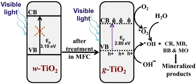
Proposed mechanism for the photocatalytic degradation of the dyes under visible light irradiation in the presence of g-TiO2 nanoparticles.
In general, high temperature, pressure, and energy input are required during synthesis and modification of nanomaterials4–9. On the other hand, in this proposed method, the modification process was quite efficient because there was no any external energy input except mechanical stirring and modification process took place at room temperature under atmospheric pressure. During the TiO2 modification process in cathode of MFC, power density increased from 54.88 mW/m2 to 70.39 mW/m2 which is a big accomplishment29. There are few reports in which various types of materials have been used in cathode for the production of electricity with a high power density. For example, Han et al.53, reported simultaneous degradation of pollutants and power generation in a MFC, where gold nanoparticles acted as a catalyst. The authors reported that a 36.56 mW/m2 power density was achieved after complete removal of the pollutant. Therefore, in this study, the less expensive TiO2, which was modified for visible light photocatalytic applications without any energy input, was used as the cathode catalyst.
Conclusions
A novel and biogenic method was used to modify the commercial TiO2 nanoparticles (w-TiO2) under ambient conditions, which resulted in an enhancement in their visible light assisted photocatalytic activities and photoelectrochemical behaviour. No structural changes occurred in g-TiO2, but Ti3+ formation, oxygen vacancies, and surface defects were identified. EIS and LSV in the dark and under visible light irradiation confirmed the visible light-induced photoactivity of the g-TiO2. Ti3+ and oxygen vacancy-induced visible light photocatalytic degradation of CR, MB, BB, and MO confirmed the improved photoactivity of the g-TiO2. This study suggests that g-TiO2 can be used as a visible light active photocatalyst as well as materials for photoelectrode. The MFC treatment can be employed to modify other metal oxides for enhanced visible light assisted photocatalytic activities and photoelectrochemical studies.
Experimental procedures
Materials used
TiO2 nanoparticles (size < 25 nm), methylene blue (MB), and brilliant blue (BB) were purchased from Sigma-Aldrich. Methyl orange (MO), Congo red (CR), and sodium sulphate were acquired from Duksan Pure Chemicals Co. Ltd. South Korea. Ethyl cellulose and α-terpineol were provided by KANTO Chemical Co., Japan and fluorine-doped transparent conducting oxide glass (FTO; F-doped SnO2 glass; 7 Ω/sq) was attained from Pilkington, USA. Altogether chemicals were of analytical grade and used as received. De-ionized water was prepared using a PURE ROUP 30 water purification system.
Methods
UV-Vis diffuse reflectance/absorbance spectroscopy (DRS) of the powdered w-TiO2 and g-TiO2 nanoparticles was performed using an ultraviolet – visible – near infrared (UV-VIS-NIR) double beam spectrophotometer (VARIAN, Cary 5000, USA) equipped with a diffuse reflectance accessory. A He-Cd laser (Kimon, 1 K, Japan) with a wavelength of 325 nm and a power of 50 mW was used as the excitation source for the photoluminescence (PL) measurements. X-ray diffraction (XRD, PANalytical, X’pert PRO-MPD, The Netherlands) was performed using Cu Kα radiation (λ = 0.15405 nm). A Rietveld refinement was conducted using Muad 2.46 software. Raman spectroscopy was performed on a HR800 UV Raman microscope (Horiba Jobin-Yvon, France). The electron paramagnetic resonance (EPR) measurements were performed using a Bruker EMX system. X-ray photoelectron spectroscopy (XPS, ESCALAB 250 XPS System, Thermo Fisher Scientific U.K.) was conducted using the following X-ray source: monochromated Al Kα, hν = 1486.6 eV, X-ray energy: 15 kV, 150 W and spot size: 500 μm. The XP spectra were fitted using the “Avantage program”. The microstructures of the w-TiO2 and g-TiO2 were observed by high resolution transmission electron microscopy (HRTEM, JEM-2100 JEOL) at an operating voltage of 200 kV combined with energy dispersive spectrometry (EDS) and a high angle annular dark field STEM (HAADF-STEM). The selected-area electron diffraction (SAED) images were recorded on a HRTEM instrument. The photocatalytic degradation and photoelectrochemical experiments (EIS and LSV) were conducted using a 400 W lamp with an irradiating intensity of 31.0 mWcm−2 (3 M, USA). The electrochemical impedance spectroscopy (EIS) and linear scan voltammetry (LSV) measurements were carried out using a potentiostat (Versa STAT 3, Princeton Research, USA) with a standard three-electrode system, in which Ag/AgCl (3.0 M KCl), a Pt gauge and fluorine-doped tin oxide (FTO) glass coated with p-TiO2, or m-TiO2 were used as the reference, counter, and working electrodes, respectively, in a 0.2 M Na2SO4 solution as the electrolyte. The working electrodes, with an effective area of 0.64 cm2, for EIS and LSV were prepared as follows. A 100 mg sample of each was suspended thoroughly by adding ethyl cellulose as a binder and α-terpineol as a solvent. The sample was mixed thoroughly for approximately 4 h and stirred at 50 °C for 4 h to obtain the paste. The obtained paste was coated on a FTO glass using the doctor-blade method, dried under a lamp for 24 h and used as the working electrode.
Modification of TiO2 nanoparticles in Microbial Fuel Cell
Commercial TiO2 nanoparticles were modified in the cathode of the MFC. The MFC was setup as reported elsewhere53. A 200 mL aqueous dispersion of w-TiO2 (50 mM) was prepared in the cathode chamber of the MFC. The preliminary pH of the aqueous dispersions was 4.40. The MFC setup was run for 20 days. After modification, the final pH of the aqueous dispersion was 3.65. The almost white w-TiO2 changed to a light gray colour upon modification. The resultant dispersion was centrifuged and a greyish powder was obtained, dried in an oven at 80 °C for 24 h and used in the different characterization techniques and applications.
Before and during the modification process, the voltage generated by the MFC was monitored regularly and recorded using a computerized multimeter. After stabilizing the MFC, the voltage obtained was ~0.3514 V and after the modification of TiO2 in the MFC cathode, the voltage obtained was ~0.3979 V, which corresponds to a power density of 54.88 mW/m2 and 70.39 mW/m2, respectively.
Photoelectrochemical studies of the w-TiO2 and g-TiO2 nanoparticles
To observe the photoelectrode performance of the w-TiO2 and g-TiO2 nanoparticles, photoelectrochemical experiments, such as EIS and LSV, were conducted under ambient conditions in the dark and under visible light irradiation in a 50 mL, 0.2 M Na2SO4 aqueous solution at room temperature. For each electrode, EIS was first performed in the dark and later under visible light irradiation (λ > 500 nm) at 0.0 V with frequencies ranging from 1–104 Hz. The photocurrent performance was attained by LSV in the dark and under visible light irradiation at a scan rate of 50 mV/s over the potential range, −1.0 to 1.0 V.
Photocatalytic degradation of CR, MB, BB and MO using w-TiO2 and g-TiO2 nanoparticles
The photocatalytic performance of the w-TiO2 and g-TiO2 nanoparticles were confirmed for the photocatalytic degradation of CR (10 mg/L), MB (10 mg/L), BB (10 mg/L), and MO (10 mg/L), as reported elsewhere21,29. For the photodecomposition experiment, a 3.0 mg sample of each photocatalyst was suspended in 20 mL of the aqueous CR, MB, BB, and MO solutions. Every single solution was sonicated for 5 min and then stirred in the dark for 10 min to complete the adsorption and desorption equilibrium on the w-TiO2 and g-TiO2 nanoparticles. The solutions were irradiated with a 400 W lamp (λ > 500 nm) and the distance between the light source and dye solution was 25 cm. The four sets of experiments for CR, MB, BB, and MO degradation were observed for 4 h. The rates of CR, MB, BB, and MO degradation were examined by taking 1.7 mL of the samples from each set at every 1 h, centrifuging the degraded solution to remove the catalyst and recording the UV-vis spectra, from which the degradation of CR, MB, BB, and MO could be calculated.
As a control experiment, the w-TiO2 nanoparticles (reference photocatalyst, Sigma-Aldrich) were used to degrade CR, MB, BB, and MO under the similar experimental conditions. Every single degradation experiment was executed in triplicate to ensure the photocatalytic activity of the w-TiO2 and g-TiO2 nanoparticles. The stability and reusability of the g-TiO2 nanoparticles were also tested in a similar way to that stated above.
Electronic supplementary material
Acknowledgements
This study was supported by the Priority Research Centres Program (Grant No: 2014R1A6A1031189), through the National Research Foundation of Korea (NRF) funded by the Ministry of Education, South Korea, and M. M. Khan would like to acknowledge the URC grant UBD/ORI/URC/RG(330)/U01 received from Universiti Brunei Darussalam, Brunei Darussalam.
Author Contributions
M.M. Khan and M.E. Khan planned the work, performed the experiments, analysed the results and wrote the manuscript. B.K. Min helps in the XRD and TEM analysis and its interpretation. M.H. Cho helps in planning the work, analysed the results and the writing manuscript.
Competing Interests
The authors declare that they have no competing interests.
Footnotes
Electronic supplementary material
Supplementary information accompanies this paper at 10.1038/s41598-018-19617-2.
Publisher's note: Springer Nature remains neutral with regard to jurisdictional claims in published maps and institutional affiliations.
Contributor Information
Mohammad Mansoob Khan, Email: mmansoobkhan@yahoo.com.
Moo Hwan Cho, Email: mhcho@ynu.ac.kr.
References
- 1.Fujishima A. Electrochemical photolysis of water at a semiconductor electrode. Nature. 1972;238:37–38. doi: 10.1038/238037a0. [DOI] [PubMed] [Google Scholar]
- 2.Hu YH. A highly efficient photocatalyst—hydrogenated black TiO2 for the photocatalytic splitting of water. Angewandte Chemie International Edition. 2012;51:12410–12412. doi: 10.1002/anie.201206375. [DOI] [PubMed] [Google Scholar]
- 3.Chen H, Nanayakkara CE, Grassian VH. Titanium dioxide photocatalysis in atmospheric chemistry. Chemical Reviews. 2012;112:5919–5948. doi: 10.1021/cr3002092. [DOI] [PubMed] [Google Scholar]
- 4.Pelaez M, et al. A review on the visible light active titanium dioxide photocatalysts for environmental applications. Applied Catalysis B: Environmental. 2012;125:331–349. doi: 10.1016/j.apcatb.2012.05.036. [DOI] [Google Scholar]
- 5.Hashimoto K, Irie H, Fujishima A. TiO2 photocatalysis: a historical overview and future prospects. Japanese journal of applied physics. 2005;44:8269. doi: 10.1143/JJAP.44.8269. [DOI] [Google Scholar]
- 6.Chen X, Liu L, Peter YY, Mao SS. Increasing solar absorption for photocatalysis with black hydrogenated titanium dioxide nanocrystals. Science. 2011;331:746–750. doi: 10.1126/science.1200448. [DOI] [PubMed] [Google Scholar]
- 7.Liu G, et al. A red anatase TiO2 photocatalyst for solar energy conversion. Energy & Environmental Science. 2012;5:9603–9610. doi: 10.1039/c2ee22930g. [DOI] [Google Scholar]
- 8.Pan X, Yang M-Q, Fu X, Zhang N, Xu Y-J. Defective TiO2 with oxygen vacancies: synthesis, properties and photocatalytic applications. Nanoscale. 2013;5:3601–3614. doi: 10.1039/c3nr00476g. [DOI] [PubMed] [Google Scholar]
- 9.Zhu Q, et al. Stable blue TiO2−x nanoparticles for efficient visible light photocatalysts. Journal of Materials Chemistry A. 2014;2:4429–4437. doi: 10.1039/c3ta14484d. [DOI] [Google Scholar]
- 10.Asahi R, Morikawa T, Ohwaki T, Aoki K, Taga Y. Visible-light photocatalysis in nitrogen-doped titanium oxides. Science. 2001;293:269–271. doi: 10.1126/science.1061051. [DOI] [PubMed] [Google Scholar]
- 11.Khan MM, Adil SF, Al-Mayouf A. Metal oxides as Photocatalysts. Journal of Saudi Chemical Society. 2015;19:462–464. doi: 10.1016/j.jscs.2015.04.003. [DOI] [Google Scholar]
- 12.Khan SU, Al-Shahry M, Ingler WB. Efficient photochemical water splitting by a chemically modified n-TiO2. Science. 2002;297:2243–2245. doi: 10.1126/science.1075035. [DOI] [PubMed] [Google Scholar]
- 13.Khan MM, Ansari SA, Amal MI, Lee J, Cho MH. Highly visible light active Ag@TiO2 nanocomposites synthesized using an electrochemically active biofilm: a novel biogenic approach. Nanoscale. 2013;5:4427–4435. doi: 10.1039/c3nr00613a. [DOI] [PubMed] [Google Scholar]
- 14.Choudhury B, Dey M, Choudhury A. Defect generation, dd transition, and band gap reduction in Cu-doped TiO2 nanoparticles. International Nano Letters. 2013;3:25. doi: 10.1186/2228-5326-3-25. [DOI] [Google Scholar]
- 15.Lee JS, You KH, Park CB. Highly photoactive, low bandgap TiO2 nanoparticles wrapped by graphene. Advanced Materials. 2012;24:1084–1088. doi: 10.1002/adma.201104110. [DOI] [PubMed] [Google Scholar]
- 16.Yang G, Jiang Z, Shi H, Xiao T, Yan Z. Preparation of highly visible-light active N-doped TiO2 photocatalyst. Journal of Materials Chemistry. 2010;20:5301–5309. doi: 10.1039/c0jm00376j. [DOI] [Google Scholar]
- 17.Tao J, Luttrell T, Batzill M. A two-dimensional phase of TiO2 with a reduced bandgap. Nature chemistry. 2011;3:296–300. doi: 10.1038/nchem.1006. [DOI] [PubMed] [Google Scholar]
- 18.Naldoni A, et al. Effect of nature and location of defects on bandgap narrowing in black TiO2 nanoparticles. Journal of the American Chemical Society. 2012;134:7600–7603. doi: 10.1021/ja3012676. [DOI] [PubMed] [Google Scholar]
- 19.Yin W-J, et al. Effective band gap narrowing of anatase TiO2 by strain along a soft crystal direction. Applied Physics Letters. 2010;96:221901. doi: 10.1063/1.3430005. [DOI] [Google Scholar]
- 20.Santara B, Giri P, Imakita K, Fujii M. Evidence for Ti interstitial induced extended visible absorption and near infrared photoluminescence from undoped TiO2 nanoribbons: an in situ photoluminescence study. The Journal of Physical Chemistry C. 2013;117:23402–23411. doi: 10.1021/jp408249q. [DOI] [Google Scholar]
- 21.Khan MM, et al. Band gap engineered TiO2 nanoparticles for visible light induced photoelectrochemical and photocatalytic studies. Journal of Materials Chemistry A. 2014;2:637–644. doi: 10.1039/C3TA14052K. [DOI] [Google Scholar]
- 22.Khan MM, Kalathil S, Lee J-T, Cho M-H. Enhancement in the photocatalytic activity of Au@TiO2 nanocomposites by pretreatment of TiO2 with UV light. Bulletin of the Korean Chemical Society. 2012;33:1753–1758. doi: 10.5012/bkcs.2012.33.5.1753. [DOI] [Google Scholar]
- 23.Liu L, Peter YY, Chen X, Mao SS, Shen D. Hydrogenation and disorder in engineered black TiO2. Physical Review Letters. 2013;111:065505. doi: 10.1103/PhysRevLett.111.065505. [DOI] [PubMed] [Google Scholar]
- 24.Chen X, et al. Properties of disorder-engineered black titanium dioxide nanoparticles through hydrogenation. Scientific Reports. 2013;3:1510. doi: 10.1038/srep01510. [DOI] [PMC free article] [PubMed] [Google Scholar]
- 25.Teng F, et al. Preparation of black TiO2 by hydrogen plasma assisted chemical vapor deposition and its photocatalytic activity. Applied Catalysis B: Environmental. 2014;148:339–343. doi: 10.1016/j.apcatb.2013.11.015. [DOI] [Google Scholar]
- 26.Leshuk T, et al. Photocatalytic activity of hydrogenated TiO2. ACS applied materials & interfaces. 2013;5:1892–1895. doi: 10.1021/am302903n. [DOI] [PubMed] [Google Scholar]
- 27.Wang G, et al. Hydrogen-treated TiO2 nanowire arrays for photoelectrochemical water splitting. Nano letters. 2011;11:3026–3033. doi: 10.1021/nl201766h. [DOI] [PubMed] [Google Scholar]
- 28.Xia T, Chen X. Revealing the structural properties of hydrogenated black TiO2 nanocrystals. Journal of Materials Chemistry A. 2013;1:2983–2989. doi: 10.1039/c3ta01589k. [DOI] [Google Scholar]
- 29.Kalathil S, Khan MM, Ansari SA, Lee J, Cho MH. Band gap narrowing of titanium dioxide (TiO2) nanocrystals by electrochemically active biofilms and their visible light activity. Nanoscale. 2013;5:6323–6326. doi: 10.1039/c3nr01280h. [DOI] [PubMed] [Google Scholar]
- 30.Liu X, et al. Green synthetic approach for Ti3+ self-doped TiO2−x nanoparticles with efficient visible light photocatalytic activity. Nanoscale. 2013;5:1870–1875. doi: 10.1039/c2nr33563h. [DOI] [PubMed] [Google Scholar]
- 31.Zuo F, et al. Self-doped Ti3+ enhanced photocatalyst for hydrogen production under visible light. Journal of the American Chemical Society. 2010;132:11856–11857. doi: 10.1021/ja103843d. [DOI] [PubMed] [Google Scholar]
- 32.Young, R. Introduction to the Rietveld method. The Rietveld Method (1993).
- 33.Das TK, Ilaiyaraja P, Mocherla PS, Bhalerao G, Sudakar C. Influence of surface disorder, oxygen defects and bandgap in TiO2 nanostructures on the photovoltaic properties of dye sensitized solar cells. Solar Energy Materials and Solar Cells. 2016;144:194–209. doi: 10.1016/j.solmat.2015.08.036. [DOI] [Google Scholar]
- 34.Park S-J, et al. In situ control of oxygen vacancies in TiO2 by atomic layer deposition for resistive switching devices. Nanotechnology. 2013;24:295202. doi: 10.1088/0957-4484/24/29/295202. [DOI] [PubMed] [Google Scholar]
- 35.Arora AK, Rajalakshmi M, Ravindran TR, Sivasubramanian V. Raman spectroscopy of optical phonon confinement nanostructured materials. J. Raman. Spectrosc. 2007;38:604–617. doi: 10.1002/jrs.1684. [DOI] [Google Scholar]
- 36.Gouadec G, Colomban P. Raman Spectroscopy of nanomaterials: How spectra relate to disorder, particle size and mechanical properties. Prog. Cryst. Growth Charact. Mater. 2007;53:1–56. doi: 10.1016/j.pcrysgrow.2007.01.001. [DOI] [Google Scholar]
- 37.Chiesa M, Paganini MC, Livraghi S, Giamello E. Charge trapping in TiO2 polymorphs as seen by Electron Paramagnetic Resonance spectroscopy. Physical Chemistry Chemical Physics. 2013;15:9435–9447. doi: 10.1039/c3cp50658d. [DOI] [PubMed] [Google Scholar]
- 38.Hoang S, Berglund SP, Hahn NT, Bard AJ, Mullins CB. Enhancing visible light photo-oxidation of water with TiO2 nanowire arrays via cotreatment with H2 and NH3: synergistic effects between Ti3+ and N. Journal of the American Chemical Society. 2012;134:3659–3662. doi: 10.1021/ja211369s. [DOI] [PubMed] [Google Scholar]
- 39.Jing L, et al. Review of photoluminescence performance of nano-sized semiconductor materials and its relationships with photocatalytic activity. Solar Energy Materials & Solar Cells. 2006;90:1773–1787. doi: 10.1016/j.solmat.2005.11.007. [DOI] [Google Scholar]
- 40.Kaniyankandy S, Ghosh HN. Efficient luminescence and photocatalytic behaviour in ultrafine TiO2 particles synthesized by arrested precipitation. Journal of Materials Chemistry. 2009;19:3523–3528. doi: 10.1039/b904589a. [DOI] [Google Scholar]
- 41.Chetri P, Choudhury A. Investigation of optical properties of SnO2 nanoparticles. Physica E: Low-dimensional Systems and Nanostructures. 2013;47:257–263. doi: 10.1016/j.physe.2012.11.011. [DOI] [Google Scholar]
- 42.Kim H-B, et al. Effects of electron beam irradiation on the photoelectrochemical properties of TiO2 film for DSSCs. Radiation Physics and Chemistry. 2012;81:954–957. doi: 10.1016/j.radphyschem.2011.11.064. [DOI] [Google Scholar]
- 43.Zhao Z, et al. Reduced TiO2 rutile nanorods with well-defined facets and their visible-light photocatalytic activity. Chemical Communications. 2014;50:2755–2757. doi: 10.1039/C3CC49182J. [DOI] [PubMed] [Google Scholar]
- 44.Etacheri, V., Seery, M., Hinder, S. & Pillai, S. Oxygen Rich Titania: A Dopant Free, A Dopant Free, High Temperature Stable, and Visible-Light Active Anatase Photocatalyst. (2011).
- 45.Kalathil S, Lee J, Cho MH. Gold nanoparticles produced in situ mediate bioelectricity and hydrogen production in a microbial fuel cell by quantized capacitance charging. ChemSusChem. 2013;6:246–250. doi: 10.1002/cssc.201200747. [DOI] [PubMed] [Google Scholar]
- 46.Yang A, et al. A simple one-pot synthesis of graphene nanosheet/SnO2 nanoparticle hybrid nanocomposites and their application for selective and sensitive electrochemical detection of dopamine. Journal of Materials Chemistry B. 2013;1:1804–1811. doi: 10.1039/c3tb00513e. [DOI] [PubMed] [Google Scholar]
- 47.Khan ME, Khan MM, Cho MH. CdS-graphene nanocomposite for efficient visible-light-driven photocatalytic and photoelectrochemical applications. Journal of Colloid and Interface Science. 2016;482:221–232. doi: 10.1016/j.jcis.2016.07.070. [DOI] [PubMed] [Google Scholar]
- 48.Khan MM, Ansari SA, Lee J, Cho MH. Enhanced optical, visible light catalytic and electrochemical properties of Au@TiO2 nanocomposites. Journal of Industrial and Engineering Chemistry. 2013;19:1845–1850. doi: 10.1016/j.jiec.2013.02.030. [DOI] [Google Scholar]
- 49.Ansari SA, et al. Oxygen vacancy induced band gap narrowing of ZnO nanostructures by an electrochemically active biofilm. Nanoscale. 2013;5:9238–9246. doi: 10.1039/c3nr02678g. [DOI] [PubMed] [Google Scholar]
- 50.Bai X, et al. Performance enhancement of ZnO photocatalyst via synergic effect of surface oxygen defect and graphene hybridization. Langmuir. 2013;29:3097–3105. doi: 10.1021/la4001768. [DOI] [PubMed] [Google Scholar]
- 51.Gan J, et al. Oxygen vacancies promoting photoelectrochemical performance of In2O3 nanocubes. Scientific reports. 2013;3:1021. doi: 10.1038/srep01021. [DOI] [PMC free article] [PubMed] [Google Scholar]
- 52.Khan ME, Khan MM, Cho MH. Ce3+-ion, surface oxygen vacancy, and visible light-induced photocatalytic dye degradation and photocapacitive performance of CeO2-Graphene nanostructures. Scientific reports. 2017;7:5928. doi: 10.1038/s41598-017-06139-6. [DOI] [PMC free article] [PubMed] [Google Scholar]
- 53.Han TH, Khan MM, Kalathil S, Lee J, Cho MH. Simultaneous enhancement of methylene blue degradation and power generation in a microbial fuel cell by gold nanoparticles. Industrial & Engineering Chemistry Research. 2013;52:8174–8181. doi: 10.1021/ie4006244. [DOI] [Google Scholar]
Associated Data
This section collects any data citations, data availability statements, or supplementary materials included in this article.


