Abstract
In recent years, phytoestrogens have been shown as useful selective estrogen receptor modulators. The estrogen-like effects of black tea (BT) and D. candidum (DC), as well as the combination of the two herbs, have remained largely elusive. This study aims to investigate the phytoestrogenic effect of BT and DC extract, and the possible mechanism. The effects on T47D (ER+ cell line) proliferation were evaluated by using MTT assay. The S phase proportion of ER+ cells was determined by using flow cytometry. The estrogen antagonist ICI 182,780 was applied to block the ER function. The activation of ER-mediated PI3K/AKT and ERK signal pathways were observed by using western blot. Expression of ERα and PGR, as well as PS2 and Cyclin D1 were detected by using western blot and real-time quantitative PCR. Firstly, our results found that BT and DC extracts promoted cell proliferation in ER-positive cells, and this effect was ER-dependent. Besides, BT and DC extracts increased the S-phase cell number. Next, PI3K, AKT and ERK pathways below ER were activated by phytoestrogen treatment, and this activation was blocked by the ER antagonist. Moreover, prolonged BT and DC treatments increased the expression of ESR1 and PGR. Consistently, the mRNA levels of not only ESR1 and PGR but also estrogen-dependent effectors ps2 and cyclin D1, were increased by phytoestrogens and blocked by ICI 182,780. Taken Together, BT and DC extracts have phytoestrogenic effects, and this may provide new ideas and experimental basis for the development and application of phytoestrogens.
Keywords: Phytoestrogens, estrogen receptor, back tea, dendrobium
Introduction
Estrogen is an important steroid hormone for growth and development of the female reproductive system. Also, it plays a role in maintaining the normal function of the cardiovascular system, skeletal system and the nervous system. During menopause, the endogenous estrogen level decreases rapidly, and concomitantly, the incidence of menopausal syndrome, osteoporosis, cardiovascular disease, senile dementia and etc. increase. Traditional estrogen replacement therapy can alleviate the above menopausal symptoms. However, long-term use of estrogen frequently results in hypercoagulable state, hypertension, edema, dementia, and even increases the risk of breast cancer, endometrial cancer, and the other gynecological cancers [1-4]. Therefore, highly effective and less toxic estrogen replacements deserve development for treatment of menopausal syndromes.
For substitution, increasing studies have taken advantages of phytoestrogens, which are extracted from the natural plants and have similar effects to the endogenous estrogen. These substances can interact with the estrogen receptor (ER) and play a two-way regulation. In the case of lower estrogen levels, they could play an estrogen-like role, while in the case of high estrogen levels, they exert an anti-estrogen role [5,6]. In recent years, phytoestrogens have been shown as useful selective estrogen receptor modulators (SERMs) in the treatment of menopausal syndromes [7-9]. Given the extensive sources of natural product, there exit great potential in the application of phytoestrogens.
Two known types of potential phytoestrogen, Black tea extract and D. candidum extract, are easily obtained in China. Black tea (BT) was reported to enhance bone regeneration in estrogen-deficient rats [10]. And for human breast cancer cell line MCF-7 (with a high estrogen signaling level), D. candidum (DC) inhibited the proliferation by inducing cell cycle arrest at G2/M phase and regulating some ER-downstream biomarkers [11]. But the estrogen-like effects of BT and DC, as well as the combination of the two herbs, have remained largely elusive. Besides, it remains to be elucidated what downstream signaling pathways in ER+ or ER- cell lines are involved in the estrogen-like activities of phytoestrogens.
To investigate the estrogen-like effects of BT and DC, we conducted this work using human breast cell lines and observed a proliferation promoting role with up-regulated estrogen signaling levels triggered by these extracts. Our findings provided an alternative of estrogen for females in the perimenopausal period, which is not only effective but also much cheaper and safer.
Materials and methods
Chemicals
Three groups of extracts were applied as follows: Black tea extract (BT), D. candidum extract (DC) and D. candidum and black tea extract (BT-DC), all of which were donated by Prof. Jun Sheng (Yunnan agricultural University).
Cell culture
The human mammary epithelial carcinoma cell lines T47D and MDA-MB-231 were obtained from the American Type Culture Collection (Manassas, VA, USA). T47D cells were cultured in Roswell Park Memorial Institute-1640 medium with 10% fetal bovine serum (FBS) and 100 IU/ml penicillin/streptomycin. MDA-MB-231 cells were cultured in Dulbecco’s modified Eagle’s medium supplemented with 10% FBS and 100 IU/ml penicillin/streptomycin. Cells were maintained at 37°C and 5% CO2 in a humidified incubator.
In different groups, different mother liquors were added in to the medium in a ratio of 1:100, and then diluted by according mediums. The BT mother liquor was 500 μg/ml and the DC mother liquor was 2 mg/ml.
MTT assay
T47D cells were seeded in a 96-well plate to a final concentration of 5000 cells/well and cultured in phenol red-free medium containing 5% charcoal-dextran-stripped FBS. Cells were respectively treated with various concentrations of BT extract (0.25 μg/ml to 10 μg/ml), DC extract (0.5 μg/ml to 20 μg/ml), and BT-DC extract (0.5 μg/ml to 20 μg/ml) for 24 h, 48 h and 72 h, either with or without ER-antagonist ICI 182,780 (Santa) at 100 nM. The positive control was added with 17β-estradiol (E2, Sigma) at 100 nM. Then the medium was removed and fresh medium was added to each well along with 10 ml of MTT solution (5 mg/ml). After 4 h incubation at 37°C, the medium was replaced by 150 ml of DMSO. The plates were read at wavelength of 490 nm using a micro-plate reader (BioTek, Winooski, VT, USA). Three reduplicate wells were used for each treatment, and the experiment was repeated three times.
Cell cycle analysis
Cell cycle distribution was evaluated using Cell cycle detection kit (BestBio, China) following the manufacturer’s instruction. Briefly, the cells were harvested 48 h after different treatments, washed twice with PBS, and fixed at 4°C for 1 h with 70% ethanol, and then stained with a propidium iodide (PI) solution (containing RNase) at 4°C for 30 min. At least 20,000 cells were analyzed by Becton Dickinson FACScan cytoflurometer (Mansfield, MA, USA). Cell cycle distribution was calculated using ModFIT cell cycle analysis software (Version 2.01.2; Becton Dickinson).
Giemsa staining
To visualize the morphology changes in mitotic period, cells (48 h after treatment) were stained with Giemsa staining Kit (Solarbio, China) according to the manufacture’s instruction. Briefly, the cells were fixed with methanol for 5 min, dried for 2 min, and then immersed in freshly prepared Giemsa stain solution for 20 min, followed by rinsing with pure water and dehydration. The stained cells were examined using a microscope with a 40× immersion objective lens.
Western blot analysis
The cells were lysed in ice-cold whole cell extraction buffer (WCEB, with protease inhibitor mixture from Roche Applied Science). After quantification, the protein samples were boiled, separated by 10% SDS polyacrylamide gel and electro-transferred to polyvinylidene fluoride (PVDF) membrane. The membrane was blocked with 5% BSA-TBST, incubated with primary antibodies against phospho-ERα (Cell Signaling Technology, Inc., USA), ERα (Santa, USA), phospho-PI3K (Cell Signaling Technology, Inc., USA), PI3K (Cell Signaling Technology, Inc., USA), phospho-AKT, AKT (Cell Signaling Technology, Inc., USA), phospho-ERK1/2 (Santa, USA) and ERK1/2 (Bioss, USA) at 4°C overnight. Then the membrane was incubated with the HRP-conjugated secondary antibodies, and visualized on Tanon-5200 Chemiluminescent Imaging System (Tanon).
Quantitative real-time PCR
Total RNA from each group of cells was extracted with TRIzol reagent (Invitrogen, USA) following the manufacturer’s instruction, and cDNA was generated with BioTeke super RT kit (BioTeke) according to the manufacture’s protocol. qRT-PCR was performed using SYBR Premix Ex TaqTM [12]. Primers are listed in Table 1.
Table 1.
RT-PCR primer sequences
| Genes | Sequences (5’-3’) | |
|---|---|---|
| ESR1 | Forward | CGCTACTGTGCAGTGTGCAAT |
| Reverse | CCTCACAGGACCAGACTCCATAA | |
| PGR | Forward | CAGCCAGAGCCCACAATACA |
| Reverse | GTTGTGCTGCCCTTCCATTG | |
| PS2 | Forward | GCCTTTGGAGCAGAGAGGAG |
| Reverse | TGTACACGTCTCTGTCTGGG | |
| Cyclin D1 | Forward | GATCAAGTGTGACCCGGACTG |
| Reverse | CCTTGGGGTCCATGTTCTGC | |
| β-actin | Forward | GCCGCCAGCTCACCAT |
| Reverse | TCGATGGGGTACTTCAGGGT |
Statistical analysis
Data were presented as mean ± SE, including at least three independent experiments. Statistical analysis was performed by one-way ANOVA for multiple group comparison. P<0.05 represents the statistical significance.
Results
Black tea and D. candidum extracts promote cell proliferation in ER-positive cells
First, we examine the effects of extracts from BT and DC, as well as their combination, on the in vitro proliferation of ER-positive cell line, T47D. Either after 24, 48 or 72 h, BT and DC showed a similar effect to E2. Specifically, the cells exposed to 0.25 to 5 μg/ml BT extracts (Figure 1A) had a significantly increased cellular viability compared with the control (24 h treatment: 0.25 μg/ml vs. control P<0.05, 0.5-5 μg/ml vs. control P<0.01. 48 h treatment: 0.5 or 5 μg/ml vs. control P<0.05, 1-2.5 μg/ml vs. control P<0.01. 72 h treatment: 0.5-2.5 μg/ml vs. control P<0.01, 5 μg/ml vs. control P<0.05). However, the proliferation promoting effect disappeared in the 10 μg/ml group. For DC treated cells (Figure 1B), the 0.5-2.5 groups at 24 h were uninfluenced while the 5-20 μg/ml groups exhibited a highly significant increase in the cellular viability (P<0.01 vs. control), at 48 h, 1-20 μg/ml groups showed a significant increase (1-5 μg/ml vs. control P<0.05, 10-20 μg/ml vs. control P<0.01), and at 72 h, all the DC treated groups had increased viability (0.5-20 μg/ml vs. control P<0.05). For combination treatments (Figure 1C), the increase was much significant at 48 or 72 h compared with the single treatments (1-10 μg/ml vs. control P<0.01 or 0.001). These results proved that BT and DC were effective phytoestrogens with E2-like roles in ER-positive cell proliferation, and the combination of BT and DC might arouse more significant effects for a continuous (more than 48 h) exposure.
Figure 1.
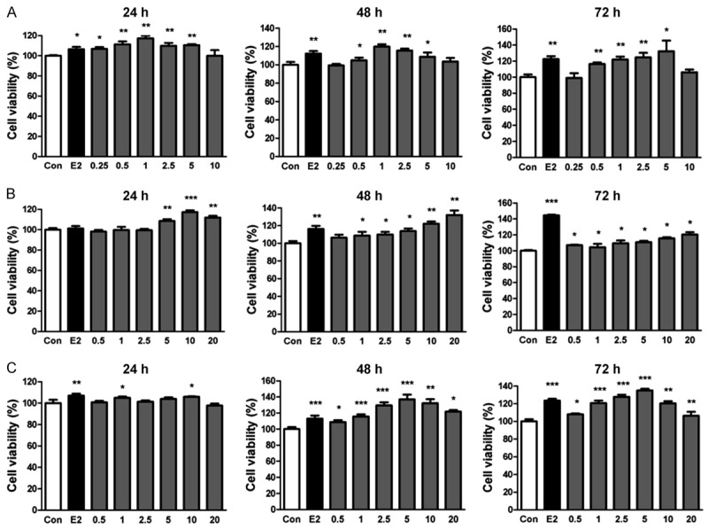
The pro-proliferative effects of phytoestrogens on Estrogen receptor (ER)-positive cell line T47D. A. Black tea extract at the concentration from 0.25 to 10 μg/ml, treating for 24 h to 72 h. B. D. candidum extract at the concentration from 0.5 to 20 μg/ml, treating for 24 h to 72 h. C. Black tea and D. candidum extract at the concentration from 0.5 to 20 μg/ml, treating for 24 h to 72 h. *P<0.05, **P<0.01, ***P<0.001.
Proliferous effects of BT and DC are ER-dependent
Next, we verified the in vitro effects of BT and DC were ER-dependent, by using the estrogen antagonist ICI 182,780 to block the ER. As shown in Figure 2, the increase of cellular viability disappeared when ICI 182,780 was present, for either BT or DC treatment (Figure 2). In support, we verified this necessity for ER-dependent functions using ER negative cell line MDA-MB-231 and found not any proliferous effects of either BT, DC or BT-DC combination (Supplementary Figure 1).
Figure 2.
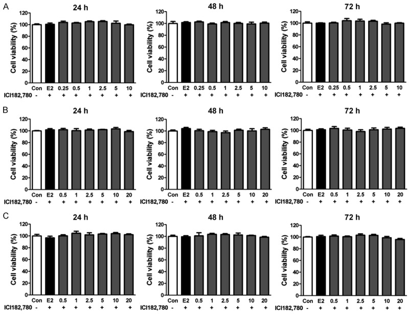
The pro-proliferative effect of phytoestrogens is blocked by the estrogen antagonist ICI 182,780. A. Black tea extract at the concentration from 0.25 to 10 μg/ml, treating for 24 h to 72 h. B. D. candidum extract at the concentration from 0.5 to 20 μg/ml, treating for 24 h to 72 h. C. Black tea and D. candidum extract at the concentration from 0.5 to 20 μg/ml, treating for 24 h to 72 h.
BT and DC extracts increase the S-phase cell number
To further support the proliferation promoting effects of the above phytoestrogens, the proportions of cells in each phase were analyzed (Figure 3A, 3B). Similar to E2, BT (1-5 μg/ml) and DC (10-20 μg/ml) increased the S-phase proportion of T47D cells. Moreover, the combination of BT and DC (5-10 μg/ml) enhanced the S-phase proportion to a similar level to E2 treated cells (Figure 3C). Also, these findings were proved by Giemsa staining and the following observation of dividing cells (Supplementary Figure 2). These results indicated that BT and DC extracts promoted mitosis through ER dependent pathways.
Figure 3.
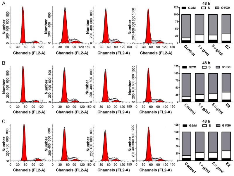
Black tea and D. candidum extracts increase the S-phase cell number after 48 h treatments. A. Cell cycle in the Black tea extract treated groups. B. Cell cycle in the D. candidum extract treated groups; C. Cell cycle in the Black tea and D. candidum extract treated groups.
BT and DC extracts activate ER signaling and the relative downstream pathways
Further, the estrogen signaling and the relative downstream pathways were investigated. Cells were respectively treated with graded concentrations of BT extract (1-5 μg/ml), DC extract (5-20 μg/ml), BT-DC extract (2.5-10 μg/ml) and E2 (100 nM) for 30 min. Analysis of ER, PI3K, AKT and ERK phosphorylation demonstrated that both BT (Figure 4A) and DC (Figure 4B), as well as BT-DC (Figure 4C), had similar effects to E2 in these pathway activation. Compared with controls, 1-5 μg BT extract was sufficient to induce highly significantly increased p-ER, p-PI3K, p-AKT and p-ERK levels (P<0.001); and 5-20 μg/ml DC extract and 2.5-10 μg/ml BT-DC extract also had an identical trend (P<0.001) in all of the above signaling pathways. Collectively, PI3K, AKT and ERK pathways below ER may play a role in the in vitro action of phytoestrogens.
Figure 4.
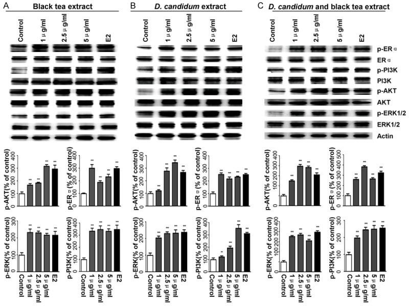
Black tea and D. candidum extracts activate ER signaling and the relative downstream pathways. A. ER signaling and relative downstream molecules expression and statistical analysis in Black tea extract treated groups. B. ER signaling and relative downstream molecules expression and statistical analysis in D. candidum extract treated groups. C. ER signaling and relative downstream molecules expression and statistical analysis in Black tea and D. candidum extract treated groups. The ER, PI3K, AKT and ERK phosphorylation are statistically shown under the Western blot bands. *P<0.05, **P<0.01, ***P<0.001.
Phytoestrogen-activated signaling could be blocked by ER antagonist ICI 182,780
In the blocking analyses, ICI 182,780 at 100 nM was applied for 1 h, followed by the treatments of BT extract (1 μg/ml), DC extract (20 μg/ml), BT-DC extracts (5 μg/ml) and E2 (100 nM) for 30 min. As Figure 5 shown, when ICI 182,780 was present, the phosphorylated ER, PI3K, AKT and ERK levels were no longer up-regulated under any of the phytoestrogen treatment (Figure 5, for all phytoestrogens, phytoestrogen vs. control P<0.001 and phytoestrogen + ICI vs. control P>0.05). These results suggested that the responses of PI3K, AKT and ERK cascades to exogenous phytoestrogens depend on the normal function of ER.
Figure 5.
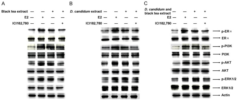
Phytoestrogen-activated signaling could be blocked by ER antagonist ICI 182,780. When ICI 182,780 was present, the phosphorylated ER, PI3K, AKT and ERK levels were no longer up-regulated under any of the phytoestrogen treatment. A. Western blot assay for Black tea extract treated groups. B. Western blot assay for D. candidum extract treated groups. C. Western blot assay for Black tea and D. candidum extract treated groups. The ER, PI3K, AKT and ERK phosphorylation are statistically shown under the western blot bands. *P<0.05, **P<0.01, ***P<0.001.
Prolonged BT and DC treatments increase the expression of ESR1 and PGR
Further, we analyzed the effects of long term exposure to BT/DC extracts on estrogen and progesterone receptors besides the acute treatments. Consistent with the above signaling, the estrogen receptor α (ESR1) and the progesterone receptor (PGR) were up-regulated in the protein expression after prolonged BT, DC and BT-DC treatments (Figure 6, P<0.001 each concentration vs. control). In parallel, the mRNA levels of ESR1 and PGR were determined by real-time qPCR. As shown in Figure 7, prolonged exposure to BT and DC extracts (included their combination) enhanced the mRNA expression of ESR1 and PGR (P<0.01 vs. control). Collectively, phytoestrogens induced estrogen and progesterone receptors expression via a prolonged activation.
Figure 6.
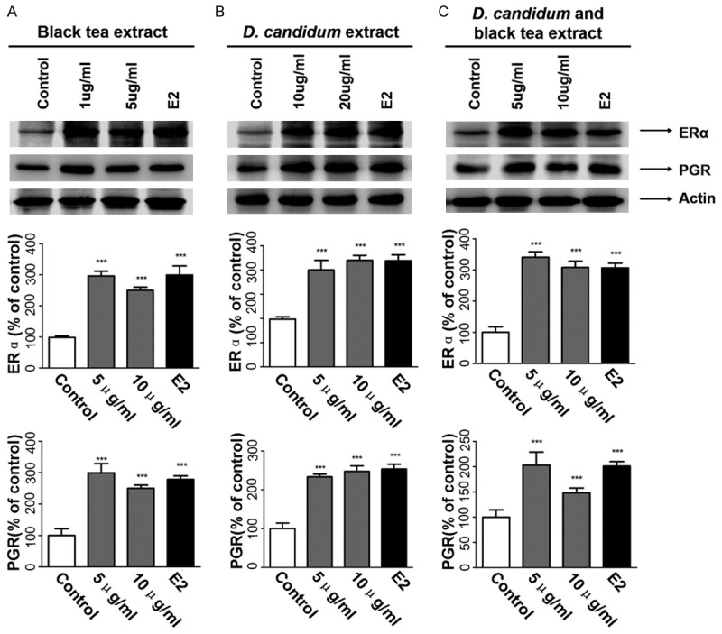
Prolonged Black tea and D. candidum treatments increase the expression of the estrogen receptor α (ERα) and progesterone receptor (PGR). A. ERα and PGR expression and statistical analysis in Black tea extract treated groups. B. ERα and PGR expression and statistical analysis in D. candidum extract treated groups. C. ERα and PGR expression and statistical analysis in Black tea and D. candidum extract treated groups. The ESR1 and PGR expression statistics are shown under the Western blot bands. *P<0.05, **P<0.01, ***P<0.001.
Figure 7.
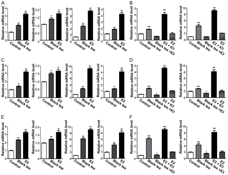
The mRNA levels of ESR1, PGR, estrogen-dependent effectors ps2 and cyclin D1 were increased by phytoestrogens and blocked by ICI 182,780. A, B. Black tea extract; C, D. D. candidum extract. E, F. Black tea and D. candidum extract. The relative mRNA levels of ESR1, PGR, ps2 and cyclin D1 are shown in each sub-figure. *P<0.05, **P<0.01, ***P<0.001.
mRNA levels of estrogen-dependent effectors ps2 and cyclin D1 were increased by phytoestrogens and blocked by ICI 182,780
Finally, we probed the further downstream effectors of estrogen-dependent cascades relating to PI3K, AKT and ERK pathways (Figure 7). Cells were respectively treated with BT extract (1 μg/ml), DC extract (20 μg/ml), and BT-DC extracts (5 μg/ml), for 48 h (with 100 nM E2 as the positive control), either with or without ICI 182,780 (100 nM). The mRNA levels were examined using real-time qPCR. First, PS2, a well-known secretory protein induced by estrogen, was up-regulated in the mRNA level after BT (Figure 7B, P<0.001 vs. control), DC (Figure 7D, P<0.001 vs. control), BT-DC (Figure 7F, P<0.001 vs. control), and E2 (Figure 7A-F, P<0.001 vs control), and this increase was blocked by ICI 182,780. Second, cyclin D1 belongs to the highly conserved cyclin family, as regulators of Cyclin-dependent kinase. This cyclin forms a complex with and functions as a regulatory subunit of CDK4 or CDK6, whose activity is required for cell cycle G1/S transition [13]. A high expression of cyclin D1 indicates an increased proliferation activity. BT (Figure 7B, P<0.001 vs. control), DC (Figure 7D, P<0.001 vs. control), BT-DC (Figure 7F, P<0.001 vs. control), and E2 (Figure 7A-F, P<0.001 vs. control) consistently increased the cyclin D1 expression; while ICI 182,780 strongly suppressed this effect (Figure 7B-F). Together, ER-dependent PS2 expression and cell cycle regulation could be highly possibly involved in the in vitro actions of phytoestrogens.
Discussions
In this article, we provide data supporting an estrogen-like role of BT and DC, as well as the downstream mechanisms. Using ER-positive cell line T47D, we observed a proliferative effect of BT and DC, which was similar to E2. We also confirmed that the ER signaling pathway was necessary for this effect by the ER inhibitor and ER-lack cells. Besides, ER relative PI3K, AKT and ERK pathways were activated by these phytoestrogens. Steroid receptors ER and PGR, the E2 triggered secretory protein PS2, and the important cell cycle regulator cyclin D1 were enhanced in expression by treatments of the above phytoestrogens, when ER signaling was normal. All these data suggest that BT and DC are effective estrogen replacements in a certain probability.
Previous studies also mentioned the estrogen like role of BT and DC. Some work claimed that BT induced increased Urinary flavonoids and phenolic acids as biomarkers [14]. Besides, BT had been reported to enhance bone regeneration in estrogen-deficient rats, and indicated benefits for the aged and menopausal women [10]. However, opposite conclusions were found that DC inhibited MCF-7 cells proliferation by inducing cell cycle arrest at G2/M phase [11], although this was also a regulatory effect of phytoestrogens.
Through ER signaling, the proliferative effects of steroids were widely observed [15]. For example, estrogens were reported to promote proliferation of the seminoma-like TCam-2 cell line through a GPER-dependent ERα36 induction [16]. Also, estrogen affected male rat enamel formation and promoted ameloblast proliferation [17]. Low doses of bisphenol A had also been shown to promote human seminoma cell proliferation via a membrane G-protein-coupled estrogen receptor [18]. Many phytoestrogens have been proved to increase cellular proliferation via ER relative pathways. A diarylheptanoid phytoestrogen from Curcuma comosa, 1,7-diphenyl-4,6-heptadien-3-ol, could accelerate the proliferation of human osteoblasts [19]. And the estrogenic activity of Pueraria mirifica was revealed for its contribution to the MCF-7 proliferation [20]. So far as we know, our study is among the very few works revealing the estrogen-like role of BT/DC and clarifying the integrated mechanisms.
Previously, steroids such as E2, via binding to cytoplasmic or membrane-associated receptors, were shown to rapidly activate intracellular signaling cascades such as MAPK and PI3K/AKT [15,21-23]. Many breast cancers express the estrogen receptor α and depend on estrogen/ERK signaling for proliferation [24]. Estrogen-induced (or even xenoestrogen-induced) ERK activation was reported to be involved in the proliferation of pituitary tumor cells [25]. And in the pregnant human myometrium, estradiol could activate ERK signaling via ERα [26]. Also, the PI3K/AKT/mTOR pathway via estrogen in ER-positive breast cancers was one of the most important growth-promoting roles [27]. For the obesity, estrogen induces endometrial cancer cell proliferation via PI3K/AKT and MAPK signaling pathways [28]. Conversely, spatholobus suberectus column extract was shown to repress the expression of phosphorylated-ER alpha (p-ERα), ERK1/2, p-ERK1/2, AKT, p-AKT, p-mTOR, PI3K, and p-PI3K, indicating the suppressed MAPK PI3K/AKT signaling pathways, and it hence inhibited ER positive breast cancer [29]. In our study, BT and DC have a similar mechanism underlying the estrogen-like effect, that activation of ER-ERK and ER-PI3K/AKT signaling could be the crucial step for phytoestrogen exerting the effects. This conclusion is consistent with most obtainable researches and highlights the most important common mechanisms between estrogen and phytoestrogens.
Another interesting finding of this work is that positive feedback exists during the prolonged BT/DC (and also E2) exposure, that ER and PGR expression were increased. Similar study about phytoestrogen extracts by Richter D et al reported that after the pumpkin seed extract treatment, estradiol production was elevated in MCF7, BeWo, and Jeg3 cells in a concentration-dependent manner, and a significant ER-α down-regulation and PGR up-regulation were observed [30]. Partially differently, we found that both PGR and ER-α were up-regulated by BT, DC and E2. This suggests different phytoestrogens may result in different expression profiling of steroid receptors. Another support from Wang et al mentioned that the mRNA expression level of ER-α was increased after prolonged treatment with E2 (when the concentration of estradiol was low), and it was significantly decreased with increasing concentrations of tamoxifen [31]. Moreover, a study using porcine luminal epithelial cells reported that high doses of E2 (>500 pg/ml) increased the cell proliferation and PGR expression [32]. These consistent evidences indicate that enhanced steroid receptor expression largely contribute to the final outcomes of phytoestrogens, and these genomic effects could be reckoned as another key mechanism underlying the estrogen-like actions.
In conclusion, our study demonstrates the phytoestrogenic effects of BT and DC. And the BT-DC combined extract has a stronger phytoestrogenic effect. The potential mechanism may be through the activation of ER-mediated PI3K/AKT and ERK signaling pathways (non-genomic effects) and up-regulation of ER downstream target gene expression (genomic effects), which find promote cell cycle activion and proliferation. This study provides a theoretical basis for the development and application of phytoestrogens (especially BT-DC combined extracts).
Acknowledgements
This study was granted by the National Natural Science Foundation of China (No.31670795) and School of Life Changbai Mountain Scholarship Foundation of Jilin Province (No.440050117010).
Disclosure of conflict of interest
None.
Supporting Information
References
- 1.Reineri S, Agati S, Miano V, Sani M, Berchialla P, Ricci L, Iannello A, Coscujuela Tarrero L, Cutrupi S, De Bortoli M. A novel functional domain of Tab2 involved in the interaction with estrogen receptor Alpha in breast cancer cells. PLoS One. 2016;11:e0168639. doi: 10.1371/journal.pone.0168639. [DOI] [PMC free article] [PubMed] [Google Scholar]
- 2.Lu ZY, Li RL, Zhou HS, Huang JJ, Qi J, Su ZX, Zhang L, Li Y, Shi YQ, Hao CN, Duan JL. Rescue of hypertension-related impairment of antiogenesis by therapeutic ultrasound. Am J Transl Res. 2016;8:3087–3096. [PMC free article] [PubMed] [Google Scholar]
- 3.Agrawal A, Robertson JF, Cheung KL, Gutteridge E, Ellis IO, Nicholson RI, Gee JM. Biological effects of fulvestrant on estrogen receptor positive human breast cancer: short, medium and long-term effects based on sequential biopsies. Int J Cancer. 2016;138:146–159. doi: 10.1002/ijc.29682. [DOI] [PMC free article] [PubMed] [Google Scholar]
- 4.Phipps AI, Doherty JA, Voigt LF, Hill DA, Beresford SA, Rossing MA, Chen C, Weiss NS. Long-term use of continuous-combined estrogen-progestin hormone therapy and risk of endometrial cancer. Cancer Causes Control. 2011;22:1639–1646. doi: 10.1007/s10552-011-9840-6. [DOI] [PMC free article] [PubMed] [Google Scholar]
- 5.Kim SH, Choi KC. Anti-cancer effect and underlying mechanism(s) of kaempferol, a phytoestrogen, on the regulation of apoptosis in diverse cancer cell models. Toxicol Res. 2013;29:229–234. doi: 10.5487/TR.2013.29.4.229. [DOI] [PMC free article] [PubMed] [Google Scholar]
- 6.Hsieh CJ, Kuo PL, Hsu YC, Huang YF, Tsai EM, Hsu YL. Arctigenin, a dietary phytoestrogen, induces apoptosis of estrogen receptor-negative breast cancer cells through the ROS/p38 MAPK pathway and epigenetic regulation. Free Radic Biol Med. 2014;67:159–170. doi: 10.1016/j.freeradbiomed.2013.10.004. [DOI] [PubMed] [Google Scholar]
- 7.Greendale GA, Tseng CH, Han W, Huang MH, Leung K, Crawford S, Gold EB, Waetjen LE, Karlamangla AS. Dietary isoflavones and bone mineral density during midlife and the menopausal transition: cross-sectional and longitudinal results from the study of women’s health across the nation phytoestrogen study. Menopause. 2015;22:279–288. doi: 10.1097/GME.0000000000000305. [DOI] [PMC free article] [PubMed] [Google Scholar]
- 8.Yang TS, Wang SY, Yang YC, Su CH, Lee FK, Chen SC, Tseng CY, Jou HJ, Huang JP, Huang KE. Effects of standardized phytoestrogen on Taiwanese menopausal women. Taiwan J Obstet Gynecol. 2012;51:229–235. doi: 10.1016/j.tjog.2012.04.011. [DOI] [PubMed] [Google Scholar]
- 9.Greendale GA, Huang MH, Leung K, Crawford SL, Gold EB, Wight R, Waetjen E, Karlamangla AS. Dietary phytoestrogen intakes and cognitive function during the menopausal transition: results from the study of women’s health across the nation phytoestrogen study. Menopause. 2012;19:894–903. doi: 10.1097/gme.0b013e318242a654. [DOI] [PMC free article] [PubMed] [Google Scholar]
- 10.Shalan NA, Mustapha NM, Mohamed S. Noni leaf and black tea enhance bone regeneration in estrogen-deficient rats. Nutrition. 2017;33:42–51. doi: 10.1016/j.nut.2016.08.006. [DOI] [PubMed] [Google Scholar]
- 11.Sun J, Guo Y, Fu X, Wang Y, Liu Y, Huo B, Sheng J, Hu X. Dendrobium candidum inhibits MCF-7 cells proliferation by inducing cell cycle arrest at G2/M phase and regulating key biomarkers. Onco Targets Ther. 2016;9:21–30. doi: 10.2147/OTT.S93305. [DOI] [PMC free article] [PubMed] [Google Scholar]
- 12.Barbosa PB, Ferreira EM, Arakaki JS, Takara LS, Moura J, Nascimento RB, Nery LE, Neder JA. Kinetics of skeletal muscle O2 delivery and utilization at the onset of heavy-intensity exercise in pulmonary arterial hypertension. Eur J Appl Physiol. 2011;111:1851–1861. doi: 10.1007/s00421-010-1799-6. [DOI] [PubMed] [Google Scholar]
- 13.Santra MK, Wajapeyee N, Green MR. F-box protein FBXO31 mediates cyclin D1 degradation to induce G1 arrest after DNA damage. Nature. 2009;459:722–725. doi: 10.1038/nature08011. [DOI] [PMC free article] [PubMed] [Google Scholar]
- 14.Mennen LI, Sapinho D, Ito H, Bertrais S, Galan P, Hercberg S, Scalbert A. Urinary flavonoids and phenolic acids as biomarkers of intake for polyphenol-rich foods. Br J Nutr. 2006;96:191–198. doi: 10.1079/bjn20061808. [DOI] [PubMed] [Google Scholar]
- 15.Fox EM, Andrade J, Shupnik MA. Novel actions of estrogen to promote proliferation: integration of cytoplasmic and nuclear pathways. Steroids. 2009;74:622–627. doi: 10.1016/j.steroids.2008.10.014. [DOI] [PMC free article] [PubMed] [Google Scholar]
- 16.Wallacides A, Chesnel A, Ajj H, Chillet M, Flament S, Dumond H. Estrogens promote proliferation of the seminoma-like TCam-2 cell line through a GPER-dependent ERalpha36 induction. Mol Cell Endocrinol. 2012;350:61–71. doi: 10.1016/j.mce.2011.11.021. [DOI] [PubMed] [Google Scholar]
- 17.Jedeon K, Loiodice S, Marciano C, Vinel A, Canivenc Lavier MC, Berdal A, Babajko S. Estrogen and bisphenol a affect male rat enamel formation and promote ameloblast proliferation. Endocrinology. 2014;155:3365–3375. doi: 10.1210/en.2013-2161. [DOI] [PubMed] [Google Scholar]
- 18.Bouskine A, Nebout M, Brucker-Davis F, Benahmed M, Fenichel P. Low doses of bisphenol a promote human seminoma cell proliferation by activating PKA and PKG via a membrane G-protein-coupled estrogen receptor. Environ Health Perspect. 2009;117:1053–1058. doi: 10.1289/ehp.0800367. [DOI] [PMC free article] [PubMed] [Google Scholar]
- 19.Tantikanlayaporn D, Robinson LJ, Suksamrarn A, Piyachaturawat P, Blair HC. A diarylheptanoid phytoestrogen from Curcuma comosa, 1,7-diphenyl-4,6-heptadien-3-ol, accelerates human osteoblast proliferation and differentiation. Phytomedicine. 2013;20:676–682. doi: 10.1016/j.phymed.2013.02.008. [DOI] [PMC free article] [PubMed] [Google Scholar]
- 20.Cherdshewasart W, Traisup V, Picha P. Determination of the estrogenic activity of wild phytoestrogen-rich pueraria mirifica by MCF-7 proliferation assay. J Reprod Dev. 2008;54:63–67. doi: 10.1262/jrd.19002. [DOI] [PubMed] [Google Scholar]
- 21.Miao Y, Edelheit A, Velmurugan S, Borovnik-Lesjak V, Radhakrishnan J, Gzamuri RJ. Estrogen fails to facilitate resuscitation from ventricular fibrillation in male rats. Am J Transl Res. 2015;7:522–534. [PMC free article] [PubMed] [Google Scholar]
- 22.Shi C, Zheng DD, Fang L, Wu F, Kwong WH, Xu J. Ginsenoside Rg1 promotes nonamyloidgenic cleavage of APP via estrogen receptor signaling to MAPK/ERK and PI3K/Akt. Biochim Biophys Acta. 2012;1820:453–460. doi: 10.1016/j.bbagen.2011.12.005. [DOI] [PubMed] [Google Scholar]
- 23.Yoshimaru T, Komatsu M, Miyoshi Y, Honda J, Sada M, Katagiri T. Therapeutic advances in BIG3-PHB2 inhibition targeting the crosstalk between estrogen and growth factors in breast cancer. Cancer Sci. 2015;106:550–558. doi: 10.1111/cas.12654. [DOI] [PMC free article] [PubMed] [Google Scholar]
- 24.Rieber M, Strasberg-Rieber M. p53 inactivation decreases dependence on estrogen/ERK signalling for proliferation but promotes EMT and susceptility to 3-bromopyruvate in ERalpha+ breast cancer MCF-7 cells. Biochem Pharmacol. 2014;88:169–177. doi: 10.1016/j.bcp.2014.01.025. [DOI] [PubMed] [Google Scholar]
- 25.Watson CS, Jeng YJ, Hu G, Wozniak A, Bulayeva N, Guptarak J. Estrogen- and xenoestrogen-induced ERK signaling in pituitary tumor cells involves estrogen receptor-alpha interactions with G protein-alphai and caveolin I. Steroids. 2012;77:424–432. doi: 10.1016/j.steroids.2011.12.025. [DOI] [PMC free article] [PubMed] [Google Scholar]
- 26.Welsh T, Johnson M, Yi L, Tan H, Rahman R, Merlino A, Zakar T, Mesiano S. Estrogen receptor (ER) expression and function in the pregnant human myometrium: estradiol via ERalpha activates ERK1/2 signaling in term myometrium. J Endocrinol. 2012;212:227–238. doi: 10.1530/JOE-11-0358. [DOI] [PubMed] [Google Scholar]
- 27.Ciruelos Gil EM. Targeting the PI3K/AKT/mTOR pathway in estrogen receptor-positive breast cancer. Cancer Treat Rev. 2014;40:862–871. doi: 10.1016/j.ctrv.2014.03.004. [DOI] [PubMed] [Google Scholar]
- 28.Zhang Z, Zhou D, Lai Y, Liu Y, Tao X, Wang Q, Zhao G, Gu H, Liao H, Zhu Y. Estrogen induces endometrial cancer cell proliferation and invasion by regulating the fat mass and obesity-associated gene via PI3K/AKT and MAPK signaling pathways. Cancer Lett. 2012;319:89–97. doi: 10.1016/j.canlet.2011.12.033. [DOI] [PubMed] [Google Scholar]
- 29.Sun JQ, Zhang GL, Zhang Y, Nan N, Sun X, Yu MW, Wang H, Li JP, Wang XM. Spatholobus suberectus column extract inhibits estrogen receptor positive breast cancer via suppressing ER MAPK PI3K/AKT pathway. Evid Based Complement Alternat Med. 2016;2016:2934340. doi: 10.1155/2016/2934340. [DOI] [PMC free article] [PubMed] [Google Scholar]
- 30.Richter D, Abarzua S, Chrobak M, Vrekoussis T, Weissenbacher T, Kuhn C, Schulze S, Kupka MS, Friese K, Briese V. Effects of phytoestrogen extracts isolated from pumpkin seeds on estradiol production and ER/PR expression in breast cancer and trophoblast tumor cells. Nutr Cancer. 2013;65:739–745. doi: 10.1080/01635581.2013.797000. [DOI] [PubMed] [Google Scholar]
- 31.Wang X, Chen Q, Huang X, Zou F, Fu Z, Chen Y, Li Y, Wang Z, Liu L. Effects of 17beta-estradiol and tamoxifen on gastric cancer cell proliferation and apoptosis and ER-alpha36 expression. Oncol Lett. 2017;13:57–62. doi: 10.3892/ol.2016.5424. [DOI] [PMC free article] [PubMed] [Google Scholar]
- 32.Kempisty B, Wojtanowicz-Markiewicz K, Ziolkowska A, Budna J, Ciesiolka S, Piotrowska H, Bryja A, Antosik P, Bukowska D, Wollenhaupt K. Association between progesterone and estradiol-17beta treatment and protein expression of pgr and PGRMC1 in porcine luminal epithelial cells: a real-time cell proliferation approach. J Biol Regul Homeost Agents. 2015;29:39–50. [PubMed] [Google Scholar]
Associated Data
This section collects any data citations, data availability statements, or supplementary materials included in this article.


