Abstract
Major histocompatibility complex class I chain-related gene B (MICB) is expressed on tumor cells and participates in natural killer (NK) cell-mediated antitumor immune response through engagement with the NKG2D receptor. This study was undertaken to identify novel microRNA (miRNA) regulators of MICB and clarify their functions in NK cell-mediated cytotoxicity to hepatocellular carcinoma (HCC) cells. Bioinformatic analysis and luciferase reporter assay were conducted to search for MICB-targeting miRNAs. Overexpression and knockdown experiments were performed to determine the roles of candidate miRNAs in the susceptibility of HCC cells to NK lysis. miR-889 was identified as a novel MICB-targeting miRNA and overexpression of miR-889 significantly inhibited the mRNA and protein expression of MICB in HepG2 and SMMC7721 HCC cells. miR-889 expression had a negative correlation with MICB mRNA levels in HCC specimens (r = −0.392, P = 0.0146). NK cell-mediated cytotoxicity was reduced in miR-889-overexpressing HCC cells, which was reversed by restoration of MICB expression. In contrast, knockdown of miR-889 led to more pronounced NK cell-mediated lysis in HCC cells. HCC cells exposed to the histone deacetylase (HDAC) inhibitor sodium valproate showed downregulation of miR-889. Enforced expression of miR-889 prevented the upregulation of MICB and enhancement of NK cell-mediated lysis by HDAC inhibitors. In conclusion, miR-889 upregulation attenuates the susceptibility of HCC cells to NK lysis and represents a potential target for improving NK cell-based antitumor therapies.
Keywords: Hepatocellular carcinoma, Immune escape, microRNA, Natural killer cells, Target gene
Introduction
Major histocompatibility complex (MHC) class I chain-related gene B (MICB) encodes a ligand for the NKG2D receptor that is abundantly expressed on the surface of natural killer (NK) cells (Lanier 2015). MICB is primarily expressed in normal tissues, but can be induced in response to stress stimuli, such as viral infection (Tong et al. 2015), genotoxic agents (Zingoni et al. 2015), and heat shock (Groh et al. 1996). It is also detected in a variety of cancer cells, including myeloma (Zingoni et al. 2015), gastric cancer (Ribeiro et al. 2016), lung cancer (Amin and Shankar 2015), and liver cancer (Yang et al. 2015). Compelling evidence indicates that MICB plays a critical role in NK cell-mediated antitumor immunity (Lanier 2015). Downregulation of MICB has been shown to accelerate tumor immune escape (Schmiedel et al. 2016). Induction of NKG2D ligands including MICB by histone deacetylase (HDAC) inhibitors such as sodium butyrate (Zhang et al. 2009) and sodium valproate (Armeanu et al. 2005) results in increases target cells to NK cell-mediated lysis. Therefore, understanding the mechanisms for the regulation of NKG2D ligands is important to develop effective NK cell-based antitumor therapies.
microRNAs (miRNAs) are a class of small, endogenous non-coding RNAs that play an important role in the regulation of target gene expression (Svoronos et al. 2016). They typically bind to complementary sequences in the 3′-untranslated regions (3′-UTR) of target mRNAs, leading to mRNA degradation or translational inhibition (Gu et al. 2009). Dysregulation of miRNAs frequently occurs in cancers, consequently affecting multiple aspects of tumor biology, such as proliferation, survival, invasion, and immune escape (Svoronos et al. 2016). Several miRNAs including miR-20a, miR-93, miR-106b (Codo et al. 2014; Xie et al. 2014), miR-302c, and miR-520c (Min et al. 2013) have been reported to target NKG2D ligands. Preclinical studies provide direct evidence that miRNAs are involved in tumor immune escape from NK cells via repression of the expression of NKG2D ligands (Codo et al. 2014; Tsukerman et al. 2012). Despite these advances, many candidate miRNAs that can target NKG2D ligands, especially MICB, have not been functionally characterized.
In this study, we identified a novel miRNA regulator of MICB, miR-889 and investigated its functions in NK cell-mediated lysis in hepatocellular carcinoma (HCC) cells. We also examined the role of miR-889 in the enhancement of NK cytotoxicity by HDAC inhibitors.
Materials and methods
Plasmids and miR-889 inhibitors
A fragment containing human miR-889 precursor was amplified from genomic DNA was cloned to pcDNA3.1(+) vector (Invitrogen, Carlsbad, CA, USA). For luciferase reporter assays, the entire MICB 3′-UTR was inserted downstream of the firefly luciferase gene in pGL3 vector (Promega, Madison, WI, USA). Mutated MICB 3′-UTR was generated using the QuikChange Site-Directed Mutagenesis kit, according to the manufacturer’s instructions (Stratagene, La Jolla, CA, USA). Full-length open reading frame of human MICB cDNA (SinoBiological, Beijing, China) was subcloned into pcDNA3.1(+) vector. Anti-miR-889 oligonucleotides and negative controls were obtained from Exiqon (Vedbæk, Denmark).
Cell transfection and treatment
Human HCC cells HepG2 and SMMC7721 were purchased from the Cell Bank of Chinese Academy of Sciences (Shanghai, China) and cultured in Dulbecco’s modified Eagle’s medium (DMEM) containing 10% fetal bovine serum (FBS; Sigma-Aldrich, St. Louis, MO, USA). HCC cells were transfected with pcDNA3.1-miR-889 plasmid (0.2 μg), anti-miR-889 inhibitors (40 nM), or their controls using Lipofectamine 2000 reagent (Invitrogen). For rescue experiments, cells were co-transfected with the pcDNA3.1-miR-889 plasmid (0.2 μg) and pcDNA3.1-MICB plasmid (1 μg) using Lipofectamine 2000. At 24 h after transfection, cells were tested for gene expression and subjected to in vitro NK cell cytotoxic assay. In some experiments, HCC cells were pre-transfected with the pcDNA3.1-miR-889 plasmid (0.2 μg) 24 h before exposure to 1 mM sodium valproate (Sigma-Aldrich) for additional 24 h (Armeanu et al. 2005).
Tissue specimens
Forty-two cases of tumor specimens were collected from HCC patients who underwent surgical resection at the First Affiliated Hospital of University of South China (Hengyang, China) between 2011 and 2013. Clinical data including tumor stage status and tumor size were retrieved from medical records. All patients involved gave written informed consent before experiments. Tissue samples were snap-frozen and stored at −80 °C until use. This study was approved by the Institutional Review Board of the University of University of South China.
RNA isolation and quantitative real-time PCR (qRT-PCR) analysis
Total RNA including miRNAs were isolated from tissue specimens and cells using TRIzol reagent, according to the manufacturer’s instructions (Invitrogen). For analysis of miR-889 expression, reverse transcription was carried out using the Taqman MicroRNA Reverse Transcription kit (Applied Biosystems, Foster City, CA, USA) and real-time PCR was performed on an Applied Biosystems 7500 Real-Time PCR System. The sequence-specific PCR primers for miR-889 and RNU44 (an internal control) were purchased from Applied Biosystems. For examination of MICB mRNA, cDNA synthesis was completed using the Superscript III Reverse Transcriptase Kit with random hexamers primers (Invitrogen). The PCR primers for MICB and β-actin (an internal control) are as follows (Ren et al. 2015): MICB: forward, 5′-ACCTTGGCTATGAACGTCACA-3′ and reverse, 5′-CCCTCTGAGACCTCGCTGCA-3′; β-actin: forward, 5′-CCACGAAACTACCTTCAACTC-3′ and reverse, 5′-TCATACTCCTGCTGCTTGCTGATCC-3′.
Luciferase reporter assay
293T cells were plated onto 96-well plates at a density of 1 × 10⁴ cells/well in triplicate and co-transfected with reporter constructs (0.2 μg) together with pcDNA3.1-miR-889 plasmid or empty vector (1 μg). The Renilla luciferase-encoding plasmid phRL-SV40 (10 ng; Promega) was also transfected to control for transfection efficiency. Twenty-four hours after transfection, cells were lysated and luciferase activities were measured using the Dual-luciferase Reporter Assay System (Promega).
Western blot analysis
Cells were lysed in radioimmunoprecipitation assay buffer containing protease inhibitors (Pierce, Rockford, IL, USA). Protein samples were separated by sodium dodecyl sulfate–polyacrylamide gel electrophoresis and transferred to nitrocellulose membranes. Mouse anti-MICB polyclonal antibody and rabbit anti-β-actin polyclonal antibody (Abcam, Cambridge, MA, USA) were used. Membranes were incubated with the primary antibodies overnight at 4 °C and then with horseradish peroxidase (HRP)-conjugated anti-rabbit or mouse IgG (Sigma-Aldrich). Signals were visualized using enhanced chemiluminescence detection reagents (GE Healthcare, Piscataway, NJ, USA).
NK cell-mediated cytotoxicity assays
The NK-92 human NK cell line was purchased from the American Type Culture Collection (ATCC, Manassas, Virginia, USA) and cultured in α-MEM containing 200 U/mL recombinant human interleukin-2 (IL-2) and 10% FBS (Sigma-Aldrich). NK-92-mediated cytotoxicity was assessed using the CytoTox 96 Non-Radioactive Cytotoxicity Assay (Promega), according to the manufacturer’s protocol. In brief, HCC cells transfected with indicated constructs were used as target cells and seeded onto 96-well plates at a density of 1 × 10⁴ cells/well. NK-92 cells were then added to the HCC culture at effector to target ratios of 8:1, 4:1, and 2:1. After incubation for 4 h at 37 °C, the supernatant was collected and examined for the activity of lactate dehydrogenase (LDH), a stable cytosolic enzyme that is released upon cell lysis. Absorbance was read at 490 nm.
Statistical analysis
Values are expressed as mean ± standard deviation. Statistical differences were analyzed with the Student’s t test or one-way analysis of variance (ANOVA). Comparison of miR-889 and MICB expression in tissue specimens was determined by the Mann–Whitney U test. The correlation between miR-889 and MICB levels was analyzed using the Pearson’s correlation coefficient test. P values of <0.05 were considered as statistically significant.
Results
miR-889 is a negative regulator of MICB
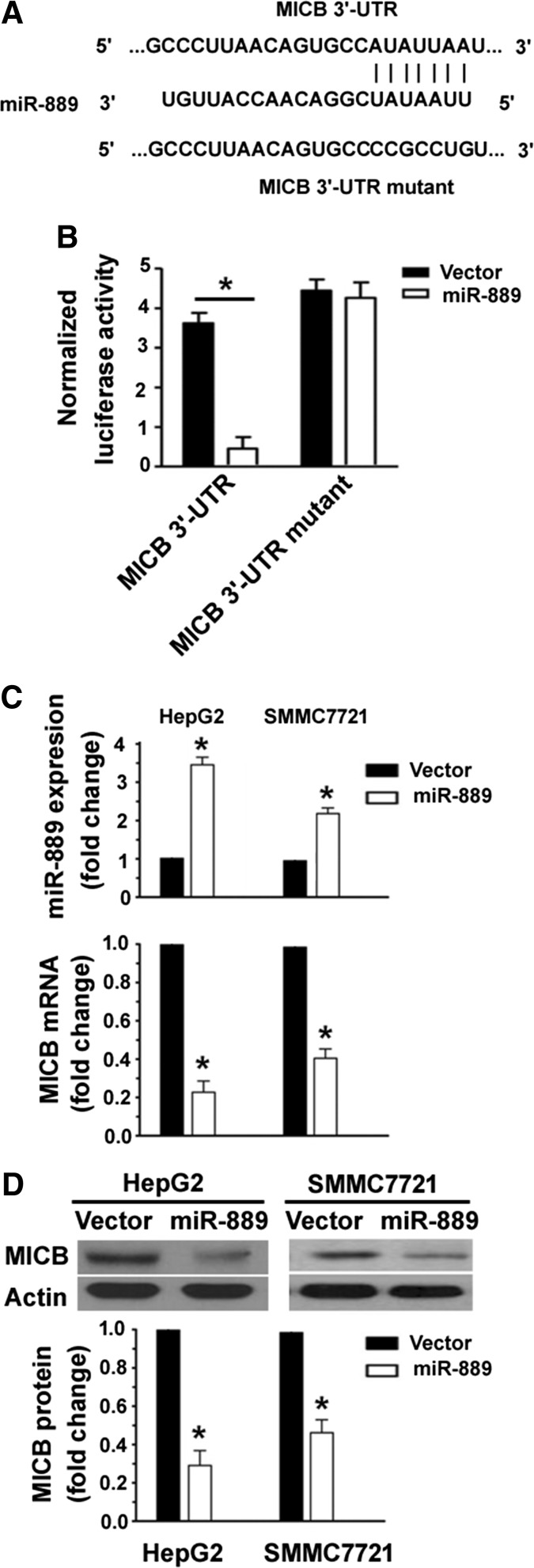
Bioinformatic analysis based on Targetscan software (http://www.targetscan.org/vert_71/) was conducted to search for potential miRNA regulators of MICB. It was predicted that there was a putative miR-889 target site in the 3′-UTR of MICB mRNA (Fig. 1a). To test the ability of miR-889 to target the 3′-UTR of MICB, luciferase reporter constructs were generated by cloning the entire MICB 3′-UTR downstream of the firefly luciferase gene and co-transfected into 293T cells together with miR-889-expressing plasmids. It was found that miR-889 significantly inhibited the luciferase reporter activities (Fig. 1b). Furthermore, mutation of the predicted miR-889 target site abolished miR-889-mediated downregulation of the MICB 3′-UTR reporters, confirming the miR-889 targeting of the MICB 3′-UTR. In addition to miR-889, several other miRNAs including miR-525-5p, miR-198, miR-217, miR-936, and miR-433 were predicted to bind to the 3′-UTR of MICB mRNA (data not shown). When the reporter plasmid carrying the entire wild-type MICB 3′-UTR were co-transfected with miRNA-expressing constructs, the reporter activities were not altered by any of the miRNAs (Fig. 1b). To confirm the negative regulation of MICB expression in HCC by miR-889, we transfected miR-889-expressing plasmids to HepG2 and SMMC7721 cells that substantially express MICB (Fang et al. 2014). As determined by qRT-PCR and Western blot analysis, overexpression of miR-889 significantly reduced the expression levels of MICB in HepG2 and SMMC7721 cells (Fig. 1c, d). Taken together, miR-889 can downregulate the expression of MICB by targeting its 3′-UTR.
Fig. 1.
miR-889 is a negative regulator of MICB. a Bioinformatic analysis based on Targetscan software suggested a putative miR-889 target site in the 3′-UTR of MICB mRNA. b Luciferase reporter assay. 293T cells were co-transfected with MICB 3′-UTR reporters together with miR-889-expressing plasmid or empty vector and luciferase activities were measured at 24 h after transfection. c qRT-PCR analysis of miR-889 and MICB mRNA in HCC cells transfected with miR-889-expressing plasmid or empty vector. d Western blot analysis of MICB protein in HCC cells transfected with miR-889-expressing plasmid or empty vector. Top representative blots. Bottom densitometric analysis of MICB protein levels. Data are representative of three independent experiments. *P < 0.05 versus cells transfected with empty vector
miR-889 is negatively correlated with MICB expression in HCC tissues
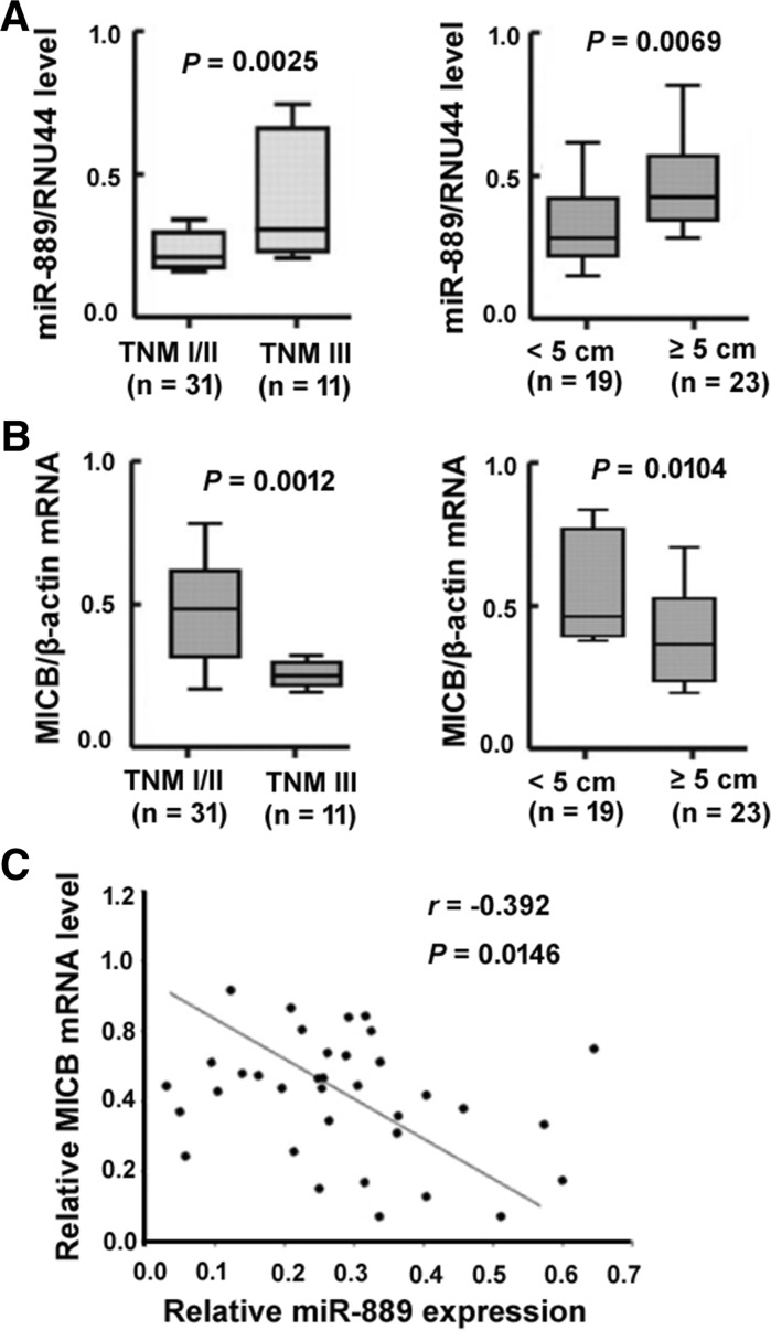
Next, we checked whether miR-889 is negatively correlated with MICB expression in HCC. To this end, we analyzed the expression of MICB and miR-889 by qRT-PCR analysis in 42 cases of HCC. The results showed that miR-889 levels were significantly increased in tumors with advanced TNM stage (P = 0.0025) or larger size (P = 0.0069) (Fig. 2a), whereas MICB mRNA levels showed an opposite trend (Fig. 2b). Moreover, miR-889 expression presented a negative correlation with MICB mRNA levels in these HCC cases (r = −0.392, P = 0.0146; Fig. 2c). These observations suggest that miR-889 may be implicated in the pathogenesis of HCC by regulating the expression of MICB.
Fig. 2.
miR-889 is negatively correlated with MICB expression in HCC tissues. a miR-889 and b MICB mRNA levels in HCC tissues according to TNM stage and tumor size were analyzed by qRT-PCR assay. c The correlation between miR-889 and MICB mRNA levels in 42 cases of HCCs was determined by the Pearson’s correlation coefficient test
miR-889 attenuates NK cell-mediated cytotoxicity by targeting MICB
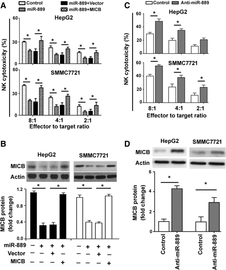
Next, we tested the hypothesis that miR-889 may confer resistance to NK cell-mediated cytotoxicity through downregulation of MICB. The miR-889-overexpressing and control HCC cells were used as target cells and co-incubated with IL-2-activated NK cells. Compared to the control group, the cytotoxicity of NK cells to HCC cells was significantly attenuated by miR-889 overexpression (Fig. 3a). However, delivery of the MICB-expressing plasmid restored the susceptibility of miR-889-overexpressing cells to NK cells. Western blot analysis validated the overexpression of MICB expression in HepG2 by transfection with the MICB-expressing plasmid (Fig. 3b). Furthermore, knockdown of miR-889 via transfection with anti-miR-889 inhibitors led to more pronounced NK cell-mediated lysis, compared to that seen in control cells (Fig. 3c), which was accompanied by increased expression of MICB (Fig. 3d). These results indicate the role for miR-889 in the regulation of HCC cell susceptibility to NK cells.
Fig. 3.
miR-889 attenuates NK cell-mediated cytotoxicity by targeting MICB. a NK cell-mediated cytotoxicity assay. HCC cells were transfected with miR-889-expressing plasmid or together with empty vector or MICB-expressing plasmid and incubated with NK-92 cells at indicated effector to target cell ratios. NK lysis was assessed by measuring LDH release. Data are representative of three independent experiments. b Western blot analysis of MICB protein in HCC cells transfected with indicated constructs. Top representative blots of three independent experiments. Bottom densitometric analysis of Western blots. c HCC cells transfected with anti-miR-889 inhibitors or control inhibitors were incubated with NK-92 cells at indicated effector to target cell ratios and subjected to NK cell-mediated cytotoxicity assay. d Measurement of MICB protein in HCC cells transfected with anti-miR-889 inhibitors or control inhibitors by Western blot analysis. Top representative blots of three independent experiments. Bottom densitometric analysis of Western blots. *P < 0.05
Downregulation of miR-889 mediates the enhancement of NK cytotoxicity by sodium valproate
Finally, we examined whether the enhancement of NK cytotoxicity by HDAC inhibitors is associated with modulation of miR-889 expression. Compared to vehicle-treated cells, there was a significant reduction in miR-889 expression in HepG2 and SMMC7721 cells after exposure to sodium valproate (Fig. 4a). Notably, overexpression of miR-889 prevented the upregulation of MICB induced by sodium valproate (Fig. 4b). Moreover, enforced expression of miR-889 rendered HCC cells more resistant to NK cell-mediated lysis, even in the presence of sodium valproate (Fig. 4c). These data suggest that downregulation of miR-889 is a novel mechanism for HDAC inhibitor-mediated enhancement of NK cell lysis.
Fig. 4.
Downregulation of miR-889 mediates the enhancement of NK cytotoxicity by sodium valproate. a Analysis of miR-889 expression in HCC cells exposed to vehicle or sodium valproate by qRT-PCR analysis. * P < 0.05 versus vehicle control. b qRT-PCR analysis of MICB mRNA expression in HCC cells pre-transfected with miR-889-expressing plasmid or vector 24 h before sodium valproate exposure. c HCC cells with the same treatments as in b were incubated with NK-92 cells at indicated effector to target cell ratios and subjected to NK cell-mediated cytotoxicity assay. Data are representative of three independent experiments. *P < 0.05
Discussion
NK cells are a major component of the host defense system against tumor development. The effector functions of NK cells are affected by the expression status of NKG2D ligands on tumor cells (Pahl and Cerwenka 2015). Several molecular regulators such as IMP3 (Schmiedel et al. 2016), miR-20a (Wang et al. 2014), and HIF-1α (Schilling et al. 2015) have been suggested to contribute to tumor immune escape via downregulation of NKG2D ligands. As a key player in gene expression regulation, the roles of miRNAs in modulation of NK cell-mediated antitumor immunity are attracting increasing attention. A few of miRNAs have been shown to target MICB mRNA, including miR-20a (Xie et al. 2014), miR-10b (Tsukerman et al. 2012), miR-373, miR-107, and miR-16 (Wu et al. 2014). In this study, we identified miR-889 as a novel MICB-targeting miRNA, as evidenced by bioinformatic prediction and luciferase reporter assay. Consistently, overexpression of miR-889 significantly suppressed endogenous MICB expression in HCC cells. It has been documented that miR-889 is deregulated in several human cancers including colorectal cancer (Molina-Pinelo et al. 2014), esophageal squamous cell carcinoma (Xu et al. 2015), and Hodgkin lymphoma (Paydas et al. 2016). Our data demonstrated that high miR-889 levels were significantly associated with advanced TNM stage and larger tumor size in HCC, suggesting its implication in tumor progression. In support of this view, we performed gain-of-function studies in HepG2 and Huh7 HCC cells and found that overexpression of miR-889 facilitated cell growth and invasion (data not shown). Given the capacity of miR-889 to repress MICB expression in HCC cell lines, we investigated their expression associations in HCC specimens. In agreement with the in vitro findings, we noted a significantly negative correlation between miR-889 and MICB expression in HCC cases, suggesting that MICB is a direct target of miR-889 in HCC.
It has been reported that miR-889 can accelerate G1/S transition and cell proliferation in esophageal squamous cell carcinoma cells (Xu et al. 2015), indicating its oncogenic roles in this malignancy. However, few studies are addressing its functions in HCC cells. In this study, we showed that overexpression of miR-889 rendered HCC cells more resistant to NK cell-mediated lysis. In contrast, knockdown of miR-889 enhanced NK lysis in HCC cells, suggesting the ability of miR-889 to govern the susceptibility of HCC cells to NK cells. Rescue studies revealed that overexpression of MICB reversed the protective effects of miR-889 against NK cell-mediated killing of HCC cells. These observations point toward that miR-889 exhibits the ability to protect HCC cells from NK cell attack, which provides a possible explanation for the relationship between miR-889 expression and more aggressive disease in HCC.
To gain more insight into the importance of miR-889 in the regulation of HCC cell susceptibility to NK cells, we checked whether this miRNA is involved in HDAC inhibitor-mediated enhancement of NK cytotoxicity. Compelling evidence has indicated that HDAC inhibitors can upregulate NKG2D ligands and consequently sensitize tumor cells to NK cell-mediated lysis (Zhang et al. 2009; Armeanu et al. 2005; Paydas et al. 2016). Notably, we found that treatment with HDAC inhibitors significantly suppressed the expression of miR-889 in HCC cells, which provides an explanation for the finding that HDAC inhibitors increased the expression of MICB in HepG2 cells (Armeanu et al. 2005). Moreover, enforced expression of miR-889 blocked the upregulation of MICB and enhancement of NK lysis by HDAC inhibitors. These findings suggest that targeting miR-889 may be a promising strategy for improving NK cell-based antitumor therapies in HCC.
In conclusion, our data provide evidence for the involvement of miR-889 in the susceptibility of HCC cells to NK cell-mediated cytotoxicity via targeting of MICB. Downregulation of miR-889 accounts for HDAC inhibitor-induced enhancement of NK lysis in HCC cells. This work suggests a biological rationale for targeting miR-889 in combination with NK cell immunotherapies in the treatment of HCC.
Acknowledgements
This work was supported in part by the Hunan Province Natural Science Foundation of China (No. 14JJ2092).
Contributor Information
Leping Zeng, Email: zengleping@hotmail.com.
Xiaoxin Jiang, Phone: 00867348279467, Email: tYHu4321568@163.com.
References
- Amin PJ, Shankar BS. Sulforaphane induces ROS mediated induction of NKG2D ligands in human cancer cell lines and enhances susceptibility to NK cell mediated lysis. Life Sci. 2015;126:19–27. doi: 10.1016/j.lfs.2015.01.026. [DOI] [PubMed] [Google Scholar]
- Armeanu S, Bitzer M, Lauer UM, Venturelli S, Pathil A, Krusch M, Kaiser S, Jobst J, Smirnow I, Wagner A, Steinle A, Salih HR. Natural killer cell-mediated lysis of hepatoma cells via specific induction of NKG2D ligands by the histone deacetylase inhibitor sodium valproate. Cancer Res. 2005;65:6321–6329. doi: 10.1158/0008-5472.CAN-04-4252. [DOI] [PubMed] [Google Scholar]
- Codo P, Weller M, Meister G, Szabo E, Steinle A, Wolter M, Reifenberger G, Roth P. MicroRNA-mediated down-regulation of NKG2D ligands contributes to glioma immune escape. Oncotarget. 2014;5:7651–7662. doi: 10.18632/oncotarget.2287. [DOI] [PMC free article] [PubMed] [Google Scholar]
- Fang L, Gong J, Wang Y, Liu R, Li Z, Wang Z, Zhang Y, Zhang C, Song C, Yang A, Ting JP, Jin B, Chen L. MICA/B expression is inhibited by unfolded protein response and associated with poor prognosis in human hepatocellular carcinoma. J Exp Clin Cancer Res. 2014;33:76. doi: 10.1186/s13046-014-0076-7. [DOI] [PMC free article] [PubMed] [Google Scholar]
- Groh V, Bahram S, Bauer S, Herman A, Beauchamp M, Spies T. Cell stress-regulated human major histocompatibility complex class I gene expressed in gastrointestinal epithelium. Proc Natl Acad Sci USA. 1996;93:12445–12450. doi: 10.1073/pnas.93.22.12445. [DOI] [PMC free article] [PubMed] [Google Scholar]
- Gu S, Jin L, Zhang F, Sarnow P, Kay MA. Biological basis for restriction of microRNA targets to the 3′ untranslated region in mammalian mRNAs. Nat Struct Mol Biol. 2009;16:144–150. doi: 10.1038/nsmb.1552. [DOI] [PMC free article] [PubMed] [Google Scholar]
- Lanier LL. NKG2D Receptor and Its Ligands in Host Defense. Cancer Immunol Res. 2015;3:575–582. doi: 10.1158/2326-6066.CIR-15-0098. [DOI] [PMC free article] [PubMed] [Google Scholar]
- Min D, Lv XB, Wang X, Zhang B, Meng W, Yu F, Hu H. Downregulation of miR-302c and miR-520c by 1,25(OH)2D3 treatment enhances the susceptibility of tumour cells to natural killer cell-mediated cytotoxicity. Br J Cancer. 2013;109:723–730. doi: 10.1038/bjc.2013.337. [DOI] [PMC free article] [PubMed] [Google Scholar]
- Molina-Pinelo S, Carnero A, Rivera F, Estevez-Garcia P, Bozada JM, Limon ML, Benavent M, Gomez J, Pastor MD, Chaves M, Suarez R, Paz-Ares L, de la Portilla F, Carranza-Carranza A, Sevilla I, Vicioso L, Garcia-Carbonero R. MiR-107 and miR-99a-3p predict chemotherapy response in patients with advanced colorectal cancer. BMC Cancer. 2014;14:656. doi: 10.1186/1471-2407-14-656. [DOI] [PMC free article] [PubMed] [Google Scholar]
- Pahl J, Cerwenka A (2017) Tricking the balance: NK cells in anti-cancer immunity. Immunobiology 222:11–20 [DOI] [PubMed]
- Paydas S, Acikalin A, Ergin M, Celik H, Yavuz B, Tanriverdi K. Micro-RNA (miRNA) profile in Hodgkin lymphoma: association between clinical and pathological variables. Med Oncol. 2016;33:34. doi: 10.1007/s12032-016-0749-5. [DOI] [PubMed] [Google Scholar]
- Ren J, Nie Y, Lv M, Shen S, Tang R, Xu Y, Hou Y, Zhao S, Wang T. Estrogen upregulates MICA/B expression in human non-small cell lung cancer through the regulation of ADAM17. Cell Mol Immunol. 2015;12:768–776. doi: 10.1038/cmi.2014.101. [DOI] [PMC free article] [PubMed] [Google Scholar]
- Ribeiro CH, Kramm K, Gálvez-Jirón F, Pola V, Bustamante M, Contreras HR, Sabag A, Garrido-Tapia M, Hernández CJ, Zúñiga R, Collazo N, Sotelo PH, Morales C, Mercado L, Catalán D, Aguillón JC, Molina MC. Clinical significance of tumor expression of major histocompatibility complex class I-related chains A and B (MICA/B) in gastric cancer patients. Oncol Rep. 2016;35:1309–1317. doi: 10.3892/or.2015.4510. [DOI] [PMC free article] [PubMed] [Google Scholar]
- Schilling D, Tetzlaff F, Konrad S, Li W, Multhoff G. A hypoxia-induced decrease of either MICA/B or Hsp70 on the membrane of tumor cells mediates immune escape from NK cells. Cell Stress Chaperones. 2015;20:139–147. doi: 10.1007/s12192-014-0532-5. [DOI] [PMC free article] [PubMed] [Google Scholar]
- Schmiedel D, Tai J, Yamin R, Berhani O, Bauman Y, Mandelboim O (2016) The RNA binding protein IMP3 facilitates tumor immune escape by downregulating the stress-induced ligands ULPB2 and MICB. Elife 5:e13426 [DOI] [PMC free article] [PubMed]
- Svoronos AA, Engelman DM, Slack FJ. OncomiR or tumor suppressor? The duplicity of microRNAs in cancer. Cancer Res. 2016;76:3666–3670. doi: 10.1158/0008-5472.CAN-16-0359. [DOI] [PMC free article] [PubMed] [Google Scholar]
- Tong HV, le Song H, Hoan NX, Cuong BK, Sy BT, Son HA, Quyet D, Binh VQ, Kremsner PG, Bock CT, Velavan TP, Toan NL. Soluble MICB protein levels and platelet counts during hepatitis B virus infection and response to hepatocellular carcinoma treatment. BMC Infect Dis. 2015;15:25. doi: 10.1186/s12879-015-0754-x. [DOI] [PMC free article] [PubMed] [Google Scholar]
- Tsukerman P, Stern-Ginossar N, Gur C, Glasner A, Nachmani D, Bauman Y, Yamin R, Vitenshtein A, Stanietsky N, Bar-Mag T, Lankry D, Mandelboim O. MiR-10b downregulates the stress-induced cell surface molecule MICB, a critical ligand for cancer cell recognition by natural killer cells. Cancer Res. 2012;72:5463–5472. doi: 10.1158/0008-5472.CAN-11-2671. [DOI] [PubMed] [Google Scholar]
- Wang B, Wang Q, Wang Z, Jiang J, Yu SC, Ping YF, Yang J, Xu SL, Ye XZ, Xu C, Yang L, Qian C, Wang JM, Cui YH, Zhang X, Bian XW. Metastatic consequences of immune escape from NK cell cytotoxicity by human breast cancer stem cells. Cancer Res. 2014;74:5746–5757. doi: 10.1158/0008-5472.CAN-13-2563. [DOI] [PubMed] [Google Scholar]
- Wu J, Zhang XJ, Shi KQ, Chen YP, Ren YF, Song YJ, Li G, Xue YF, Fang YX, Deng ZJ, Xu X, Gao J, Tang KF. Hepatitis B surface antigen inhibits MICA and MICB expression via induction of cellular miRNAs in hepatocellular carcinoma cells. Carcinogenesis. 2014;35:155–163. doi: 10.1093/carcin/bgt268. [DOI] [PubMed] [Google Scholar]
- Xie J, Liu M, Li Y, Nie Y, Mi Q, Zhao S. Ovarian tumor-associated microRNA-20a decreases natural killer cell cytotoxicity by downregulating MICA/B expression. Cell Mol Immunol. 2014;11:495–502. doi: 10.1038/cmi.2014.30. [DOI] [PMC free article] [PubMed] [Google Scholar]
- Xu Y, He J, Wang Y, Zhu X, Pan Q, Xie Q, Sun F. miR-889 promotes proliferation of esophageal squamous cell carcinomas through DAB2IP. FEBS Lett. 2015;589:1127–1135. doi: 10.1016/j.febslet.2015.03.027. [DOI] [PubMed] [Google Scholar]
- Yang H, Lan P, Hou Z, Guan Y, Zhang J, Xu W, Tian Z, Zhang C. Histone deacetylase inhibitor SAHA epigenetically regulates miR-17-92 cluster and MCM7 to upregulate MICA expression in hepatoma. Br J Cancer. 2015;112:112–121. doi: 10.1038/bjc.2014.547. [DOI] [PMC free article] [PubMed] [Google Scholar]
- Zhang C, Wang Y, Zhou Z, Zhang J, Tian Z. Sodium butyrate upregulates expression of NKG2D ligand MICA/B in HeLa and HepG2 cell lines and increases their susceptibility to NK lysis. Cancer Immunol Immunother. 2009;58:1275–1285. doi: 10.1007/s00262-008-0645-8. [DOI] [PMC free article] [PubMed] [Google Scholar]
- Zingoni A, Cecere F, Vulpis E, Fionda C, Molfetta R, Soriani A, Petrucci MT, Ricciardi MR, Fuerst D, Amendola MG, Mytilineos J, Cerboni C, Paolini R, Cippitelli M, Santoni A. Genotoxic stress induces senescence-associated ADAM10-dependent release of NKG2D MIC ligands in multiple myeloma cells. J Immunol. 2015;195:736–748. doi: 10.4049/jimmunol.1402643. [DOI] [PubMed] [Google Scholar]


