1. Introduction
Chronic heart failure (HF) is the most common cause for hospital admissions in Western countries. It is the end result of various cardiovascular diseases, and obesity, diabetes, hypercholesterolaemia, and smoking are among the most important risk factors for these diseases. While the prevalence of smoking has already decreased and will further decline in most developed countries,1 the prevalence of obesity2 and diabetes3 is still on the rise, which will have an important impact on the prevalence of HF in decades to come. The treatment of diabetes has long been a dilemma since many glucose-lowering agents increase the risk of cardiovascular disease and, in particular, of HF.4 Empagliflozin is an inhibitor of the sodium glucose cotransporter 2 (SGLT-2); inhibiting SGLT-2 decreases urinary glucose extraction, thereby reducing serum glucose and glycated haemoglobin (HbA1c) levels. Surprisingly, this drug substantially reduced the risk of HF and even total mortality of patients with diabetes at high cardiovascular risk in the EMPA-REG OUTCOME trial.5 These results spurred new enthusiasm in the medical community that potentially drugs that affect metabolism may be of benefit in patients not only with diabetes but also with HF.6
Independent of its effects on the vasculature, diabetes directly affects cardiac morphology and function and can lead to diabetic cardiomyopathy.7 Patients with HF have either reduced (HFrEF) or preserved ejection fraction (HFpEF), and in particular in patients with HFpEF, metabolic dysfunction is thought to play an important pathophysiological role by triggering inflammation that is an upstream mechanism of cardiomyocyte stiffness and cardiac fibrosis.8
While in patients with HFrEF, inhibiting neuroendocrine activation and slowing heart rate reduce morbidity and mortality, this is not the case in patients with HFpEF.9 This may indicate that the pathophysiology of HFpEF may be different from that of HFrEF and not just an earlier time point in a continuum of HF progression. In light of these concepts, the aim of this Spotlight Issue of Cardiovascular Research is to highlight (i) how metabolic alterations, which occur during obesity and diabetes, impact cardiac biology and function and (ii) how metabolic alterations in the context of cardiac hypertrophy and failure of other causes (hypertension, ischaemia, etc.) contribute to myocardial remodelling and dysfunction.
2. Substrate utilization
The normal heart utilizes primarily fatty acids (∼70%) and to a lesser extent glucose (∼30%). However, it is an omnivore that can adapt its substrate utilization to the respective availability, facilitated by the regulatory feedback loops of the Randle cycle.10 In patients with HFrEF, alterations of substrate utilization occur at different stages of the syndrome, and it has been a controversy whether these alterations are adaptive or maladaptive.10 Ritterhoff and Tian11 focus on these metabolic alterations but also on substrates whose involvement and importance in normal and failing hearts has become apparent only more recently, such as branched chain amino acids and ketone bodies. Furthermore, the consequences of the accumulation of metabolic intermediates that can induce signalling in their own right by epigenetic regulation, post-translational protein modifications (such as GlcNAcylation), lipotoxicity and/or redox signaling are also discussed.
In patients with diabetes, the heart loses its metabolic flexibility related to insulin resistance and peroxisome proliferator activated receptor α (PPARα) signalling, virtually shutting down glucose utilization. Further to PPARα effects, post-translational acetylation of mitochondrial proteins rewires metabolic flux towards fatty acid oxidation in diabetes, thereby further aggravating the metabolic inflexibility of the diabetic heart.12 In this Special Issue, Chong et al.13 delineate the detailed alterations of substrate utilization that occur in the diabetic heart and also touch on the relevance of ketones and lactate as metabolic substrates. Together with the review by Ritterhoff and Tian11, this provides a comprehensive view on substrate utilization and its alterations in diabetes and HF and therefore may allow to identify similarities but also important differences between these disease states. Moreover, several drugs that can interfere with substrate utilization and, in particular, with fatty acid oxidation (such as trimetazidin, perhexilline, and ranolazine) are available but have not been comprehensively tested in patients with HF yet. Furthermore, by gaining new insight into substrate metabolism, novel targets and/or ways to provide substrates may be revealed. A recent promising example is that nutritional ketosis can substantially improve endurance performance in atheletes.14
3. Inflammation
Excessive nutrients, as occur with obesity can trigger inflammatory signalling pathways. Inflammation is recognized as an important contributor to diabetes, cardiac hypertrophy, and HF. In this Spotlight Issue, there are two reviews addressing this important topic. Nishida and Otsu15 discuss the role of inflammation in metabolic cardiomyopathy and Frati et al.16 provide an overview of the molecular mechanisms linking inflammation to diabetic cardiomyopathy. In a previous issue of Cardiovascular Research, Kalfon et al.17 reported on the protective role of activating transcription factor 3 (ATF3), which dampens inflammation, insulin sensitivity, and maladaptive cardiac remodelling in response to a high-fat diet.
4. Cell–cell communication
Very recently, Wang et al.18 identified a protective cell–cell communication pathway that temporarily protects from the development of diabetic cardiomyopathy. In response to high glucose, endothelial cells release heparanase that is taken up by cardiac myoctyes, where it stimulates the expression of anti-apoptotic genes. This protective mechanism is lost during more long-standing diabetes, where heparanase may contribute to lipotoxicity and thereby potentially to the development of diabetic cardiomyopathy.18 Furthermore, Wan and Rodrigues19 from the same lab recently gave a comprehensive overview on endothelial–cardiac myocyte cross-talk in diabetic cardiomyopathy, highlighting important differences in substrate utilization between these cell types. Furthermore, they also review how one cell type can influence the fate of the respective other in a complex intercellular signalling network that may harbour some promising targets for the treatment of diabetic cardiomyopathy.
5. Atrial fibrillation
Diabetes is also an important risk factor for atrial fibrillation (AF). The serine protease cathepsin A (CatA) is up-regulated in diabetes and involved in the degradation of extracellular peptides. In a recent issue of Cardiovascular Research, Linz et al.20 observed that transgenic overexpression of CatA increases atrial fibrosis and susceptibility to AF. Vice versa, pharmacological blockade of CatA ameliorated structural and electrical remodelling of atria in a diabetes rat model, reducing the susceptibility to AF. Therefore, CatA antagonism may be a novel therapeutic option in patients with diabetes to prevent and/or treat AF.
6. Gender aspects
Female sex appears to predispose to metabolic cardiomyopathies: Heart disease is five times more common in diabetic women but only twice as common in diabetic men. Sex differences in baseline cardiac metabolism and calcium handling have also been described. Murphy et al.21 discuss the signalling pathways that play a role in these sex differences in metabolic cardiomyopathies.
In patients with HF, cardiolipin, a key phospholipid of the inner mitochondrial membrane, becomes oxidized and thereby its function impaired. This can lead to a disassembly of the complexes of the respiratory chain, which can lead to more production of reactive oxygen species (ROS) and less adenosine triphosphate.22 SS-31 is a novel peptide drug that specifically binds to cardiolipin and can ameliorate mitochondrial ROS and maladaptive remodelling in several animal models of HF.22,23 Therefore, cardiolipin function and dysfunction have gained increasing interest in the scientific community dealing with cardiovascular diseases. The Barth syndrome is a rare X-linked recessive disorder that causes cardiomyopathy, immune defects, and fatigue in young boys. It is caused by a mutation of the gene encoding tafazzin, the terminal enzyme in the biosynthesis of cardiolipin. Dudek and Maack24 review the current literature and highlight which consequences the ensuing cardiolipin remodelling has for mitochondrial, cellular and in particular, cardiac function. Besides gaining insight into one rare, yet classical ‘mitochondrial cardiomyopathy’, these insights will also allow conclusions for more general forms of cardiomyopathies due to the mentioned cardiolipin defects in HF patients.22
7. Conclusions
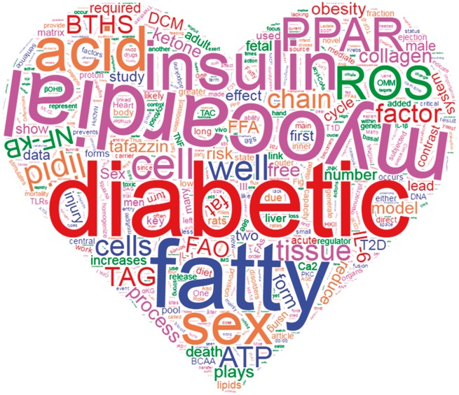
In conclusion, metabolic cardiomyopathies are an important and growing health concern. This Spotlight Issue covers traditional (metabolism) and newer (inflammation and sex differences) topics in metabolic cardiomyopathies and provides fresh insights into a growing and—in light of the current epidemiological developments—urgently needed area of research and treatment (Figure 1).
Figure 1.
Cloud of all the words included in the papers of this Special issue on metabolic cardiomyopathies.
Acknowledgements
C.M. is supported by the Deutsche Forschungsgemeinschaft (Heisenberg Professur, Ma2528/7-1 and SFB-894) and the Corona Stiftung. E.M. is supported by the NHLBI, NIH intramural program.
Conflict of interest: none declared.
References
- 1. Bilano V, Gilmour S, Moffiet T, d'Espaignet ET, Stevens GA, Commar A, Tuyl F, Hudson I, Shibuya K.. Global trends and projections for tobacco use, 1990–2025: an analysis of smoking indicators from the WHO Comprehensive Information Systems for Tobacco Control. Lancet. 2015;385:966–976. [DOI] [PubMed] [Google Scholar]
- 2. Ng M, Fleming T, Robinson M, Thomson B, Graetz N, Margono C, Mullany EC, Biryukov S, Abbafati C, Abera SF, Abraham JP, Abu-Rmeileh NME, Achoki T, AlBuhairan FS, Alemu ZA, Alfonso R, Ali MK, Ali R, Guzman NA, Ammar W, Anwari P, Banerjee A, Barquera S, Basu S, Bennett DA, Bhutta Z, Blore J, Cabral N, Nonato IC, Chang J-C, Chowdhury R, Courville KJ, Criqui MH, Cundiff DK, Dabhadkar KC, Dandona L, Davis A, Dayama A, Dharmaratne SD, Ding EL, Durrani AM, Esteghamati A, Farzadfar F, Fay DFJ, Feigin VL, Flaxman A, Forouzanfar MH, Goto A, Green MA, Gupta R, Hafezi-Nejad N, Hankey GJ, Harewood HC, Havmoeller R, Hay S, Hernandez L, Husseini A, Idrisov BT, Ikeda N, Islami F, Jahangir E, Jassal SK, Jee SH, Jeffreys M, Jonas JB, Kabagambe EK, Khalifa SEAH, Kengne AP, Khader YS, Khang Y-H, Kim D, Kimokoti RW, Kinge JM, Kokubo Y, Kosen S, Kwan G, Lai T, Leinsalu M, Li Y, Liang X, Liu S, Logroscino G, Lotufo PA, Lu Y, Ma J, Mainoo NK, Mensah GA, Merriman TR, Mokdad AH, Moschandreas J, Naghavi M, Naheed A, Nand D, Narayan KMV, Nelson EL, Neuhouser ML, Nisar MI, Ohkubo T, Oti SO, Pedroza A, Prabhakaran D, Roy N, Sampson U, Seo H, Sepanlou SG, Shibuya K, Shiri R, Shiue I, Singh GM, Singh JA, Skirbekk V, Stapelberg NJC, Sturua L, Sykes BL, Tobias M, Tran BX, Trasande L, Toyoshima H, van de Vijver S, Vasankari TJ, Veerman JL, Velasquez-Melendez G, Vlassov VV, Vollset SE, Vos T, Wang C, Wang X, Weiderpass E, Werdecker A, Wright JL, Yang YC, Yatsuya H, Yoon J, Yoon S-J, Zhao Y, Zhou M, Zhu S, Lopez AD, Murray CJL, Gakidou E.. Global, regional, and national prevalence of overweight and obesity in children and adults during 1980–2013: a systematic analysis for the Global Burden of Disease Study 2013. Lancet. 2015;384:766–781. [DOI] [PMC free article] [PubMed] [Google Scholar]
- 3. Collaboration NCDRF. Worldwide trends in diabetes since 1980: a pooled analysis of 751 population-based studies with 4·4 million participants. Lancet. 2016;387:1513–1530. [DOI] [PMC free article] [PubMed] [Google Scholar]
- 4. Udell JA, Cavender MA, Bhatt DL, Chatterjee S, Farkouh ME, Scirica BM.. Glucose-lowering drugs or strategies and cardiovascular outcomes in patients with or at risk for type 2 diabetes: a meta-analysis of randomised controlled trials. Lancet Diabetes Endocrinol. 2015;3:356–366. [DOI] [PubMed] [Google Scholar]
- 5. Zinman B, Wanner C, Lachin JM, Fitchett D, Bluhmki E, Hantel S, Mattheus M, Devins T, Johansen OE, Woerle HJ, Broedl UC, Inzucchi SE.. Empagliflozin, cardiovascular outcomes, and mortality in type 2 diabetes. N Engl J Med. 2015;373:2117–2128. [DOI] [PubMed] [Google Scholar]
- 6. Marx N, McGuire DK.. Sodium-glucose cotransporter-2 inhibition for the reduction of cardiovascular events in high-risk patients with diabetes mellitus. Eur Heart J. 2016;37:3192–3200. [DOI] [PubMed] [Google Scholar]
- 7. Seferović PM, Paulus WJ.. Clinical diabetic cardiomyopathy: a two-faced disease with restrictive and dilated phenotypes. Eur Heart J. 2015;36:1718–1727. [DOI] [PubMed] [Google Scholar]
- 8. Paulus WJ, Tschöpe C.. A novel paradigm for heart failure with preserved ejection fraction: comorbidities drive myocardial dysfunction and remodeling through coronary microvascular endothelial inflammation. J Am Coll Cardiol. 2013;62:263–271. [DOI] [PubMed] [Google Scholar]
- 9. Ponikowski P, Voors AA, Anker SD, Bueno H, Cleland JG, Coats AJ, Falk V, Gonzalez-Juanatey JR, Harjola VP, Jankowska EA, Jessup M, Linde C, Nihoyannopoulos P, Parissis JT, Pieske B, Riley JP, Rosano GM, Ruilope LM, Ruschitzka F, Rutten FH, van der Meer P, Authors/Task Force M, Document R. 2016 ESC Guidelines for the diagnosis and treatment of acute and chronic heart failure: The Task Force for the diagnosis and treatment of acute and chronic heart failure of the European Society of Cardiology (ESC). Developed with the special contribution of the Heart Failure Association (HFA) of the ESC. Eur J Heart Fail. 2016;18:891–975. [DOI] [PubMed] [Google Scholar]
- 10. Nickel A, Löffler J, Maack C.. Myocardial energetics in heart failure. Basic Res Cardiol 2013;108:358. [DOI] [PubMed] [Google Scholar]
- 11. Ritterhoff J, Tian R.. Metabolism in cardiomyopathy: every substrate matters. Cardiovasc Res. 2017;113:411--421. [DOI] [PMC free article] [PubMed] [Google Scholar]
- 12. Vazquez EJ, Berthiaume JM, Kamath V, Achike O, Buchanan E, Montano MM, Chandler MP, Miyagi M, Rosca MG.. Mitochondrial complex I defect and increased fatty acid oxidation enhance protein lysine acetylation in the diabetic heart. Cardiovasc Res. 2015;107:453–465. [DOI] [PMC free article] [PubMed] [Google Scholar]
- 13. Chong CR, Clarke K, Levelt E.. Metabolic remodelling in diabetic cardiomyopathy. Cardiovasc Res. 2017;113:422--430. [DOI] [PMC free article] [PubMed] [Google Scholar]
- 14. Cox Pete J, Kirk T, Ashmore T, Willerton K, Evans R, Smith A, Murray Andrew J, Stubbs B, West J, McLure Stewart W, King MT, Dodd Michael S, Holloway C, Neubauer S, Drawer S, Veech Richard L, Griffin Julian L, Clarke K.. Nutritional ketosis alters fuel preference and thereby endurance performance in athletes. Cell Metab. 2016;24:256–268. [DOI] [PubMed] [Google Scholar]
- 15. Nishida K, Otsu K.. Inflammation and metabolic cardiomyopathy. Cardiovasc Res. 2017;113:389--398. [DOI] [PubMed] [Google Scholar]
- 16. Frati G, Schirone L, Chimenti I, Yee D, Biondi-Zoccai G, Volpe M, Sciarretta S.. An overview of the inflammatory signaling mechanisms in the myocardium underlying the development of diabetic cardiomyopathy. Cardiovasc Res. 2017;13:378--388. [DOI] [PubMed] [Google Scholar]
- 17. Kalfon R, Koren L, Aviram S, Schwartz O, Hai T, Aronheim A.. ATF3 expression in cardiomyocytes preserves homeostasis in the heart and controls peripheral glucose tolerance. Cardiovasc Res .2016;113:134–146. [DOI] [PubMed] [Google Scholar]
- 18. Wang F, Jia J, Lal N, Zhang D, Chiu AP-L, Wan A, Vlodavsky I, Hussein B, Rodrigues B.. High glucose facilitated endothelial heparanase transfer to the cardiomyocyte modifies its cell death signature. Cardiovasc Res. 2016;112:622–625. [DOI] [PMC free article] [PubMed] [Google Scholar]
- 19. Wan A, Rodrigues B.. Endothelial cell–cardiomyocyte crosstalk in diabetic cardiomyopathy. Cardiovasc Res. 2016;111:172–183. [DOI] [PMC free article] [PubMed] [Google Scholar]
- 20. Linz D, Hohl M, Dhein S, Ruf S, Reil J-C, Kabiri M, Wohlfart P, Verheule S, Böhm M, Sadowski T, Schotten U.. Cathepsin A mediates susceptibility to atrial tachyarrhythmia and impairment of atrial emptying function in Zucker diabetic fatty rats. Cardiovasc Res. 2016;110:371–380. [DOI] [PubMed] [Google Scholar]
- 21. Murphy E, Amanakis G, Fillmore N, Parks RJ, Sun J.. Sex differences in metabolic cardiomyopathy. Cardiovasc Res. 2017;113:370--377. [DOI] [PMC free article] [PubMed] [Google Scholar]
- 22. Szeto HH. First-in-class cardiolipin-protective compound as a therapeutic agent to restore mitochondrial bioenergetics. Br J Pharmacol. 2014;171:2029–2050. [DOI] [PMC free article] [PubMed] [Google Scholar]
- 23. Nickel AG, von Hardenberg A, Hohl M, Löffler JR, Kohlhaas M, Becker J, Reil JC, Kazakov A, Bonnekoh J, Stadelmaier M, Puhl SL, Wagner M, Bogeski I, Cortassa S, Kappl R, Pasieka B, Lafontaine M, Lancaster CR, Blacker TS, Hall AR, Duchen MR, Kaestner L, Lipp P, Zeller T, Müller C, Knopp A, Laufs U, Böhm M, Hoth M, Maack C.. Reversal of mitochondrial transhydrogenase causes oxidative stress in heart failure. Cell Metab. 2015;22:472–484. [DOI] [PubMed] [Google Scholar]
- 24. Dudek J, Maack C.. Barth syndrome cardiomyopathy. Cardiovasc Res. 2017;113: 399--410 [DOI] [PubMed] [Google Scholar]


