Abstract
Background
Juvenile dermatomyositis (JDM) is an auto-immune muscle disease which presents with skin manifestations and muscle weakness. At least 10% of the patients with JDM present with acquired lipodystrophy. Laminopathies are caused by mutations in the lamin genes and cover a wide spectrum of diseases including muscular dystrophies and lipodystrophy. The p.T10I LMNA variant is associated with a phenotype of generalized lipodystrophy that has also been called atypical progeroid syndrome.
Case presentation
A previously healthy female presented with bilateral proximal lower extremity muscle weakness at age 4. She was diagnosed with JDM based on her clinical presentation, laboratory tests and magnetic resonance imaging (MRI). She had subcutaneous fat loss which started in her extremities and progressed to her whole body. At age 7, she had diabetes, hypertriglyceridemia, low leptin levels and low body fat on dual energy X-ray absorptiometry (DEXA) scan, and was diagnosed with acquired generalized lipodystrophy (AGL). Whole exome sequencing (WES) revealed a heterozygous c.29C > T; p.T10I missense pathogenic variant in LMNA, which encodes lamins A and C. Muscle biopsy confirmed JDM rather than muscular dystrophy, showing perifascicular atrophy and perivascular mononuclear cell infiltration. Immunofluroscence of skin fibroblasts confirmed nuclear atypia and fragmentation.
Conclusions
This is a unique case with p.T10I LMNA variant displaying concurrent JDM and AGL. This co-occurrence raises the intriguing possibility that LMNA, and possibly p.T10I, may have a pathogenic role in not only the occurrence of generalized lipodystrophy, but also juvenile dermatomyositis. Careful phenotypic characterization of additional patients with laminopathies as well as individuals with JDM is warranted.
Electronic supplementary material
The online version of this article (10.1186/s40842-018-0058-3) contains supplementary material, which is available to authorized users.
Keywords: Laminopathies, Myositis, LMNA, Whole exome sequencing, P.T10I, Muscle biopsy
Background
Juvenile dermatomyositis (JDM) is an autoimmune inflammatory disease, which typically affects the proximal skeletal muscles and presents with characteristic cutaneous manifestations in the first 18 years of life [1]. Among other multi-system manifestations, association with abnormal fat distribution and metabolic abnormalities has been recognized to fit the phenotype of lipodystrophy. The reported prevalence of lipodystrophy in patients with JDM is 8–40% In a large multi-center study where 353 patients with JDM were evaluated for having lipodystrophy, 28 JDM patients were found to have lipodystrophy, either generalized or partial [1].
Acquired generalized lipodystrophy (AGL) is characterized by progressive loss of subcutaneous adipose tissue, which starts before adolescence. The fat loss affects the whole body and results in low serum leptin levels [1, 2]. As a consequence, patients experience various metabolic complications such as hypertriglyceridemia, hyperphagia, severe insulin resistance leading to diabetes, and non-alcoholic steatohepatitis. These metabolic derangements often result in serious complications such as recurrent episodes of acute pancreatitis, premature cardiovascular disease, and hepatic cirrhosis [1, 3].
Mutations in the LMNA gene cause a wide spectrum of diseases called laminopathies which include muscular dystrophies such as limb girdle muscular dystrophy type 1 (LGMD); progeroid syndromes like Hutchinson Gilford progeria syndrome (HGPS); and familial partial lipodystrophy (FPLD) which is a lipodystrophy syndrome [4, 5]. LMNA p.T10I results from a missense mutation (c.29C > T) in exon 1 and leads to a mutant lamin A protein. A total of nine cases presenting with this mutation, including the case reported herein, have recently been described in a case series as part of a global collaboration [6] with 5 cases presenting with a phenotype suggestive of AGL [4, 5, 7] and initially deemed to have either atypical progeroid syndrome or AGL. However, no other case with this mutation was reported to have lipodystrophy and JDM concurrently and none have undergone a muscle biopsy to the best of our knowledge.
Here, we present a unique patient harboring the heterozygous c.29C > T (p.T10I) mutation in lamin A/C who presented originally with JDM followed by what was diagnosed as AGL. This case provides evidence that the spectrum of muscle abnormalities caused by laminopathies may be wider than previously recognized.
Methods
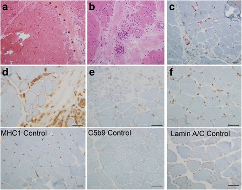
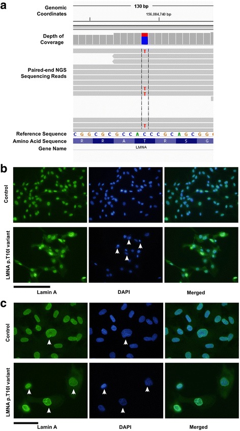
Our patient’s metabolic characteristics were previously reported in a paper describing metabolic response of lipodystrophy patients to metreleptin. Methods for metabolic characterization were also included in this report [2]. Informed assent and consent were obtained from the patient and her mother for genetic testing and fibroblast collection, as well as from the control patient with Duchenne muscular dystrophy under approval from the Institutional Review Board of the University of Michigan Medical School. Histopathological examination of fresh frozen muscle biopsy specimen (Fig. 1) was performed using standard Hematoxyin Eosin(HE) staining and immunostains for CD3/CD20, CD163/CD4, C5b9, MHC1, Lamin A/C, following standard procedures with specific antibodies (Additional file 1: Appendix 1). Whole exome sequencing (WES) (Fig. 2) and confirmation via Sanger sequencing were performed as previously described [8] (Additional file 1: Appendix 2). Skin fibroblast cultures obtained from the patient and a control Duchenne muscular dystrophy patient were analyzed via indirect immunofluorescence with the lamin A antibody [9].
Fig. 1.

a Hematoxylin Eosin (HE) staining shows prominent perifascicular atrophy (asterisk). b HE staining highlights mononuclear infiltration around a blood vessel and infiltrating the endomysium. c CD163/CD4 immunostaining highlights several CD4 T-cells (red) in the perivascular and endomysial space with associated macrophages (brown) (d) MHC1 immunostaining shows abnormal sarcolemmal membrane overexpression in myocytes, note the normal control below should only highlight capillaries (e) C5b9 immunostaining highlights complement deposition in capillaries associated with affected fibers; note normal control does not show any capillary immunostain (f) Lamin A/C staining shows nuclear staining comparable to normal control. Scale bars: 50 μm
Fig. 2.

p.T10I LMNA mutation disrupts nuclear structure. a Integrated Genomics Viewer (IGV) screenshot of 130 bp paired-end whole exome sequencing reads, represented by horizontal gray bars, demonstrated a single nucleotide change (C to T) at genomic position of chromosome 1 g.156,084,738 (GRCh37/hg19) resulting in a missense alteration at codon 29 of LMNA replacing a threonine (T) with an isoleucine (I): NM_170707.3(LMNA):c.29C > T (p.T10I) (b) Skin fibroblast cell cultures of our patient with heterozygous p.T10I LMNA mutation and a Duchenne muscular dystrophy patient were analyzed by immunoflourescence using an antibody to lamin A. Cell cultures were stained with primary lamin A antibody, followed by 4′,6-diamidino-2 phenylindole (DAPI) nuclear staining and with the Alexa 488 goat anti-rabbit IgG. Scale bar: 200 μm. c Higher magnification images of the nuclear staining shown in (b); scale bar: 50 μm. Arrows in panel (b) indicate fragmented nuclei; arrows in panel (c) indicate examples of atypically shaped nuclei and/or nuclei with ruffles of lamin A staining
Case presentation
The patient is a 17 year-old African-American female, who had normal fat development until age 4 when she started losing weight and muscle strength. At age 6, she presented with bilateral proximal lower extremity weakness and had elevated levels of erythrocyte sedimentation rate (ESR), C-reactive protein (CRP), creatinine kinase (CK), lactate dehydrogenase (LDH), aspartate aminotransferase (AST) and aldolase. T2-weighted (magnetic resonance imaging).
MRI images of her lower extremities showed hyperintensity in her quadriceps muscles consistent with myositis. On the basis of her clinical presentation, laboratory results and MRI, she was diagnosed with JDM. Her presentation was reported previously [2]. On physical examination, she had generalized loss of subcutaneous fat including her face, a high-arched palate, micrognathia, a small mid-face and bilateral parotid enlargement. She also had progressive contractures in most upper extremity joints, calcinosis on her knuckles and mottled skin throughout her upper body. She had hepatosplenomegaly and non-alcoholic steatohepatitis. Her leptin level was < 0.7 ng/dL, and her DEXA scan showed 9.3% body fat. She was diagnosed with AGL, with associated diabetes and polycystic ovarian syndrome. In the next 7 years, she had severe elevations in triglycerides > 10,000 mg/dL and multiple episodes of acute pancreatitis and diabetic ketoacidosis. Echocardiogram performed at age 9 showed moderate aortic stenosis and atrial septal defect, ostium secundum type. She also had ovarian cysts and nephromegaly due to bilateral renal cysts. Despite being on methotrexate for JDM, her serum muscle enzyme levels fluctuated. For her lipodystrophy, she was treated with metreleptin [2].
Whole exome sequencing done at age 16 identified a heterozygous missense mutation (c.29C > T) in LMNA resulting in p.T10I substitution which is considered to be pathogenic (Fig. 2). This was not maternally inherited. The patient’s father had sudden cardiac death at age 49, so a blood sample was not available. He was reported to be thin, especially on the face and was diabetic in the last 2 years of his life. Further he had history of thyroid cancer as conveyed by patient and her mother. The patient’s WES data was interrogated for pathogenic variants in the genes that might be related to JDM, such as TNF, HLADRB1, IL1RN, IL1A, IL1B, ISG15, BLK and CCL21. No pathogenic variants were found in these genes. Of note, PLCL1 gene was not covered in the patient’s WES data, hence was not screened. A muscle biopsy was obtained from her right quadriceps muscle which confirmed the earlier diagnosis of JDM, including perifascicular atrophy, perivascular mononuclear cell infiltrates and upregulation of sarcolemmal MHC1 (Fig. 1). Immunofluorescence staining of skin fibroblasts from the patient and a control patient with Duchenne muscular dystrophy which is used as a non- LMNA related control both displayed positive nuclear staining for lamin A (Fig. 2). However, LMNA mutant nuclei exhibited greater nuclear atypia and fragmentation compared to the control fibroblasts from an unrelated myopathy without LMNA mutations. Our results in Fig. 2 are consistent with previous reports that scored atypical nuclei which reported that control fibroblasts have occaisonal atypical nuclei in ~ 19% of cells while the number of atypical nuclei in fibroblasts from patients with lamin A/C mutations were much more common (33–92%) [8]. It should be noted that it is very reasonable to use Duchenne muscular dystrophy (DMD) as a control, and although there is one report of a case study of a DMD patient with altered nuclear structure similar to LMNA related muscular dystrophy, further analysis showed this patient also carried a mutation in Nesprin-1, and patients with other mutations in dystrophin showed no significant differences in nuclear structure from non-disease human fibroblasts [10].
Discussion
Here we report a patient with concurrent JDM and AGL with some atypical features. She was found to have a p.T10I mutation in the lamin A/C region encoding the N-terminal head; such mutations were previously associated with generalized lipodystrophy in patients classified as having atypical progeroid syndrome [4, 5, 7]. To our knowledge, she is the first with this mutation to present with both generalized lipodystrophy and biopsy proven JDM. In fact, the nine cases observed including our case were summarized in a new paper recently and none of the others presented with muscle disease [6]. Because different lamin A/C mutations cause various muscle disorders as well as lipodystrophy phenotypes [5], we propose that the presence of JDM and AGL might be linked to the p.T10I mutation in this patient.
The wide spectrum of diseases within the laminopathies is attributed to different mutations in LMNA leading to various modifications in the tertiary structure of the lamin A/C proteins. In addition to disruptions in the nuclear structure leading to fragility, some mutations are known to interfere with cell division, alter interactions with certain transcription factors, or modify gene expression (8). It was proposed earlier that the p.T10I mutation, which is located in the extreme amino terminus of the lamin A protein, might have a causal role in detachment of chromatin from the nuclear lamina as well as disruption of lamin polymerization [5, 8]. Four previous reports associate inflammatory myositis with LMNA variants, suggesting that the muscle disease of laminopathy is not restricted to muscular dystrophy and may be more complicated [11–14].
While muscle biopsies are now rarely performed in suspected cases of LMNA mutations causing muscular dystrophy, previous reports have shown that some LMNA muscular dystrophy patients have marked inflammatory cell infiltrates, although without noted upregulation of MHC [15]. More careful analysis of potential causes of the muscle pathology may reveal greater overlap between the lipodystrophy syndromes, myositis, and muscle diseases associated with lamin A/C mutations than previously suggested. Based on previous work, JDM appears to be a heterogeneous phenotype and to date, no clear molecular etiology has emerged although HLA associations and some genome wide association study (GWAS) loci have been reported [16]. Our case and a few other published reports [12–14] raise the possibility that nuclear lamins may be implicated in the development of inflammatory myopathies. We propose that deeper muscular phenotyping of laminopathies in patients who present with this genotype should be undertaken to understand if we are correct especially if abnormalities in muscle enzymes can be demonstrated. The selective genetic modifiers that result in different tissue manifestations despite having the same mutation remain to be defined.
Conclusion
LMNA mutations are associated with an array of diseases called laminopathies which include muscular dystrophy and lipodystrophy, among other diseases. However, no certain causal relation between LMNA mutations and inflammatory muscle diseases has been established. We posit that both inflammatory muscle disease and generalized lipodystrophy in this patient are due to the lamin A/C T10I variant, although a multifactorial etiology cannot be excluded. This case broadens the spectrum of laminopathies; more importantly, it provides the first potential molecular link to the known association between JDM and lipodystrophy syndromes. Further studies probing the interaction between nuclear envelope proteins and the immunotranscriptome are needed to unlock the mysteries of this long recognized but unexplained association.
Additional file
Appendix 1. Antibodies used for immunohistochemistry. Histopathological examination using fresh frozen muscle biopsy specimen from the patient was performed using the immunostains for: MHC1, Lamin A/C, C5b9, CD163/CD4, CD3/CD20. The antibodies used and respective protocols are shown. Appendix 2 Sanger sequencing chromatogram completed by a CLIA certified laboratory. Data from Sanger sequencing was used to confirm the patient’s whole exome sequencing (WES) results. Similar to the WES data, Sanger sequencing chromatogram revealed a heterozygous c.29 C > T mutation in exon 1. In the figure, both the wild type and the patient’s data with this mutation are shown. (PDF 618 kb)
Acknowledgements
We are grateful to our patient and her mother for providing us with the opportunity to learn from her. Her severe metabolic state allowed us to pursue bringing leptin studies to the University of Michigan and provided the inspiration to continue this line of work. We dedicate this work to her and others like her who struggle with rare diseases around the globe. We are also grateful to all medical staff who contributed to her care at Mott Children’s Hospital and her primary care physician Dr. Tisa Johnson, Dr. Nevin Ajluni, and Adam H. Neidert who helped in the care of the patient and the leptin therapy study. We also thank the following sources of philanthropic gifts for Lipodystrophy Research at the University of Michigan: Mr. And Mrs. James Sopha, the White Point Foundation of Turkey (Istanbul, Turkey) and Ionis Pharmaceuticals (Carlsbad, CA). Dr. Innis acknowledges support of the Morton S. and Henrietta K. Sellner Professorship in Human Genetics.
Funding
This work was supported by NIH grants R01 DK088114, R21DK098776, UL1TR000433, DK034933, Nutrition Obesity Research Centers grant: P30 DK089503.
Abbreviations
- AGL
Acquired generalized lipodystrophy
- AST
Aspartate aminotransferase
- BLK
Blymphocyte kinase
- c.29C > T
Replacement of a cytosine with a thymidine at codon 29
- C5b9
Complement membrane attack complex
- CCL21
Chemokine (C-C motif) ligand 21
- CD
Cluster of differentiation 3
- CK
Creatinine kinase
- CRP
C-reactive protein
- DEXA
Dual energy X-ray absorptiometry
- ESR
Erythrocyte sedimentation rate
- FPLD
Familial partial lipodystrophy
- GWAS
Genome wide association study
- HGPS
Hutchinson Gilford progeria syndrome
- HLA DRB1
Human leukocyte antigen DRB1
- IL1A
Interleukin 1 alpha
- IL1B
Interleukin 1 beta
- IL1RN
Interleukin 1 receptor antagonist
- ISG15
Interferon stimulated gene 15
- JDM
Juvenile dermatomyositis
- LDH
Lactate dehydrogenase
- LGMD
Limb girdle muscular dystrophy
- MHC1
Major histocompatibility complex class I
- MRI
Magnetic resonance imaging
- p.T10I
Replacement of threonine with isoleucine at position 10
- PLCL1
Phospholipase C Like 1
- TNF
Tumor necrosis factor
- WES
Whole exome sequencing
Authors’ contributions
Clinical data were collected and interpreted by EAO, MS, SK, MR, GFB and SCP. WES data were reviewed and interpreted by AR, MKT, PT, JWI and EAO. Fibroblast cell lines were established and LMNA/control staining 199 were performed by AC and DEM. MBO provided critical scientific advice. Funding for studies were obtained by DEM and EAO. The manuscript was drafted by MS, SK and EAO. Artwork was compiled by RM. All authors read, edited and approved the final manuscript.
Ethics approval and consent to participate
Informed assent and consent were obtained from the patient and her mother for genetic testing and fibroblast collection, as well as from the control patient with Duchenne muscular dystrophy under approval from the Institutional Review Board of the University of Michigan Medical School.
Consent for publication
Consent for publication has been obtained from the patient and her mother, as well as the control subject.
Availability of data and materials: The datasets used and/or analysed during the current study are available from the corresponding author on reasonable request.
Competing interests
EAO received grant support from and served as an advisor to Amylin Pharmaceuticals LLC, Bristol-Myers-Squibb, and AstraZeneca. She currently receives grant support and is an advisor to Aegerion Pharmaceuticals, Akcea Therapeutics and Ionis Pharmaceuticals. Other authors have no financial relationships relevant to this article to disclose.
Publisher’s Note
Springer Nature remains neutral with regard to jurisdictional claims in published maps and institutional affiliations.
Footnotes
Electronic supplementary material
The online version of this article (10.1186/s40842-018-0058-3) contains supplementary material, which is available to authorized users.
References
- 1.Bingham A, Mamyrova G, Rother KI, Oral E, Cochran E, Premkumar A, Kleiner D, James-Newton L, Targoff IN, Pandey JP, Carrick DM, Sebring N, O'Hanlon TP, Ruiz-Hidalgo M, Turner M, Gordon LB, Laborda J, Bauer SR, Blackshear PJ, Imundo L, Miller FW, Rider LG. Childhood myositis heterogeneity study G. Predictors of acquired lipodystrophy in juvenile-onset dermatomyositis and a gradient of severity. Medicine (Baltimore) 2008;87:70–86. doi: 10.1097/MD.0b013e31816bc604. [DOI] [PMC free article] [PubMed] [Google Scholar]
- 2.Lebastchi J, Ajluni N, Neidert A, Oral EA. A report of three cases with acquired generalized lipodystrophy with distinct autoimmune conditions treated with Metreleptin. J Clin Endocrinol Metab. 2015;100:3967–3970. doi: 10.1210/jc.2015-2589. [DOI] [PMC free article] [PubMed] [Google Scholar]
- 3.Ajluni N, Meral R, Neidert AH, Brady GF, Buras E, McKenna B, DiPaola F, Chenevert TL, Horowitz JF, Buggs-Saxton C, Rupani AR, Thomas PE, Tayeh MK, Innis JW, Omary MB, Conjeevaram H, Oral EA. Spectrum of disease associated with partial lipodystrophy: lessons from a trial cohort. Clin Endocrinol. 2017;86:698–707. doi: 10.1111/cen.13311. [DOI] [PMC free article] [PubMed] [Google Scholar]
- 4.Garg A, Subramanyam L, Agarwal AK, Simha V, Levine B, D'Apice MR, Novelli G, Crow Y. Atypical progeroid syndrome due to heterozygous missense LMNA mutations. J Clin Endocrinol Metab. 2009;94:4971–4983. doi: 10.1210/jc.2009-0472. [DOI] [PMC free article] [PubMed] [Google Scholar]
- 5.Mory PB, Crispim F, Kasamatsu T, Gabbay MA, Dib SA, Moises RS. Atypical generalized lipoatrophy and severe insulin resistance due to a heterozygous LMNA p.T10I mutation. Arquivos brasileiros de endocrinologia e metabologia. 2008;52:1252–1256. doi: 10.1590/S0004-27302008000800008. [DOI] [PubMed] [Google Scholar]
- 6.Hussain I, Patni N, Ueda M, Sorkina E, Valerio CM, Cochran E, Brown RJ, Peeden J, Tikhonovich Y, Tiulpakov A, Stender SRS, Klouda E, Tayeh MK, Innis J, Meyer A, Lal P, Godoy-Matos AF, Teles MG, Adams-Huet B, Rader DJ, Hegele RA, Oral EA, Garg AA. Novel Generalized Lipodystrophy-associated Progeroid Syndrome due to recurrent heterozygous LMNA p.T10I Mutation. J Clin Endocrinol Metab. 2017;103(3):1005–14. doi: 10.1210/jc.2017-02078. [DOI] [PMC free article] [PubMed] [Google Scholar]
- 7.Cardona-Hernandez R, Suarez-Ortega L, Torres M. Difficult to manage diabetes mellitus associated with generalized congenital lipodystrophy. Report of two cases. Anales de pediatria (Barcelona, Spain : 2003) 2011;74:126–130. doi: 10.1016/j.anpedi.2010.09.020. [DOI] [PubMed] [Google Scholar]
- 8.Csoka AB, Cao H, Sammak PJ, Constantinescu D, Schatten GP, Hegele RA. Novel Lamin a/C gene (LMNA) mutations in atypical progeroid syndromes. J Med Genet. 2004;41:304–308. doi: 10.1136/jmg.2003.015651. [DOI] [PMC free article] [PubMed] [Google Scholar]
- 9.Kwan R, Brady GF, Brzozowski M, Weerasinghe SV, Martin H, Park MJ, Brunt MJ, Menon RK, Tong X, Yin L, Stewart CL, Omary MB. Hepatocyte-specific deletion of mouse Lamin a/C leads to male-selective steatohepatitis. Cellular and molecular gastroenterology and hepatology. 2017;4:365–383. doi: 10.1016/j.jcmgh.2017.06.005. [DOI] [PMC free article] [PubMed] [Google Scholar]
- 10.Taranum S, Vaylann E, Meinke P, Abraham S, Yang L, Neumann S, Karakesisoglou I, Wehnert M, Noegel AA. LINC complex alterations in DMD and EDMD/CMT fibroblasts. Eur J Cell Biol. 2012;91:614–628. doi: 10.1016/j.ejcb.2012.03.003. [DOI] [PMC free article] [PubMed] [Google Scholar]
- 11.Greenberg SA, Pinkus JL, Amato AA. Nuclear membrane proteins are present within rimmed vacuoles in inclusion-body myositis. Muscle Nerve. 2006;34:406–416. doi: 10.1002/mus.20584. [DOI] [PubMed] [Google Scholar]
- 12.Komaki H, Hayashi YK, Tsuburaya R, Sugie K, Kato M, Nagai T, Imataka G, Suzuki S, Saitoh S, Asahina N, Honke K, Higuchi Y, Sakuma H, Saito Y, Nakagawa E, Sugai K, Sasaki M, Nonaka I, Nishino I. Inflammatory changes in infantile-onset LMNA-associated myopathy. Neuromuscular disorders : NMD. 2011;21:563–568. doi: 10.1016/j.nmd.2011.04.010. [DOI] [PubMed] [Google Scholar]
- 13.Moraitis E, Foley AR, Pilkington CA, Manzur AY, Quinlivan R, Jacques TS, Phadke R, Compeyrot-Lacassagne S. Infantile-onset LMNA-associated muscular dystrophy mimicking juvenile idiopathic inflammatory myopathy. J Rheumatol. 2015;42:1064–1066. doi: 10.3899/jrheum.140554. [DOI] [PubMed] [Google Scholar]
- 14.Luo YB, Mitrpant C, Johnsen R, Fabian V, Needham M, Fletcher S, Wilton SD, Mastaglia FL. Investigation of splicing changes and post-translational processing of LMNA in sporadic inclusion body myositis. Int J Clin Exp Pathol. 2013;6:1723–1733. [PMC free article] [PubMed] [Google Scholar]
- 15.Quijano-Roy S, Mbieleu B, Bonnemann CG, Jeannet PY, Colomer J, Clarke NF, Cuisset JM, Roper H, De Meirleir L, D'Amico A, Ben Yaou R, Nascimento A, Barois A, Demay L, Bertini E, Ferreiro A, Sewry CA, Romero NB, Ryan M, Muntoni F, Guicheney P, Richard P, Bonne G, Estournet B. De novo LMNA mutations cause a new form of congenital muscular dystrophy. Ann Neurol. 2008;64:177–186. doi: 10.1002/ana.21417. [DOI] [PubMed] [Google Scholar]
- 16.Miller FW, Cooper RG, Vencovsky J, Rider LG, Danko K, Wedderburn LR, Lundberg IE, Pachman LM, Reed AM, Ytterberg SR, Padyukov L, Selva-O’ Callaghan A, Radstake TR, Isenberg DA, Chinoy H, Ollier WE, O'Hanlon TP, Peng B, Lee A, Lamb JA, Chen W, Amos CI, Gregersen PK. Genome-wide association study of dermatomyositis reveals genetic overlap with other autoimmune disorders. Arthritis Rheum. 2013;65:3239–3247. doi: 10.1002/art.38137. [DOI] [PMC free article] [PubMed] [Google Scholar]
Associated Data
This section collects any data citations, data availability statements, or supplementary materials included in this article.
Supplementary Materials
Appendix 1. Antibodies used for immunohistochemistry. Histopathological examination using fresh frozen muscle biopsy specimen from the patient was performed using the immunostains for: MHC1, Lamin A/C, C5b9, CD163/CD4, CD3/CD20. The antibodies used and respective protocols are shown. Appendix 2 Sanger sequencing chromatogram completed by a CLIA certified laboratory. Data from Sanger sequencing was used to confirm the patient’s whole exome sequencing (WES) results. Similar to the WES data, Sanger sequencing chromatogram revealed a heterozygous c.29 C > T mutation in exon 1. In the figure, both the wild type and the patient’s data with this mutation are shown. (PDF 618 kb)


