

Abstract
An experimental application of fluorescence correlation spectroscopy is presented for the detection and identification of fluorophores and auto-Abs in solution. The recording time is between 2 and 60 sec. Because the actual number of molecules in the unit volume (confocal detection volume of about 1 fl) is integer or zero, the fluorescence generated by the molecules is discontinuous when single-molecule sensitivity is achieved. We first show that the observable probability, N, to find a single fluorescent molecule in the very tiny space element of the unit volume is Poisson-distributed below a critical bulk concentration c*. The measured probability means we have traced, for example, 5 × 10¹⁰ fluorophore molecules per ml of bulk solution. The probability is related to the average frequency, C, that the volume of detection contains a single fluorescent molecule and to the concentration, c, of the bulk solution. The analytical sensitivity of an assay is calculated from the average frequency C. In the Goodpasture experiment, we determined as analytical sensitivity a probability of 99.1% of identifying one single immune complex. Under these conditions, a single molecule event is proven. There exist no instrumental assumptions of our approach on which the experiment itself, the theoretical background, or the conclusion are based. Our results open up a broad field for analytics and diagnostics in solution, especially in immunology.
In conventional fluorescence spectroscopy, in contrast to fluorescence correlation spectroscopy (FCS), large volumes of detection are illuminated. Thus, the average fluorescence intensities are measured against a high background noise of scattered light and autofluorescence light of the medium. The sensitivity and resolution are limited to about 1 nM. To overcome these disadvantages, FCS has a confocal optical arrangement with a well focused laser beam defining a femtoliter “cavity” (1–3). Hence, FCS is able to measure fluctuations of the fluorescence intensity with negligible background noise. Historically, FCS was developed as an alternative way of measuring the translational (4–6) and rotational (7) diffusion coefficients and chemical reaction rates of molecules in solution.
FCS detects the random Brownian motion of fluorescent-labeled molecules diffusing across a volume focused by the laser beam. Fluorescent light is emitted during the transient time when the fluorescent particles migrate in and out of the tiny femtoliter “cavity” (confocal volume of detection). Today, new applications of FCS are being developed to replace the current methods used in molecular biology, pharmacology, and immunology (8–14). In this article, we first develop an analysis procedure to investigate one single fluorescent molecule in solution within the confocal volume of detection and without burst analysis of intensity traces.
Materials and Methods
Reagents.
The fluorescent dye rhodamine green was obtained from Molecular Probes (R-6113). The absorption maximum is at 503 nm and the fluorescence emission maximum is at 528 nm. The fluorescence emission was measured between 505 and 550 nm. The fluorescent dye Cy5 (monofunctional) was purchased from Amersham Pharmacia Biotech (PA25001). The absorption maximum is at 650 nm and the fluorescence emission maximum is at 670 nm. The fluorescence emission was measured above 650 nm. The samples were diluted in bidistilled water (Fresenius, Vienna).
All FCS measurements were carried out in chambered cover glass (Nalge-Nunc, IL). The measurement volume was 20 μl.
Immunological Sample.
A series of serum samples was collected from subjects with Goodpasture syndrome. The anti-glomerular basement membrane auto-Abs of IgG-type (BMA Abs) were sandwiched between the rhodamine green-labeled Goodpasture antigen (alpha 3 chain of type IV collagen) and a mouse anti-human IgG Ab specific for the Fcv fragment with the fluorescent tag of Cy5.
Optical Setup.
The optical setup of the fluorescence correlation spectrometer ConfoCor 2 (Zeiss) is schematically shown in Fig. 1. The instrument implements the optical principle of confocal arrangement, i.e., the focus inside the sample and the pinhole is placed at optical conjugate points (1–3). The residual reflected light or the fluorescence light within the focal plane of the sample is projected onto the pinhole by the same objective.
Figure 1.

Optical apparatus. The laser beams (488-nm line and/or 633-nm) are focused with epi-illumination optics to small spots in the sample solution. The solution is on the stage of an inverted microscope (Axiovert 100). It is contained by a chambered cover glass. Fluorescence light collected by the high numerical aperture objective passes through the main (dichroic) beam splitter. The main (dichroic) beam splitter reflects the excitation light toward the objective and transmits the fluorescence light originating from the sample. The light is directed to pinholes that block scattered excitation light and fluorescence light from outside the focal spot. Fluctuations in the filtered light are monitored by photodiodes. AOTF, acusto optical tunable filter; APD, avalanche photo diode.
An argon-ion laser (458, 488, or 514 nm) and two helium-neon lasers (543 and 633 nm) can be used for the excitation of the sample molecules (Fig. 1). In our experiments the 488-nm laser line and the 633-nm laser line were applied by using excitation filters. The main (dichroic) beam splitter reflects the excitation light. The laser light is focused by the microscope objective (C-Apochromat ×40/1.2 W Korr) at the focal plane within the sample. The sample, contained by a chambered cover glass (sterile borosilicate; Nalge), is placed on the stage of the inverted microscope (Axiovert 100; Zeiss). The observed confocal volume elements depend on the intensity profiles around the focal spot of the excitation laser beams. The detection profiles determine the quality of auto- and cross-correlation measurements. The fluorescence originating from the sample is collected by the objective (numerical aperture 1.2). The main (dichroic) beam splitter transmits the emitted fluorescence and the pinhole optics focus the transmitted fluorescence on the pinholes. After passing a secondary beam splitter, residual laser excitation light and Raman-scattered light are removed by additional band-pass filters (emission filters). The emissions are recorded between 505 and 550 nm (green detection channel) and above 650 nm (red detection channel). The green and red fluorescences are focused on actively quenched avalanche photodiodes (photon-counting mode; EG & G, MA). A personal computer (PC) running under Microsoft windows nt is interfaced to the photon-detecting electronics. The PC counts the number of photoelectron pulses and is used to calculate the auto- and cross-correlation data.
FCS Analysis.
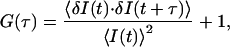
FCS avoids the averaging of the data in a large volume as it can record the fluorescence signals from a very tiny volume element of about 1 fl (1 × 10⁻¹⁵ l). The single fluorescent molecules diffuse in and out of the laser focus emitting bursts of fluorescence quanta during their transient time. These fluorescence-intensity fluctuations are recorded over time. The signal to noise ratio is usually larger than 106 (2). The fluorescence quanta can be statistically analyzed by auto- and crosscorrelating the time-resolved signals. The FCS auto- and cross-correlation functions are derived from a model of free diffusion of molecules in solution (4, 15). The correlation functions express the average concentration fluctuation of molecules at time t with a concentration fluctuation of the same molecules or of any other molecules at a later time (t + τ) in the volume element V. The fluorescence signal I(t) emitted by the molecules passing through V fluctuates around mean values < I(t) >. The normalized correlation functions G(τ) of fluorescence fluctuations, assuming a random stationary signal, is
 |
1 |
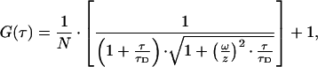
where δI(t) = I(t) − < I(t) > is the spontaneous fluctuation of fluorescence. The evolution in time is obtained by integrating over the local and time-dependent molecule concentration in the intensity profile of the illumination volume. After Fourier transformation, the fluctuations δI(t) of the photon count rate I(t) are characterized by
 |
2 |
where N is the observed averaged and absolute number of fluorescent molecules in auto-correlation mode; in cross-correlation mode, the N value has to be expanded to obtain the observed averaged and absolute number N of the two-color molecules. The detailed analysis of the absolute number of two-color species in the presence of unspecific molecules carrying only one color (either green or red) is given in refs. 13, 14, 16, and 17. τD is the characteristic translational diffusion time of the fluorescent molecules. ω is the radius and z is the half-length of the elliptically shaped blue, red, or overlapping confocal volume elements. The analysis of deviations between experimental auto- or crosscorrelations and Eq. 2 was performed with software based on the Marquardt nonlinear least-squares parameterization for calculating the normalized mean square (18). In each analysis, the deviations were random and close to 1.0.
Poisson Analysis of Single-Molecule Detection in Solution: Solution-Phase Single-Molecule FCS (SPSM-FCS)
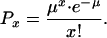
The Poisson distribution describes the probability that an event occurs when the frequency of that event is very low. If the event x happens at an average frequency μ, then the probability (Px) that a single event takes place is
 |
3 |
e is the base of the natural logarithm. The number of fluorescent molecules in solution is always an integer or zero. Let us assume that the solution contains an average number C of specific fluorescent molecules per unit volume V (confocal volume of detection in auto- or cross-correlation mode). We describe the relationship between the bulk concentration c (the average number C of specific fluorescent molecules in the unit volume) and the probability that the unit volume contains a single (specific) fluorescent molecule by the Poisson distribution. x represents the “actual” number of specific fluorescent molecules in the unit volume at an average frequency μ = C. The value x is always a small integer (here x = 1; for motivation see Fig. 2) and does not always equal C. Then, the probability P1 that the confocal volume of detection contains a single (specific) fluorescent molecule (x = 1) is
 |
4 |
that is
 |
5 |
for
 |
6 |
Under the condition of Eq. 6, we can immediately write
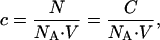 |
7 |
where c is the average concentration of the specific fluorescent molecule in the bulk solution, NA the Avogadro's number with 6.022 × 10²³ mol−1, and V the confocal volume of detection (unit volume). The probability that the unit volume V contains two (specific) fluorescent molecules (x = 2) is given by
 |
8 |
Figure 2.

A blue confocal detection volume is shown schematically. It contains one fluorescent molecule (fluorophore molecule). The distance from the center in axial direction is ωblue. The half-length is zblue. The dimensions of the cylindrically shaped volume element are obtained from the measured auto-correlation function and the measured diffusion coefficient of rhodamine green at the blue excitation wavelength of 488 nm.
Results
The Brownian motion of the fluorophore molecules rhodamine green was detected as fluctuations of the emitted fluorescence intensity. The recording time in our experiments was 2 sec. The experimental results obtained are summarized in Fig. 3 A–F. The measured average photon counts per fluorescent molecule and per second were 121.230 ± 4.172 kHz (mean ± SD, n = 6). Thus, the fluorophore molecules caused only the fluctuations of the fluorescence intensity. By using the optical system, the emitted fluorescence intensity was measured without background noise. The measured and calculated auto-correlation functions G(τ) of the fluorophore rhodamine green gave the characteristic diffusion time of 28 μsec. The time course of G(τ) in Fig. 3 B–F does not change from that in Fig. 3A, thereby indicating that G(0) increased. The maximum amplitude of G(τ) was inversely proportional to the average number of molecules N in the observation volume (confocal volume of detection). The lowest value for N that could be measured without background noise was 0.0085. The Poisson analysis of single-molecule detection in solution was performed with the data of Fig. 3 and the results are shown in Table 1. The average frequency C, that the volume of detection contained a single fluorophore molecule, was calculated by means of Eq. 4 and compared with the measured value of N. C was almost equal to the experimental value G(0) if N was less than 0.05, as is the case in Fig. 3 C–F. Next, we took the P2 parameter as the criterion for single-molecule analytical sensitivity.
Figure 3.

Auto-correlations of the rhodamine green fluorescence fluctuations accumulated with sample times of 2 sec. The measured curves are depicted in black. The red curves represent auto-correlation functions of the form of Eq. 2. The physical process of diffusion of rhodamine green molecules is considered only (red curves). G(0) increases for smaller bulk concentrations (A–F), but the average photon counts per fluorescent molecule and per sec are 121.230 ± 4.172 kHz (mean ± SD, n = 6). Concentrations c, in nM, of the fluorescent molecules in the bulk solutions are A, 20.116; B, 0.815; C, 0.398; D, 0.269; E, 0.180; and F, 0.109.
Table 1.
Synopsis of Poisson-distribution analysis of single-molecule detection in solution (SPSM-FCS)
| Mean fluorescence intensity, kHz | Absolute photon counts per molecule and per second, × 103 | Nmeasured* | Ccalculated† | P2‡ |
|---|---|---|---|---|
| 247.229 | 122.209 | 2.023 | ND | ND |
| 9.565 | 116.646 | 0.082 | 0.090 | 3.70 × 10⁻³ |
| 4.896 | 122.400 | 0.040 | 0.042 | 8.46 × 10⁻⁴ |
| 3.210 | 118.889 | 0.027 | 0.028 | 3.81 × 10⁻⁴ |
| 2.138 | 118.778 | 0.018 | 0.018 | 1.59 × 10⁻⁴ |
| 1.413 | 128.455 | 0.011 | 0.011 | 5.98 × 10⁻⁵ |
It is assumed that a single fluorescent molecule (e.g., fluorophore) represents the “actual” number of fluorescent molecules in the unit volume (confocal volume of detection) at an average frequency C. Then, Nmeasured is taken as the Poisson probability P1 that the confocal volume of detection V contains a single fluorescent molecule. ND, not defined.
The N values were directly obtained by the FCS experiments according to Eq. 2. The concentrations of the bulk solutions were 20.116, 0.815, 0.398, 0.269, 0.180, and 0.109 nM, respectively.
The average frequency C that the volume of detection contains a single fluorophore molecule is calculated by means of Eq. 4.
The Poisson probability that the confocal volume of detection Vg contains two fluorophore molecules is calculated for each value of C by means of Eq. 8.
To verify the applicability of our new approach of SPSM-FCS, we determined antiglomerular basement membrane auto-Abs of IgG-type in serum of patients with Goodpasture syndrome. For coincidence detection, the assay was split up into a sandwich. In Fig. 4A, the circulating Goodpasture auto-Abs were quantified in cross-correlation mode by binding to the rhodamine green-labeled antigen and a secondary Ab with the Cy5 tag. By using this method, it was possible to distinguish the specific binding interactions of the BMA Abs caused by the sandwiched immune complex carrying both colors in the same molecule. We measured an averaged absolute number of the complexed auto-Abs of N = 0.125 (Fig. 4). The absolute number was related to the average frequency C that the volume of detection contained a single complexed auto-Ab with C = 0.146 (see Table 1 for the calculation). For this case, our P2 parameter was 9.09 × 10⁻³. In other words, the probability that the confocal volume element contained one single complexed auto-Ab was 99.1%.
Figure 4.

Cross-correlation of the Goodpasture auto-Abs in patient serum. The sample times are 60 sec. The measured curves are depicted in black. The red curves represent the crosscorrelated fluorescence signals of the form of Eq. 2. (A) The crosscorrelated fluorescence signals of the two-color complex are superimposed by the unspecific crosstalk of the free dye rhodamine green and the free rhodamine green-labeled antigen in the preparation, which can be corrected for (see B). The absolute number of two-color molecules N in the sample was calculated according to the analysis developed for the first time in refs. 13, 14, 16, and 17. In the cross-correlation mode, we determined N = 0.138 for the two-color immune complex. This is about the same absolute number of the auto-Ab complex determined in the red autocorrelations of N = 0.111 with a relative fraction of 82.7% compared with 17.3% of free Cy5 (not shown). (B) The control serum of a healthy person who has no Goodpasture syndrome. The crosscorrelated fluorescence signals reflect the unspecific crosstalk of the free dye rhodamine green and the free rhodamine green-labeled antigen.
Discussion
FCS is used to study a wide range of physical, biochemical, and molecular biological properties of molecules including reactions between biological molecules (8, 9, 11, 19). FCS is a powerful tool for analyzing conformational fluctuations of single nucleic acid molecules in solution (20) and on surfaces (21), and of single enzyme molecules on surface (22). The introduction of the confocal volume element in FCS has provided researchers with the technology to measure molecular diffusion processes, chemical kinetics, and dynamics (1–3). Besides its prominence in physics, chemistry, and biotechnology, FCS is very important in biological and medical sciences.
We have measured the emitted fluorescence signals of single rhodamine green molecules. During the lifetime of the excited electronic singlet state, the rhodamine green molecules emit quantum bursts of fluorescent light. Under continuous wave excitation, the fluorescence process is cyclic as long as the fluorophores are not irreversibly photobleached (destroyed). We were able to focus our attention on single molecules rather than on averages over many molecules. It was possible because the FCS illuminates a sufficiently small volume and optically separates the emitted fluorescence from scattered excitation light and other interfering luminescence (see Fig. 1). The values of the count rate per fluorescent molecule show that the fluctuations in fluorescence intensity are arising only from single fluorophore particles. Further, the data of Fig. 3 reveal that FCS can correctly identify fluorophore molecules down to concentrations in the lower pM range with very high photon counts per fluorescent molecule and per sec. Correlation of the fluorescence fluctuations from single rhodamine green molecules gives rise to the number values N of fluorescent molecules shown in Table 1. The values are obtained from the inverse amplitude of G(0). The correlated fluctuations of emitted light quanta are measured instead of averaged. It leads to increased sensitivity when the fluorescence arises from a single emission source. The molecules randomly distribute in solution and this random behavior is not based on measurement errors. We use here our analysis for detecting single molecules, SPSM-FCS. Because the actual number of molecules in the confocal volume of detection (unit volume) is always an integer or zero, the fluorescence generated by the molecules is discontinuous when single-molecule sensitivity is achieved (Fig. 2). We show that the probability to find a single molecule in the very tiny volume element is Poisson-distributed under assay conditions sensitive enough to detect less than about 1 nM of a fluorophore (see Table 1), meaning below a critical bulk concentration c*. For example, at the lowest measured probability of 0.0085 found without background noise, we have traced 5 × 10¹⁰ specific molecules per milliliter of bulk solution. For the average frequencies C obtained from the experimental values G(0), we have calculated the Poisson probability P2 that the confocal volume of detection contains two fluorescent molecules. This is the correct parameter to use when determining the analytical sensitivity of an assay for single molecules in terms of the bulk concentration c. The molecules do not have to be quantified in a more concentrated form, or in flow and trapping experiments. The results demonstrated that single-molecule analytical sensitivity is achieved (P2 values in Table 1), and that the Poisson-distribution analysis of single-molecule detection in terms of the bulk concentration c is an effective and elegant way to monitor assay sensitivity.
Next, we combined our new analytical approach named SPSM-FCS with the specificity of an antigen-Ab system for the immunological diagnosis of Goodpasture syndrome. Our SPSM-FCS is the alternative to single-molecule detection of immobilized molecules by spectroscopic methods including FCS. We are able to show statistically even one single immune complex in diluted solutions. Under the condition of performing the experiments below a critical bulk concentration of the fluorescent molecules of about 1 nM, the average frequency C that the volume of detection contains a two-color fluorescent molecule is obtained from the FCS experiment. There are no instrumental assumptions of our approach on which the experiment itself, the theoretical background, or the conclusion are based. Therefore, important analytical information about many clinical parameters can be obtained straightforwardly and directly when this pioneering diagnostic tool is used. SPSM-FCS is of great impact for the development of new ultrasensitive assays in immunology and molecular medicine.
Abbreviations
- FCS
fluorescence correlation spectroscopy
- SPSM-FCS
single-phase single-molecule FCS
References
- 1.Rigler R, Mets U. Int Soc Opt Eng Laser Spectrosc Biomol. 1992;1921:239–248. [Google Scholar]
- 2.Rigler R, Mets U, Widengren J, Kask P. Eur Biophys J. 1993;22:169–175. [Google Scholar]
- 3.Mets U, Rigler R. J Fluoresce. 1994;4:259–264. doi: 10.1007/BF01878461. [DOI] [PubMed] [Google Scholar]
- 4.Magde D, Elson E L, Webb W W. Phys Rev Lett. 1972;29:705–711. [Google Scholar]
- 5.Elson E L, Magde D. Biopolymers. 1974;13:1–27. doi: 10.1002/bip.1974.360130103. [DOI] [PubMed] [Google Scholar]
- 6.Koppel D W. Phys Rev. 1974;10:1938–1945. [Google Scholar]
- 7.Ehrenberg M, Rigler R. Chem Phys. 1974;4:390–401. [Google Scholar]
- 8.Eigen M, Rigler R. Proc Natl Acad Sci USA. 1994;91:5740–5747. doi: 10.1073/pnas.91.13.5740. [DOI] [PMC free article] [PubMed] [Google Scholar]
- 9.Rigler R. J Biotechnol. 1995;41:177–186. doi: 10.1016/0168-1656(95)00054-t. [DOI] [PubMed] [Google Scholar]
- 10.Földes-Papp Z, Angerer B, Ankenbauer W, Baumann G, Birch-Hirschfeld E, Börjling S, Conrad S, Hinz M, Rigler R, Seliger H, Thyberg P, Kleinschmidt A K. In: Fractals in Biology and Medicine. Losa G A, Merlini T F, Nonnenmacher E R, Weibel E R, editors. II. New York: Springer; 1998. pp. 238–254. [Google Scholar]
- 11.Földes-Papp Z, Kinjo M. Fluorescence Correlation Spectroscopy: Theory and Applications. In: Elson E L, Rigler R, editors. Springer Series in Physical Chemistry. Vol. 65. Boston: Springer; 2001. pp. 25–64. [Google Scholar]
- 12.Földes-Papp Z, Thyberg P, Björling S, Holmgren A, Rigler R. Nucleosides Nucleotides. 1997;16:781–787. [Google Scholar]
- 13.Földes-Papp Z, Angerer B, Thyberg P, Hinz M, Wennmalm S, Ankenbauer W, Seliger H, Holmgren A, Rigler R. J Biotechnol. 2001;86:203–224. doi: 10.1016/s0168-1656(00)00414-4. [DOI] [PubMed] [Google Scholar]
- 14.Földes-Papp Z, Angerer B, Ankenbauer W, Rigler R. J Biotechnol. 2001;86:237–253. doi: 10.1016/s0168-1656(00)00416-8. [DOI] [PubMed] [Google Scholar]
- 15.Schwille P, Meyer-Almes F-J, Rigler R. Biophys J. 1997;72:1878–1886. doi: 10.1016/S0006-3495(97)78833-7. [DOI] [PMC free article] [PubMed] [Google Scholar]
- 16.Rigler R, Földes-Papp Z, Meyer-Almes F-J, Sammet C, Völcker M, Schnetz A. J Biotechnol. 1998;63:97–109. doi: 10.1016/s0168-1656(98)00079-0. [DOI] [PubMed] [Google Scholar]
- 17.Földes-Papp Z, Rigler R. Biol Chem. 2001;382:473–478. doi: 10.1515/BC.2001.057. [DOI] [PubMed] [Google Scholar]
- 18.Marquardt D W. J Soc Indus Appl Math. 1963;11:431–441. [Google Scholar]
- 19.Maiti S, Haupts U, Webb W W. Proc Natl Acad Sci USA. 1997;94:11753–11757. doi: 10.1073/pnas.94.22.11753. [DOI] [PMC free article] [PubMed] [Google Scholar]
- 20.Edman L, Mets U, Rigler R. Proc Natl Acad Sci USA. 1996;93:6710–6715. doi: 10.1073/pnas.93.13.6710. [DOI] [PMC free article] [PubMed] [Google Scholar]
- 21.Wennmalm S, Edman L, Rigler R. Proc Natl Acad Sci USA. 1997;94:10641–10645. doi: 10.1073/pnas.94.20.10641. [DOI] [PMC free article] [PubMed] [Google Scholar]
- 22.Edman L, Földes-Papp Z, Wennmalm S, Rigler R. Chem Phys. 1999;247:11–22. [Google Scholar]