ABSTRACT
In 2015, a laboratory of the United States Department of Defense (DoD) inadvertently shipped preparations of gamma-irradiated spores of Bacillus anthracis that contained live spores. In response, a systematic evidence-based method for preparing, concentrating, irradiating, and verifying the inactivation of spore materials was developed. We demonstrate the consistency of spore preparations across multiple biological replicates and show that two different DoD institutions independently obtained comparable dose-inactivation curves for a monodisperse suspension of B. anthracis spores containing 3 × 10¹⁰ CFU. Spore preparations from three different institutions and three strain backgrounds yielded similar decimal reduction (D10) values and irradiation doses required to ensure sterility (DSAL) to the point at which the probability of detecting a viable spore is 10−6. Furthermore, spores of a genetically tagged strain of B. anthracis strain Sterne were used to show that high densities of dead spores suppress the recovery of viable spores. Together, we present an integrated method for preparing, irradiating, and verifying the inactivation of spores of B. anthracis for use as standard reagents for testing and evaluating detection and diagnostic devices and techniques.
IMPORTANCE The inadvertent shipment by a U.S. Department of Defense (DoD) laboratory of live Bacillus anthracis (anthrax) spores to U.S. and international destinations revealed the need to standardize inactivation methods for materials derived from biological select agents and toxins (BSAT) and for the development of evidence-based methods to prevent the recurrence of such an event. Following a retrospective analysis of the procedures previously employed to generate inactivated B. anthracis spores, a study was commissioned by the DoD to provide data required to support the production of inactivated spores for the biodefense community. The results of this work are presented in this publication, which details the method by which spores can be prepared, irradiated, and tested, such that the chance of finding residual living spores in any given preparation is 1/1,000,000. These irradiated spores are used to test equipment and methods for the detection of agents of biological warfare and bioterrorism.
KEYWORDS: Bacillus anthracis, gamma irradiation, select agent, spore, inactivation, sterility assurance level, sterilization
INTRODUCTION
Like other species of the genus Bacillus, Bacillus anthracis, the causative agent of the disease anthrax, forms hardy metabolically dormant endospores upon the exhaustion of nutrient supplies. These spores are highly resistant to desiccation, temperature extremes, predation, exposure to UV light, and ionizing radiation and can survive for extended periods under conditions unfavorable to the survival of vegetative bacteria (1–3). The environmental persistence, overall hardiness, and the relative simplicity of production methods also have made B. anthracis a favorite among nations that have historically pursued biological warfare, including the United States (pre-1972), the former Soviet Union, Iraq, and several other states that have been suspected of harboring biological weapons programs (4, 5).
Because of its known history as a biological weapon, B. anthracis spores have also been utilized as instruments of bioterrorism, notably by Aum Shinrikyo (6) and by the perpetrator of the 2001 anthrax attacks (7, 8) through the United States Postal Service. Therefore, the development of methods to detect and quantify B. anthracis spores in samples has been of great interest to the public health and military sectors. However, the rigid relatively impermeable spore structure (9–11) also renders the spores refractory to various molecular and biochemical detection methods (12), as harsh mechanical disruption is required to extract nucleic acids in particular, complicating the development of detection modalities. In addition to the intrinsic physical properties of the spores, costly biosafety level 3 (BSL3) procedures are generally required during the development and testing of new detection and decontamination methods. Therefore, methods of inactivating spores without destroying their physical and molecular characteristics are highly desired and have been utilized over the past decades (13–16).
Methods for inactivating spores have historically included wet heat, chemical treatment methods, and irradiation, many of which are reviewed elsewhere (13, 16). Wet heat forces steam under high pressure through the otherwise relatively impermeable spore envelope, resulting in the denaturation of protein components. As a result, wet heat methods compromise the integrity of epitopes recognized by antibody-based detection methods (17, 18). Likewise, chemical methods of decontamination (primarily using bleach, hydrogen peroxide, or ethylene oxide) result in extensive free-radical damage that destroys important components of the spore, preventing germination (19, 20). Because of its high penetrating power and more-selective effects on large macromolecular targets (e.g., DNA), gamma irradiation has become a favored method of inactivating biological materials in implantable medical devices, food, and in the sterilization of laboratory equipment (21–25), as irradiation can reduce the risk of contamination and provide assurance of sterility with minimal remaining risks.
Gamma irradiation was first reported as a suitable method for irradiating B. anthracis spores in 1959 (14). Subsequent work showed that spores of various Bacillus species typically exhibited decimal reduction values ([D10] the dose required to reduce the population by one log) of between 1.8 and 5.5 kGy, with marginal effects on spore ultrastructure, physical properties, and the response to antibody- or PCR-based detection assays (13, 18, 26–29). Because of the perceived efficacy of irradiation in yielding a safe yet responsive product that could mimic the essential properties of the live agent, irradiated spores of B. anthracis became a critical reagent for the development of systems and devices to detect agents of biological warfare and bioterrorism. Many of the experiments describing irradiation procedures, doses, and the determination of D10 values for B. anthracis were summarized in the analysis by the U.S. Department of Defense (DoD) of the inadvertent shipments of live, virulent B. anthracis spores to unregistered laboratories in 2015 (30). However, as the DoD report indicated, the conditions under which the spores were grown, prepared, concentrated, and handled were inconsistent from one experiment to another, preventing the direct comparison of these data. Thus, the need for reproducible standardized methods for producing such materials was highlighted.
In many inactivation procedures, the risk that live organisms are present after sterilization is approximated by the parameter known as the sterility assurance level (SAL) (31). For medical devices and pharmaceutical products sterilized with radiation, acceptable SALs range from 10−3 to 10−6, depending on the sensitivity of the product itself to the effects of ionizing radiation (24). The SAL represents the probability that a viable organism will be found in a sample subjected to a given dose of radiation (e.g., a SAL of 10−6 indicates that one sample in 106 identically treated samples will contain a viable organism). The SAL is typically extrapolated from the linear portion of dose-inactivation curves (“kill curves”) generated with a defined bioburden—usually an appropriate, well-characterized surrogate organism (25)—and depends on the linearity of the inactivation curve up to the point of complete inactivation. To our knowledge, the concept of the SAL has not been applied to the inactivation of B. anthracis spore preparations or to the preparation of biological materials per se (14, 15, 18, 19, 26, 32, 33), particularly for DoD laboratories charged with preparing and distributing inactivated materials (30).
In this work, we present a method for preparing and inactivating B. anthracis spores to a SAL of 10−6 using gamma irradiation. In an interlaboratory comparison performed in parallel at the United States Army Edgewood Chemical Biological Center (ECBC) and the United States Army Medical Research Institute of Infectious Disease (USAMRIID), we establish dose-inactivation curves and decimal reduction values for multiple biological replicates using two different irradiation platforms and use those data to extrapolate radiation doses sufficient to inactivate spores of attenuated B. anthracis Sterne and fully virulent B. anthracis strain Ames to a SAL of 10−6. Finally, we characterize and define methods for detecting low levels of viable spores amid a background of inactivated spores. If followed stringently, the procedure for preparing and irradiating spores presented here should prevent any further inadvertent distribution of live B. anthracis spores and ensure that distributing laboratories remain in compliance with laws and regulations governing biological select agents.
RESULTS
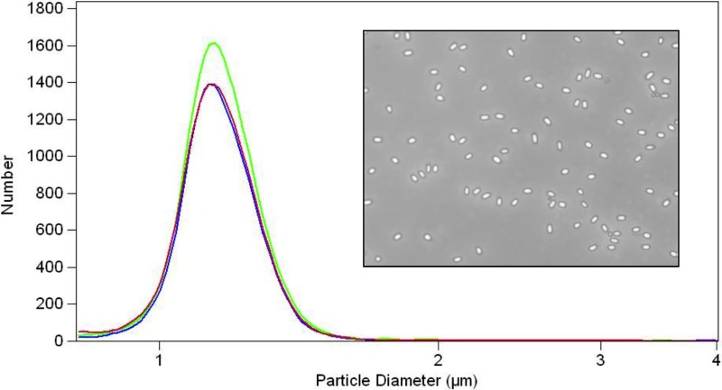
Each independent spore preparation was completed on an independent date and surpassed the quality/quantity requirements stipulated for the preparation of B. anthracis spores as a standard reagent for biodefense research. These requirements are delineated in Table 1. We reasoned that each spore preparation should be derived from a well-sporulating parent strain with a known passage history relative to the original isolate. Ideally, spore cultures should exhibit final spore titers of at least 1 × 10⁸ CFU/ml of sporulation medium (target is 1 × 10⁹ CFU/ml) prior to spore harvest and purification. The entire spore preparation was not heat shocked, but an aliquot was removed for heat shocking. An aliquot of at least 1 × 10⁷ spores from each spore preparation exhibited heat resistance (65°C, 30 min) using a standard quantitative plate assay. The spore mode size should be consistently between 1.0- and 1.5-μm volume-equivalent spherical diameter determined using a Beckman-Coulter Multisizer (BCM) (Fig. 1). The spores were at least 95% pure as judged by light microscopy counting of at least 100 particles per spore preparation (Fig. 1, inset). Spore dispersion was confirmed by examining at least 100 spores with light microscopy (Fig. 1, inset) and at least 500 spores with particle analysis via the BCM. The BCM measurements from three independent preparations showed reproducible, single, uniformly distributed particle size peaks, demonstrating that there was no detectable spore clumping.
TABLE 1.
Specifications for standardized spore preparation for B. anthracis strains and surrogates
| Property | Requirement | Method |
|---|---|---|
| Titer | ≥108 heat-resistant CFU/ml in initial sporulation medium; 1010 CFU/ml in final preparation | Heat shock 107 spores for 30 min at 65°C; quantitate by serial dilution plating on TSA medium |
| Size | 1.0- to 1.5-μm vol-equivalent spherical diameter | Measure ≥500 spores using a BCM |
| Purity | ≥95% pure spores with minimal vegetative cell debris | Examine ≥100 particles per spore preparation by light microscopy; spores are bright refractive particles |
| Clumping or aggregation | Unclumped individual spores | Prepare and dilute spores in buffer containing 0.1% Tween 80; examine ≥100 spores by light microscopy and ≥500 spores by particle analysis via BCM |
FIG 1.
Standardized spore preparation of B. anthracis Sterne. Particle size distribution analysis of three independent spore preparations prepared according to methods described in this work. Inset shows monodisperse phase-bright spores visualized by phase-contrast microscopy.
Determination of inactivation kinetics and sterilizing dose of B. anthracis.
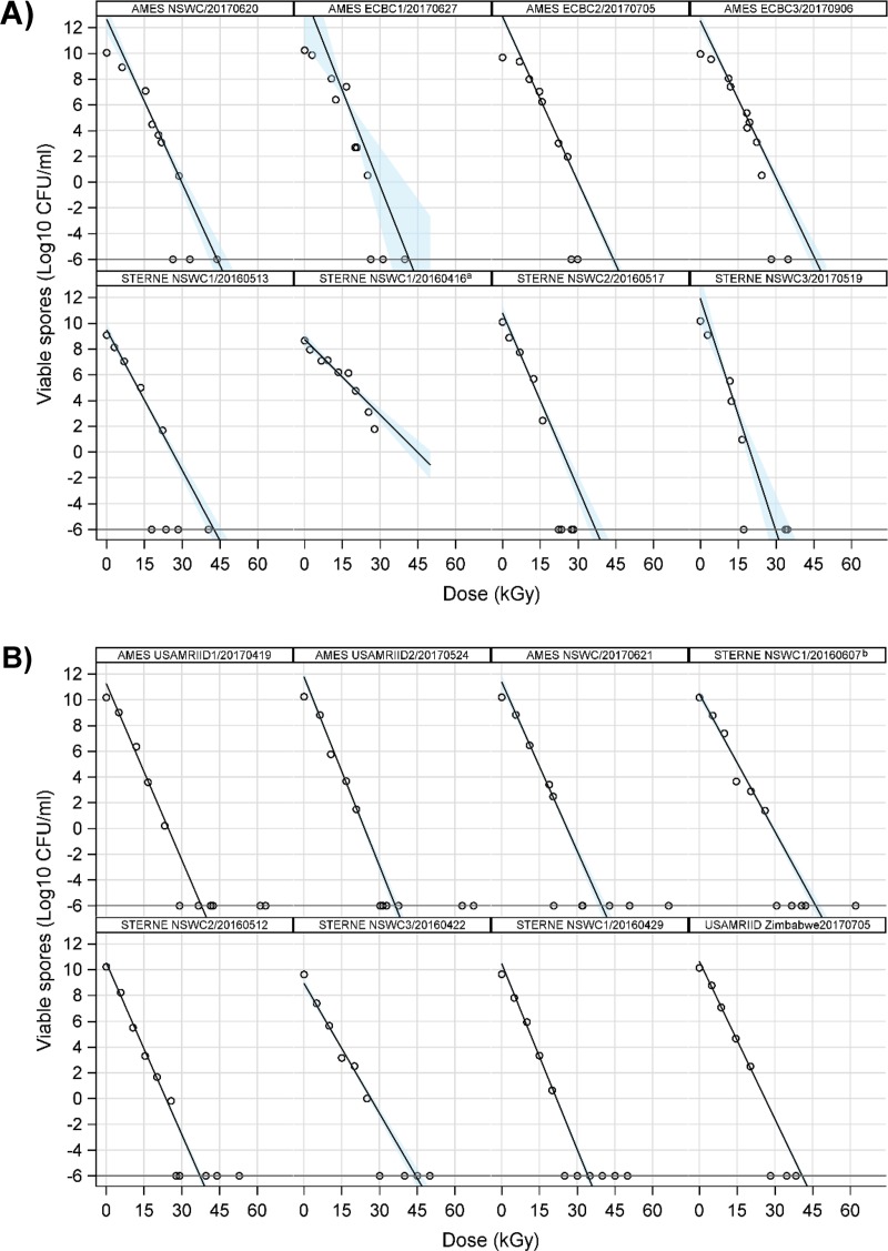
To determine the batch-to-batch variability of radiation sensitivity and whether the irradiator configuration affects inactivation, we conducted dose-inactivation studies on 3-ml aliquots of three independently prepared batches of B. anthracis strain Sterne and B. anthracis Ames at 1010 CFU/ml. Because B. anthracis Sterne lacks pXO2 and is exempt from select agent regulations, we utilized the Sterne strain as a surrogate for virulent B. anthracis to develop the procedure prior to validating the results using fully virulent strains. Dose inactivation of a geographically diverse isolate, B. anthracis Zimbabwe, was also tested. Spore preparations were treated incrementally with ∼5-kGy increments, and the population of viable spores remaining was determined by serial dilution and plating. The actual radiation doses delivered were monitored by measuring the increase in absorbance of radiochromic film dosimeters that had been attached to each 15-ml conical tube containing the spore aliquots. Dose-inactivation curves from USAMRIID and ECBC are shown in Fig. 2. The curves were similar across all batches studied and were similar between both institutions and among three differing genetic backgrounds. From the slopes of the plots and the intercepts, D10 and SALs of 10−6 were determined, which are listed in Table 2. We conducted a statistical analysis to determine the average dose for each institute and the 95% confidence intervals for each of the parameters. D10 values for each batch ranged from 1.66 to 2.98 kGy, and DSAL ranged between 29.8 and 46.2 kGy, with the upper bounds of the 95% confidence intervals (CIs) of 3.55 kGy and 52.4 kGy for D10 and SAL(10−6), respectively (Table 3). Our statistical comparison revealed neither significant differences between spore batches nor significant differences between institutions (i.e., differences in irradiators and experimental processing), with the exception of a modest but statistically significant difference between the mean DSALs determined for the Ames strain between ECBC and USAMRIID (43.77 versus 37.95 kGy, respectively; P = 0.0089). Overall, averaged over 15 inactivation curves covering three different strain backgrounds, the mean D10 was 2.32 ± 0.09 kGy, and the mean DSAL was 40.10 ± 1.20 kGy (Table 4).
FIG 2.
Summary of dose-inactivation curves from 16 independent inactivation experiments performed at ECBC (A) and USAMRIID (B). Except as noted, spore suspensions in experiments performed at USAMRIID were maintained on ice during irradiation. Shaded areas represent the 95% confidence intervals. a, diluent was PBS with no Tween 80; b, ice omitted.
TABLE 2.
Results of individual dose-inactivation experiments
| Strain | Irradiator | Spore preparation/irradiation date (yr-mo-day) | kGy (95% CI)a |
|
|---|---|---|---|---|
| D10b | DSALc | |||
| AMES | ECBCd | NSWC/2017-06-20 | 2.35 (2.03, 2.67) | 43.86 (40.23, 47.48) |
| ECBC1/2017-06-27 | 2.02 (1.10, 2.95) | 41.53 (30.19, 52.86) | ||
| ECBC2/2017-07-05 | 2.33 (2.22, 2.45) | 44.05 (42.66, 45.44) | ||
| ECBC3/2017-09-06 | 2.46 (2.23, 2.70) | 45.65 (42.89, 48.41) | ||
| USAMRIID | USAMRIID1/2017-04-19 | 2.19 (2.18, 2.20) | 37.79 (37.55, 38.04) | |
| USAMRIID2/2017-05-24 | 2.04 (1.92, 2.16) | 36.32 (34.93, 37.72) | ||
| NSWC/2017-06-21 | 2.28 (2.12, 2.45) | 39.75 (37.96, 41.53) | ||
| STERNE | ECBCd | NSWC1/2016-05-13 | 2.75 (2.48, 3.01) | 42.59 (39.67, 45.50) |
| NSWC1/2016-04-16f | 5.11 (4.29, 5.94) | —e | ||
| NSWC2/2016-05-17 | 2.20 (1.95, 2.45) | 37.02 (34.06, 39.98) | ||
| NSWC3/2017-05-19 | 1.66 (1.23, 2.10) | 29.83 (25.18, 34.48) | ||
| USAMRIID | NSWC1/2016-04-22f,g | 2.81 (2.63, 2.99) | 46.22 (44.38, 48.06) | |
| NSWC2/2016-04-29f,g | 2.25 (2.16, 2.34) | 37.24 (36.29, 38.19) | ||
| NSWC3/2016-05-12 | 2.98 (2.76, 3.19) | 44.45 (42.27, 46.64) | ||
| NSWC1/2016-06-07d | 2.08 (1.99, 2.17) | 34.30 (33.37, 35.22) | ||
| ZIMBABWE | USAMRIID | USAMRIIDZimbabwe/2017-07-05 | 2.45 (2.39, 2.51) | 40.86 (40.17, 41.56) |
CI, confidence interval.
D10, decimal reduction value. Dose required for 10-fold reduction.
DSAL, sterility assurance level. Dose required to achieve a SAL of 10−6.
No ice utilized during irradiation.
—, statistics not estimable from the observed data.
Tween 80 not present in diluent buffer.
Dosimetry not utilized.
TABLE 3.
Interlaboratory comparison dose-inactivation experimentsa
| Comparison | Mean kGy (95% CI) |
Difference (P value) | |
|---|---|---|---|
| Ames | Sterne | ||
| D10 | |||
| ECBC | 2.29 (1.99, 2.59) | 2.20 (0.86, 3.55) | 0.09 (0.8089) |
| USAMRIID | 2.17 (1.87, 2.48) | 2.53 (1.84, 3.21) | −0.36 (0.1967) |
| Difference (P value) | 0.12 (0.3511) | −0.32 (0.4441) | |
| DSAL | |||
| ECBC | 43.77 (41.07, 46.48) | 36.48 (20.59, 52.37) | 7.29 (0.1818) |
| USAMRIID | 37.95 (33.69, 42.22) | 40.55 (31.49, 49.62) | −2.60 (0.4415) |
| Difference (P value) | 5.82 (0.0089) | −4.07 (0.4303) | |
Excluding experiment NSWC1/20160416. P values indicate results of Welch's t tests.
TABLE 4.
D10 and DSAL overall summarya
| Statistic | n | kGy |
||
|---|---|---|---|---|
| Mean | SEM | SD | ||
| D10 | 15 | 2.32 | 0.09 | 0.34 |
| DSAL | 15 | 40.10 | 1.20 | 4.65 |
Excluding experiment NSWC1/20160416.
Analysis of spore integrity/morphology.
To determine whether extensive damage to the spore structure had occurred as a result of gamma irradiation, we examined the spores under phase-contrast microscopy. Unirradiated spores were highly refractile (as expected), whereas irradiated Sterne spores had lost much of their refractility (Fig. 3). The spore darkening suggests at least partial loss of Ca2+-dipicolinate and partial rehydration of the spore core. A comparison of various doses (up to 50 kGy) of unirradiated and irradiated spores by transmission electron microscopy revealed no gross structural damage or morphological alterations (i.e., coat fragmentation or significant damage to the exosporia).
FIG 3.
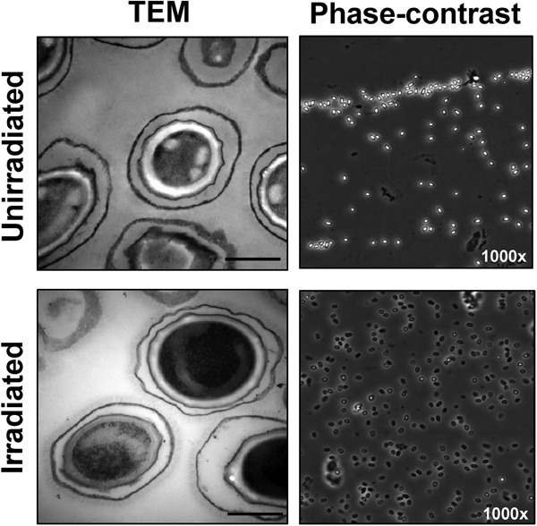
Spore ultrastructure analysis. Irradiated and unirradiated B. anthracis Sterne spores were visualized by transmission electron microscopy and phase-contrast microscopy (50- and 40-kGy doses, respectively). Bars in electron microscopy represent 500 nm. Phase-contrast images were taken using an oil immersion objective at ×1,000 magnification.
At gamma irradiation doses of 15 to 25 kGy, the morphology of recovered colonies exhibited a progressively dramatic variation (Fig. 4). While colonies originating from unirradiated spores displayed uniform morphology, spore preparations that had been irradiated with ∼15 kGy yielded heterogeneous colonies in both size and morphology. When spores treated with ∼20 kGy were plated, the morphologies were highly variable. Some colonies appeared to expand in a filamentous or “dendritic” pattern, whereas others retained the typical flat, circular rough morphology with undulate edges.
FIG 4.
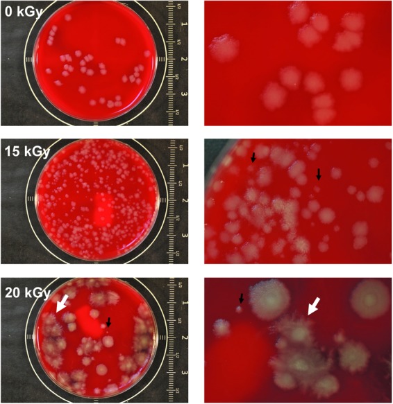
Abnormal morphologies of B. anthracis Sterne colonies recovered from samples irradiated with near-sterilizing doses of 15 and 20 kGy (nominal dose) of gamma radiation. Unirradiated and irradiated spores were diluted to yield countable numbers of colonies upon plating. Experiment shown is representative of three independent biological replicates irradiated and plated at two different institutions. Black arrows represent small colony morphotypes, and white arrows represent “dendritic” morphotype.
Detection of viable spores in irradiated materials.
Since spore germination at high densities has been shown to be impaired due to the presence of high levels of the alanine racemase enzyme (34), we also sought to test whether direct plating yields accurate counts of viable spores, particularly when viable spores are plated along with large numbers of dead spores. We hypothesized in particular that high densities of nonviable spores would inhibit the recovery of viable spores due in part to the remaining activity of alanine racemase in the irradiated/dead spores. To test this hypothesis, we spiked irradiated Sterne spores at densities of 1010 CFU/ml with known quantities (∼2,000 CFU/ml) of viable unirradiated spores of a strain of B. anthracis Sterne that contains a short genetic tag or “barcode” in the chromosome. The recovery of viable spores by plating was reduced by approximately half in the presence of 109 inactivated spores (Fig. 5A). Some of the recovered colonies exhibited a dendritic morphology reminiscent of colonies recovered from near the sterility point (Fig. 5A, inset). We then tested whether higher concentrations of dead spores would further inhibit the recovery of viable spores. When irradiated spores were concentrated to 1011 CFU/ml prior to spiking (resulting in a mass of 1010 inactivated spores in 100 μl samples), recoveries were reduced by 80 to 95% (Fig. 5B). In contrast, the recovery of viable spores was not appreciably inhibited when the density of irradiated spores plated with the viable spores was less than 108 (data not shown). All colonies recovered in this experiment (n = 88) were shown by barcode-specific PCR to contain the barcode (data not shown). When we pretreated the viable barcoded spores with the germinants alanine and inosine (10 mM each germinant) prior to adding them to the dead spore mass, a near complete recovery of spiked spores was achieved (Fig. 5C). However, the addition of the germinants after the viable spores were added to the dead spore mass did not promote a complete recovery of the viable spores (not shown).
FIG 5.
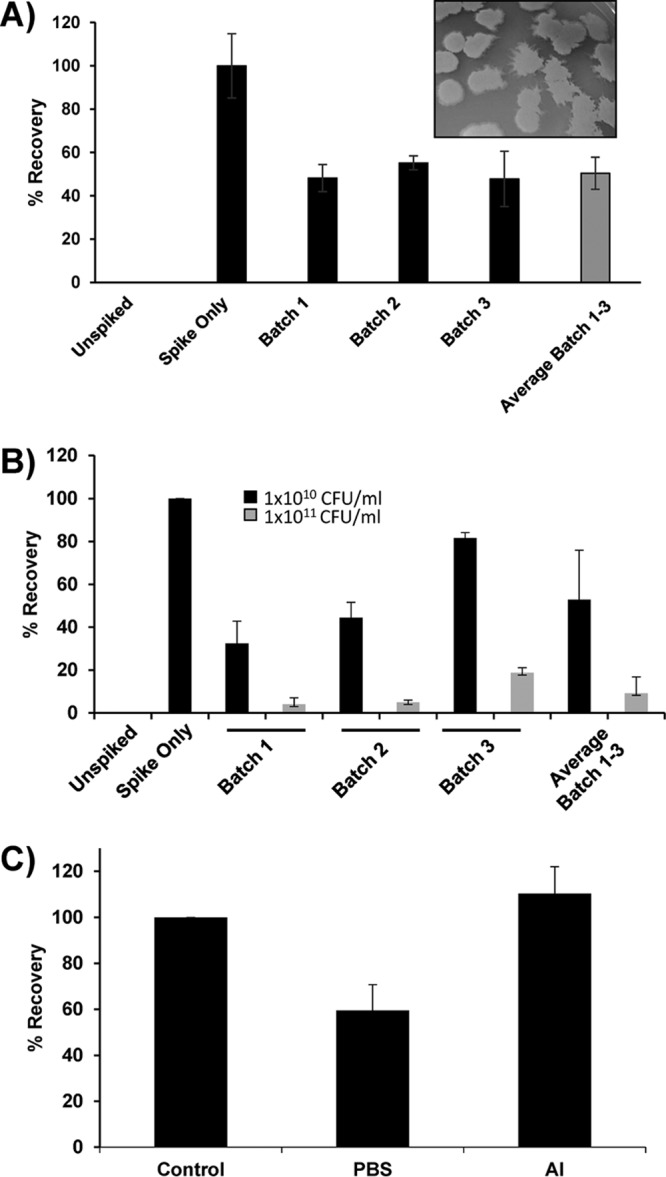
Recovery of viable spores is inhibited by the presence of high densities of dead spores. (A) Viable spores were spiked to a concentration of ∼2,000 CFU/ml into 100 μl of irradiated spores at 1010 CFU/ml or diluent buffer and plated onto TSAB. (Inset) Abnormal morphology of recovered colonies. (B) Effects of concentrating dead spores to 1011 CFU/ml on viable spore recovery; 100 μl of each spore suspension was spiked with 2,000 CFU/ml of barcoded B. anthracis Sterne spores and plated on TSAB. (C) Effects of germinants and hold time on spore recovery. Alanine and inosine (AI) were added (10 mM each) to viable spores prior to spiking into slurry of dead spores. PBS was added in lieu of alanine and inosine as a control. Results are the averages from two independent experiments. Error bars represent standard deviations. Addition of AI to dead spore mass prior to addition of live spores did not promote recovery of viable spores (not shown).
Dilution of irradiated spores into liquid culture.
We also tested whether dilution of the irradiated spore mass into liquid broth would aid in the recovery of viable spores. One hundred percent of the irradiated aliquots (one from each spore preparation) were diluted 1:500 (vol/vol) into three culture flasks containing 500 ml of tryptic soy broth (TSB) and were grown in shaking cultures at 37°C for 2 weeks. The optical density at 600 nm (OD600) of the cultures decreased within 24 h by approximately 50%, which corresponded to the loss of spore refractility in the irradiation-exposed spores. The OD600 of the suspensions then remained steady at ∼0.03 for 2 weeks, suggesting that no viable spores were detectable in the samples. Furthermore, no viable spores could be cultured from the culture flasks at any point following inoculation.
Evaluation of effect of postirradiation incubation on potential spore reactivation.
After the unintended shipment of viable spores by the DoD, it was proposed that spores could potentially possess mechanisms to enable recovery after exposure to radiation, a hypothesis colloquially referred to as the “zombie spore hypothesis” (30). Under this hypothesis, spores “recover” due to DNA repair mechanisms that are active prior to the initiation of the germination program. We tested this hypothesis in several different experiments. Initially, we examined the impact of short-term postirradiation storage at 4°C and at room temperature. After realizing that multiple institutions have different procedures for viability testing, it became clear that the time between irradiation and viability confirmation could be a confounding variable. To address this, we performed viability testing on samples within 30 min of irradiation and then placed the irradiated material at 4°C for long-term storage. We retested the viability of the irradiated spores at 2 and 4 weeks postirradiation. No increase in titer was noted for lethally irradiated materials (Fig. 6A). As described in Fig. 6B and C, there was evidence for a slight increase in viability but only at very specific suboptimal doses of radiation (approximately 10 to 20 kGy). The same trends were observed with spores of both the Sterne strain (Fig. 6B) and the Ames strain (Fig. 6C) of B. anthracis.
FIG 6.
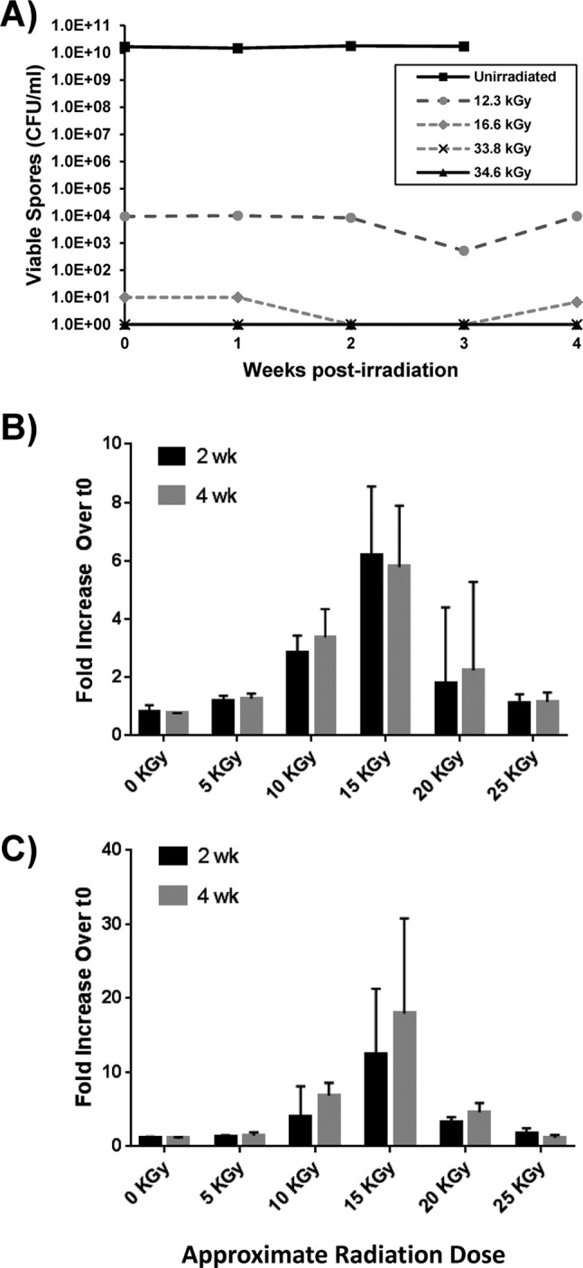
Effects of extended hold times on recovery of spores in irradiated samples of B. anthracis Sterne and Ames spores. (A) Representative absolute CFU titers of Sterne spores after postirradiation holds of 1 to 4 weeks at 4°C. (B) Increase in viable counts in comparison to t0 initial counts performed immediately after exposure to radiation (fold increase). Postirradiation storage of Sterne spores for 2 to 4 weeks at 4°C leads to a relative increase in viable spore titer. A modest increase in recovered viable CFU was observed in some cases when suboptimal doses of radiation were administered (i.e., 10 to 20 kGy). (C) The same effects were observed for spores of the fully virulent Ames strain. In cases where there was no detectable growth, a value of 1 CFU/ml was used to calculate the respective fold increase, as that was the limit of detection of the plating assay. Error bars represent standard deviations. Data are averages for at least two (0- and 5-kGy doses) or at least three (≥10-kGy doses) experiments.
We also examined the impacts of extreme holding times of greater than 1 year. B. anthracis Sterne samples that had been exposed to 35 or 40 kGy and then held for greater than 1 year at 4°C were suspended in TSB or brain heart infusion (BHI) broth to look for any evidence of spore viability. Eight samples were tested at USAMRIID and six samples were tested at ECBC, and there was no evidence of viable B. anthracis in any of the samples (data not shown). Lastly, we examined Sterne spores that were irradiated, frozen at −80°C, and then thawed; again, neither USAMRIID nor ECBC observed any restored viability in this scenario (data not shown).
Impact of radiation on enzyme activity and DNA integrity.
We sought to determine the effects of gamma irradiation on the integrity of known spore components. We reasoned that large macromolecules such as DNA would be more susceptible to degradation than smaller targets such as proteins. We therefore examined the activity of alanine racemase, an important spore enzyme that is localized in both the exosporium and the spore coat, and the integrity of DNA from B. anthracis Sterne spores. The alanine racemase activity was suggested to play a role in the “masking” of viable spores during viability testing. To address this concern, we wanted to establish baseline effects of radiation on the alanine racemase activity associated with B. anthracis spores. As presented in Fig. 7A, the enzymatic activity associated with spore preparations was assayed using whole spores that were either boiled (positive control for decrease in enzymatic activity), exposed to 0 kGy, or exposed to 50 kGy. The spores that were exposed to 50 kGy were either irradiated on wet ice or at the ambient temperature of the irradiator, representing USAMRIID's and ECBC's irradiator configurations, respectively. A dose of 50 kGy had a significant effect on the alanine racemase activity associated with B. anthracis Sterne spores irradiated at room temperature (P < 0.01) or on wet ice (P = 0.01) compared to that of untreated spores (0 kGy). There was also a statistically significant difference observed when comparing the alanine racemase activity associated with spores irradiated at ambient temperature and that of spores irradiated on wet ice (P < 0.01). The enzymatic activity was determined within 24 h of irradiation and again at 14 days postirradiation. There was no recovery of alanine racemase activity during this hold time at 4°C, suggesting that there was no apparent repair of enzyme function associated with time after 14 d at 4°C.
FIG 7.
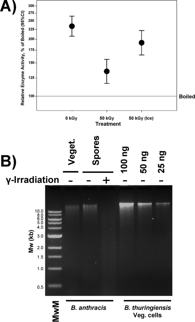
Spore irradiation affects DNA and enzymatic integrity. (A) Alanine racemase activity in unirradiated spores (0 kGy), spores irradiated at room temperature (RT), and spores irradiated on wet ice (ICE). The data depict relative enzymatic activities compared to boiled spores. Two individual spore preparations were used to calculate enzymatic activities. For each test-treatment (radiation on ice or ambient temperature), n = 4 or for the unirradiated-0 kGy samples, n = 8, and the bars depict 95% confidence intervals (CI). A dose of 50 kGy had a significant effect on the alanine racemase activity associated with B. anthracis Sterne spores irradiated at room temperature (P < 0.01) or on wet ice (P = 0.01) compared to that in untreated spores (0 kGy). There was also a statistically significant difference observed when comparing the alanine racemase activity associated with spores irradiated at ambient temperature and that of spores irradiated on wet ice (P < 0.01). The enzymatic activity was determined within 24 h of irradiation and again at 14 days postirradiation; there was no recovery of alanine racemase activity during this hold time at 4°C. (B) Agarose gel electrophoresis of genomic DNA isolated from vegetative bacilli (Veget.), unirradiated spores, and irradiated spores. Genomic DNA isolated from B. thuringiensis vegetative cells is also shown as a quantitation standard. MwM, 1-kb molecular weight marker. Gel was loaded to optimize visualization of the degree of DNA arising from irradiation.
To determine the degree of fragmentation of DNA in irradiated samples, we analyzed genomic DNA isolated from vegetative cells, spores, and irradiated spores. As expected, the preparations from spores and vegetative cells yielded DNA of a high molecular weight. DNA from spores also had some lower-molecular-weight components. In contrast, DNA from irradiated spores was highly sheared with little high-molecular-weight DNA visible (Fig. 7B).
Effects of gamma irradiation on spore detection assays.
We isolated genomic DNA from irradiated and unirradiated spores and performed quantitative PCR (qPCR) on equal quantities of genomic DNA. No difference in the sensitivities of the assays employed was noted (data not shown). These results were consistent with previous reports (17, 18, 29). Likewise, lateral flow immunoassays specific for B. anthracis were obtained from the Defense Biological Product Assurance Office (DBPAO). No differences in the limits of detection of these assays were noted when irradiated spores were utilized, although the intensity of the positive signal was reduced when irradiated spores were applied to the test strips of the lateral flow assay.
DISCUSSION
In this work, we present an integrated end-to-end procedure for the standardized production of irradiated spores of B. anthracis. We were able to apply the quantitative concept of the sterility assurance level to the residual risk of recovering a viable spore in preparations of irradiated spores.
The spore preparation method described here yields a high concentration (approximately 1010 CFU/ml) of viable spores in a monodisperse suspension of uniformly sized particles. It is based on previous work in our laboratories with B. thuringiensis Al Hakam and B. anthracis ΔSterne (35–39). The inclusion of 0.1% Tween 80 in our suspension buffer and all diluent buffers to reduce aggregation minimizes the effects of clumping on downstream analyses and reduces any protective effects that aggregation may have during exposure to gamma radiation. Spores of exosporium-containing Bacillus spp. tend to agglomerate and stick to surfaces (40) due to spore hydrophobicity (41–45), which can be specifically attributed to the exosporium. Species lacking exosporia, such as Bacillus subtilis or B. atrophaeus (35, 46), are considerably less prone to aggregation and produce consistent results, as shown by the ability not only to produce three virtually identical batches using multiple shake flasks per batch but to do so at three different institutions, with comparable results in three different genetic backgrounds.
Likewise, the irradiation procedure described here produced almost superimposable results at two different institutions with two differently configured irradiators. USAMRIID's irradiator is configured such that multiple 60Co source elements surround the sample, producing a uniform field of radiation, whereas ECBC's irradiator has three independently operable sources at the rear of an approximately cubic chamber, producing a field that attenuates with increasing distance from the source. In addition, USAMRIID's irradiator configuration permitted the use of ice to keep the sample cold, whereas this was not possible in ECBC's irradiator due to the size limitation of the chamber. In both cases, the D10 and DSAL values extrapolated from the inactivation curves were nearly identical, although the differences in irradiator configurations may contribute to some of the higher variability observed in the ECBC runs. By utilizing independent biological replicates, we also accounted for batch-to-batch variability as a potential contributor to differences in radiation sensitivity, provided that the conditions for the production of the initial spore material are kept consistent.
Unlike previous reports (15, 17, 18, 26, 33), our method incorporates the known inhibitory effects on germination of plating spores at high spore densities into our standard procedure for quantitating residual viable spores (34). This experiment was motivated by our findings that viable spores were not recovered at doses of at least 30 kGy, whereas the material distributed prior to 2015 by the DoD had been irradiated with considerably higher doses yet was shown to contain low concentrations of viable B. anthracis spores (30). It is unlikely that the absence of detectable viable spores in our study is due to a complete inhibition of germination of remaining viable spores, since the dead spore mass plated in the generation of the kill curves only marginally inhibited the recovery of viable spores from deliberately spiked samples. At the upper end of the dose range utilized in the kill curves, we would have expected only ∼50% inhibition when 100 μl of the inactivated material was plated (Fig. 5A). This inhibition of germination could be due to spatial constraints limiting the contact of viable spores with the TSB or, more likely, due to the residual alanine racemase activity associated with the dead spore mass. The plating of spores at higher densities than those we routinely utilized in this study (e.g., 1011 CFU/ml, such as those formerly produced by the DoD [30]) resulted in 80 to 95% inhibition of the recovery of the barcoded spiked spores (Fig. 5B). It is theoretically possible that residual viable spores in the material formerly produced by the DoD may have escaped initial detection due to the inhibition of germination and/or outgrowth by the dense mass of dead spores. However, the differences between our conditions and those used to grow, prepare, and irradiate the material formerly distributed by the DoD make a direct comparison of the two scenarios impossible. Our results indicate that, due to the inhibitory effect of the dead spore mass, approximately 20% of the undiluted inactivated material would need to be plated to achieve the equivalent of the suggested 10% sample for determining nonviability (47). Alternately, our results indicate that diluting the inactivated spores by a factor of 10 prior to plating for viability generally eliminates the inhibitory effect of the dead spore mass.
It is also unlikely that B. anthracis spores irradiated to a SAL of 10−6 can recover from radiation damage after extended periods of storage before plating. Our DNA profiles show extreme degrees of fragmentation (Fig. 7), a type of damage that is typically repaired by nonhomologous end joining (NHEJ) in irradiated spores of Bacillus subtilis. NHEJ is thought to occur during germination with the resumption of metabolic activity (48, 49) and would not be active in ungerminated spores. The extent of damage (i.e., the upper limit on the number of DNA strand breaks) that a spore can incur before being rendered nonviable is unknown, but our data suggest that the extensive fragmentation observed is not repairable by the B. anthracis DNA repair pathways. When viable spores can be detected immediately postirradiation, the number of recoverable CFU from stored (“held”) samples increases only marginally within approximately 1 week of irradiation. However, it is not clear whether this observation is due to the actual germination and proliferation of bacilli within the sample or whether the increase in recoverable CFU represents the ongoing disaggregation of clusters of spores that may have protected a minuscule fraction of the spores from irradiation. We consider the latter possibility unlikely due to the monodisperse nature of our spore suspensions, but we cannot formally exclude the possibility that a small number of spores are present in clusters that are not readily found in the microscopic surveys performed here. In contrast, when viable spores were not detected immediately postirradiation, even extended hold times of up to 4 weeks at room temperature or 4°C or over a year at 4°C did not promote a significant recovery of viable spores from these materials. We therefore believe that, under the conditions described here, irradiation to a SAL of 10−6 is sufficient to render a 3-ml suspension containing 3 × 10¹⁰ spores inactive and nonviable.
Because the spore preparation described here is intended for use across the chemical and biological defense enterprise as a standard reagent to test DoD-fielded and candidate detection systems, it was necessary that the requisite properties of the spores be preserved through the irradiation process. The damage to spores in the irradiation experiments likely results from a combination of direct radiation and oxidative and thermal damage, the latter of which may be mitigated by maintaining the spores on ice during the procedure (Fig. 7B). We have documented several altered colony morphologies associated with apparent gamma radiation damage (Fig. 4). The “dendritic” morphology may be due in part to the effects of the mass of dead spores, as colonies recovered from unirradiated barcoded spores exhibited a dendritic morphotype when plated at high densities on dead spores. However, the small colony morphotypes likely arise from irradiation-induced damage. While the exact causes of the phenotypic changes are unknown, their observation is important as an indication that the dose of gamma radiation delivered is clearly suboptimal but likely approaching the lethal dose which would result in high-confidence SALs. On the basis of our results with a standard hand-held lateral-flow immunoassay, sensitivity is not substantially compromised when irradiated material is applied to the assay tickets nor is the utility of the material diminished for detection using qPCR assays, in accordance with previous work (17, 18, 29). We cannot exclude the possibility that spores may be damaged in ways not accounted for in this study, particularly with respect to membrane and coat components. Of particular concern may be the integrity of the polymeric components of the spore coat and the structure of the exosporium. Notably, the spore germ cell wall and cortex consist of high-molecular-weight peptidoglycan components that would form large macromolecular targets for radiation or free-radical damage (3, 9, 50, 51). These larger molecular complexes would likely be more sensitive to radiation than individual proteins, since radiation sensitivity is a function of molecular target size (52). Our previous work indicated that irradiated B. atrophaeus spores lose the ability to bind malachite green spore stain (29), suggesting that irradiation can impact the integrity of the spore coat, and the loss of refractility observed in this study suggests that irradiated spores may have suffered similar damage to one or more components of the spore envelope.
We experienced a single instance (out of a total of 65 samples irradiated at >30 kGy) in which viable spores were recovered after irradiation. An analysis of the procedure used to dilute and plate for viable spores in that experiment revealed that the samples had been diluted and plated beginning with the unirradiated materials and concluding with the highest dose points. Subsequently, we ensured that the samples were plated in order of increasing expected titer of viable spores, and no viable organisms were detected in the high-dose samples from subsequent irradiation experiments. Given the high titers of viable spores in these samples, cross-contamination of inactivated samples is possible—a 1-nl droplet of spore materials prepared as described here would contain 104 viable spores, a level which would contaminate a 3-ml aliquot of inactivated spores to a level of 3 × 10³ CFU/ml. The latter titer is well within the range observed in the samples analyzed for the DoD report (reference 30 and S. Sozhamannan, personal communication). On the basis of the consistency of our dose-inactivation curves and the number of samples successfully inactivated in this study, we believe that previous “irradiation failures” (26, 30) may actually have been the result of inadvertent contamination with extremely small volumes of high-titer spore material. Therefore, the principles of aseptic technique must be practiced stringently, with particular attention to any potential exposure of irradiated spore suspensions to materials or surfaces (e.g., pipettors) containing viable spores. At a minimum, irradiated materials should be manipulated in a rigorously cleaned biosafety cabinet in which no viable spores have been handled prior to the experiment. To further reduce the risk of cross-contamination of material destined for removal from containment facilities, a dedicated set of pipettors should be utilized for irradiated material that should be distinct from any pipettor used for preparing or characterizing the unirradiated spore preparation. Finally, we recommend that both the irradiated material itself and any cultures used to check for viability be transferred from the biosafety cabinet prior to any manipulation of viable spores.
We believe that it is critical to note that the irradiation procedure presented here is applicable only to those conditions described in this study. Significant deviations from the procedure described here (e.g., changes in spore concentration, volume of spore suspension, medium or diluent composition, irradiation dose rate, etc.) could potentially result in unpredicted effects on radiation sensitivity. Indeed, the resistance of B. subtilis spores to UV, wet heat, and hydrogen peroxide can be altered by changing the growth conditions (53, 54). Thus, prior to producing irradiated spore material in a new laboratory setting or following changes to the spore preparation methods, it is recommended that the D10 and DSAL values determined in this work be independently verified and that 100% sampling of the initial batches of produced material be conducted to ascertain that no viable spores escape either irradiation or detection.
MATERIALS AND METHODS
Bacillus anthracis strains.
The Sterne strain (34F2) was obtained from the Unified Culture Collection at USAMRIID (Frederick, MD): Unified Culture Collection Identifier AGD 0000844 BACI012. The parent strain for the construction of a barcoded Sterne variant was obtained from S. Leppla (National Institutes of Allergy and Infectious Diseases, Bethesda, MD). The Ames strain utilized in this study was obtained from the USAMRIID collection or the Lovelace Research Institute. The Zimbabwe strain utilized in this study was obtained from the USAMRIID culture collection. Strains were routinely cultured on tryptic soy agar ([TSA] Difco), TSA containing 5% sheep's blood ([TSAB] Remel), or on brain heart infusion (BHI) agar (Difco). Spectinomycin and kanamycin were utilized (when required) at 250 and 20 μg/ml, respectively.
Strain construction.
Barcode sequence and oligonucleotide primers used for the construction of the barcoded B. anthracis Sterne strain are listed in Table 5. Btk-Barcode1 is a 148-bp unique DNA sequence designed in silico using the Barcoder algorithm (M. Lux, C. Bernhards, S. Katoski, and H. S. Gibbons, unpublished data) and synthesized by Integrated DNA Technologies Inc. A neutral chromosomal location for barcode insertion, between the convergently transcribed genes BAS2302 and BAS2303 of the B. anthracis Sterne chromosome (RefSeq accession number NC_005945.1), was selected using the TargetFinder algorithm (details to be published elsewhere). We adopted selection rules from reference 55 in combination with the PATRIC database (56) and transcriptome sequencing (RNA-seq) data (57, 58) to ensure the absence of annotations and transcription within the region. Approximately 500 bp flanking each side of the barcode insertion point were separately PCR amplified from the B. anthracis Sterne 34F2 chromosome. Btk-Barcode1 was PCR amplified from the synthesized plasmid (pSMART-Btk1). The three amplicons were joined by overlap extension PCR (59) using primers Ba1_UpFlank_F and Ba1_DownFlank_R (Table 5) and mediated by the overlapping DNA ends generated during the initial PCR step. The resulting DNA fragment contained Btk-Barcode1 flanked on both sides by B. anthracis DNA. Following gel extraction (QIAquick gel extraction kit; Qiagen), the DNA fragment was cloned into plasmid pRP1028 (60) by digestion with the restriction enzymes BamHI-HF and KpnI-HF (New England BioLabs Inc.) and ligation. This pRP1028 derivative was introduced into B. anthracis Sterne 34F2, and Btk1-Barcode1 was incorporated into the chromosome using the markerless allelic exchange strategy described previously (60). Btk-Barcode1 insertion at the desired site was confirmed by PCR amplification and sequencing. Whole-genome sequencing (MiSeq; Illumina) was also performed to verify the absence of substantial off-target mutations. When used to spike irradiated materials, barcoded strains were prepared and the titers determined according to the procedure described below, and they were then diluted in phosphate-buffered saline (PBS)-0.1% Tween 80 and added to irradiated spore suspensions to a final concentration of ∼2,000 CFU/ml. In some experiments, the barcoded spores were exposed to l-alanine and inosine (10 mM) prior to being spiked into the dead spore mass. In other experiments, l-alanine (100 mM final concentration) and inosine (6.7 mM final concentration) were added to the dead spore mass before the latter was spiked with the barcoded spores.
TABLE 5.
Oligonucleotide sequences utilized in this study
| Primer or sequence name | Sequence (5′→3′)a | Description/use |
|---|---|---|
| Ba1_UpFlank_F | CTGAGGATCCGATGCACAACTGCCATCC | Amplification of ∼500-bp fragment upstream of barcode insertion point and amplification during overlap extension PCR |
| Ba1_UpFlank_R | GACAGAGCTCCTTATCTGCTTTCTCTGTATATTTTTTAAG | Amplification of ∼500-bp fragment upstream of barcode insertion point |
| Ba1_Barcode1_F | GCAGATAAGGAGCTCTGTCTCCAACTC | Amplification of Btk-Barcode1 |
| Ba1_Barcode1_R | GGTTCTTCAGCTAGCGGTTTGAGGCAG | Amplification of Btk-Barcode1 |
| Ba1_DownFlank_F | CCGCTAGCTGAAGAACCTACCTGCTTC | Amplification of ∼500-bp fragment downstream of barcode insertion point |
| Ba1_DownFlank_R | CGTGGTACCGTTGAAGTAATTGGTACAG | Amplification of ∼500-bp fragment downstream of barcode insertion point and amplification during overlap extension PCR |
| Ba1_UpOut | CGTACTAGCCCCTCTGATGATG | Amplification of entire barcode insertion region/sequencing |
| Ba1_DownOut | ACCGCAGTCCGTTCTAATATCA | Amplification of entire barcode insertion region/sequencing |
| Btk-Barcode1 | GAGCTCTGTCTCCAACTCCCAGTATCTTATTTTCTTAATAGATTATATCAAGACTATTAATTTTACCCGACGCAGGACCCCACTAAGTGATTTCAATATTTAGAAATTATTGTAAAATATCTTAACTTCGCTGCCTCAAACCGCTAGC | Barcode is flanked by SacI and NheI restriction sites and was obtained from plasmid pSMART-Btk1 |
Restriction sites are in bold. Bases in italics represent regions that align with the B. anthracis Sterne chromosome. Underlined sequences align with Btk-Barcode1.
Preparation of B. anthracis spores.
Spores were prepared in broth and characterized as previously published (35, 39, 61, 62). The sporulation medium was 2.5% nutrient broth (NB) amended with CCY salts (35, 61, 63, 64) at pH 7.0, as described previously (38). A 2.63% solution of nutrient broth and 30× KPO4 buffer (pH 7.0) (CCY buffer) were sterilized as independent components. CCY divalent cations were sterile filtered and stored at −80°C. Nutrient broth and CCY buffer were combined before the addition of CCY divalent cations to mitigate divalent cation-phosphate precipitation. TSA was streaked with primary glycerol stocks of B. anthracis that were frozen in long-term storage at −80°C. After incubating for 16 ± 2 h at 37°C, a single colony from a TSA plate was transferred to 10 ml of sporulation medium (preheated to 37°C) and vortexed for 30 s. Preaerated and preheated sporulation medium (200 ml medium in 1-1 baffled flasks with filter caps; Corning 431403) was inoculated with 0.6 ml from the 10 ml of inoculum. The Erlenmeyer flasks were then incubated at 34°C with shaking (300 rev · min−1) for 72 ± 2 h in a New Brunswick Scientific shaker/incubator. Sporulated cultures were amended with 35.5 ml of 20% Tween 80 (final concentration, 3%) and incubated an additional 24 ± 2 h at 34°C at 300 rev · min−1 to disperse (“unclump”) spores. The spores were harvested by centrifugation at 2,000 × g at 20°C for 10 min. Spores were washed twice with 200 ml of 3% Tween 80 at room temperature (22 ± 4°C) for 24 ± 2 h at 200 rev · min−1. The spores were resuspended in 10 to 20 ml of 0.1% Tween 80 as previously published (35–37, 40, 65) to a final concentration of ∼1 × 10¹⁰ CFU/ml, aliquoted into 3-ml portions, and then characterized via light microscopy and BCM analysis (39, 61, 62). The titers of heat-resistant spores were obtained by heating the spore preparations to 65°C for 30 min and plating serial dilutions of untreated versus heat-treated spore preparations. BCM analysis was used to assess spore clumping, determine spore size, and quantify spore cleanliness. TSAB was used to verify that the starting strains and subsequent isolates were non-beta-hemolytic. B. anthracis Ames strain spores were made as described above, with the exception being the Ames strain and Zimbabwe strain spores produced at USAMRIID were incubated with shaking at 200 rev · min−1.
Irradiation of spores.
Three-milliliter aliquots of spores in 15-ml conical tubes were packaged either into a Safe-T-Pak STP-100 category A reusable shipping system (Saf-T-Pak, Inc., Hanover, MD) at ECBC or into triple-sealed plastic bags at USAMRIID and irradiated in a JL Shepherd irradiator (ECBC, model 484; USAMRIID, model 109-68). The differences in irradiation procedures at this stage arose because spore preparation and irradiation at ECBC were carried out in separate buildings, requiring transport of select agent materials in appropriate containers between facilities. In most cases, the samples at USAMRIID were maintained on ice during irradiation, whereas the temperature of the samples could not be regulated at ECBC due to the space constraints of the irradiator. The irradiation times to achieve the desired dose were determined by the position of the sample in the irradiator and extrapolating the administered dose on the basis of the age of the respective cobalt sources.
Radiation dosimetry.
In most experiments presented here, B3 WINdose radiochromic film dosimeters (GEX Corporation, Centennial, CO, USA) were applied to the outsides of 15-ml conical tubes. Following irradiation, the dosimeters were heat fixed at 60°C for 15 min. The films were read at 525 nm on a spectrophotometer (Genesys 20, Thermo Scientific) containing a dosimeter holder. Using a reference table provided by GEX, the absorbance value was translated to irradiation dose received.
Electron microscopy.
Standard methods for transmission electron microscopy (TEM) were employed. B. anthracis spore samples receiving various doses of irradiation were fixed with 1% glutaraldehyde and 4% formaldehyde in 0.1 M phosphate buffer. Postfixation was performed for 1 h at room temperature in phosphate buffer containing 1% osmium tetroxide and contrasted in ethanolic uranyl acetate before dehydration in a graded series of ethanol rinses and propylene oxide. The samples were embedded in EMbed-812 embedding medium (Electron Microscopy Sciences, Hatfield, PA) overnight at room temperature, and the samples were then sectioned into 90-nm sections. These sections were counterstained with uranyl and lead salts. All TEM samples were examined using a JEOL 1011 transmission electron microscope.
Detection and enumeration of viable spores.
Following irradiation, the spore suspensions were serially diluted 1:10 (vol/vol) in PBS containing 0.1% Tween 80, briefly vortexed, and plated in triplicates (100 μl) onto TSAB. The plates were incubated at 37°C, and plates with well-separated colonies were enumerated after 24 and 48 h. Plates with no growth were incubated for up to 30 days and monitored for the appearance of colonies. For each lot of spores, the log10 of the concentration of viable spores at 48 h of incubation was plotted versus the dose of irradiation received to produce a kill curve. The D10 value, the dose that kills 90% of the spore population, was calculated as the negative reciprocal of the slope following linear regression analysis. The irradiation dose to achieve a SAL of 10−6 was extrapolated from the regression plot.
In some experiments, viable spores of a genetically tagged strain of B. anthracis Sterne (see above) were spiked into the irradiated spore suspension to a final concentration of ∼2,000 CFU/ml. In spiking experiments, 10 μl of barcoded spores at 2 × 10⁴ CFU/ml in PBS-0.1% Tween 80 were added to 100 μl of irradiated spore suspension or, as a control, to 100 μl of diluent buffer. The spiked samples were held at room temperature for 4 h before being plated onto TSAB. The plates were enumerated following an overnight incubation at 37°C.
Three independent batches of irradiated B. anthracis Sterne spores were evaluated for viability using 100% sampling of one (3-ml) aliquot irradiated at 40 kGy. The aliquot was diluted to a volume of 15 ml in tryptic soy broth (TSB) and split evenly into three 2-l nonbaffled disposable sterile flasks with filtered caps, each with 500 ml TSB, to obtain cultures with an OD600 of approximately 0.05. The cultures were incubated at 37°C with shaking at 225 rpm for 2 weeks. The OD600 was monitored at 0, 1, 3, 7, and 14 days postinoculation. Microscopic morphology under ×100 magnification was monitored at days 0, 1, and 14, and the viability was assessed by plating on solid medium on days 7 and 14. For viability assessment by plating, 1 ml of culture was centrifuged for 10 min at 3,000 × g, 900 μl of supernatant was removed, and the remaining 100 μl was used to resuspend the spores prior to plating the entirety on sheep TSAB plates. The plates were incubated at 37°C and evaluated for 1 week to ensure nonviability of the spores.
Evaluation of postirradiation hold time and temperature on viability assessment.
Four tubes from each of the three NSWC batches of B. anthracis Sterne were prepared for irradiation at either ECBC or USAMRIID as described above. The turntable of the irradiator at ECBC was nonfunctional; therefore, the container was rotated manually by 90° at 5-kGy intervals. One tube from each batch was removed following irradiation with nominal doses of 15, 20, 25, and 40 kGy. The actual doses for each experiment were verified by dosimetry as described above. In some experiments, the samples were divided following irradiation. Half of the material was held at room temperature, and the other half of the material was held at 4°C. All of the material was plated in triplicates on TSAB plates at several weeks post irradiation (hold time is indicated in each figure). All plates were incubated at 37°C and were counted 24 and 48 h after plating. Additionally, irradiated Sterne spores were maintained at 4°C for over 1 year.
Evaluation of freeze-thaw cycle effects on viability assessment.
B. anthracis samples that were used to generate the kill curve data were used to evaluate the effect of a freeze-thaw cycle on irradiated B. anthracis spores. Three hundred-microliter aliquots of ∼40-kGy irradiated spores were placed at −80°C overnight. The next day, the samples were thawed at room temperature, plated in triplicates on TSAB plates (100 μl per plate), and incubated at 37°C. The plates were observed at 24 and 48 h for growth.
Alanine racemase activity assay.
Assays were performed to measure alanine racemase on the surface of B. anthracis spores (66, 67). Two individual preparations of B. anthracis Sterne spores were diluted to a working stock concentration of 1 × 10⁸ CFU/ml. Each diluted spore stock was aliquoted into 12 separate screw-cap tubes. Four tubes of each stock were boiled for 1 h. Four separate tubes of each stock were irradiated at 50 kGy, on either wet ice or at room temperature, and the remaining aliquots were untreated and stored at 4°C. Boiled control samples were boiled the same day on which the experimental samples were irradiated. After the sample preparation, a reaction mixture was prepared with the following final concentrations of reagents in water for injection: 10 mM tricine (pH 8.5), 10 mM β-NAD sodium salt (β-NAD), 0.15 U/ml l-alanine dehydrogenase, and 0.1 mM d-alanine. This reaction mixture was made fresh for every assay and kept on ice under dark conditions before use. A 90-μl aliquot of the reaction mixture was added to the experimental wells in a Costar black/clear-bottom microtiter plate. A 120-μl volume of the reaction mixture was added to the plate as a medium control, and 120 μl of Tris buffer (50 mM Tris, pH 8.0) was added to separate background wells. Spores were dispensed into a separate Falcon 3007 clear U-bottom plate to prevent the reaction from starting until all media had been added to the appropriate wells. The spores (30 μl) were then transferred to the experimental wells. After 15 min at room temperature, the plate was inserted into a SpectraMax i3x multimode detection platform. The fluorescence was measured every 20 min for 1 h (excitation, 340 nm; emission, 460 nm).
Statistical analysis.
For each trial, the relationship between radiation dose (kGy) and colony count (CFU) was estimated under the generalized linear regression model having a structural component as log(average colony count) = α + β × dose + log(volume × dilution), with errors following a negative binomial distribution. The free parameters α and β were estimated from the data and represent the intercept and slope of the log-linear model, respectively. This model is equivalent to the standard single-target single-hit model as discussed by Bodgi et al. (68). The dose required for each 10-fold reduction in the colony count (D10) is estimated as log(0.1)/β. The dose required to achieve an expected colony count of 10−6 is estimated as [log(10−6) − α]/β. The confidence intervals were based on large-sample delta-method approximation. The analysis was implemented in SAS PROC NLMIXED SAS3 version 9.4. A few outliers were removed prior to model fitting. In the NSWC/20170620 experiment, the 15.4-kGy dose level, dilutions −6 and −7 were removed along with a single observation labeled as likely hemolysis. These were technical errors, marked as such in the data. In the ECBC2/20170705 colonies, outliers were observed in a single aliquot that had been irradiated with 44.4 kGy. As these were likely due to contamination with unirradiated spores (see Results and Discussion), these data were not included in the fit and were marked as a technical error in the source data files. Several of the experiments showed a pronounced shouldering effect at low doses, wherein the dose response did not take effect at doses near 0 kGy. To improve the overall model fit at higher dose levels, the 0-kGy points were not considered for the purpose of model fitting. The statistical significances of strain and institute effects were obtained by entering the unweighted estimates of D10 and DSAL into Welch's t test. No adjustment was applied for multiple comparisons. The alanine racemase assays were processed as follows. For each preparation, plate blanks were subtracted, technical replicates were averaged, and the maximum linear rate (RFU per min.) was estimated by ordinary least-squares linear regression. The rates were compared between treatment groups by an analysis of variance (ANOVA) model, controlling for the effect of spore stock. Arithmetic mean rates are reported as ratios to boiled control, with confidence intervals obtained by standard delta method approximations to the variance of the ratio of statistically independent arithmetic means. The analysis is implemented in SAS version 9.4 SAS PROC GLIMMIX.
ACKNOWLEDGMENTS
We thank Angelo Madonna and the members of the Bacillus anthracis Irradiation and Viability Technical Working Groups for valuable discussions during the design of the study. We thank Mark Karavis and Alvin Liem (ECBC) for assistance with whole-genome sequencing and informatics.
Funding was provided by the Office of the Deputy Assistant Secretary of Defense for Chemical and Biological Defense and by the Defense Threat Reduction Agency/National Research Council Post-doctoral Fellowship Program (to C.B.B.).
REFERENCES
- 1.Nicholson WL, Munakata N, Horneck G, Melosh HJ, Setlow P. 2000. Resistance of Bacillus endospores to extreme terrestrial and extraterrestrial environments. Microbiol Mol Biol Rev 64:548–572. doi: 10.1128/MMBR.64.3.548-572.2000. [DOI] [PMC free article] [PubMed] [Google Scholar]
- 2.Klobutcher LA, Ragkousi K, Setlow P. 2006. The Bacillus subtilis spore coat provides “eat resistance” during phagocytic predation by the protozoan Tetrahymena thermophila. Proc Natl Acad Sci U S A 103:165–170. doi: 10.1073/pnas.0507121102. [DOI] [PMC free article] [PubMed] [Google Scholar]
- 3.Driks A. 2009. The Bacillus anthracis spore. Mol Aspects Med 30:368–373. doi: 10.1016/j.mam.2009.08.001. [DOI] [PubMed] [Google Scholar]
- 4.Regis E. 1999. The biology of doom: the history of America's secret germ warfare project, 1st ed Henry Holt, New York, NY. [Google Scholar]
- 5.Leitenberg M, Zilinskas RA. 2012. The Soviet biological weapons program: a history. Harvard University Press, Cambridge, MA. [Google Scholar]
- 6.Keim P, Smith KL, Keys C, Takahashi H, Kurata T, Kaufmann A. 2001. Molecular investigation of the Aum Shinrikyo anthrax release in Kameido, Japan. J Clin Microbiol 39:4566–4567. doi: 10.1128/JCM.39.12.4566-4567.2001. [DOI] [PMC free article] [PubMed] [Google Scholar]
- 7.Anonymous. 2010. Amerithrax investigative summary. U.S. Department of Justice, Washington, DC. [Google Scholar]
- 8.Rasko DA, Worsham PL, Abshire TG, Stanley ST, Bannan JD, Wilson MR, Langham RJ, Decker RS, Jiang L, Read TD, Phillippy AM, Salzberg SL, Pop M, Van Ert MN, Kenefic LJ, Keim PS, Fraser-Liggett CM, Ravel J. 2011. Bacillus anthracis comparative genome analysis in support of the Amerithrax investigation. Proc Natl Acad Sci U S A 108:5027–5032. doi: 10.1073/pnas.1016657108. [DOI] [PMC free article] [PubMed] [Google Scholar]
- 9.Driks A. 2002. Maximum shields: the assembly and function of the bacterial spore coat. Trends Microbiol 10:251–254. doi: 10.1016/S0966-842X(02)02373-9. [DOI] [PubMed] [Google Scholar]
- 10.Bozue JA, Welkos S, Cote CK. 2015. The Bacillus anthracis exosporium: what's the big “hairy” deal? Microbiol Spectr 3:TBS-0021-2015. doi: 10.1128/microbiolspec.TBS-0021-2015. [DOI] [PubMed] [Google Scholar]
- 11.Stewart GC. 2015. The exosporium layer of bacterial spores: a connection to the environment and the infected host. Microbiol Mol Biol Rev 79:437–457. doi: 10.1128/MMBR.00050-15. [DOI] [PMC free article] [PubMed] [Google Scholar]
- 12.Picard FJ, Gagnon M, Bernier MR, Parham NJ, Bastien M, Boissinot M, Peytavi R, Bergeron MG. 2009. Internal control for nucleic acid testing based on the use of purified Bacillus atrophaeus subsp. globigii spores. J Clin Microbiol 47:751–757. doi: 10.1128/JCM.01746-08. [DOI] [PMC free article] [PubMed] [Google Scholar]
- 13.Russell AD. 1982. The destruction of bacterial spores. Academic Press, Cambridge, MA. [Google Scholar]
- 14.Horne T, Turner GC, Willis AT. 1959. Inactivation of spores of Bacillus anthracis by gamma-radiation. Nature 183:475–476. doi: 10.1038/183475b0. [DOI] [PubMed] [Google Scholar]
- 15.Manchee RJ, Watson S. 1991. Inactivation of vegetative bacteria and spores of Bacillus anthracis by gamma-irradiation: CBDE Porton Down technical note no. 1090 Defence Science and Technology Laboratory (Dstl), Porton Down, Salisbury, United Kingdom. [Google Scholar]
- 16.Spotts Whitney EA, Beatty ME, Taylor TH Jr, Weyant R, Sobel J, Arduino MJ, Ashford DA. 2003. Inactivation of Bacillus anthracis spores. Emerg Infect Dis 9:623–627. doi: 10.3201/eid0906.020377. [DOI] [PMC free article] [PubMed] [Google Scholar]
- 17.Dang JL, Heroux K, Kearney J, Arasteh A, Gostomski M, Emanuel PA. 2001. Bacillus spore inactivation methods affect detection assays. Appl Environ Microbiol 67:3665–3670. doi: 10.1128/AEM.67.8.3665-3670.2001. [DOI] [PMC free article] [PubMed] [Google Scholar]
- 18.Dauphin LA, Newton BR, Rasmussen MV, Meyer RF, Bowen MD. 2008. Gamma irradiation can be used to inactivate Bacillus anthracis spores without compromising the sensitivity of diagnostic assays. Appl Environ Microbiol 74:4427–4433. doi: 10.1128/AEM.00557-08. [DOI] [PMC free article] [PubMed] [Google Scholar]
- 19.Melly E, Cowan AE, Setlow P. 2002. Studies on the mechanism of killing of Bacillus subtilis spores by hydrogen peroxide. J Appl Microbiol 93:316–325. doi: 10.1046/j.1365-2672.2002.01687.x. [DOI] [PubMed] [Google Scholar]
- 20.Young SB, Setlow P. 2003. Mechanisms of killing of Bacillus subtilis spores by hypochlorite and chlorine dioxide. J Appl Microbiol 95:54–67. doi: 10.1046/j.1365-2672.2003.01960.x. [DOI] [PubMed] [Google Scholar]
- 21.Aparecida da Silva Aquino K. 2012. Sterilization by gamma irradiation, p 171–206. In Adrovic F. (ed), Gamma radiation. InTech, Rijeka, Croatia. [Google Scholar]
- 22.Trampuz A, Piper KE, Steckelberg JM, Patel R. 2006. Effect of gamma irradiation on viability and DNA of Staphylococcus epidermidis and Escherichia coli. J Med Microbiol 55:1271–1275. doi: 10.1099/jmm.0.46488-0. [DOI] [PubMed] [Google Scholar]
- 23.Tauxe RV. 2001. Food safety and irradiation: protecting the public from foodborne infections. Emerg Infect Dis 7:516–521. doi: 10.3201/eid0707.017706. [DOI] [PMC free article] [PubMed] [Google Scholar]
- 24.Association for the Advancement of Medical Instrumentation. 2011. Sterilization of health care products—requirements and guidance for selecting a sterility assurance level (SAL) for products labeled “sterile.” Association for the Advancement of Medical Instrumentation, Arlington, VA. [Google Scholar]
- 25.Association for the Advancement of Medical Instrumentation. 2006. Sterilization of health care products—radiation—part 2: establishing the sterilization dose. Association for the Advancement of Medical Instrumentation, Arlington, VA. [Google Scholar]
- 26.Bowen JE, Manchee RJ, Watson S, Turnbull PC. 1996. Inactivation of Bacillus anthracis vegetative cells and spores by gamma irradiation. Salisbury Med Bull Suppl 87:70–72. [Google Scholar]
- 27.Parisi AN, Antoine AD. 1975. Characterization of Bacillus pumilus E601 spores after single sublethal gamma irradiation treatments. Appl Microbiol 29:34–39. [DOI] [PMC free article] [PubMed] [Google Scholar]
- 28.Neely W, Blevins W, Littrell D, Federle S. 1997. Inactivation of biological agent simulants by gamma radiation from cobalt-60. Air Force Material Command, Eglin Air Force Base, FL. [Google Scholar]
- 29.Broomall SM, Ait Ichou M, Krepps MD, Johnsky LA, Karavis MA, Hubbard KS, Insalaco JM, Betters JL, Redmond BW, Rivers BA, Liem AT, Hill JM, Fochler ET, Roth PA, Rosenzweig CN, Skowronski EW, Gibbons HS. 2016. Whole-genome sequencing in microbial forensic analysis of gamma-irradiated microbial materials. Appl Environ Microbiol 82:596–607. doi: 10.1128/AEM.02231-15. [DOI] [PMC free article] [PubMed] [Google Scholar]
- 30.Majidi V. 2015. Review committee report: inadvertent shipment of live Bacillus anthracis spores by DoD. Office of the Secretary of Defense, OASD(NCB), U.S. Department of Defense, Washington, DC. [Google Scholar]
- 31.Akers J, Agalloco J. 1997. Sterility and sterility assurance. PDA J Pharm Sci Technol 51:72–77. [PubMed] [Google Scholar]
- 32.Herr PR. 2008. United States Postal Service: information on the irradiation of federal mail in the Washington, DC area. Government Accountability Office, Washington, DC. [Google Scholar]
- 33.Niebuhr SE, Dickson JS. 2003. Destruction of Bacillus anthracis strain Sterne 34F2 spores in postal envelopes by exposure to electron beam irradiation. Lett Appl Microbiol 37:17–20. doi: 10.1046/j.1472-765X.2003.01337.x. [DOI] [PubMed] [Google Scholar]
- 34.Titball RW, Manchee RJ. 1987. Factors affecting the germination of spores of Bacillus anthracis. J Appl Bacteriol 62:269–273. doi: 10.1111/j.1365-2672.1987.tb02408.x. [DOI] [PubMed] [Google Scholar]
- 35.Buhr TL, Young AA, Minter ZA, Wells CM, McPherson DC, Hooban CL, Johnson CA, Prokop EJ, Crigler JR. 2012. Test method development to evaluate hot, humid air decontamination of materials contaminated with Bacillus anthracis ΔSterne and B. thuringiensis Al Hakam spores. J Appl Microbiol 113:1037–1051. doi: 10.1111/j.1365-2672.2012.05423.x. [DOI] [PubMed] [Google Scholar]
- 36.Buhr TL, Young AA, Barnette HK, Minter ZA, Kennihan NL, Johnson CA, Bohmke MD, DePaola M, Cora-Lao M, Page MA. 2015. Test methods and response surface models for hot, humid air decontamination of materials contaminated with dirty spores of Bacillus anthracis ΔSterne and Bacillus thuringiensis Al Hakam. J Appl Microbiol 119:1263–1277. doi: 10.1111/jam.12928. [DOI] [PubMed] [Google Scholar]
- 37.Buhr TL, Wells CM, Young AA, Minter ZA, Johnson CA, Payne AN, McPherson DC. 2013. Decontamination of materials contaminated with Bacillus anthracis and Bacillus thuringiensis Al Hakam spores using PES-Solid, a solid source of peracetic acid. J Appl Microbiol 115:398–408. doi: 10.1111/jam.12253. [DOI] [PubMed] [Google Scholar]
- 38.Prokop EJ, Crigler JR, Wells CM, Young AA, Buhr TL. 2014. Response surface modeling for hot, humid air decontamination of materials contaminated with Bacillus anthracis ΔSterne and Bacillus thuringiensis Al Hakam spores. AMB Express 4:21. doi: 10.1186/s13568-014-0021-3. [DOI] [PMC free article] [PubMed] [Google Scholar]
- 39.Buhr TL, Young AA, Minter ZA, Wells CM, Shegogue DA. 2011. Decontamination of a hard surface contaminated with Bacillus anthracis ΔSterne and B. anthracis Ames spores using electrochemically generated liquid-phase chlorine dioxide (eClO2). J Appl Microbiol 111:1057–1064. doi: 10.1111/j.1365-2672.2011.05122.x. [DOI] [PubMed] [Google Scholar]
- 40.Camp DW, Montgomery NK. 2008. How good labs can get wrong results—keys to accurate and reproducible quantitation of Bacillus anthracis spore sampling or extraction efficiency. Abstr 3rd Natl Conf Environmental Sampling and Detection for Bio-Threat Agents, Las Vegas, NV. [Google Scholar]
- 41.Doyle RJ, Nedjat-Haiem F, Singh JS. 1984. Hydrophobic characteristics of Bacillus spores. Curr Microbiol 10:329–332. doi: 10.1007/BF01626560. [DOI] [Google Scholar]
- 42.Koshikawa T, Yamazaki M, Yoshimi M, Ogawa S, Yamada A, Watabe K, Torii M. 1989. Surface hydiophobicity of spores of Bacillus spp. J Gen Microbiol 135:2717–2722. [DOI] [PubMed] [Google Scholar]
- 43.Husmark U, Ronner U. 1990. Forces involved in adhesion of Bacillus cereus spores to solid surfaces under different environmental conditions. J Appl Bacteriol 69:557–562. doi: 10.1111/j.1365-2672.1990.tb01548.x. [DOI] [PubMed] [Google Scholar]
- 44.Rönner U, Husmark U, Henriksson A. 1990. Adhesion of Bacillus spores in relation to hydrophobicity. J Appl Bacteriol 69:550–556. doi: 10.1111/j.1365-2672.1990.tb01547.x. [DOI] [PubMed] [Google Scholar]
- 45.Faille C, Jullien C, Fontaine F, Bellon-Fontaine MN, Slomianny C, Benezech T. 2002. Adhesion of Bacillus spores and Escherichia coli cells to inert surfaces: role of surface hydrophobicity. Can J Microbiol 48:728–738. doi: 10.1139/w02-063. [DOI] [PubMed] [Google Scholar]
- 46.Charlton S, Moir AJ, Baillie L, Moir A. 1999. Characterization of the exosporium of Bacillus cereus. J Appl Microbiol 87:241–245. doi: 10.1046/j.1365-2672.1999.00878.x. [DOI] [PubMed] [Google Scholar]
- 47.United States Centers for Disease Control and Prevention. 14 August 2017. Revised FSAP policy statement: inactivated Bacillus anthracis and Bacillus cereus bv. anthracis. https://www.selectagents.gov/policystatement_bacillus.html Accessed 12 December 2017.
- 48.Wang ST, Setlow B, Conlon EM, Lyon JL, Imamura D, Sato T, Setlow P, Losick R, Eichenberger P. 2006. The forespore line of gene expression in Bacillus subtilis. J Mol Biol 358:16–37. doi: 10.1016/j.jmb.2006.01.059. [DOI] [PubMed] [Google Scholar]
- 49.Moeller R, Raguse M, Reitz G, Okayasu R, Li Z, Klein S, Setlow P, Nicholson WL. 2014. Resistance of Bacillus subtilis spore DNA to lethal ionizing radiation damage relies primarily on spore core components and DNA repair, with minor effects of oxygen radical detoxification. Appl Environ Microbiol 80:104–109. doi: 10.1128/AEM.03136-13. [DOI] [PMC free article] [PubMed] [Google Scholar]
- 50.Kohler LJ, Quirk AV, Welkos SL, Cote CK. 2018. Incorporating germination-induction into decontamination strategies for bacterial spores. J Appl Microbiol 124:2–14. doi: 10.1111/jam.13600. [DOI] [PubMed] [Google Scholar]
- 51.Driks A. 1999. Bacillus subtilis spore coat. Microbiol Mol Biol Rev 63:1–20. [DOI] [PMC free article] [PubMed] [Google Scholar]
- 52.Osborne JC, Miller JH, Kempner ES. 2000. Molecular mass and volume in radiation target theory. Biophys J 78:1698–1702. doi: 10.1016/S0006-3495(00)76721-X. [DOI] [PMC free article] [PubMed] [Google Scholar]
- 53.Levinson HS, Hyatt MT. 1964. Effect of sporulation medium on heat resistance, chemical composition, and germination of Bacillus megaterium spores. J Bacteriol 87:876–886. [DOI] [PMC free article] [PubMed] [Google Scholar]
- 54.Moeller R, Wassmann M, Reitz G, Setlow P. 2011. Effect of radioprotective agents in sporulation medium on Bacillus subtilis spore resistance to hydrogen peroxide, wet heat and germicidal and environmentally relevant UV radiation. J Appl Microbiol 110:1485–1494. doi: 10.1111/j.1365-2672.2011.05004.x. [DOI] [PubMed] [Google Scholar]
- 55.Buckley P, Rivers B, Katoski S, Kim MH, Kragl FJ, Broomall S, Krepps M, Skowronski EW, Rosenzweig CN, Paikoff S, Emanuel P, Gibbons HS. 2012. Genetic barcodes for improved environmental tracking of an anthrax simulant. Appl Environ Microbiol 78:8272–8280. doi: 10.1128/AEM.01827-12. [DOI] [PMC free article] [PubMed] [Google Scholar]
- 56.Wattam AR, Abraham D, Dalay O, Disz TL, Driscoll T, Gabbard JL, Gillespie JJ, Gough R, Hix D, Kenyon R, Machi D, Mao C, Nordberg EK, Olson R, Overbeek R, Pusch GD, Shukla M, Schulman J, Stevens RL, Sullivan DE, Vonstein V, Warren A, Will R, Wilson MJ, Yoo HS, Zhang C, Zhang Y, Sobral BW. 2014. PATRIC, the bacterial bioinformatics database and analysis resource. Nucleic Acids Res 42:D581–D591. doi: 10.1093/nar/gkt1099. [DOI] [PMC free article] [PubMed] [Google Scholar]
- 57.Passalacqua KD, Varadarajan A, Ondov BD, Okou DT, Zwick ME, Bergman NH. 2009. Structure and complexity of a bacterial transcriptome. J Bacteriol 191:3203–3211. doi: 10.1128/JB.00122-09. [DOI] [PMC free article] [PubMed] [Google Scholar]
- 58.Passalacqua KD, Varadarajan A, Weist C, Ondov BD, Byrd B, Read TD, Bergman NH. 2012. Strand-specific RNA-seq reveals ordered patterns of sense and antisense transcription in Bacillus anthracis. PLoS One 7:e43350. doi: 10.1371/journal.pone.0043350. [DOI] [PMC free article] [PubMed] [Google Scholar]
- 59.Horton RM, Hunt HD, Ho SN, Pullen JK, Pease LR. 1989. Engineering hybrid genes without the use of restriction enzymes: gene splicing by overlap extension. Gene 77:61–68. doi: 10.1016/0378-1119(89)90359-4. [DOI] [PubMed] [Google Scholar]
- 60.Plaut RD, Stibitz S. 2015. Improvements to a markerless allelic exchange system for Bacillus anthracis. PLoS One 10:e0142758. doi: 10.1371/journal.pone.0142758. [DOI] [PMC free article] [PubMed] [Google Scholar]
- 61.Buhr TL, McPherson DC, Gutting BW. 2008. Analysis of broth-cultured Bacillus atrophaeus and Bacillus cereus spores. J Appl Microbiol 105:1604–1613. doi: 10.1111/j.1365-2672.2008.03899.x. [DOI] [PubMed] [Google Scholar]
- 62.McCartt AD, Gates SD, Jeffries JB, Hanson RK, Joubert LM, Buhr TL. 2011. Response of Bacillus thuringiensis Al Hakam endospores to gas dynamic heating in a shock tube. Z Phys Chem (N F) 225:1367–1377. doi: 10.1524/zpch.2011.0183. [DOI] [Google Scholar]
- 63.Stewart GS, Johnstone K, Hagelberg E, Ellar DJ. 1981. Commitment of bacterial spores to germinate. A measure of the trigger reaction. Biochem J 198:101–106. [DOI] [PMC free article] [PubMed] [Google Scholar]
- 64.Atrih A, Foster SJ. 2001. Analysis of the role of bacterial endospore cortex structure in resistance properties and demonstration of its conservation amongst species. J Appl Microbiol 91:364–372. doi: 10.1046/j.1365-2672.2001.01394.x. [DOI] [PubMed] [Google Scholar]
- 65.Buhr TL, Young AA, Bensman M, Minter ZA, Kennihan NL, Johnson CA, Bohmke MD, Borgers-Klonkowski E, Osborn EB, Avila SD, Theys AM, Jackson PJ. 2016. Hot, humid air decontamination of a C-130 aircraft contaminated with spores of two acrystalliferous Bacillus thuringiensis strains, surrogates for Bacillus anthracis. J Appl Microbiol 120:1074–1084. doi: 10.1111/jam.13055. [DOI] [PubMed] [Google Scholar]
- 66.Kanodia S, Agarwal S, Singh P, Agarwal S, Singh P, Bhatnagar R. 2009. Biochemical characterization of alanine racemase—a spore protein produced by Bacillus anthracis. BMB Rep 42:47–52. doi: 10.5483/BMBRep.2009.42.1.047. [DOI] [PubMed] [Google Scholar]
- 67.Anthony KG, Strych U, Yeung KR, Shoen CS, Perez O, Krause KL, Cynamon MH, Aristoff PA, Koski RA. 2011. New classes of alanine racemase inhibitors identified by high-throughput screening show antimicrobial activity against Mycobacterium tuberculosis. PLoS One 6:e20374. doi: 10.1371/journal.pone.0020374. [DOI] [PMC free article] [PubMed] [Google Scholar]
- 68.Bodgi L, Canet A, Pujo-Menjouet L, Lesne A, Victor JM, Foray N. 2016. Mathematical models of radiation action on living cells: from the target theory to the modern approaches. A historical and critical review. J Theor Biol 394:93–101. doi: 10.1016/j.jtbi.2016.01.018. [DOI] [PubMed] [Google Scholar]


