Abstract
The vascular plant pathogen Verticillium nonalfalfae causes Verticillium wilt in several important crops. VnaSSP4.2 was recently discovered as a V. nonalfalfae virulence effector protein in the xylem sap of infected hop. Here, we expanded our search for candidate secreted effector proteins (CSEPs) in the V. nonalfalfae predicted secretome using a bioinformatic pipeline built on V. nonalfalfae genome data, RNA-Seq and proteomic studies of the interaction with hop. The secretome, rich in carbohydrate active enzymes, proteases, redox proteins and proteins involved in secondary metabolism, cellular processing and signaling, includes 263 CSEPs. Several homologs of known fungal effectors (LysM, NLPs, Hce2, Cerato-platanins, Cyanovirin-N lectins, hydrophobins and CFEM domain containing proteins) and avirulence determinants in the PHI database (Avr-Pita1 and MgSM1) were found. The majority of CSEPs were non-annotated and were narrowed down to 44 top priority candidates based on their likelihood of being effectors. These were examined by spatio-temporal gene expression profiling of infected hop. Among the highest in planta expressed CSEPs, five deletion mutants were tested in pathogenicity assays. A deletion mutant of VnaUn.279, a lethal pathotype specific gene with sequence similarity to SAM-dependent methyltransferase (LaeA), had lower infectivity and showed highly reduced virulence, but no changes in morphology, fungal growth or conidiation were observed. Several putative secreted effector proteins that probably contribute to V. nonalfalfae colonization of hop were identified in this study. Among them, LaeA gene homolog was found to act as a potential novel virulence effector of V. nonalfalfae. The combined results will serve for future characterization of V. nonalfalfae effectors, which will advance our understanding of Verticillium wilt disease.
Introduction
Soil-borne vascular plant pathogens, members of the Verticillium genus [1], cause Verticillium wilt in several economically important crops, including tomato, potato, cotton, hop, sunflower and woody perennials [2,3]. Studies of Verticillium–host interactions and disease processes, particular those caused by V. dahliae, have significantly contributed to the understanding of Verticillium spp. pathogenicity, although more research is needed for successful implementation of Verticillium wilt disease control [4–6].
Plant-colonizing fungi secrete a number of effectors, consisting among others of hydrolytic enzymes, toxic proteins and small molecules, to alter the host cell structure and function, thereby facilitating infection and/or triggering defense responses [7]. The assortment of effector molecules is complex and highly dynamic, reflecting the fungal pathogenic lifestyle [8] and leading to pathogen perpetuation and development of disease. Research into plant-pathogen interactions has significantly advanced, with an increasing number of sequenced microbial genomes, which have enabled computational prediction of effectors and subsequent functional and structural characterization of selected candidates. However, the prediction of fungal effectors, mainly small secreted proteins, which typically lack conserved sequence motifs and structural folds, is challenging and largely based on broad criteria, such as the presence of a secretion signal, no similarities with other protein domains, relatively small size, high cysteine content and species-specificity [8–10]. Using these features to mine predicted secretomes for candidate effectors has been valuable, but has not produced a one-size-fits-all solution [11]. Various approaches that combine several bioinformatics tools and also consider features such as diversifying selection, genome location and expression in planta [12–15], have had mixed outcomes. The EffectorP application has recently been presented as the first machine learning approach to predicting fungal effectors with over 80% sensitivity and specificity [16].
The genome sequences of five Verticillium species (V. dahliae, V. alfalfae, V. tricorpus, V. longisporum and V. nonalfalfae) and their strains have been published [17–22], providing a wealth of genomic information for various studies. Klosterman et al. [17] queried V. dahliae (strain VdLs.17) and V. alfalfae (strain VaMs.102) genomes for potential effectors and other secreted proteins based on subcellular localization and the presence of signal peptide. A similar number of secreted proteins was found in both genomes (780 and 759 for V. dahliae and V. alfalfae, respectively), and comparable to other fungi. These secretomes were further examined for effector candidates, obtaining 127 for V. dahliae and 119 for V. alfalfae proteins, based on the assumption that fungal effectors are small cysteine-rich proteins (SSCPs) with fewer than 400 amino acids and more than four cysteine residues. Siedl et al. [19] later re-examined the secretomes of two V. dahliae strains (VdLs.17 and JR2) and V. alfalfae (VaMs.102), and predicted a higher number of secreted proteins and smaller number of SSCPs, due to improved gene annotation and restricted criteria for SSCPs. Interestingly, in their comparison of highly pathogenic Verticillium species with saprophytic and weak pathogen V. tricorpus, a similar content of the secretome (cca 8.5%) in their respective proteomes was obtained. Orthologs of known effectors of F. oxysporum, C. fulvum or oomycete Phytophtora infestans were not found in V. dahliae and V. alfalfae genomes [17], except for C. fulvum LysM effector Ecp6 [23] (7 and 6 genes in V. dahliae and V. alfalfae, respectively) and C. fulvum virulence factor Ecp2 [24]. Several LysM effectors, widespread fungal proteins recognized by the LysM domain, have also been characterized as suppressors of PTI (PAMP-triggered immunity) through their chitin binding ability [25–28]. Reexamination of Verticillium LysM effectors has corroborated only three LysM effectors as core proteins in the genomes of V. dahliae strains with a function other than fungal pathogenicity, and one strain specific (VdLs.17) virulence associated LysM protein [18,27]. An increase in NLP (necrosis and ethylene-inducing protein (NEP-1)-like proteins) gene homologs has also been found among secreted proteins in Verticillium genomes (8 and 7 in V. dahliae and V. alfalfae, respectively) [17,19]. Zhou et al. [29] and Santhanam et al. [30] showed that only two of them displayed cytotoxic activity in tomato, cotton, Arabidopsis and Nicotiana benthamiana, while reduced virulence has been demonstrated for the deletion mutant of VdNLP1 and VdNLP2 in tomato and Arabidopsis.
Although numerous secreted proteins with unknown function or sequence similarity have been reported for Verticillium spp, only a handful have been characterized. Virulence effector Ave1 is a 134 aa long secreted protein with 4 conserved cysteines and an Expansin-like EG45 domain, discovered by comparative genomics in V. dahliae race 1 strains [31]. Ave1 recognition by tomato receptor-like protein Ve1 triggered immune signaling pathways leading to resistance to V. dahliae race 1 strains [32,33]. The other reported V. dahliae effector protein, PevD1, induced a hypersensitive response in tobacco [34,35] and triggered innate immunity in cotton plants, as demonstrated by upregulation of defense-related genes, metabolic substance deposition and cell wall modifications [36]. Zhang et al. [37] recently characterized a novel effector protein, VdSCP7, as a host nucleus targeting protein, which induced a plant immune response and altered the plant’s susceptibility to fungal and oomycete pathogens.
In addition to V. dahliae effectors, a small secreted protein, VnaSSP4.2, with an important role in fungal virulence, has been discovered in xylem sap during V. nonalfalfae infection of hops [38]. V. nonalfalfae is another pathogenic species of the genus Verticillium, although with a narrower host range than V. dahlia [1]. However, it causes Verticillium wilt and plant death in several important crops [4]. The occurrence of different virulent strains has been well documented in hop (Humulus lupulus L.), in which two pathotypes of V. nonalfalfae with different aggressiveness have been isolated, causing mild (fluctuating) and lethal (progressive) disease forms [39–43]. The disease is demonstrated by plant wilting, foliar chlorosis and necrosis, vascular browning and rapid plant withering and dieback in the lethal disease form [43]. Sporadic outbreaks of the V. nonalfalfae lethal pathotype in European hop gardens are of major concern, since there is no effective disease control except host resistance and strict phytosanitary measures. Despite the high economic losses caused by the lethal Verticillium wilt, the development of highly aggressive V. nonalfalfae pathotypes, as well as the genetics of hop resistance, remains enigmatic.
In the present study, a comprehensive biological database was generated using data from recently sequenced V. nonalfalfae genomes [22], transcriptomic and proteomic research of fungal growth on xylem stimulating medium [44] and RNA-seq studies of V. nonalfalfae–hop interactions [45]. A customized bioinformatics platform was used to set up a pipeline for prediction and characterization of the V. nonalfalfae secretome and select the best CSEPs for functional studies. From a total of 263 CSEPs in the final dataset, the gene expression of the 44 highest ranking CSEPs was assessed by spatio-temporal RT-qPCR profiling of infected hop. Furthermore, deletion mutants of five selected CSEPs were analyzed in pathogenicity assays, with one of them exhibiting reduced virulence on hop plants. Our findings should assist further characterization of V. nonalfalfae effectors in an attempt to understand the molecular mechanisms of Verticillium wilt disease.
Results
The V. nonalfalfae in silico secretome is rich in carbohydrate-active enzymes, proteases and CSEPs
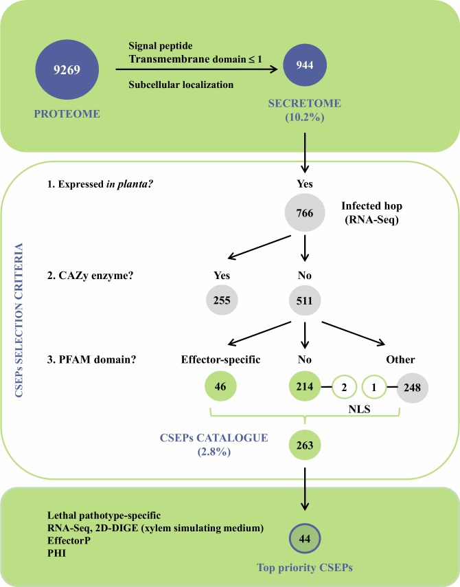
The V. nonalfalfae genome comprises 9,269 predicted protein-encoding genes. Among these putative proteins, 944 are classically secreted proteins with signal peptide and no more than one transmembrane domain, representing 10.2% of the V. nonalfalfae predicted proteome (Fig 1 and S1 Table). The accuracy of prediction was evaluated by comparing this dataset to a set of 91 unique sequences obtained by proteomic analysis (2D-DIGE) of V. nonalfalfae proteins secreted in xylem simulating medium [44], resulting in a 81.3% match (S1 Table). Using TMHMM and Phobius [46] for transmembrane (TM) domain prediction, 801 proteins without a TM domain and 161 proteins harboring one TM domain were determined (S1 Table). Based on subcellular localization predictions, 709 extracellular proteins ('extr' > 17) were acquired with WoLF PSORT [47], 450 proteins residing in the apoplast were determined with ApoplastP [48], while Localizer [49] identified 52 proteins harboring a chloroplast targeting signal and 12 proteins with a signal sequence for localization in mitochondria (S1 Table).
Fig 1. Bioinformatics pipeline for secretome prediction, identification and characterization of V. nonalfalfae CSEPs.
V. nonalfalfae predicted proteome was first filtered based on signal peptide, transmembrane domains and subcellular localization to determine classically secreted proteins. This total predicted secretome was then refined to proteins expressed in planta and carbohydrate active enzymes were removed so that only proteins with effector-specific PFAM domains, NLS signal or no PFAM domains were retained in the final dataset of 263 CSEPs. These were narrowed down to 44 top-priority candidates based on the results of RNA-Seq and 2D-DIGE analyses, sequence similarity searching to known effectors in the PHI database and EffectorP prediction.
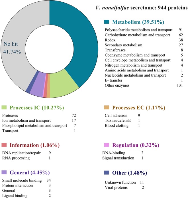
After similarity searches to known proteins in various databases, hypothetical functions were assigned to 727 (77%) putatively secreted V. nonalfalfae proteins. A superfamily annotation scheme [50] was used to classify the V. nonalfalfae secretome into seven functional groups (Fig 2). In the 'Metabolism' group, pectin lyase-like proteins and proteins with a cellulose-binding domain were over-represented within the 'Polysaccharide metabolism and transport' category, while (trans)glycosidases and six-hairpin glycosidases were predominant in the 'Carbohydrate metabolism and transport' category. Other abundant proteins in the 'Metabolism' group were reductases and heme-dependent peroxidases in the 'Redox' category, Concavalin A-like lectins/glucanases in the 'Secondary metabolism' category and 'Transferases'. The major constituents of the 'Intracellular processes' group were 'Proteases' (especially acid proteases, Zn-dependent exopeptidases, subtilisin-like proteases and metalloproteases), cupredoxins in the 'Ion metabolism and transport' category and phospholipases in the 'Phospholipid metabolism and transport' category. Proteins involved in 'Cell adhesion' were over-represented within 'Extracellular processes', while most proteins in the 'Information' group classified into the 'DNA replication/repair' category. In the 'General' group, FAD-binding/transporter-associated domain-like proteins and proteins with a FAD/NAD(P)-binding domain were major constituents in the 'Small molecule binding' category. Analysis of the EuKaryotic Orthologous Groups (KOG) (S2 Table) revealed a high number of proteins in the 'Cellular processing and signaling' category, associated with posttranslational modification, protein turnover, chaperones and signal transduction mechanisms, as well as cell wall/membrane/envelope biogenesis and intracellular trafficking, secretion, and vesicular transport, while the protein composition of the 'Metabolism' category mirrored that of the Superfamily.
Fig 2. Classification of the V. nonalfalfae predicted secreted proteins into functional groups based on the Superfamily annotation scheme.
Groups were classified after [50].
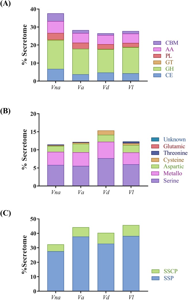
Since 'Carbohydrate metabolic process' and 'Peptidase activity' were overrepresented terms after Blast2GO analysis of the V. nonalfalfae secretome (S1 Fig), they were investigated more thoroughly. Almost one third of the putative V. nonalfalfae secretome was CAZymes, of which 255 were expressed in planta and distributed as follows: glycoside hydrolases (129 GH), carbohydrate esterases (47 CE), redox enzymes that act in conjunction with CAZymes (49 AA; proteins with auxiliary activities), proteins with carbohydrate-binding modules (32 CBM), polysaccharide lyases (25 PL), and glycosyltransferases (4 GT). This repertoire of CAZymes was compared to other plant pathogenic Verticillium species (Fig 3A) and it was demonstrated that V. nonalfalfae had statistically more putative secreted CEs than V. alfalfae, in particular those involved in deacetylation of xylans and xylo-oligosaccharides. Moreover, the V. nonalfalfae secretome consisted of more putative secreted GHs than V. dahliae with major differences found in the GH3 group consisting primarily of stereochemistry-retaining β-glucosidases [51], the GH5 group of enzymes acting on β-linked oligo- and polysaccharides, and glycoconjugates [52], and the GH43 group of enzymes for the debranching and degradation of hemicellulose and pectin polymers [53]. In addition, the V. nonalfalfae secretome was enriched in putative secreted proteins with CBMs, when compared to V. alfalfae, V. longisporum and V. dahlia. This was largely due to cellulose-binding module CBM1 attached to various enzymes from families CE1, CE5, CE15, GH5, GH6, GH7, GH10, GH11, GH12, GH45, GH74, PL1, PL3 and AA9, and to some extent due to chitin-binding module CBM50 found in LysM effector proteins and subgroup C chitinases [54].
Fig 3. Relative abundance of carbohydrate-active enzymes (CAZymes), peptidases and small secreted proteins within secretomes of plant pathogenic Verticillium species.
(A) Comparison of CAZymes from different classes is presented in percentages of predicted fungal secretome. CE, carbohydrate esterase; GH, glycoside hydrolase; GT, glycosyl transferase; PL, polysaccharide lyase; CBM, proteins with carbohydrate-binding modules; AA, proteins with auxiliary activities. (B) Comparison of various classes of peptidases depicted as percentage of predicted fungal secretome. (C) Relative abundance of small secreted proteins and small secreted cysteine rich proteins in predicted fungal secretomes. SSPs, small secreted proteins with less than 300 aa; SSCPs, small secreted proteins with more than 5% cysteine content and at least 4 Cys residues [55]; Vna, V. nonalfalfae; Va, V. alfalfae; Vd, V. dahliae; Vl, V. longisporum.
Similarity searching of V. nonalfalfae putative secreted proteins against peptidases in the MEROPS database revealed 12 in planta expressed aspartic peptidases, 2 cysteine peptidases, 27 metallopeptidases, 44 serine peptidases and 1 threonine peptidase. The highest representation of V. nonalfalfae putative secreted peptidases was in the M14A (carboxypeptidase A1), S08A (subtilisin Carlsberg) and A01A (pepsin A) subfamilies (S3 Table). Comparison of putative secreted peptidases between plant pathogenic Verticillium species (Fig 3B) revealed a similar distribution of peptidases among V. nonalfalfae, V. alfalfae and V. longisporum, while V. dahliae had a statistically different distribution of metallopeptidases, cysteine and serine peptidases. Other enzymatic activities of putatively secreted V. nonalfalfae proteins according to the KEGG analysis can be found in S4 Table.
Querying Verticillium in silico secretomes for small secreted proteins (SSPs) of less than 300 aa and small secreted cysteine rich (SSCPs) proteins with more than 5% cysteine content and at least 4 Cys residues [55] showed 5–10% lower abundance of SSPs and 2–3% fewer SSCPs in the V. nonalfalfae secretome than in the secretomes of other plant pathogenic Verticillium species (Fig 3C). Since small secreted proteins are the least characterized portion of fungal secretomes and many have been shown to act as effectors, our secretome analysis further focused on filtering secreted proteins for expression in planta to identify CSEPs relevant to V. nonalfalfae infection of hop. Genome-wide transcriptome analysis of the V. nonalfalfae interaction with hop [45] revealed that 766 (81%) transcripts in the V. nonalfalfae in silico secretome were expressed in infected hop samples. They showed distinct expression patterns related to different stages of infection (6, 12, 18 and 30 dpi), hop cultivar (susceptible 'Celeia' or resistant 'Wye Target') and plant tissue (roots or shoots) (S2 Fig). From this dataset, all CAZymes except CBMs were omitted from further analysis, resulting in 529 putatively secreted in planta expressed proteins (Fig 1), of which 308 had sequence similarity to PFAM domains (S5 Table). These included, among others, effector-specific PFAM domains, such as LysM effectors [27], Necrosis inducing proteins (NPP1) [56], Hce2 (Homologs of Cladosporium fulvum Ecp2 effector) effector proteins [57], Cerato-platanins [58], Cyanovirin-N lectins [59], hydrophobins [60] and CFEM (Common in Fungal Extracellular Membranes) domain containing proteins [61]. Our final dataset of CSEPs comprised a total of 263 proteins without functional PFAM domains, and proteins bearing known effector-specific PFAM domains, representing 2.8% of the putative proteome. Among them, we determined also 3 CSEPs with a nuclear localization signal (NLS), implying their activity in the plant nucleus, 3 CSEPs specific to the lethal strain of V. nonalfalfae and 69 probable effector proteins (S6 Table) as predicted by EffectorP [16]. Similarity searching of CSEPs to experimentally verified pathogenicity, virulence and effector genes from fungal, oomycete and bacterial pathogens in the Pathogen-host interaction (PHI) database [62] revealed proteins matching AVR effectors (5 hits) and known effector proteins displaying reduced virulence (11 hits) or unaffected pathogenicity (4 hits) (S6 Table).
V. nonalfalfae CSEPs display distinct gene expression profiles during infection of hop
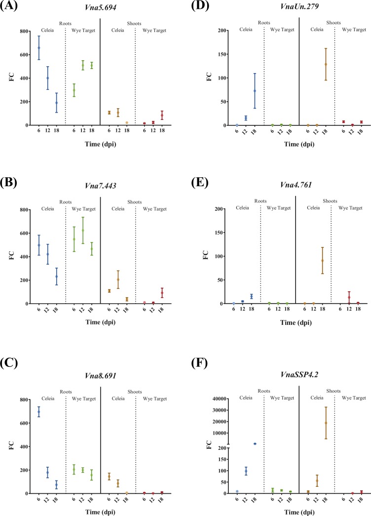
Establishing successful colonization of a host plant requires effective and timely delivery of the fungal pathogen’s effectors. Using quantitative real-time PCR, the expression of the 44 top-priority CSEPs selected according to their likelihood of being effectors (see Methods for selection criteria), was investigated in root and shoot samples of Verticillium wilt susceptible ('Celeia') and resistant ('Wye Target') hop at 6, 12 and 18 days after inoculation with V. nonalfalfae. In a preliminary experiment, the average expression of the selected CSEPs in pooled root samples at different time points was examined (S7 Table). The three highest expressed CSEPs that were also selected by Effector P prediction, and two lethal pathotype specific CSEPs, were then profiled using biological replicates (Fig 4). We included the VnaSSP4.2 gene, encoding a small secreted protein, in the gene expression analysis as a positive control for virulence-associated V. nonalfalfae effector [38].
Fig 4. Gene expression profiles of selected V. nonalfalfae CSEPs in the roots and shoots of infected hop.
FC, fold change in gene expression was determined by quantitative real-time PCR using topoisomerase and splicing factor 3a2 as endogenous controls and fungal samples grown on ½ CD medium as a reference. Means ± SEM (n = 5) are presented. Statistical significance was determined with the t-test using the Holm-Sidak approach, with α = 5%. 'Celeia', Verticillium wilt susceptible hop; 'Wye Target', Verticillium wilt resistant hop; dpi, days post inoculation.
The expression levels of genes Vna5.694, Vna7.443 and Vna8.691 were greater in the roots than in the shoots of both hop varieties. Gene expression of Vna5.694 (Fig 4A), encoding a small (81 aa) secreted cysteine-rich protein of unknown function and displaying the highest similarity to V. longisporum CRK15920 protein, decreased with time in the roots of susceptible hop, while its expression in the resistant hop increased. A similar trend of expression was also observed in the shoots of both hop varieties. Gene expression of Vna7.443 (Fig 4B), which produces a secreted protein (276 aa without cysteines) with the highest similarity to V. longisporum CRJ82870 protein, was comparable to Vna5.694; it decreased in the roots of susceptible hop and peaked at 12 dpi in the roots of resistant hop. A peak of expression at 12 dpi was also observed in the shoots of susceptible hop, while its expression increased with time in the shoots of resistant hop. Expression of the Vna8.691 gene (Fig 4C), coding for a small secreted protein (95 aa without cysteines) of unknown function with the highest similarity to V. longisporum CRK10461 protein, was highest in the roots of susceptible hop at 6 dpi and then decreased with time of infection. The same trend was also observed in the shoots of susceptible hop. Expression of Vna8.691 in the roots of resistant plants was constant, and around 200-fold higher than that in ½ CD medium, whereas no expression was detected in the shoots. Interestingly, two lethal pathotype specific genes VnaUn.279 (Fig 4D), encoding a small secreted protein (92 aa without cysteines) with the highest similarity to V. dahliae VdLs.17 EGY23483 protein, and Vna4.761 (Fig 4E), encoding a 186 aa protein with 8 cysteines and the highest similarity to V. longisporum CRK16219 protein, had similar gene expression patterns as the virulence-associated V. nonalfalfae effector VnaSSP4.2 (Fig 4F). They were expressed only in the roots and shoots of susceptible hop (with expression levels increasing with time of infection) and barely detected in the resistant plants.
Identification of novel virulence effector of V. nonalfalfae
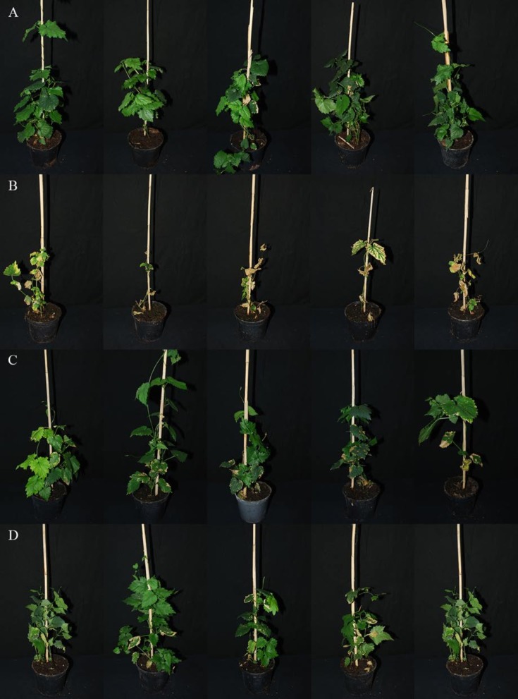
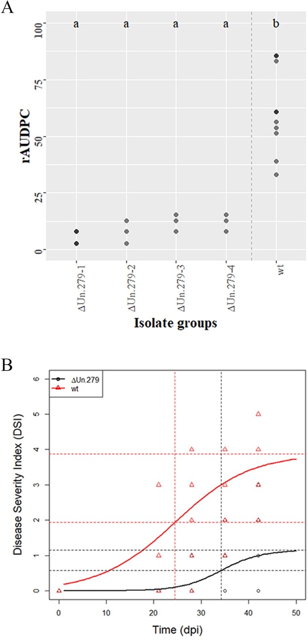
Since all five selected CSEPs were specifically expressed during plant colonization, a reverse genetics approach was used to test their contribution to the virulence of V. nonalfalfae in hop. Knockout mutants of VnaUn.279 displayed only minor wilting symptoms in the susceptible hop (Fig 5), while vegetative growth, fungal morphology and sporulation were not affected. Analysis of the relative area under the disease progress curve (rAUDPC) [63] indicated that four independent VnaUn.279 knockout mutants displayed statistically significantly lower values of rAUDPC than the wild type fungus (Fig 6A). To understand the progress of disease in time, statistical modelling was undertaken. For illustrative purposes only, disease severity index (DSI) values [42] for ΔVnaUn.279 and wild type fungus were modelled by logistic growth model (Fig 6B). The variability of disease progression in individual hop plants is probably due to the specific nature of Verticillium colonization, in which only a few attached hyphae randomly penetrate the root intercellularly [64]. As demonstrated by the inflection point of the ΔVnaUn.279 logistic curve, development of disease symptoms was delayed for 10 days compared to the wild type fungus. Based on the asymptote values, the wilting symptoms were considerably less severe in the mutant than in the wild type fungus. Additionally, fungal biomass assessment with qPCR revealed that 32% of plants were infected with V. nonalfalfae ΔVnaUn.279 mutants compared to at least 80% for the wild type fungus. These results indicate that deletion of VnaUn.279 not only severely reduced V. nonafalfae virulence but also significantly affected the fungal infectivity via a yet unknown mechanism.
Fig 5. Pathogenicity testing in susceptible hop 'Celeia'.
Pictures were taken 31 days post inoculation of hop roots with V. nonalfalfae conidia suspension. (A) mock-inoculated plants; (B) plants inoculated with wild type V. nonalfalfae T2 strain; (C and D), plants inoculated with V. nonalfalfae ΔVnaUn.279 mutant strains (two replicates).
Fig 6. V. nonalfalfae knockout mutant ΔVnaUn.279 showed severely reduced virulence.
(A) Relative area under the disease progress curve (rAUDPC) is presented for knockout mutant ΔVnaUn.279 and for wild type (wt) V. nonalfalfae. Darker dots depict double values. A Kruskal-Wallis test followed by multiple comparison test resulted in two groups: a for ΔVnaUn.279 and b for wild type. (B) Disease severity index (DSI) values were fitted by a simple logistic growth model for mutant ΔVnaUn.279 and for wild type V. nonalfalfae hop isolates. The upper horizontal line is the asymptote (black for ΔVnaUn.279, red for wt); the vertical lines show the inflection points (black for ΔVnaUn.279, red for wt) at which the predicted DSI is one half of the asymptote.
The other tested V. nonafalfae knockout mutants (Vna5.694, Vna7.443 and Vna8.691 in S3 Fig) showed unaffected pathogenicity and no statistical differences in rAUDPC values relative to the wild type. Deletion of Vna4.761 was not achieved, due to its functional redundancy, since two additional copies with over 95% sequence identity have been found after Blastn search against V. nonalfalfae reference genome at two different genomic locations.
Discussion
Fungal pathogens have evolved diverse strategies to interact with host plants and secrete various effector molecules to overcome plant defense mechanisms. A recently published genome of xylem-invading Sordariomycete fungus Verticillium nonalfalfae [22], a transcriptome study of infected hop [45] and obtained proteomic data of fungal growth on xylem simulating medium [44] have provided an opportunity to screen for proteins that may contribute to fungal virulence in hop. In the current study, a customized bioinformatics pipeline was designed to predict the classical V. nonalfalfae secretome and then to refine the secretome based on experimental data to identify CSEPs.
The relative secretome size of V. nonalfalfae (10.2%) conforms to the nutritional lifestyle of plant pathogens with larger secretome sizes (from 2.9% to 12.1% of the proteomes, with an average of 7.4%) and fits in the phylogenetic context with other Pezizomycotina (from 3.7% to 12.1% of the proteomes, with an average of 7.3%) [55]. The majority of proteins (69.5%) in the V. nonalfalfae secretome were less than 500 aa residues. In contrast to some plant pathogenic Pezizomycotina, which had remarkable 10–15% enrichment of proteins of up to 100 aa residues [55], only 1.8% of such proteins were found in the V. nonalfalfae secretome.
The composition of the V. nonalfalfae predicted secretome, rich in carbohydrate active enzymes (33%), proteases (11%), lipases/cutinases (4.6%) and oxidoreductases (4%), reflected its nutritional lifestyle as a hemibiotroph and plant vascular pathogen. Hemibiotrophic fungi undergo two phases during the infection process; an initial biotrophic phase, with characteristic expression of small secreted proteins without functional annotation (SSPs), is followed by a necrotrophic stage, which is generally associated with the expression of plant cell wall-degrading enzymes (CWDEs) [8]. Similar to V. dahliae and V. alfalfae genomes [65], the V. nonalfalfae genome encodes more CWDEs per number of secreted proteins than other plant pathogenic fungi [8]. Pectinases, xylanases, cellulases, glucanases, proteases, cutinases and lipases are major classes of CWDEs [66] and play important roles during plant colonization. They may facilitate penetration of the plant roots to reach the xylem vessels, degrade pectin gels and tyloses, formed in response to infection, to spread inside vessels, breakdown insoluble wall polymers to acquire nutrients and contribute to the release of survival structures from dead plant material [65]. In addition to contributing to virulence [67], some CWDEs are recognized as pathogen-associated molecular patterns (PAMPs), which provoke PAMP-triggered immunity [68,69]. On the other hand, V. dahliae carbohydrate-binding module family 1 domain-containing proteins may suppress glycoside hydrolase 12 protein-triggered immunity in plants [70]. As in the case of V. dahliae [65], the V. nonalfalfae predicted proteome contains numerous glycoside hydrolases and polysaccharide lyases, most of which are secreted [44]; however, only five glycosyl transferases were determined in the predicted secretome. In contrast to V. dahliae, there was almost double the number of carbohydrate esterases in the V. nonalfalfae proteome and over half were predicted to be secreted. Other abundant proteins in the V. nonalfalfae predicted secretome were acid proteases, subtilisin-like proteases and zinc-dependent metalloproteases. These enzymes probably participate in amino acid acquisition, manipulation of host defenses by degradation of pathogenesis-related proteins, including plant chitinases, and act as virulence factors or as elicitors of defense responses [71,72]. A significant number of lipases, phospholipases and cutinases were determined in the V. nonalfalfae predicted secretome and identified in a previous proteomic study [44]. In addition to supplying energy for pathogen growth, lipid hydrolysis is crucial for the production of certain signaling molecules, such as oxylipins, which manipulate the host lipid metabolism and alter plant defense responses [73]. The role of cutinases in pathogenicity is controversial and has been associated with the dissolution of the plant cuticle during penetration, suppression of callose formation, spore attachment and surface signaling [74,75]. Another group of abundant enzymes in V. nonalfalfae predicted and experimentally determined secretomes were oxidoreductases, in particular FAD-dependent oxidoreductases and GMC oxidoreductases. Oxidoreductases are probably secreted for protection against host-produced reactive oxygen species, such as the generation of H2O2, which was detected after infection with V. dahliae in cotton roots [76] and in tomato plants [77]. On the other hand, fungal pathogens can actively contribute to the ROS level in host plants [78]. In V. dahliae, NADPH oxidase complex (Nox), composed of the catalytic subunit VdNoxB and tetraspanin VdPls1, is responsible for the production of ROS and the formation of penetration peg within the hyphopodium [64]. Moreover, VdNoxB regulates the cytoskeletal organization of the VdSep5-septin ring that separates the hyphopodium from invasive hyphae and forms a specialized fungus-host penetration interface, where small secreted proteins preferentially accumulate [79].
Since small secreted proteins (SSPs) are the least characterized fungal secreted proteins and some have been reported as effectors, we particularly focused on this group of proteins. Various criteria for the determination of SSPs have been reported [12,80,81] but, for comparison purposes, we adopted a definition [55] that considers SSPs proteins with a mature length of ≤ 300 aa residues and proteins with a relative cysteine content of ≥5%, as well as ≥4 cysteine residues, to be small secreted cysteine-rich proteins (SSCP). According to these criteria, the V. nonalfalfae predicted secretome contains 310 SSPs (32.8% of the predicted secretome) and 46 (4.9% of the secretome) of those belong to SSCPs. These numbers are lower than the average contents of SSPs (49%) and SSCPs (6.7%) determined in fungi of class 2 secretome size (500–1100 secreted proteins) and average contents of SSPs (47%) and SSCPs (7.5%) in the Pezizomycotina group [55]. In a recent study, SSPs with a mature length of ≤ 300 aa residues were identified in 136 fungal species and compared in terms of taxa and lifestyles [82]. On average, hemibiotrophs and necrotrophs had higher proportions of secreted enzymes, while biotrophs, symbionts and certain hemibiotrophs had the most abundant SSPs. Furthermore, higher numbers of species-specific SSPs (over 100) were associated with biotrophs and symbionts than necrotrophs and saprotrophs (around 30), suggesting that these effectors coevolved with their hosts, while the range was widest for hemibiotrophs. However, no species-specific SSPs have been discovered in V. nonalfalfae, while 13 and 19 have been reported in V. albo-atrum and V. dahliae, respectively [82].
Further analysis of the refined V. nonalfalfae secretome, comprised of 263 CSEPs, focused in particular on homologs of known effectors from other plant pathogens. Searching for LysM effectors using CAZy module (CBM50) and PFAM domain PF01476 revealed that the V. nonalfalfae secretome contains four in planta expressed proteins with 2–6 LysM domains, of which Vna2.979 is an ortholog of VDAG_00902 (S6 Table), a core LysM effector of V. dahliae [27]. The NLPs are a group of widespread conserved effectors that can trigger immune responses and cell death [56]. Similar to other Verticillium spp. [19,65], NLP genes are expanded in the V. nonalfalfae genome, with seven genes orthologous to V. dahliae NLP1-9 and having no ortholog to VdNLP6. All five NLPs with homologs in the PHI database (S6 Table) were expressed in hop; the most abundant was Vna7.239 (VdNLP9), in particular in the roots of susceptible hop (S3 Fig), suggesting some role in the plant colonization process. Four fungal hydrophobins, characterized by high levels of hydrophobicity and the presence of eight conserved cysteine residues [60], were found in the refined V. nonalfalfae secretome and had homologs in the PHI database (S6 Table). Although all were expressed in hop, only expression of Vna7.87 was abundant and root-specific. Interestingly, a role in the development of microsclerotia [83] was demonstrated for type II hydrophobin VDH1 from V. dahliae, but it was not required for pathogenicity. Further mining of CSEPs for known effectors revealed that in planta expressed Vna2.8 and Vna3.54 are homologs of AVR-Pita1, a neutral zinc metalloprotease from Magnaporthe oryzae [84], and Vna1.1274 was similar to MgSM1, a putative small protein of the Cerato-platanin family [85]. Three V. nonalfalfae CSEPs, Vna10.263 and Vna5.719 with high and ubiquitous expression in hop, and Vna9.246 specifically expressed only in susceptible hop, had similarity to Candida albicans RBT4, secreted pathogenesis-related proteins [86], while one CSEP was an ortholog of urea amidolyase (DUR1,2), which enables utilization of urea as a sole nitrogen source [87]. Highly expressed secreted protein Vna4.130 had similarity to EMP1, extracellular matrix protein 1 from Magnaporthe grisea, which was required for appressorium formation and pathogenicity [88]. Vna7.617, which was putatively secreted and abundantly expressed in susceptible hop, had an ortholog in Fusarium oxysporum membrane mucin Msb2, which regulates invasive growth and plant infection upstream of Fmk1 MAPK [89].
The remaining V. nonalfalfae CSEPs (92%) were hypothetical, predicted and conserved hypothetical proteins with no functional annotation and their temporal gene expression patterns in susceptible and resistant hop were explored to provide some clues to their function (S7 Table and S2 Fig). In addition to the already reported effector VnaSSP4.2 [38], several other CSEPs with distinct expression patterns and high levels of expression were found. Among five CSEPs selected for gene functional analysis using a reverse genetics approach, four were predicted as effectors by EffectorP [16] and one had an ortholog in the PHI database, displaying sequence similarity to fungal effector LaeA, a regulator of secondary metabolism [90] and morphogenetic fungal virulence factor [91]. After comparing the pathogenicity of wild type fungus to CSEPs knockout mutants (Fig 6 and S3 Fig), we discovered that the later CSEP, encoded by lethal pathotype specific gene VnaUn.279, is a novel virulence factor of V. nonalfalfae. ΔVnaUn.279 mutants had diminished infectivity and exhibited severely reduced virulence in hop. Reduced virulence was also reported for a number of LaeA deletion mutants from human pathogen A. fumigatus [91], plant pathogenic fungi, including A. flavus, C. heterostrophus and several Fusarium species [92], as well as entomopathogenic fungus Beauveria bassiana [93]. Alltogether, these findings justify further investigation of the biological role of VnaUn.279 in V. nonalfalfae pathogenicity.
Despite the other selected CSEPs mutants not displaying any virulence associated phenotype, based on their expression profiles, they probably participate in other physiological processes during V. nonalfalfae infection of hop. Additionally, certain CSEPs may be recognized (and subsequently termed Avr effectors) by plant resistance proteins (R proteins), which are intracellular nucleotide-binding leucine rich repeat (NLR) receptors, via direct (receptor-mediated binding) or indirect (accessory protein-mediated) interactions, resulting in effector triggered immunity (ETI) [94,95]. To support this hypothesis, further testing of CSEPs mutants in the resistant hop cultivar is required and could result in identification of corresponding hop resistance proteins. These may then be exploited in Verticillium wilt control by introducing new genetic resistance traits into hop breeding, as already successfully implemented in certain other crops [96].
Conclusions
After comprehensively investigating the predicted V. nonalfalfae secretome using a diverse bioinformatics approaches and integrating multiple lines of evidence (genomics, transcriptomics and proteomics), several candidate secreted effector proteins were identified among protein-encoding genes. These are of high interest to scientists working on Verticillium wilt and, more generally, on pathogen effectors. Since the majority were non-annotated protein sequences, two strategies were adopted to gather clues about their function. With spatio-temporal gene expression profiling, we identified those candidate effectors that have important roles during V. nonalfalfae colonization of hop, while pathogenicity assays with effector knockout mutants revealed the candidates that contribute to fungal pathogenicity in hop. In conclusion, a new virulence effector of V. nonalfalfae, encoded by lethal-pathotype specific gene VnaUn.279, was identified and will be subject to future functional and structural studies.
Material and methods
Microbial strains and cultivation
Sordariomycete fungus Verticillium nonalfalfae [1] was obtained from the Slovenian Institute of Hop Research and Brewing fungal collection. Two isolates from infected hop were used, differing in aggressiveness: lethal pathotype (isolate T2) and mild pathotype (isolate Rec) [97]. Fungal mycelium was cultured at room temperature on a half concentration of Czapek Dox broth (½ CD), supplemented with 1 g/L malt extract, 1 g/L peptone (all from Duchefa, The Netherlands) and 1 g/L yeast extract (Sigma Life Science, USA). For solid media, 15 g/L agar (Duchefa, The Netherlands) was added to ½ CD with supplements. Alternatively, potato dextrose agar (PDA; Biolife Italiana Srl, Italy) or Xylem simulating medium (XSM; [98]) was used.
Escherichia coli DH5α, used for amplification of vector constructs, was cultivated in LB medium with 50 mg/L kanamycin (Duchefa, The Netherlands) at 37°C.
Agrobacterium tumefaciens (LBA4404) transformation was performed in YM medium [99] containing 100 mg/L streptomycin and 50 mg/L kanamycin (both from Duchefa, The Netherlands) at 30°C. The co-cultivation of transformed A. tumefaciens and V. nonalfalfae was carried out on IMAS plates [99] at room temperature.
Functional annotation of V. nonalfalfae gene models and RNA-Seq analysis
Using a customized Genialis Platform (Genialis, Slovenia; https://www.genialis.com/genialis-platform/), the gene models of the V. nonalfalfae reference genome [22] were translated into putative proteins with the ExPASy Translate tool [100] and ORF Finder [101]. The general characteristics of putative proteins (molecular weight, number of amino acids (aa), percentage of cysteines and isoelectric point) were predicted by the ProtParam tool [100]. Functional annotation of the predicted proteins was performed with HMMER searches [102] against CAZy [103,104], Pfam [105], and Superfamily [106], as well as with BLAST searches [107] against NCBI, KOG [108], MEROPS [109] and PHI databases [62], followed by Blast2GO [110] and KEGG [111,112] analyses. The overrepresentation of GO terms in the V. nonalfalfae in-silico secretome compared to proteome was assessed using a hypergeometric distribution test (HYPGEOM.DIST function in Excel) with a p-value < 0.05 and FDR < 0.05.
RNA-sequencing of V. nonalfalfae mild and lethal pathotypes was performed by IGA Technology Service (Udine, Italy) using Illumina Genome Analyzer II. For this purpose, total RNA, enriched for the polyA mRNA fraction, was isolated in three biological replicates from fungal mycelia of mild and lethal strains grown in xylem-simulating media according to [98]. Illumina raw sequence reads were deposited at NCBI (Bioproject PRJNA283258). RNA-Seq analysis was performed using CLC Genomics Workbench tools (Qiagen, USA). Differentially expressed genes between lethal and mild fungal pathotype were identified as those with fold change FC ≥ 1.5 or FC ≤ - 1.5 (p ≤ 0.05; FDR ≤ 0.05).
From our previous RNA-Seq data of compatible and incompatible interactions between hop and V. nonalfalfae [45], fungal transcripts expressed at a least one time point (6, 12, 18 and 30 dpi) and one hop cultivar (susceptible 'Celeia' or resistant 'Wye Target') were filtered out and data were presented as a matrix of log2CPM (counts per million–number of reads mapped to a gene model per million reads mapped to the library) expression values. These genes were considered as expressed in planta.
Secretome prediction and comparison
The V. nonalfalfae in silico secretome was determined using a customized Genialis Platform (Genialis, Slovenia) according to the method described in [113], which reportedly gives 83.4% accuracy for fungal secreted proteins. It combines SignalP4.1 [114], WolfPsort [47] and Phobius [46] for N-terminal signal peptide prediction, TMHMM (http://www.cbs.dtu.dk/services/TMHMM/) for eliminating membrane proteins (allowing one transmembrane (TM) domain in the first 60 aa) and PS-Scan [115] for removing proteins with ER targeting sequence (Prosite: PS00014). In addition, we used LOCALIZER [49] to predict effector protein localization to chloroplasts and mitochondria, while proteins with a nuclear localization signal were determined with NucPred (likelihood score >0.80) [116] and PredictNLS [117]. Localization of effector proteins to the apoplast was predicted by ApoplastP [48] based on enrichment in small amino acids and cysteines, as well as depletion in glutamic acid.
To compare the composition of the V. nonalfalfae secretome to other closely related plant pathogenic Verticillium species, protein coding sequences of Verticillium dahliae JR2, Verticillium longisporum GCA_001268145 and Verticillium alfalfae VaMs.102 from the Ensembl Fungi database (http://fungi.ensembl.org/info/website/ftp/index.html) were used and secretome predictions, HMMER searches against CAZy database and blastp searches against the MEROPS database were performed using the same pipeline as for V. nonalfalfae. Two way ANNOVA followed by Tukey’s multiple comparisons test (p-value < 0.05) in GraphPad Prism 7.03 (GraphPad Software, Inc., USA) was used to find differences between sets of fungal proteins.
Refinement of V. nonalfalfae secretome and selection of CSEPs
A refinement of total V. nonalfalfae in silico secretome (Fig 1) was done to maintain only proteins, transcripts of which were expressed in planta according to the RNA-Seq analysis. Proteins with carbohydrate enzymatic activities (CAZy screening) were excluded from further analysis and additional filtering was applied based on the presence of PFAM domains. CSEPs were identified as proteins having known effector-specific domains [9,12,13,16,118], NLS signal or as proteins with no PFAM domains.
In the second step, the number of secreted proteins was narrowed down using the following criteria: proteins determined in the lethal pathotype-specific region identified by comparative genomics of mild and lethal V. nonalfalfae strains from three geographic regions [22]; proteins differentially expressed in lethal compared to mild V. nonalfalfae strains grown in xylem-simulating media as determined by RNA-Seq; V. nonalfalfae secreted proteins analyzed by 2D-DIGE and identified by MALDI-TOF/TOF MS [44]; proteins with sequence similarity to experimentally verified pathogenicity, virulence and effector genes in the PHI (Pathogen-host Interaction) database [62] and putative effector proteins predicted by EffectorP software [16].
Quantitative real time PCR and fungal biomass quantification
Susceptible 'Celeia' and resistant 'Wye Target' hop varieties were inoculated by the root dipping method [119] with V. nonalfalfae spores. Total RNA was isolated from the roots and stems of infected plants (6, 12 and 18 dpi) or mock-inoculated plants using a MagMAX total RNA isolation kit (Life Technologies, USA). The quality and quantity of RNA was assessed on an Agilent 2100 Bioanalyzer (Agilent Technologies, Germany). Total RNA was transcribed to cDNA with a High Capacity cDNA Reverse Transcription kit (Applied Biosystems, USA). Real-time PCR reactions were performed on 7500 Fast Real Time PCR Systems (Life Technologies, USA) using the FastStart SYBR Green master mix (Roche, Switzerland) and primers (S8 Table) designed by Primer Express 3.0 software (Thermo Fisher Scientific, USA). For each of the 44 top priority CSEPs, gene expression was analyzed on pooled samples containing the roots of five individual plants, in two technical replicates. The highest expressed CSEPs were also analyzed in five biological and two technical replicates per sample group. The CSEPs’ gene expression was calculated by the comparative CT method [120]. The cDNA from V. nonalfalfae mycelium cultivated on ½ CD was used as a reference sample. V. nonalfalfae DNA topoisomerase (VnaUn.148) and splicing factor 3a2 (Vna8.801) were selected as the best endogenous control genes according to GeNorm analysis [121] and fungal biomass normalization. For the latter, fungal DNA was extracted from infected hop using CTAB and quantified by qPCR as described in [122]. The expression of control genes was compared to fungal biomass in infected hop using Pearson’s correlation coefficient.
Construction of knockout vectors and preparation of V. nonalfalfae knockout mutants
CSEPs knockout mutants were prepared according to Frandsen’s protocol [123]. The plasmid vector pRF-HU2 was first linearized with Nt.BbvCI and PacI (New England BioLabs, USA) and purified with Illustra GFX PCR DNA and Gel Band Purification Kit (GE Healthcare, UK). Homologous gene sequences were then amplified with PCR using a PfuTurbo Cx Hotstart DNA polymerase (Agilent Technologies, USA) and the following settings: 95°C for 2 minutes, 30 cycles: 95°C for 30s, 55°C for 30s, 72°C for 1 minute; 75°C for 10 minutes. PCR products were purified with Illustra GFX PCR DNA and a Gel Band Purification Kit (GE Healthcare, UK) and ligated to linearized vector pRF-HU2 with USER enzyme (New England BioLabs, USA). Vector constructs were multiplied in E. coli DH5α cells and isolated with a High Pure Plasmid Isolation Kit (Roche, Life Science, USA).
V. nonalfalfae knockout mutants were generated with Agrobacterium tumefaciens mediated transformation (ATMT) [124] using acetosyringone (Sigma Aldrich, USA). The knockout vector constructs were electroporated with Easyject Prime (EQUIBIO, UK) into electro-competent Agrobacterium tumefaciens (LBA4404) cells. Positive colonies with a correct construct orientation were verified by colony PCR. Co-culture of transformed A. tumefaciens and V. nonalfalfae was carried out on IMAS media as described in [99]. Colonies were transferred on a cellophane membrane (GE Healthcare, UK) to primary and secondary ½ CD selection medium with 150 mg/L timentin (Duchefa, The Netherlands) and 75 mg/L hygromycin (Duchefa, The Netherlands). Genomic DNA was isolated according to [125] from the remaining colonies and the knockout was verified with PCR. Transformed V. nonalfalfae conidia were stored in 30% glycerol at -80°C until testing.
Pathogenicity evaluation of V. nonalfalfae knockout mutants in hop
Before pathogenicity tests were carried out, fungal growth and conidiation were inspected as described previously [126]. Ten to fifteen plants of the Verticillium wilt susceptible hop cultivar 'Celeia' were inoculated at phenological stage BBCH 12 by 10-min root dipping in a conidia suspension of V. nonalfalfae knockout mutants as described previously [126]. Conidia of the wild type V. nonalfalfae lethal pathotype served as a positive control and sterile distilled water was used as a mock control. Verticillium wilting symptoms were assessed four to five times post inoculation using a disease severity index (DSI) with a 0–5 scale [42], and rAUDPC was calculated according to [63]. After symptom assessment, fungal re-isolation test and qPCR using V. nonalfalfae specific primers (S8 Table) were performed to confirm infection of hops.
Statistics
The R package [127] was used for the statistical analysis of the pathogenicity assay of knockout mutants. Due to the different variability of rAUDPC values for the 'isolate' groups, a non-parametric approach was pursued. A Kruskal-Wallis test was used, followed by multiple comparison test with Bonferroni correction. To understand how the time post inoculation with V. nonalfalfae affects hop health, a simple logistic growth model [128] was fitted to DSI values for the groups under study.
Supporting information
Gene IDs marked in bold represent 263 proteins in the CSEPs catalogue. 2D-DIGE, proteins secreted by mild and lethal strains of V. nonalfalfae growing in xylem simulating medium (XSM) were analysed by 2D-DIGE and identified by MALDI-TOF/TOF MS [44]; RNA-Seq, differential fungal gene expression (fold change, FC ≥ 1.5 or FC ≤ - 1.5) of V. nonalfalfae lethal versus mild fungal pathotype growing in XSM; LOCALIZER, a tool for subcellular localization prediction of both plant and effector proteins in the plant cell [49]; ApoplastP, a tool for prediction of effectors and plant proteins in the apoplast using machine learning [48].
(XLSX)
KOG, a database of euKaryotic Orthologous Groups from NCBI that allows identification of ortholog and paralog proteins [108].
(XLSX)
MEROPS, a database of peptidases and the proteins that inhibit them [109].
(XLSX)
KEGG, the Kyoto Encyclopedia of Genes and Genomes is a database resource for understanding high-level functions and utilities of the biological systems [111].
(XLSX)
PFAM, a collection of protein families, represented by multiple sequence alignments and hidden Markov models (HMMs) [105].
(XLSX)
EffectorP, a machine learning prediction program for fungal effectors [16]; PHI, Pathogen-Host Interaction database, which contains expertly curated molecular and biological information on genes proven to affect the outcome of pathogen-host interactions [62].
(XLSX)
Gene expression was calculated by the comparative CT method [120]. Hop plants were inoculated by the root dipping method with V. nonalfalfae conidia and sampled at 6, 12 and 18 days post inoculation. Analyzed samples contained the roots of five individual plants from either susceptible hop variety 'Celeia' (CE) or resistant 'Wye Target' (WT). cDNA from V. nonalfalfae mycelium cultivated on ½ Czapek Dox (CD) medium was used as a reference sample. V. nonalfalfae DNA topoisomerase (VnaUn.148) and splicing factor 3a2 (Vna8.801) were used as endogenous control genes. Numbers represent log2 fold changes in the expression of genes in infected plants at indicated time points compared to gene expression in ½ CD medium. na, not available.
(XLSX)
a, oligonucleotide designed according to template [123]; b, oligonucleotide that amplifies the promoter region of the target gene; c, oligonucleotide that amplifies the terminator region of the target gene; d, oligonucleotide that amplifies the target gene for knockout (KO) selection; e, oligonucleotide that amplifies genomic and vector sequences for KO selection; f, V. nonalfalfae lethal pathotype-specific marker [97].
(XLSX)
GO terms for biological process (A) and molecular function (B) are presented at GO level 4. Relative abundance of gene ontology (GO) terms determined by Blast2GO is expressed in percentages of predicted fungal secretome and proteome, respectively.
(TIF)
Fungal transcripts were first identified by mapping of reads with at least 90% sequence identity and 90% sequence coverage to the V. nonalfalfae reference genome [22] using CLC Workbench. Normalization by trimmed mean of M values (TMM) [129] was performed to eliminate composition biases between libraries. Read counts were converted into log2-counts-per-million (logCPM) values and a cutoff of CPM >1 was chosen. Color scale bar represents the logCPM values, with darker red color meaning higher expression values.
(PDF)
Relative area under the disease progress curve (rAUDPC) is presented for the three V. nonalfalfae deletion mutants (each in four replicates) and the wild type fungus. Depicted are mean values ± SEM (n = 10). Analysis of variance was first performed by Levene’s test, followed by Dunnett’s test to compare each treatment (knockout) with a single control (wild type); however, no statistical differences were found at α level of 5%.
(TIF)
Acknowledgments
We express our sincere gratitude to Dr. Vasja Progar and the Genialis team for their help with bioinformatics. We acknowledge advice on data normalization in RT-qPCR experiments by Dr. Nataša Štajner from the University of Ljubljana. We thank Martin Cregeen for English language editing.
Data Availability
All relevant data are within the paper and its Supporting Information files.
Funding Statement
This work was supported by Slovenian Research Agency (http://www.arrs.gov.si/en/index.asp) grants P4-0077 (BJ), J4-8220 (BJ) and 342250 (KM). The funder had no role in study design, data collection and analysis, decision to publish, or preparation of the manuscript.
References
- 1.Inderbitzin P, Bostock RM, Davis RM, Usami T, Platt HW, Subbarao KV. Phylogenetics and taxonomy of the fungal vascular wilt pathogen Verticillium, with the descriptions of five new species. PLoS One 2011;6: e28341 doi: 10.1371/journal.pone.0028341 [DOI] [PMC free article] [PubMed] [Google Scholar]
- 2.Pegg GF, Brady BL. Verticillium Wilts. New York: CABI Pub.; 2002. [Google Scholar]
- 3.Baker R, Bragard C, Candresse T, Gilioli G, Grégoire JC, Holb I et al. Scientific Opinion on the pest categorisation of Verticillium albo-atrum sensu stricto Reinke and Berthold, V. alfalfae Inderb., HW Platt, RM Bostock, RM Davis & KV Subbarao, sp. nov., and V. nonalfalfae Inderb., HW Platt, RM Bostock, RM Davis & KV Subbar. EFSA Journal. 2014;12(12): 3927 doi: 10.2903/j.efsa.20143927 [Google Scholar]
- 4.Inderbitzin P, Subbarao KV. Verticillium Systematics and Evolution: How Confusion Impedes Verticillium Wilt Management and How to Resolve It. Phytopathology. 2014;104: 564–574. doi: 10.1094/PHYTO-11-13-0315-IA [DOI] [PubMed] [Google Scholar]
- 5.Klimes A, Dobinson KF, Klosterman SJ, Thomma BPHJ. Genomics Spurs Rapid Advances in our Understanding of the Basic Biology of Vascular Wilt Pathogens in the Genus Verticillium. Annu Rev Phytopathol. 2015;53: 181–198. doi: 10.1146/annurev-phyto-080614-120224 [DOI] [PubMed] [Google Scholar]
- 6.Daayf F. Verticillium wilts in crop plants: Pathogen invasion and host defence responses. Can J Plant Pathol. 2015;37: 8–20. [Google Scholar]
- 7.Kamoun S. The Secretome of Plant-Associated Fungi and Oomycetes. The Mycota. Berlin, Heidelberg: Springer Berlin Heidelberg; 2009. [Google Scholar]
- 8.Lo Presti L, Lanver D, Schweizer G, Tanaka S, Liang L, Tollot M, et al. Fungal effectors and plant susceptibility. Annu Rev Plant Biol. 2015;66: 513–545. doi: 10.1146/annurev-arplant-043014-114623 [DOI] [PubMed] [Google Scholar]
- 9.Stergiopoulos I, de Wit PJGM. Fungal effector proteins. Annu Rev Phytopathol. 2009;47: 233–263. doi: 10.1146/annurev.phyto.112408.132637 [DOI] [PubMed] [Google Scholar]
- 10.de Jonge R. In Silico Identification and Characterization of Effector Catalogs. Methods in molecular biology (Clifton, NJ). New York: Humana press; 2012. [DOI] [PubMed] [Google Scholar]
- 11.Sperschneider J, Williams AH, Hane JK, Singh KB, Taylor JM. Evaluation of Secretion Prediction Highlights Differing Approaches Needed for Oomycete and Fungal Effectors. Front Plant Sci. 2015;6: 1168 doi: 10.3389/fpls.2015.01168 [DOI] [PMC free article] [PubMed] [Google Scholar]
- 12.Saunders DGO, Win J, Cano LM, Szabo LJ, Kamoun S, Raffaele S. Using hierarchical clustering of secreted protein families to classify and rank candidate effectors of rust fungi. PLoS One. 2012;7: e29847 doi: 10.1371/journal.pone.0029847 [DOI] [PMC free article] [PubMed] [Google Scholar]
- 13.Guyon K, Balagué C, Roby D, Raffaele S. Secretome analysis reveals effector candidates associated with broad host range necrotrophy in the fungal plant pathogen Sclerotinia sclerotiorum. BMC Genomics. 2014;15: 336 doi: 10.1186/1471-2164-15-336 [DOI] [PMC free article] [PubMed] [Google Scholar]
- 14.Sonah H, Deshmukh RK, Bélanger RR. Computational Prediction of Effector Proteins in Fungi: Opportunities and Challenges. Front Plant Sci. 2016;7: 126 doi: 10.3389/fpls.2016.00126 [DOI] [PMC free article] [PubMed] [Google Scholar]
- 15.Gibriel HAY, Thomma BPHJ, Seidl MF. The Age of Effectors: Genome-Based Discovery and Applications. Phytopathology. 2016;106: 1206–1212. doi: 10.1094/PHYTO-02-16-0110-FI [DOI] [PubMed] [Google Scholar]
- 16.Sperschneider J, Gardiner DM, Dodds PN, Tini F, Covarelli L, Singh KB, et al. EffectorP: predicting fungal effector proteins from secretomes using machine learning. New Phytol. 2016;210: 743–761. doi: 10.1111/nph.13794 [DOI] [PubMed] [Google Scholar]
- 17.Klosterman SJ, Subbarao KV, Kang S, Veronese P, Gold SE, Thomma BPHJ, et al. Comparative Genomics Yields Insights into Niche Adaptation of Plant Vascular Wilt Pathogens. PLoS Pathog. 2011;7: e1002137 doi: 10.1371/journal.ppat.1002137 [DOI] [PMC free article] [PubMed] [Google Scholar]
- 18.De Jonge R, Bolton MD, Kombrink A, Van Den Berg GCM, Yadeta KA, Thomma BPHJ. Extensive chromosomal reshuffling drives evolution of virulence in an asexual pathogen. Genome Res. 2013;23: 1271–1282. doi: 10.1101/gr.152660.112 [DOI] [PMC free article] [PubMed] [Google Scholar]
- 19.Seidl MF, Faino L, Shi-Kunne X, van den Berg GCM, Bolton MD, Thomma BPHJ. The Genome of the Saprophytic Fungus Verticillium tricorpus Reveals a Complex Effector Repertoire Resembling That of Its Pathogenic Relatives. Mol Plant-Microbe Interact. 2015;28: 362–373. doi: 10.1094/MPMI-06-14-0173-R [DOI] [PubMed] [Google Scholar]
- 20.Faino L, Seidl MF, Datema E, van den Berg GCM, Janssen A, Wittenberg AHJ, et al. Single-Molecule Real-Time Sequencing Combined with Optical Mapping Yields Completely Finished Fungal Genome. MBio. 2015;6: e00936–15. doi: 10.1128/mBio.00936-15 [DOI] [PMC free article] [PubMed] [Google Scholar]
- 21.Depotter JRL, Seidl MF, van den Berg GCM, Thomma BPHJ, Wood TA. A distinct and genetically diverse lineage of the hybrid fungal pathogen Verticillium longisporum population causes stem striping in British oilseed rape. Environ Microbiol. 2017;19: 3997–4009. doi: 10.1111/1462-2920.13801 [DOI] [PubMed] [Google Scholar]
- 22.Jakše J, Jelen V, Radišek S, de Jonge R, Mandelc S, Majer A, et al. Genome sequence of xylem-invading Verticillium nonalfalfae lethal strain. Genome Announc. 2018;6: e01458–17. doi: 10.1128/genomeA.01458-17 [DOI] [PMC free article] [PubMed] [Google Scholar]
- 23.Sánchez-Vallet A, Saleem-Batcha R, Kombrink A, Hansen G, Valkenburg D-J, Thomma BPHJ, et al. Fungal effector Ecp6 outcompetes host immune receptor for chitin binding through intrachain LysM dimerization. Elife. 2013;2: e00790 doi: 10.7554/eLife.00790 [DOI] [PMC free article] [PubMed] [Google Scholar]
- 24.Laugé R. The in planta-produced extracellular proteins ECP1 and ECP2 of Cladosporium fulvum are virulence factors. Mol Plant Microbe Interact. 1997;10: 725–734. [Google Scholar]
- 25.Mentlak TA, Kombrink A, Shinya T, Ryder LS, Otomo I, Saitoh H, et al. Effector-mediated suppression of chitin-triggered immunity by Magnaporthe oryzae is necessary for rice blast disease. Plant Cell. 2012;24: 322–335. doi: 10.1105/tpc.111.092957 [DOI] [PMC free article] [PubMed] [Google Scholar]
- 26.Takahara H, Hacquard S, Kombrink A, Hughes HB, Halder V, Robin GP, et al. Colletotrichum higginsianum extracellular LysM proteins play dual roles in appressorial function and suppression of chitin-triggered plant immunity. New Phytol. 2016;211: 1323–1337. doi: 10.1111/nph.13994 [DOI] [PubMed] [Google Scholar]
- 27.Kombrink A, Rovenich H, Shi-Kunne X, Rojas-Padilla E, van den Berg GCM, Domazakis E, et al. Verticillium dahliae LysM effectors differentially contribute to virulence on plant hosts. Mol Plant Pathol. 2017;18: 596–608. doi: 10.1111/mpp.12520 [DOI] [PMC free article] [PubMed] [Google Scholar]
- 28.de Jonge R, Peter van Esse H, Kombrink A, Shinya T, Desaki Y, Bours R, et al. Conserved Fungal LysM Effector Ecp6 Prevents Chitin-Triggered Immunity in Plants. Science. 2010;329: 953–955. doi: 10.1126/science.1190859 [DOI] [PubMed] [Google Scholar]
- 29.Zhou B-J, Jia P-S, Gao F, Guo H-S. Molecular Characterization and Functional Analysis of a Necrosis- and Ethylene-Inducing, Protein-Encoding Gene Family from Verticillium dahliae. Mol Plant-Microbe Interact. 2012;25: 964–975. doi: 10.1094/MPMI-12-11-0319 [DOI] [PubMed] [Google Scholar]
- 30.Santhanam P, van Esse HP, Albert I, Faino L, Nürnberger T, Thomma BPHJ. Evidence for functional diversification within a fungal NEP1-like protein family. Mol Plant Microbe Interact. 2013;26: 278–286. doi: 10.1094/MPMI-09-12-0222-R [DOI] [PubMed] [Google Scholar]
- 31.de Jonge R, van Esse HP, Maruthachalam K, Bolton MD, Santhanam P, Saber MK, et al. Tomato immune receptor Ve1 recognizes effector of multiple fungal pathogens uncovered by genome and RNA sequencing. Proc Natl Acad Sci. 2012;109(13): 5110–5115. doi: 10.1073/pnas.1119623109 [DOI] [PMC free article] [PubMed] [Google Scholar]
- 32.Fradin EF, Abd-El-Haliem A, Masini L, van den Berg GCM, Joosten MHAJ, Thomma BPHJ. Interfamily Transfer of Tomato Ve1 Mediates Verticillium Resistance in Arabidopsis. Plant Physiol. 2011;156: 2255–2265. doi: 10.1104/pp.111.180067 [DOI] [PMC free article] [PubMed] [Google Scholar]
- 33.Fradin EF, Zhang Z, Rovenich H, Song Y, Liebrand TWH, Masini L, et al. Functional analysis of the tomato immune receptor Ve1 through domain swaps with its non-functional homolog Ve2. PLoS One. 2014;9: e88208 doi: 10.1371/journal.pone.0088208 [DOI] [PMC free article] [PubMed] [Google Scholar]
- 34.Liu W, Zeng H, Liu Z, Yang X, Guo L, Qiu D. Mutational analysis of the Verticillium dahliae protein elicitor PevD1 identifies distinctive regions responsible for hypersensitive response and systemic acquired resistance in tobacco. Microbiol Res. 2014;169: 476–482. doi: 10.1016/j.micres.2013.08.001 [DOI] [PubMed] [Google Scholar]
- 35.Wang B, Yang X, Zeng H, Liu H, Zhou T, Tan B, et al. The purification and characterization of a novel hypersensitive-like response-inducing elicitor from Verticillium dahliae that induces resistance responses in tobacco. Appl Microbiol Biotechnol. 2012;93: 191–201. doi: 10.1007/s00253-011-3405-1 [DOI] [PubMed] [Google Scholar]
- 36.Bu B, Qiu D, Zeng H, Guo L, Yuan J, Yang X. A fungal protein elicitor PevD1 induces Verticillium wilt resistance in cotton. Plant Cell Rep. 2014;33: 461–470. doi: 10.1007/s00299-013-1546-7 [DOI] [PubMed] [Google Scholar]
- 37.Zhang L, Ni H, Du X, Wang S, Ma XW, Nürnberger T, et al. The Verticillium-specific protein VdSCP7 localizes to the plant nucleus and modulates immunity to fungal infections. New Phytol. 2017;215: 368–381. doi: 10.1111/nph.14537 [DOI] [PubMed] [Google Scholar]
- 38.Flajšman M, Mandelc S, Radišek S, Štajner N, Jakše J, Košmelj K, et al. Identification of novel virulence-associated proteins secreted to xylem by Verticillium nonalfalfae during colonization of hop plants. Mol Plant Microbe Interact. 2016;29: 362–373. doi: 10.1094/MPMI-01-16-0016-R [DOI] [PubMed] [Google Scholar]
- 39.Keyworth WG. Verticillium wilt of the hop (Humulus lupulus). Ann Appl Biol. 1942;29: 346–357. [Google Scholar]
- 40.Sewell GWF, Wilson JF. The nature and distribution of Verticillium albo-atrum strains highly pathogenic to the hop. Plant Pathol. 1984;33: 39–51. [Google Scholar]
- 41.Talboys PW. Resistance to vascular wilt fungi. Proc R Soc London Ser B Biol Sci. 1972;181: 319–332. [Google Scholar]
- 42.Radišek S, Jakše J, Simončič A, Javornik B. Characterization of Verticillium albo-atrum Field Isolates Using Pathogenicity Data and AFLP Analysis. Plant Dis. 2003;87: 633–638. [DOI] [PubMed] [Google Scholar]
- 43.Radišek S, Jakše J, Javornik B. Genetic variability and virulence among Verticillium albo-atrum isolates from hop. Eur J plant Pathol. 2006;116: 301–314. [Google Scholar]
- 44.Mandelc S, Javornik B. The secretome of vascular wilt pathogen Verticillium albo-atrum in simulated xylem fluid. Proteomics. 2015;15: 787–797. doi: 10.1002/pmic.201400181 [DOI] [PubMed] [Google Scholar]
- 45.Progar V, Jakše J, Štajner N, Radišek S, Javornik B, Berne S. Comparative transcriptional analysis of hop responses to infection with Verticillium nonalfalfae. Plant Cell Rep. 2017;36: 1599–1613. doi: 10.1007/s00299-017-2177-1 [DOI] [PMC free article] [PubMed] [Google Scholar]
- 46.Käll L, Krogh A, Sonnhammer ELL. A combined transmembrane topology and signal peptide prediction method. J Mol Biol. 2004;338: 1027–1036. doi: 10.1016/j.jmb.2004.03.016 [DOI] [PubMed] [Google Scholar]
- 47.Horton P, Park K-J, Obayashi T, Fujita N, Harada H, Adams-Collier CJ, et al. WoLF PSORT: protein localization predictor. Nucleic Acids Res. 2007;35: W585–587. doi: 10.1093/nar/gkm259 [DOI] [PMC free article] [PubMed] [Google Scholar]
- 48.Sperschneider J, Dodds PN, Singh KB, Taylor JM. ApoplastP: prediction of effectors and plant proteins in the apoplast using machine learning. 2017;4: 1764–1778. [DOI] [PubMed] [Google Scholar]
- 49.Sperschneider J, Catanzariti AM, Deboer K, Petre B, Gardiner DM, Singh KB, et al. LOCALIZER: Subcellular localization prediction of both plant and effector proteins in the plant cell. 2017;7: 44598 doi: 10.1038/srep44598 [DOI] [PMC free article] [PubMed] [Google Scholar]
- 50.Wilson D, Pethica R, Zhou Y, Talbot C, Vogel C, Madera M, et al. SUPERFAMILY—sophisticated comparative genomics, data mining, visualization and phylogeny. Nucleic Acids Res. 2009;37: D380–386. doi: 10.1093/nar/gkn762 [DOI] [PMC free article] [PubMed] [Google Scholar]
- 51.Macdonald SS, Blaukopf M, Withers SG. N-acetylglucosaminidases from CAZy family GH3 are really glycoside phosphorylases, thereby explaining their use of histidine as an acid/Base catalyst in place of glutamic acid. J Biol Chem. 2015;290: 4887–4895. doi: 10.1074/jbc.M114.621110 [DOI] [PMC free article] [PubMed] [Google Scholar]
- 52.Aspeborg H, Coutinho PM, Wang Y, Brumer H, Henrissat B. Evolution, substrate specificity and subfamily classification of glycoside hydrolase family 5 (GH5). BMC Evol Biol. 2012;12: 186 doi: 10.1186/1471-2148-12-186 [DOI] [PMC free article] [PubMed] [Google Scholar]
- 53.Mewis K, Lenfant N, Lombard V, Henrissat B. Dividing the large glycoside hydrolase family 43 into subfamilies: A motivation for detailed enzyme characterization. Appl Environ Microbiol. 2016;82: 1686–1692. doi: 10.1128/AEM.03453-15 [DOI] [PMC free article] [PubMed] [Google Scholar]
- 54.Akcapinar GB, Kappel L, Sezerman OU, Seidl-Seiboth V. Molecular diversity of LysM carbohydrate-binding motifs in fungi. Curr Genet. 2015;61: 103–113. doi: 10.1007/s00294-014-0471-9 [DOI] [PMC free article] [PubMed] [Google Scholar]
- 55.Krijger J-J, Thon MR, Deising HB, Wirsel SG. Compositions of fungal secretomes indicate a greater impact of phylogenetic history than lifestyle adaptation. BMC Genomics. 2014;15: 722 doi: 10.1186/1471-2164-15-722 [DOI] [PMC free article] [PubMed] [Google Scholar]
- 56.Gijzen M, Nürnberger T. Nep1-like proteins from plant pathogens: recruitment and diversification of the NPP1 domain across taxa. Phytochemistry. 2006;67: 1800–1807. doi: 10.1016/j.phytochem.2005.12.008 [DOI] [PubMed] [Google Scholar]
- 57.Stergiopoulos I, Kourmpetis YAI, Slot JC, Bakker FT, De Wit PJGM, Rokas A. In Silico Characterization and Molecular Evolutionary Analysis of a Novel Superfamily of Fungal Effector Proteins. Mol Biol Evol. 2012;29: 3371–3384. doi: 10.1093/molbev/mss143 [DOI] [PubMed] [Google Scholar]
- 58.Baccelli I. Cerato-platanin family proteins: one function for multiple biological roles? Front Plant Sci. 2014;5: 769 doi: 10.3389/fpls.2014.00769 [DOI] [PMC free article] [PubMed] [Google Scholar]
- 59.Koharudin LMI, Viscomi AR, Montanini B, Kershaw MJ, Talbot NJ, Ottonello S, et al. Structure-function analysis of a CVNH-LysM lectin expressed during plant infection by the rice blast fungus Magnaporthe oryzae. Structure. 2011;19: 662–674. doi: 10.1016/j.str.2011.03.004 [DOI] [PMC free article] [PubMed] [Google Scholar]
- 60.Bayry J, Aimanianda V, Guijarro JI, Sunde M, Latgé J-P. Hydrophobins—Unique Fungal Proteins. PLoS Pathog. 2012;8: e1002700 doi: 10.1371/journal.ppat.1002700 [DOI] [PMC free article] [PubMed] [Google Scholar]
- 61.Kulkarni RD, Kelkar HS, Dean RA. An eight-cysteine-containing CFEM domain unique to a group of fungal membrane proteins. Trends in Biochemical Sciences. 2003;28(3): 118–121. doi: 10.1016/S0968-0004(03)00025-2 [DOI] [PubMed] [Google Scholar]
- 62.Urban M, Pant R, Raghunath A, Irvine AG, Pedro H, Hammond-Kosack KE. The Pathogen-Host Interactions database (PHI-base): additions and future developments. Nucleic Acids Res. 2015;43: D645–D655. doi: 10.1093/nar/gku1165 [DOI] [PMC free article] [PubMed] [Google Scholar]
- 63.Simko I, Piepho H-P. The area under the disease progress stairs: calculation, advantage, and application. Phytopathology. 2012;102: 381–389. doi: 10.1094/PHYTO-07-11-0216 [DOI] [PubMed] [Google Scholar]
- 64.Zhao Y-L, Zhou T-T, Guo H-S. Hyphopodium-Specific VdNoxB/VdPls1-Dependent ROS-Ca2+ Signaling Is Required for Plant Infection by Verticillium dahliae. PLOS Pathog. 2016;12: e1005793 doi: 10.1371/journal.ppat.1005793 [DOI] [PMC free article] [PubMed] [Google Scholar]
- 65.Klosterman SJ, Subbarao K V, Kang S, Veronese P, Gold SE, Thomma BPHJ, et al. Comparative genomics yields insights into niche adaptation of plant vascular wilt pathogens. PLoS Pathog. Public Library of Science; 2011;7: e1002137 doi: 10.1371/journal.ppat.1002137 [DOI] [PMC free article] [PubMed] [Google Scholar]
- 66.Di Pietro A, Roncero MIG, Roldán MCR. From Tools of Survival to Weapons of Destruction: The Role of Cell Wall-Degrading Enzymes in Plant Infection. In: Deising HB, editor. Plant Relationships. Berlin, Heidelberg: Springer Berlin Heidelberg; 2009. pp. 181–200. [Google Scholar]
- 67.Kubicek CP, Starr TL, Glass NL. Plant cell wall-degrading enzymes and their secretion in plant-pathogenic fungi. Annu Rev Phytopathol. 2014;52: 427–451. doi: 10.1146/annurev-phyto-102313-045831 [DOI] [PubMed] [Google Scholar]
- 68.Ma Z, Song T, Zhu L, Ye W, Wang Y, Shao Y, et al. A Phytophthora sojae Glycoside Hydrolase 12 Protein Is a Major Virulence Factor during Soybean Infection and Is Recognized as a PAMP. Plant Cell. 2015;27: 2057–2072. doi: 10.1105/tpc.15.00390 [DOI] [PMC free article] [PubMed] [Google Scholar]
- 69.Zhang L, Kars I, Essenstam B, Liebrand TWH, Wagemakers L, Elberse J, et al. Fungal Endopolygalacturonases Are Recognized as Microbe-Associated Molecular Patterns by the Arabidopsis Receptor-Like Protein RESPONSIVENESS TO BOTRYTIS POLYGALACTURONASES1. Plant Physiol. 2014;164: 352–364. doi: 10.1104/pp.113.230698 [DOI] [PMC free article] [PubMed] [Google Scholar]
- 70.Gui Y-J, Chen J-Y, Zhang D-D, Li N-Y, Li T-G, Zhang W-Q, et al. Verticillium dahliae manipulates plant immunity by glycoside hydrolase 12 proteins in conjunction with carbohydrate-binding module 1. Environ Microbiol. 2017;19: 1914–1932. doi: 10.1111/1462-2920.13695 [DOI] [PubMed] [Google Scholar]
- 71.Jashni MK, Mehrabi R, Collemare J, Mesarich CH, de Wit PJGM. The battle in the apoplast: further insights into the roles of proteases and their inhibitors in plant–pathogen interactions. Front Plant Sci. 2015;6: 584 doi: 10.3389/fpls.2015.00584 [DOI] [PMC free article] [PubMed] [Google Scholar]
- 72.Chandrasekaran M, Thangavelu B, Chun SC, Sathiyabama M. Proteases from phytopathogenic fungi and their importance in phytopathogenicity. J Gen Plant Pathol. 2016;82: 233–239. [Google Scholar]
- 73.Zeilinger S, Gupta VK, Dahms TES, Silva RN, Singh HB, Upadhyay RS, et al. Friends or foes? Emerging insights from fungal interactions with plants. FEMS Microbiol Rev. 2016;40(2): 182–207. doi: 10.1093/femsre/fuv045 [DOI] [PMC free article] [PubMed] [Google Scholar]
- 74.Serrano M, Coluccia F, Torres M, L’Haridon F, Métraux J-P. The cuticle and plant defense to pathogens. Front Plant Sci. 2014;5: 274 doi: 10.3389/fpls.2014.00274 [DOI] [PMC free article] [PubMed] [Google Scholar]
- 75.Blümke A, Falter C, Herrfurth C, Sode B, Bode R, Schäfer W, et al. Secreted Fungal Effector Lipase Releases Free Fatty Acids to Inhibit Innate Immunity-Related Callose Formation during Wheat Head Infection. Plant Physiol. 2014;165: 346–358. doi: 10.1104/pp.114.236737 [DOI] [PMC free article] [PubMed] [Google Scholar]
- 76.Xie C, Wang C, Wang X, Yang X. Proteomics-based analysis reveals that Verticillium dahliae toxin induces cell death by modifying the synthesis of host proteins. J Gen Plant Pathol. 2013;79: 335–345. [Google Scholar]
- 77.Gayoso C, Pomar F, Novo-Uzal E, Merino F, de Ilárduya OM. The Ve-mediated resistance response of the tomato to Verticillium dahliae involves H2O2, peroxidase and lignins and drives PAL gene expression. BMC Plant Biol. 2010;10: 232 doi: 10.1186/1471-2229-10-232 [DOI] [PMC free article] [PubMed] [Google Scholar]
- 78.Heller J, Tudzynski P. Reactive Oxygen Species in Phytopathogenic Fungi: Signaling, Development, and Disease. Annu Rev Phytopathol. 2011;49: 369–390. doi: 10.1146/annurev-phyto-072910-095355 [DOI] [PubMed] [Google Scholar]
- 79.Zhou T-T, Zhao Y-L, Guo H-S, Kikuchi S, Arioka M. Secretory proteins are delivered to the septin-organized penetration interface during root infection by Verticillium dahliae. PLOS Pathog. 2017;13: e1006275 doi: 10.1371/journal.ppat.1006275 [DOI] [PMC free article] [PubMed] [Google Scholar]
- 80.Ma L-J, van der Does HC, Borkovich KA, Coleman JJ, Daboussi M-J, Di Pietro A, et al. Comparative genomics reveals mobile pathogenicity chromosomes in Fusarium. Nature. 2010;464: 367–373. doi: 10.1038/nature08850 [DOI] [PMC free article] [PubMed] [Google Scholar]
- 81.Gan P, Ikeda K, Irieda H, Narusaka M, O’Connell RJ, Narusaka Y, et al. Comparative genomic and transcriptomic analyses reveal the hemibiotrophic stage shift of Colletotrichum fungi. New Phytol. 2013;197: 1236–1249. doi: 10.1111/nph.12085 [DOI] [PubMed] [Google Scholar]
- 82.Kim K-T, Jeon J, Choi J, Cheong K, Song H, Choi G, et al. Kingdom-Wide Analysis of Fungal Small Secreted Proteins (SSPs) Reveals their Potential Role in Host Association. Front Plant Sci. 2016;7: 186 doi: 10.3389/fpls.2016.00186 [DOI] [PMC free article] [PubMed] [Google Scholar]
- 83.Klimes A, Dobinson KF. A hydrophobin gene, VDH1, is involved in microsclerotial development and spore viability in the plant pathogen Verticillium dahliae. Fungal Genet Biol. 2006;43: 283–294. doi: 10.1016/j.fgb.2005.12.006 [DOI] [PubMed] [Google Scholar]
- 84.Jia Y, Zhou E, Lee S, Bianco T. Coevolutionary Dynamics of Rice Blast Resistance Gene Pi-ta and Magnaporthe oryzae Avirulence Gene AVR-Pita 1. Phytopathology. Phytopathology; 2016;106: 676–683. doi: 10.1094/PHYTO-02-16-0057-RVW [DOI] [PubMed] [Google Scholar]
- 85.Yang Y, Zhang H, Li G, Li W, Wang X, Song F. Ectopic expression of MgSM1, a Cerato-platanin family protein from Magnaporthe grisea, confers broad-spectrum disease resistance in Arabidopsis. Plant Biotechnol J. 2009;7: 763–777. doi: 10.1111/j.1467-7652.2009.00442.x [DOI] [PubMed] [Google Scholar]
- 86.Röhm M, Lindemann E, Hiller E, Ermert D, Lemuth K, Trkulja D, et al. A family of secreted pathogenesis-related proteins in Candida albicans. Mol Microbiol. 2013;87: 132–151. doi: 10.1111/mmi.12087 [DOI] [PubMed] [Google Scholar]
- 87.Navarathna DHMLP, Lionakis MS, Lizak MJ, Munasinghe J, Nickerson KW, Roberts DD. Urea Amidolyase (DUR1,2) Contributes to Virulence and Kidney Pathogenesis of Candida albicans. PLoS One. 2012;7: e48475 doi: 10.1371/journal.pone.0048475 [DOI] [PMC free article] [PubMed] [Google Scholar]
- 88.Ahn N, Kim S, Choi W, Im K-H, Lee Y-H. Extracellular matrix protein gene, EMP1, is required for appressorium formation and pathogenicity of the rice blast fungus, Magnaporthe grisea. Mol Cells. 2004;17: 166–173. [PubMed] [Google Scholar]
- 89.Perez-Nadales E, Di Pietro A. The Membrane Mucin Msb2 Regulates Invasive Growth and Plant Infection in Fusarium oxysporum. Plant Cell. 2011;23: 1171–1185. doi: 10.1105/tpc.110.075093 [DOI] [PMC free article] [PubMed] [Google Scholar]
- 90.Bok JW, Keller NP. LaeA, a regulator of secondary metabolism in Aspergillus spp. Eukaryot Cell. 2004;3: 527–535. doi: 10.1128/EC.3.2.527-535.2004 [DOI] [PMC free article] [PubMed] [Google Scholar]
- 91.Bok JW, Balajee SA, Marr KA, Andes D, Nielsen KF, Frisvad JC, et al. LaeA, a regulator of morphogenetic fungal virulence factors. Eukaryot Cell. American Society for Microbiology (ASM); 2005;4: 1574–1582. doi: 10.1128/EC.4.9.1574-1582.2005 [DOI] [PMC free article] [PubMed] [Google Scholar]
- 92.Sarikaya-Bayram Ã, Palmer JM, Keller N, Braus GH, Bayram Ã. One Juliet and four Romeos: VeA and its methyltransferases. Front Microbiol. 2015;6: 1 doi: 10.3389/fmicb.2015.00001 [DOI] [PMC free article] [PubMed] [Google Scholar]
- 93.Qin Y, Ortiz-Urquiza A, Keyhani NO. A putative methyltransferase, mtrA, contributes to development, spore viability, protein secretion and virulence in the entomopathogenic fungus Beauveria bassiana. Microbiology. 2014;160: 2526–2537. doi: 10.1099/mic.0.078469-0 [DOI] [PubMed] [Google Scholar]
- 94.Dodds PN, Rathjen JP. Plant immunity: towards an integrated view of plant-pathogen interactions. Nat Rev Genet. 2010;11: 539–548. doi: 10.1038/nrg2812 [DOI] [PubMed] [Google Scholar]
- 95.Cui H, Tsuda K, Parker JE. Effector-triggered immunity: from pathogen perception to robust defense. Annu Rev Plant Biol. 2015;66: 487–511. doi: 10.1146/annurev-arplant-050213-040012 [DOI] [PubMed] [Google Scholar]
- 96.Vleeshouwers VGAA Oliver RP. Effectors as Tools in Disease Resistance Breeding Against Biotrophic, Hemibiotrophic, and Necrotrophic Plant Pathogens. Mol Plant-Microbe Interact. 2014;27: 196–206. doi: 10.1094/MPMI-10-13-0313-IA [DOI] [PubMed] [Google Scholar]
- 97.Radišek S, Jakše J, Javornik B. Development of pathotype-specific SCAR markers for detection of Verticillium albo-atrum isolates from hop. Plant Dis. 2004;88: 1115–1122. [DOI] [PubMed] [Google Scholar]
- 98.Neumann MJ, Dobinson KF. Sequence tag analysis of gene expression during pathogenic growth and microsclerotia development in the vascular wilt pathogen Verticillium dahliae. Fungal Genet Biol. 2003;38: 54–62. [DOI] [PubMed] [Google Scholar]
- 99.Frandsen RJN, Frandsen M, Giese H. Plant Fungal Pathogens. 2012;835: 17–45. [DOI] [PubMed] [Google Scholar]
- 100.Gasteiger E, Gattiker A, Hoogland C, Ivanyi I, Appel DR, Bairoch A. ExPASy: the proteomics server for in-depth protein knowledge and analysis. Nucleic Acids Res. 2003;31: 3784–3788. [DOI] [PMC free article] [PubMed] [Google Scholar]
- 101.Wheeler DL, Church DM, Federhen S, Lash AE, Madden TL, Pontius JU, et al. Database resources of the National Center for Biotechnology. Nucleic Acids Res. 2003;31: 28–33. [DOI] [PMC free article] [PubMed] [Google Scholar]
- 102.Finn RD, Clements J, Eddy SR. HMMER web server: interactive sequence similarity searching. Nucleic Acids Res. 2011;39: W29–37. doi: 10.1093/nar/gkr367 [DOI] [PMC free article] [PubMed] [Google Scholar]
- 103.Cantarel BL, Coutinho PM, Rancurel C, Bernard T, Lombard V, Henrissat B. The Carbohydrate-Active EnZymes database (CAZy): an expert resource for Glycogenomics. Nucleic Acids Res. 2009;37: D233–238. doi: 10.1093/nar/gkn663 [DOI] [PMC free article] [PubMed] [Google Scholar]
- 104.Lombard V, Golaconda Ramulu H, Drula E, Coutinho PM, Henrissat B. The carbohydrate-active enzymes database (CAZy) in 2013. Nucleic Acids Res. 2014;42: D490–495. doi: 10.1093/nar/gkt1178 [DOI] [PMC free article] [PubMed] [Google Scholar]
- 105.Finn RD, Bateman A, Clements J, Coggill P, Eberhardt RY, Eddy SR, et al. Pfam: The protein families database. Nucleic Acids Res. 2014;42: 222–230. [DOI] [PMC free article] [PubMed] [Google Scholar]
- 106.Gough J, Karplus K, Hughey R, Chothia C. Assignment of homology to genome sequences using a library of hidden Markov models that represent all proteins of known structure. J Mol Biol. 2001;313: 903–919. doi: 10.1006/jmbi.2001.5080 [DOI] [PubMed] [Google Scholar]
- 107.Altschul SF, Gish W, Miller W, Myers EW, Lipman DJ. Basic local alignment search tool. J Mol Biol. 1990;215: 403–410. doi: 10.1016/S0022-2836(05)80360-2 [DOI] [PubMed] [Google Scholar]
- 108.Tatusov RL, Fedorova ND, Jackson JD, Jacobs AR, Kiryutin B, Koonin E V, et al. The COG database: an updated version includes eukaryotes. BMC Bioinformatics. 2003;4: 41 doi: 10.1186/1471-2105-4-41 [DOI] [PMC free article] [PubMed] [Google Scholar]
- 109.Rawlings ND, Barrett AJ, Bateman A. MEROPS: the database of proteolytic enzymes, their substrates and inhibitors. Nucleic Acids Res. 2012;40: D343–D350. doi: 10.1093/nar/gkr987 [DOI] [PMC free article] [PubMed] [Google Scholar]
- 110.Conesa A, Götz S, García-Gómez JM, Terol J, Talón M, Robles M. Blast2GO: a universal tool for annotation, visualization and analysis in functional genomics research. Bioinformatics. 2005;21: 3674–3676. doi: 10.1093/bioinformatics/bti610 [DOI] [PubMed] [Google Scholar]
- 111.Kanehisa M, Goto S. KEGG: kyoto encyclopedia of genes and genomes. Nucleic Acids Res. 2000;28: 27–30. [DOI] [PMC free article] [PubMed] [Google Scholar]
- 112.Kanehisa M, Goto S, Sato Y, Kawashima M, Furumichi M, Tanabe M. Data, information, knowledge and principle: back to metabolism in KEGG. Nucleic Acids Res. 2014;42: D199–205. doi: 10.1093/nar/gkt1076 [DOI] [PMC free article] [PubMed] [Google Scholar]
- 113.Min XJ. Evaluation of Computational Methods for Secreted Protein Prediction in Different Eukaryotes. 2010;3: 143–147. [Google Scholar]
- 114.Petersen TN, Brunak S, von Heijne G, Nielsen H. SignalP 4.0: discriminating signal peptides from transmembrane regions. Nat Methods. 2011;8: 785–786. doi: 10.1038/nmeth.1701 [DOI] [PubMed] [Google Scholar]
- 115.de Castro E, Sigrist CJA, Gattiker A, Bulliard V, Langendijk-Genevaux PS, Gasteiger E, et al. ScanProsite: detection of PROSITE signature matches and ProRule-associated functional and structural residues in proteins. Nucleic Acids Res. 2006;34: W362–W365. doi: 10.1093/nar/gkl124 [DOI] [PMC free article] [PubMed] [Google Scholar]
- 116.Brameier M, Krings A, MacCallum RM. NucPred—predicting nuclear localization of proteins. Bioinformatics. 2007;23: 1159–1160. doi: 10.1093/bioinformatics/btm066 [DOI] [PubMed] [Google Scholar]
- 117.Cokol M, Nair R, Rost B. Finding nuclear localization signals. EMBO Rep. 2000;1: 411–415. doi: 10.1093/embo-reports/kvd092 [DOI] [PMC free article] [PubMed] [Google Scholar]
- 118.Heard S, Brown NA, Hammond-Kosack K. An Interspecies Comparative Analysis of the Predicted Secretomes of the Necrotrophic Plant Pathogens Sclerotinia sclerotiorum and Botrytis cinerea. PLoS One. 2015;10: e0130534 doi: 10.1371/journal.pone.0130534 [DOI] [PMC free article] [PubMed] [Google Scholar]
- 119.Mandelc S, Timperman I, Radišek S, Devreese B, Samyn B, Javornik B. Comparative proteomic profiling in compatible and incompatible interactions between hop roots and Verticillium albo-atrum. Plant Physiol Biochem. 2013;68: 23–31. doi: 10.1016/j.plaphy.2013.03.017 [DOI] [PubMed] [Google Scholar]
- 120.Schmittgen TD, Livak KJ. Analyzing real-time PCR data by the comparative C(T) method. Nat Protoc. 2008;3: 1101–1108. [DOI] [PubMed] [Google Scholar]
- 121.Schlotter YM, Veenhof EZ, Brinkhof B, Rutten VPMG, Spee B, Willemse T, et al. A GeNorm algorithm-based selection of reference genes for quantitative real-time PCR in skin biopsies of healthy dogs and dogs with atopic dermatitis. Vet Immunol Immunopathol. 2009;129: 115–118. doi: 10.1016/j.vetimm.2008.12.004 [DOI] [PubMed] [Google Scholar]
- 122.Cregeen S, Radišek S, Mandelc S, Turk B, Štajner N, Jakše J, et al. Different Gene Expressions of Resistant and Susceptible Hop Cultivars in Response to Infection with a Highly Aggressive Strain of Verticillium albo-atrum. Plant Mol Biol Rep. 2015;33: 689–704. doi: 10.1007/s11105-014-0767-4 [DOI] [PMC free article] [PubMed] [Google Scholar]
- 123.Frandsen RJN, Andersson JA, Kristensen MB, Giese H. Efficient four fragment cloning for the construction of vectors for targeted gene replacement in filamentous fungi. BMC Mol Biol. 2008;9: 70 doi: 10.1186/1471-2199-9-70 [DOI] [PMC free article] [PubMed] [Google Scholar]
- 124.Knight CJ, Bailey AM, Foster GD. Agrobacterium-mediated transformation of the plant pathogenic fungus Verticillium albo-atrum. J Plant Pathol. 2009;91: 745–750. [Google Scholar]
- 125.Möller EM, Bahnweg G, Sandermann H, Geiger HH. A simple and efficient protocol for isolation of high molecular weight DNA from filamentous fungi, fruit bodies, and infected plant tissues. Nucleic Acids Res. 1992;20: 6115–6116. [DOI] [PMC free article] [PubMed] [Google Scholar]
- 126.Flajšman M, Radišek S, Javornik B. Pathogenicity Assay of Verticillium nonalfalfae on Hop Plants. Bio-protocol. 2017;7: e2171 doi: 10.21769/BioProtoc.2171 [DOI] [PMC free article] [PubMed] [Google Scholar]
- 127.R Core Team. R: A Language and Environment for Statistical Computing. Vienna: The R Foundation for Statistical Computing; 2016. [Google Scholar]
- 128.Pinheiro JC, Bates DM. Mixed-effects models in S and S-PLUS. New York: Springer; 2000. [Google Scholar]
- 129.Robinson MD, Oshlack A. A scaling normalization method for differential expression analysis of RNA-seq data. Genome Biol. BioMed Central; 2010;11: R25 doi: 10.1186/gb-2010-11-3-r25 [DOI] [PMC free article] [PubMed] [Google Scholar]
Associated Data
This section collects any data citations, data availability statements, or supplementary materials included in this article.
Supplementary Materials
Gene IDs marked in bold represent 263 proteins in the CSEPs catalogue. 2D-DIGE, proteins secreted by mild and lethal strains of V. nonalfalfae growing in xylem simulating medium (XSM) were analysed by 2D-DIGE and identified by MALDI-TOF/TOF MS [44]; RNA-Seq, differential fungal gene expression (fold change, FC ≥ 1.5 or FC ≤ - 1.5) of V. nonalfalfae lethal versus mild fungal pathotype growing in XSM; LOCALIZER, a tool for subcellular localization prediction of both plant and effector proteins in the plant cell [49]; ApoplastP, a tool for prediction of effectors and plant proteins in the apoplast using machine learning [48].
(XLSX)
KOG, a database of euKaryotic Orthologous Groups from NCBI that allows identification of ortholog and paralog proteins [108].
(XLSX)
MEROPS, a database of peptidases and the proteins that inhibit them [109].
(XLSX)
KEGG, the Kyoto Encyclopedia of Genes and Genomes is a database resource for understanding high-level functions and utilities of the biological systems [111].
(XLSX)
PFAM, a collection of protein families, represented by multiple sequence alignments and hidden Markov models (HMMs) [105].
(XLSX)
EffectorP, a machine learning prediction program for fungal effectors [16]; PHI, Pathogen-Host Interaction database, which contains expertly curated molecular and biological information on genes proven to affect the outcome of pathogen-host interactions [62].
(XLSX)
Gene expression was calculated by the comparative CT method [120]. Hop plants were inoculated by the root dipping method with V. nonalfalfae conidia and sampled at 6, 12 and 18 days post inoculation. Analyzed samples contained the roots of five individual plants from either susceptible hop variety 'Celeia' (CE) or resistant 'Wye Target' (WT). cDNA from V. nonalfalfae mycelium cultivated on ½ Czapek Dox (CD) medium was used as a reference sample. V. nonalfalfae DNA topoisomerase (VnaUn.148) and splicing factor 3a2 (Vna8.801) were used as endogenous control genes. Numbers represent log2 fold changes in the expression of genes in infected plants at indicated time points compared to gene expression in ½ CD medium. na, not available.
(XLSX)
a, oligonucleotide designed according to template [123]; b, oligonucleotide that amplifies the promoter region of the target gene; c, oligonucleotide that amplifies the terminator region of the target gene; d, oligonucleotide that amplifies the target gene for knockout (KO) selection; e, oligonucleotide that amplifies genomic and vector sequences for KO selection; f, V. nonalfalfae lethal pathotype-specific marker [97].
(XLSX)
GO terms for biological process (A) and molecular function (B) are presented at GO level 4. Relative abundance of gene ontology (GO) terms determined by Blast2GO is expressed in percentages of predicted fungal secretome and proteome, respectively.
(TIF)
Fungal transcripts were first identified by mapping of reads with at least 90% sequence identity and 90% sequence coverage to the V. nonalfalfae reference genome [22] using CLC Workbench. Normalization by trimmed mean of M values (TMM) [129] was performed to eliminate composition biases between libraries. Read counts were converted into log2-counts-per-million (logCPM) values and a cutoff of CPM >1 was chosen. Color scale bar represents the logCPM values, with darker red color meaning higher expression values.
(PDF)
Relative area under the disease progress curve (rAUDPC) is presented for the three V. nonalfalfae deletion mutants (each in four replicates) and the wild type fungus. Depicted are mean values ± SEM (n = 10). Analysis of variance was first performed by Levene’s test, followed by Dunnett’s test to compare each treatment (knockout) with a single control (wild type); however, no statistical differences were found at α level of 5%.
(TIF)
Data Availability Statement
All relevant data are within the paper and its Supporting Information files.


