Abstract
The association of junctional epidermolysis bullosa with pyloric atresia (JEB-PA) and aplasia cutis congenita (ACC) was described by El Shafie et al. (J Pediatr Surg 14(4):446–449, 1979) and Carmi et al. (Am J Med Genet 11:319–328, 1982). Most patients die in the first weeks of life, and no curative treatment options are available so far. We describe a patient with JEB-PA and ACC (OMIM # 226730) who was treated for extensive areas of ACC by Integra®-Dermal Regeneration Template and split-thickness skin grafting (STSG). Clinically, the dermal template changed into well-vascularized neodermis, and after STSG, full take of the transplants was detected. No infections of the huge ACC areas were seen. Further studies must validate this treatment option in severe and acute cases of JEB-PA with ACC. Based on clinical findings, we postulate that placement of Integra®-Dermal Regeneration Template with STSG could be a new treatment option for patients having JEB-PA with ACC to prevent severe infection, compartment-syndrome-like conditions, and deformities. Based on literature findings, we assume that Integra®-Dermal Regeneration Template with STSG could even be able to prevent new blistering and thereby be a treatment option in cases of ACC and JEB.
Keywords: Aplasia cutis congenita, Carmi syndrome, Epidermolysis bullosa, Integra®-Dermal Regeneration Template, Skin graft
Introduction
Coappearance of junctional epidermolysis bullosa (JEB) with congenital gastrointestinal atresia, most frequently pyloric atresia (PA), and aplasia cutis congenita (ACC) represents a disease pattern that is often described as Carmi syndrome. It follows an autosomal recessive pattern of inheritance [1, 3, 5].
Pyloric atresia has incidence of less than 1 in 100,000 live births and accounts for less than 1 % of all gastrointestinal atresia, but is well understood.
ACC is a rare clinical manifestation comprising a heterogeneous group of disorders that can be associated with different subtypes of JEB and a number of malformation syndromes. It frequently presents on the extremities and the scalp with the clinical finding of complete absence of skin. The overall incidence of ACC is stated as 1 in 3000 newborns [9, 15, 18, 21]. Genetic studies have revealed a couple of genes that may play a crucial role in skin morphogenesis, and mutations in corresponding gene regions may result in overlapping and similar skin defects as seen in ACC [15].
Epidermolysis bullosa (EB) is a heterogeneous group of skin fragility syndromes with the diagnostic hallmark of blistering and erosions of the skin. The incidence of inherited junctional forms of EB has been calculated at 2.04–19.60 per 1 million live births [7, 21].
The most common classification divides EB into four categories depending on the side of tissue separation within the cutaneous basement membrane zone [8]. It is now known that mutations in at least 19 different genes expressed within the cutaneous basement membrane zone underlie the classic forms of EB. In JEB-PA, expression of α6β4 integrin is reduced or completely absent. The α6β4–plectin interaction is of great importance, acting as an initiation step in assembly of hemidesmosomes [19, 23]. ITGA6 and ITGB4 are the encoding genes [5]. To date, there are no established cures for any subtype of EB, with only treatment options to prevent infection and treat chronic wounds having been described. Genetic therapies are the focus for future therapy but are not yet ready to apply. Current therapies focus on symptomatic treatment [24]. Living-cell-based wound dressings such as Apligraf (Organogenesis, Canton, MA) and Dermagraft (Shire Regenerative Medicine, La Jolla, CA) have been proved to be effective in adults, especially for correction of contractions and treatment of chronic wounds, but are expensive and need special treatment ahead of application. Instead, various types of collagen-containing and acellular wound dressings such as Integra® have been used successfully to improve healing of chronic wounds in EB and also in treatment of ACC, but have not yet been assessed in a formal clinical trial [11]. Integra®, a bilayer artificial skin, is a completely noncellular artificial matrix. The outer silicone layer simulates the epidermis, serving to control moisture loss and prevent invasion by microorganisms. The inner layer is a three-dimensional porous matrix of cross-linked collagen and glycosaminoglycan of bovine origin that acts as a dermal regeneration template. Following migration of endothelial cells and fibroblasts, this results in formation of neodermis that, histologically and functionally, is very similar to normal dermis. Other benefits of Integra® include minimal pain and discomfort, favorable scarring, and the ability to cover lesions without sacrificing tissues surrounding the wound. A second surgical procedure is necessary for removal of the silicone layer and split-thickness skin grafting. This is often done after three to four weeks with thin autografts (STSG, not meshed) [10]. Long-term results for treatment of EB and ACC with Integra® and with Integra® following skin transplantation are pending.
Clinical Course and Treatment Strategies
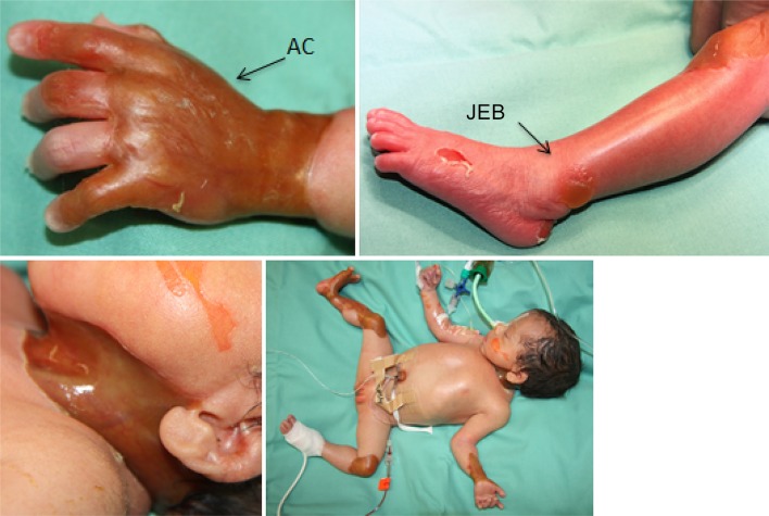
We report a female preterm newborn with generalized junctional epidermolysis bullosa (JEB) and pyloric atresia (PA) together with aplasia cutis congenita (ACC) (Fig. 1). Informed consent was obtained from the participant’s parents for inclusion in the study. The baby was born with weight of 1780 g from consanguineous parents at 34 weeks of gestational age. Prenatal laboratory tests were unremarkable. The parents were consanguine (first cousins) and had one healthy child. Two children of a grandaunt died with congenital skin defects not further described. Prenatal ultrasound revealed upper gastrointestinal atresia, most likely duodenal or pyloric atresia. Cesarean section was carried out because of premature labor and pelvic presentation of the child. At delivery, the neonate presented numerous erosions and tense bullae over the face and trunk. Aplasia cutis congenita was located circularly over the neck, left forearm and hand, left knee, and right lower leg and foot (Fig. 2). Nails, eyelashes, and scalp hair were of normal appearance; Abdominal X-rays demonstrated paucity of bowel gas suspicious for pyloric atresia, which was confirmed at surgical repair.
Fig. 1.
Initial presentation with junctional epidermolysis bullosa (JEB), pyloric atresia (PA), and aplasia cutis congenital (ACC)
Fig. 2.
Tangential excision of ACC membranes and placement of Integra® on left knee and right leg
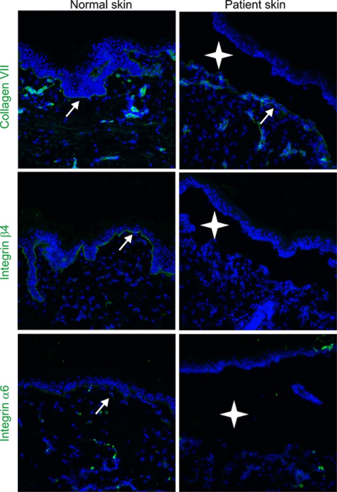
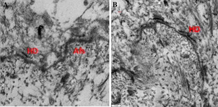
Surgical repair of PA was performed by open gastroduodenostomy on day 2. To avoid an additional burden of repeated operations, ACC was treated by tangential excision with removal of the membranous ACC at the same time due to concerns that the circumferential involvement of the neck could cause perfusion restriction of the head due to compression of cervical vessels. Furthermore, there was concern that, due to the circumferential involvement of the arm and leg, a compartment-like syndrome could arise. In addition, deformities due to the more rigid and nonflexible membrane on the arm and leg should have been corrected. To avoid subsequent infection, the wound surface was reduced by covering with Integra®-Dermal Regeneration Template as bilayer membrane (Fig. 2). Skin biopsies taken during the surgery for PA and ACC confirmed presence of a junctional cleavage plane. Immunohistochemistry showed normal expression of type VII collagen, but total loss of β4 and α6 integrin all along the skin basement membrane zone (Fig. 3), and transmission electron microscopy showed defective hemidesmosomes within the basal layer (Fig. 4). These findings proved the diagnosis of junctional epidermolysis bullosa.
Fig. 3.
Immunofluorescence antigen mapping performed on skin sections from a healthy control (Co) and our patient. Confocal microscopy was used for visualization. Integrin-a6, integrin-b4, and collagen VII appear in green. Positions of blisters are depicted by a cross, and nuclei appear in blue. Scale bars 1/4 100 mm
Fig. 4.
Ultramicroscopy showing blistering and a basal-layer with single hemidesmosomes before and after skin transplantation. HD hemidesmosomes, Afs actin filaments a basal-layer before Integra® and Skingraft Transplantation; b basal-layer 41 days after Integra® and split thickness skin grafting
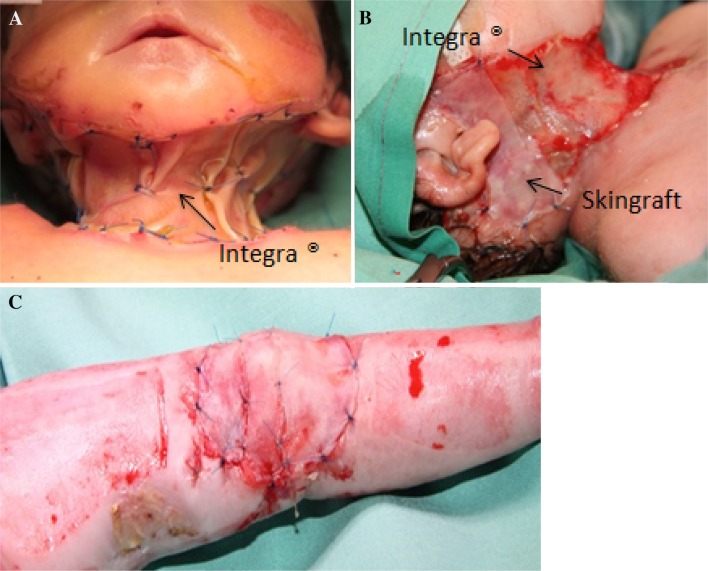
The postoperative clinical course was uneventful. Major blistering of other areas could be avoided due to a gentle care handling policy. The Integra®-Dermal Regeneration Template clinically converted to sufficient neodermis, shown by the typical color change. There were neither lesions in the neodermis nor infections or areas with insufficient neovascularization. After removal of the silicone sheet on day 36, split-thickness skin grafting (0.1 mm, sheet graft, not meshed) was performed, starting with the neck and left knee (Fig. 5). The left thigh and lower leg served as the donor site. Skin grafts showed complete take clinically, and all grafted areas showed no signs of local infection on day 41. Transmission electron microscopy still showed defective hemidesmosomes in transplanted areas after transplantation, consistent with the underlying disease.
Fig. 5.
Clinical course: a Integra® layer on day 6, b well-vascularized Integra®, partially covered with a split-thickness skin graft, c grafted left knee (donor sites left thigh and left lower leg)
In follow-up, the child demonstrated widespread blistering but not in the transplanted areas. Three days postoperative STSG, the child deteriorated quickly. Signs of infection of the grafted areas or the remaining Integra®-covered areas were not seen (Fig. 6), nor were signs of systemic infection or septicemia. On day 41, the child died of right heart failure after intensive therapy, clinically hypothesized to be a consequence of a pulmonary embolism. The parents refused further examination and autopsy.
Fig. 6.
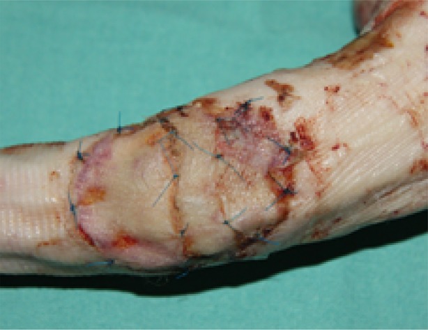
Clinically good take on day 4 after split-thickness skin grafting
Discussion
The diagnosis of Carmi syndrome was made based on clinical signs and histomorphological investigation with detection of total loss of α6- and β4-integrins within the basal layer (Fig. 3). Furthermore, detection of defective hemidesmosomes on transmission electron microscopy (TEM) confirmed the suspected diagnosis. Since the parents denied genetic analysis of ITGA6 and ITGB4 genes, we could not prove the presumptive mutation after immunohistochemistry [22].
The PA was corrected successfully on day 2 with uneventful gastrointestinal follow-up.
Postnatally, the ACC circumferentially affected areas at the left forearm and hand and right lower leg, foot, and neck already showed a deformed, contractive appearance. In the majority of cases, areas of ACC heal spontaneously [20]. While rebuilding into scar tissue, ACC membranes may become dry and shrink, similar to burn eschar [4]. This can cause compartment-like conditions in circumferential areas, as seen also in burns [2]. Because of the already developing deformity (Fig. 1), the impending risk of a compartment syndrome, the risk of perfusion restriction of the head, and concern regarding severe infections in areas of ACC, the decision was taken for early surgical intervention. The membranes were removed, and after tangential excision, Integra®-Dermal Regeneration Template was applied to cover the defects. After good vascularization of the neodermis, STSG was performed without any complications. Despite the known JEB, harvesting the grafts from the left leg could be done without difficulties and no further blistering occurred during handling of the tissue. Clinical examination showed no local signs of infection at the skin-grafted areas, with a very sufficient take (Fig. 6).
Earlier publications demonstrated successful use of full-thickness skin grafts in finger reconstruction for dystrophic EB [13]. Although some studies postulate that tissue modeling after Integra® with STSG is quite similar to original skin, histological analyses in other studies indicated that the long-term rearrangement of collagen fibers in transplanted areas resembles the patterning of scar tissue [14, 16]. Considering these statements, we hoped to influence the clinical appearance of JEB in the grafted areas whilst treating the ACC. Unfortunately, specimen of our patient could not proof this theory. It must be mentioned that the patient died only 4 days after STSG and that biopsies were taken post mortem.
Recent publications state a scarring pattern in collagen filaments after Integra® and STSG, thus leading to the assumption that, by dermal scarring, the lack of hemidesmosomes could be compensated by replacement with collagen fibers. We think that this process could stabilize the basement layer and thereby prevent blistering in the grafted areas. Complete remodeling of the skin is normally seen after 3–4 weeks posttransplantation [17]. The patient died after 41 days of life and 4 days after skin graft, so further clinical follow-up was not possible to prove the benefit of our treatment strategy for JEB in this case.
To the best of the authors’ knowledge, this is the first report of a therapy with Integra®-Dermal Regeneration Template and STSG in JEB-PA and aplasia cutis congenita and the first attempt to illustrate the local benefit of the treated areas regarding JEB. We think that blistering could be avoided after dermal restructuring by scaring modulation with collagen fibers at the basal layer and that Integra® with autologous split-thickness skin grafting could be used for local treatment of JEB in children in special cases. An animal study has been started to verify the influence of our treatment on JEB.
How useful skin transplantation could be in diseases affecting the whole integument must be discussed critically. Modern gene therapy methods seem to be promising as future therapy models, as already published by Hirsch et al. [12]. However, genetic therapies are still highly experimental with just single cases treated, limited to large centers with gene therapy laboratories, and the costs are very high. In contrast, surgical strategies such as that presented herein are easier to perform and far less cost intensive.
Conclusions
In cases of JEB-PA where ACC is present, use of Integra® and autograft could be a good solution to improve healing and avoid scarring with deformities.
Acknowledgements
We thank the participants of the study.
Funding
No funding or sponsorship was received for this study or publication of this article. The article processing charges were funded by the authors.
Authorship
All named authors meet the International Committee of Medical Journal Editors (ICMJE) criteria for authorship for this article, take responsibility for the integrity of the work as a whole, and have given their approval for this version to be published.
Disclosures
Julian Trah, Christina Has, Ingrid Hausser, Heinz Kutzner, Konrad Reinshagen, and Ingo Königs have nothing to disclose.
Compliance with ethics guidelines
Informed consent was obtained from the participant for being included in the study.
Open Access
This article is distributed under the terms of the Creative Commons Attribution-NonCommercial 4.0 International License (http://creativecommons.org/licenses/by-nc/4.0/), which permits any noncommercial use, distribution, and reproduction in any medium, provided you give appropriate credit to the original author(s) and the source, provide a link to the Creative Commons license, and indicate if changes were made.
Footnotes
Enhanced digital features
To view enhanced digital features for this article go to 10.6084/m9.figshare.6139190.
References
- 1.Birnbaum RY, Landau D, Elbedour K, Ofir R, Birk OS, Carmi R. Deletion of the first pair of fibronectin type III repeats of the integrin beta-4 gene is associated with epidermolysis bullosa, pyloric atresia and aplasia cutis congenita in the original Carmi syndrome patients. Am J Med Genet A. 2008;146A:1063–1066. doi: 10.1002/ajmg.a.31903. [DOI] [PubMed] [Google Scholar]
- 2.Burke JF, Yannas IV, Quinby WC, Bondoc CC, Jung WK. Successful use of a physiologically acceptable artificial skin in the treatment of extensive burn injury. Ann Surg. 1981;194:413–428. doi: 10.1097/00000658-198110000-00005. [DOI] [PMC free article] [PubMed] [Google Scholar]
- 3.Carmi R, Sofer S, Karplus M, Ben-Yakar Y, Mahler D, Zirkin H, Bar-Ziv J. Aplasia cutis congenita in two sibs discordant for pyloric atresia. Am J Med Genet. 1982;11:319–328. doi: 10.1002/ajmg.1320110308. [DOI] [PubMed] [Google Scholar]
- 4.Cherubino M, Maggiulli F, Dibartolo R, Valdatta L. Treatment of multiple wounds of aplasia cutis congenita on the lower limb: a case report. J Wound Care. 2016;25:760–762. doi: 10.12968/jowc.2016.25.12.760. [DOI] [PubMed] [Google Scholar]
- 5.Chung HJ, Uitto J. Epidermolysis bullosa with pyloric atresia. Dermatol Clin. 2010;28:43–54. doi: 10.1016/j.det.2009.10.005. [DOI] [PMC free article] [PubMed] [Google Scholar]
- 6.El Shafie M, Stidham GL, Klippel CH, Katzman GH, Weinfeld IJ. Pyloric atresia and epidermolysis bullosa letalis: a lethal combination in two premature newborn siblings. J Pediatr Surg. 1979;14(4):446–9. doi: 10.1016/S0022-3468(79)80012-3. [DOI] [PubMed] [Google Scholar]
- 7.Fine JD. Epidemiology of inherited epidermolysis bullosa based on incidence and prevalence estimates from based on incidence and prevalence estimates from the national epidermolysis bullosa registry. JAMA Dermatol. 2016;152(11):1231–8. doi: 10.1001/jamadermatol.2016.2473. [DOI] [PubMed] [Google Scholar]
- 8.Fine JD, Bruckner-Tuderman L, Eady RA, Bauer EA, Bauer JW, Has C, Heagerty A, Hintner H, Hovnanian A, Jonkman MF, Leigh I, Marinkovich MP, Martinez AE, McGrath JA, Mellerio JE, Moss C, Murrell DF, Shimizu H, Uitto J, Woodley D, Zambruno G. Inherited epidermolysis bullosa: updated recommendations on diagnosis and classification. J Am Acad Dermatol. 2014;70(6):1103–26. doi: 10.1016/j.jaad.2014.01.903. [DOI] [PubMed] [Google Scholar]
- 9.Frieden IJ. Aplasia cutis congenita: a clinical review and proposal for classification. J Am Acad Dermatol. 1986;14:646–660. doi: 10.1016/S0190-9622(86)70082-0. [DOI] [PubMed] [Google Scholar]
- 10.González Alaña I, Torrero López JV, Martín Playá P, Gabilondo Zubizarreta FJ. Combined use of negative pressure wound therapy and Integra® to treat complex defects in lower extremities after burns. Ann Burns Fire Disasters. 2013;26:90–93. [PMC free article] [PubMed] [Google Scholar]
- 11.Gorell ES, Leung TH, Khuu P, Lane AT. Purified type I collagen wound matrix improves chronic wound healing in patients with recessive dystrophic epidermolysis bullosa. Pediatr Dermatol. 2015;32:220–225. doi: 10.1111/pde.12492. [DOI] [PubMed] [Google Scholar]
- 12.Hirsch T, Rothoeft T, Teig N, Bauer JW, Pellegrini G, De Rosa L, Scaglione D, Reichelt J, Klausegger A, Kneisz D, et al. Regeneration of the entire human epidermis using transgenic stem cells. Nature. 2017;551:327–332. doi: 10.1038/nature24487. [DOI] [PMC free article] [PubMed] [Google Scholar]
- 13.Ikeda S, Yaguchi H, Ogawa H. Successful surgical management and long-term follow-up of epidermolysis bullosa. Int J Dermatol. 1994;33:442–445. doi: 10.1111/j.1365-4362.1994.tb04049.x. [DOI] [PubMed] [Google Scholar]
- 14.Lee SM, Stewart CL, Miller CJ, Chu EY. The histopathologic features of Integra® Dermal Regeneration Template. J Cutan Pathol. 2015;42:368–369. doi: 10.1111/cup.12488. [DOI] [PubMed] [Google Scholar]
- 15.Marneros AG. Genetics of aplasia cutis reveal novel regulators of skin morphogenesis. J Invest Dermatol. 2015;135:666–672. doi: 10.1038/jid.2014.413. [DOI] [PubMed] [Google Scholar]
- 16.Moiemen N, Yarrow J, Hodgson E, Constantinides J, Chipp E, Oakley H, Shale E, Freeth M. Long-term clinical and histological analysis of Integra dermal regeneration template. Plast Reconstr Surg. 2011;127:1149–1154. doi: 10.1097/PRS.0b013e31820436e3. [DOI] [PubMed] [Google Scholar]
- 17.Moiemen NS, Vlachou E, Staiano JJ, Thawy Y, Frame JD. Reconstructive surgery with Integra dermal regeneration template: histologic study, clinical evaluation, and current practice. Plast Reconstr Surg. 2006;117:160S–174S. doi: 10.1097/01.prs.0000222609.40461.68. [DOI] [PubMed] [Google Scholar]
- 18.Okoye BO, Parikh DH, Buick RG, Lander AD. Pyloric atresia: five new cases, a new association, and a review of the literature with guidelines. J Pediatr Surg. 2000;35:1242–1245. doi: 10.1053/jpsu.2000.8762. [DOI] [PubMed] [Google Scholar]
- 19.de Pereda JM, Lillo MP, Sonnenberg A. Structural basis of the interaction between integrin alpha6beta4 and plectin at the hemidesmosomes. EMBO J. 2009;28:1180–1190. doi: 10.1038/emboj.2009.48. [DOI] [PMC free article] [PubMed] [Google Scholar]
- 20.Perry BM, Maughan CB, Crosby MS, Hadenfeld SD. Aplasia cutis congenita type V: a case report and review of the literature. Int J Dermatol. 2017;56:e118–e121. doi: 10.1111/ijd.13611. [DOI] [PubMed] [Google Scholar]
- 21.Pfendner E, Uitto J, Fine JD. Epidermolysis bullosa carrier frequencies in the US population. J Invest Dermatol. 2001;116:483–484. doi: 10.1046/j.1523-1747.2001.01279-11.x. [DOI] [PubMed] [Google Scholar]
- 22.Pulkkinen L, Rouan F, Bruckner-Tuderman L, Wallerstein R, Garzon M, Brown T, Smith L, Carter W, Uitto J. Novel ITGB4 mutations in lethal and nonlethal variants of epidermolysis bullosa with pyloric atresia: missense versus nonsense. Am J Hum Genet. 1998;63:1376–1387. doi: 10.1086/302116. [DOI] [PMC free article] [PubMed] [Google Scholar]
- 23.Schaapveld RQ, Borradori L, Geerts D, van Leusden MR, Kuikman I, Nievers MG, Niessen CM, Steenbergen RD, Snijders PJ, Sonnenberg A. Hemidesmosome formation is initiated by the beta4 integrin subunit, requires complex formation of beta4 and HD1/plectin, and involves a direct interaction between beta4 and the bullous pemphigoid antigen 180. J Cell Biol. 1998;142:271–284. doi: 10.1083/jcb.142.1.271. [DOI] [PMC free article] [PubMed] [Google Scholar]
- 24.Tabor A, Pergolizzi JV, Jr, Marti G, Harmon J, Cohen B, Lequang JA. Raising awareness among healthcare providers about epidermolysis bullosa and advancing toward a cure. J Clin Aesthet Dermatol. 2017;10(5):36–48. [PMC free article] [PubMed] [Google Scholar]


