eLife logoLink to eLife
. 2018 Jul 2;7:e36073. doi: 10.7554/eLife.36073

A positive-feedback-based mechanism for constriction rate acceleration during cytokinesis in Caenorhabditis elegans

Renat N Khaliullin 1,, Rebecca A Green 1, Linda Z Shi 2, J Sebastian Gomez-Cavazos 1, Michael W Berns 2, Arshad Desai 1, Karen Oegema 1,
Editors: Mohan K Balasubramanian3, Anna Akhmanova4
PMCID: PMC6063732  PMID: 29963981

Abstract

To ensure timely cytokinesis, the equatorial actomyosin contractile ring constricts at a relatively constant rate despite its progressively decreasing size. Thus, the per-unit-length constriction rate increases as ring perimeter decreases. To understand this acceleration, we monitored cortical surface and ring component dynamics during the first cytokinesis of the Caenorhabditis elegans embryo. We found that, per unit length, the amount of ring components (myosin, anillin) and the constriction rate increase with parallel exponential kinetics. Quantitative analysis of cortical flow indicated that the cortex within the ring is compressed along the axis perpendicular to the ring, and the per-unit-length rate of cortical compression increases during constriction in proportion to ring myosin. We propose that positive feedback between ring myosin and compression-driven flow of cortex into the ring drives an exponential increase in the per-unit-length amount of ring myosin to maintain a high ring constriction rate and support this proposal with an analytical mathematical model.

Research organism: C. elegans

Introduction

During cytokinesis in animal cells, constriction of an equatorial actomyosin ring cinches the mother cell surface to generate a dumbbell-shaped structure with an intercellular bridge that connects the two daughter cells (Fededa and Gerlich, 2012; Green et al., 2012). This cellular shape change is brought about by a cortical contractile ring that assembles around the cell equator following chromosome segregation in anaphase. To ensure that each cell inherits a single genomic complement, contractile ring assembly is directed by the small GTPase RhoA. RhoA is activated in an equatorial zone (the ‘Rho zone’) in response to signaling by the anaphase spindle (Green et al., 2012; Jordan and Canman, 2012; Piekny et al., 2005) and patterns the equatorial cortex by recruiting contractile ring components from the cytoplasm (Vale et al., 2009; Yumura, 2001; Zhou and Wang, 2008). RhoA activates Rho kinase, which promotes the assembly and recruitment of myosin II (Matsumura et al., 2011) and the cytokinesis formin that assembles the long actin filaments that make up the ring (Otomo et al., 2005). Contractile rings also contain membrane-associated septin filaments (Bridges and Gladfelter, 2015) and the filament cross linker anillin (D'Avino, 2009; Piekny and Maddox, 2010). Recent work in the Caenorhabditis elegans embryo suggests that the equatorial cortex is compressed along the pole-to-pole axis after this initial patterning, leading to the alignment of actin filament bundles as the ring forms (Reymann et al., 2016). After its assembly, the ring begins to constrict in the around-the-ring direction. Constriction is thought to be coupled to the progressive disassembly of the ring (i.e. loss of components in proportion to reduction in length; Carvalho et al., 2009; Murrell et al., 2015; Schroeder, 1990).

Ring constriction must complete within a short cell cycle window during mitotic exit (Canman et al., 2000; Martineau et al., 1995; Straight et al., 2003). Timely constriction relies on the conserved ability of contractile rings to maintain a relatively constant overall closure rate despite their progressively decreasing perimeter (Biron et al., 2004; Bourdages et al., 2014; Calvert et al., 2011; Carvalho et al., 2009; Ma et al., 2012; Mabuchi, 1994; Pelham and Chang, 2002; Zumdieck et al., 2007). This property implies that the per-unit-length constriction rate increases as the rings get smaller.

Here, we explore the mechanisms underlying constriction rate acceleration during the first division of the C. elegans embryo. Since the contractile ring is an integral part of the larger cell cortex, we investigated the possibility that interactions between the ring and surrounding cortex contribute to constriction rate acceleration. By generating a 4D map of cortical surface dynamics, we show that the surface area of the cortex at the cell poles expands in response to the tension generated by the constricting ring to provide the increased cortical surface area required to generate the daughter cells. The ability of the polar cortex to expand in response to tension also allows ring myosin to compress cortical surface along the pole-to-pole axis perpendicular to the ring, leading to a continuous flow of cortical surface into the ring as it constricts. We show that, per unit length, the amount of ring components (myosin, anillin), the constriction rate, and the rate of cortical surface compression within the ring all increase with parallel exponential kinetics, suggesting control by positive feedback. Based on our experimental observations, we propose that feedback between ring myosin and compression-driven flow of cortex into the ring (Compression Feedback) drives the exponential increase in the per-unit-length amount of ring myosin, which in turn increases the per-unit-length constriction rate to keep the overall constriction rate high as the ring closes. We further show that an analytical mathematical formulation of the proposed feedback can recapitulate the observed dynamics of the ring during constriction.

Results

The cortex at the cell poles expands in response to tension generated by the constricting ring without limiting the constriction rate

During the first division of the C. elegans embryo, the surface area of the cell increases by ~40% to accommodate the shape change that generates the daughter cells. Work in multiple systems has shown that the molecular components of the cortex, cortex-associated granules in the cytoplasm, and particles adhered to cell surface receptors all move in a coordinated fashion during cytokinesis (Cao and Wang, 1990; Dan, 1954; Dan and Dan, 1940; Dan et al., 1938; DeBiasio et al., 1996; Fishkind et al., 1996; Hird and White, 1993; Reymann et al., 2016; Swann and Mitchison, 1958; Wang et al., 1994). In a classic set of experiments, Dan and colleagues monitored the pattern of cortical surface expansion and compression during cytokinesis by measuring the distance between surface adhered particles and/or pigmented cortex-associated cytoplasmic granules in sea urchin embryos. This analysis revealed that ring constriction is accompanied by a wave of cortical surface expansion that initiates at the cell poles (Dan et al., 1938; Dan and Ono, 1954; Dan et al., 1937; Swann and Mitchison, 1958). Note that expansion is an increase in cortical surface area and compression is reduction in cortical surface area. These terms do not imply a specific mechanism for how the increase or reduction occurs. For example, expansion can be accompanied by deposition of new cortical components that allow the cortex to maintain the same density, or by stretching and thinning of the existing cortex; similarly, compression can be accompanied by an increase in the density of cortical components or by their disassembly. Also, note that the analysis of cell surface dynamics described above refers to the movement, expansion and compression of the cortex and associated structures. How deposition of plasma membrane, the fluid lipid layer that overlies the cortex, is controlled and where it occurs is a separate question that we will not address here.

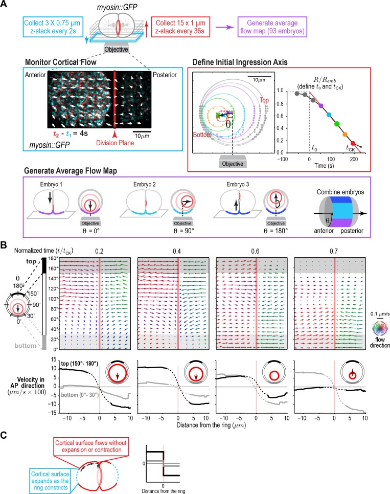
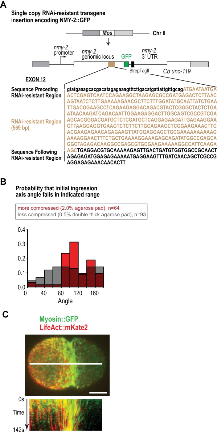
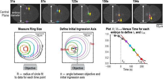
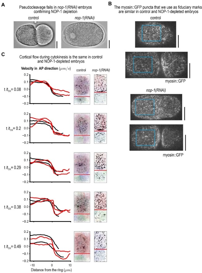
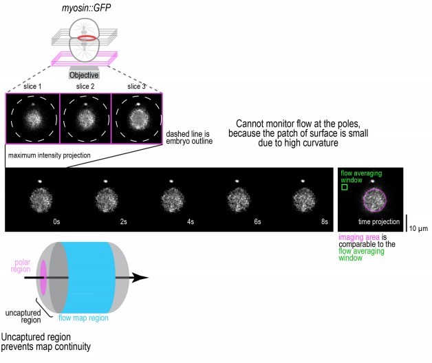
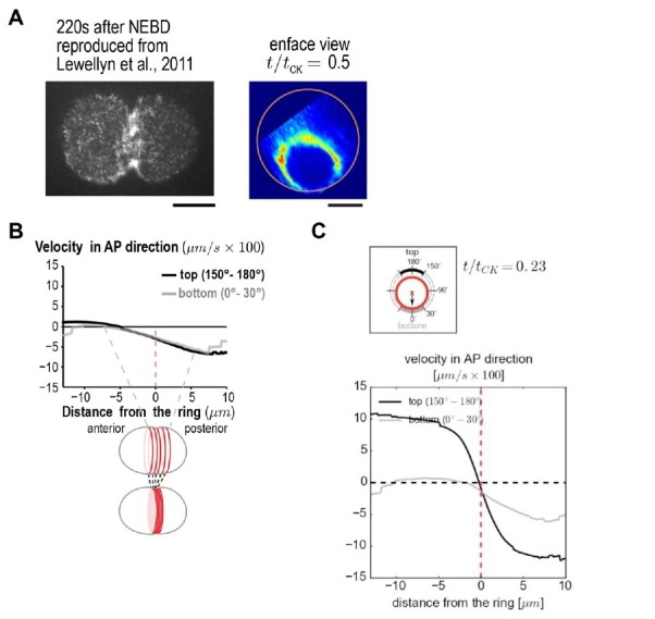
Although the experiments performed by Dan and colleagues (Dan et al., 1938; Dan and Ono, 1954; Dan et al., 1937; Swann and Mitchison, 1958) in sea urchin embryos provided a rough map of where expansion occurs, they did not allow quantification of the extent of change in cortical surface area or provide a map of cortical surface movements. To generate a quantitative map of cortical surface dynamics during the first division of the C. elegans embryo, we employed an updated version of the classical approach in which we used myosin foci, rather than surface adhered particles, as fiduciary marks. We imaged the cortex at high time resolution (2 s intervals, cyan box in Figure 1A, Video 1) in embryos expressing a GFP fusion with the heavy chain of non-muscle myosin II (NMY-2; hereafter myosin::GFP; Figure 1—figure supplement 1A,B). In addition to its RhoA-dependent enrichment in the contractile ring, myosin is present in small puncta, distributed over the entire cortex, that flow together with actin filaments (LifeAct::mKate2, Figure 1—figure supplement 1C), validating their utility as fiduciary marks for monitoring cortical movements. To temporally and spatially align data collected in different embryos, ring constriction was also monitored at lower time resolution in the same embryos (36 s intervals, Figure 1A, Figure 1—figure supplement 2). Because the contractile ring closes asymmetrically within the division plane (Maddox et al., 2007; Figure 1A, Figure 1—figure supplement 2), cortical dynamics are not cylindrically symmetric. Therefore, we generated an average 4D map of cortical movement by computationally combining data from 93 embryos imaged in random rotational orientations (Figure 1A, Figure 1—figure supplement 2). We defined the top of the embryo as the side where the furrow ingresses first, the bottom as the opposite side, and referenced positions around the embryo circumference by the angle θ relative to the initial ingression axis (Figure 1A). For temporal alignment, we fit a line to normalized ring size (R¯:=R/Remb) versus time between 30 and 80% closure for each embryo and extrapolated this line to 1 and 0 to define t0 (cytokinesis onset) and tCK (time of cytokinesis), respectively (Figure 1A, Figure 1—figure supplement 2). Cortical movement could not be monitored in the division plane, because it is hidden inside the cell, or at the cell poles, due to their high curvature. Thus, this approach provided a quantitative picture of cortical movement in the central 2/3 of the embryo, with the exception of the division plane, throughout cytokinesis (Figure 1B; Video 2).

Figure 1. A quantitative map of cortical surface dynamics during the first cytokinesis in the C. elegans embryo reveals that the cortical surface at the cell poles expands as the ring constricts.

(A) (top) Schematic of the experimental procedure. (middle, left) Superposition of images of the cortex acquired 4 s apart. Arrows indicate cortical flow (magnified 2.5X). (middle, right) The initial ingression axis, t0, and tCK were defined as shown for a representative embryo. The angle θ specifies the position of the imaged cortex relative to the initial furrow ingression axis. Image and quantification are representative of the 93 imaged embryos. (bottom) Angular position was used to combine data from 93 embryos to generate an average flow map. (B) (top) Average flow at the indicated timepoints. Arrows show direction and magnitude of the displacement in 1 s (magnified 20X). (middle) Graphs are average velocity in the A-P direction versus position along the A-P axis for the cortex on the top (black) and bottom (grey) of the embryo (shaded in flow maps). Surface movement changes direction across the division plane, the apparent velocity gradient close to the division plane is a projection artifact due to the fact that the cortical surface turns inwards as it approaches the furrow from either side (dotted regions on velocity curves). (C) Schematics show the predicted cortical velocity profile along the AP axis if surface is gained at the poles; velocity would be constant in magnitude within the flow map region with opposite directions on the two sides of the ring, as is experimentally observed.

Figure 1.

Figure 1—figure supplement 1. Actin and myosin move together with the cortical surface during cytokinesis.

Figure 1—figure supplement 1.

(A) Schematic of the single-copy nmy-2::gfp transgene inserted into a specific locus on chromosome II. Cb unc-119, the unc-119 coding region from the related nematode C. briggsae, was used as a transformation marker. The transgene was re-encoded while maintaining amino acid sequence in the indicated region to render it resistant to RNAi targeting the endogenous gene for other experiments, this feature was not used in the experiments in this manuscript. (B) Compression biases the direction of contractile ring closure. Graph plotting the probability that the angle between the objective axis and the initial ingression axis falls in the indicated range for embryos mounted with more (red) or less (grey) compression. Due to this bias, embryos were mounted using the low compression conditions shown in grey. (C) Actin and myosin move together with the cortical surface during cytokinesis. The white line in the center of the image (top) indicates the region used for the kymograph (bottom). Image is representative of five imaged embryos. Scale bar is 10 µm.
Figure 1—figure supplement 2. An automated method for monitoring contractile ring closure.

Figure 1—figure supplement 2.

(top) Central plane images of the embryo in Figure 1A. Panels on the lower left and lower right are reproduced from Figure 1A for comparison. An automated algorithm was used to identify the edges of the embryo (dashed lines) and the position of the contractile ring (colored circles) in each z-plane. Yellow arrows mark the direction of furrow ingression and illustrate how the furrow initially ingresses from the top and then changes directions to ingress from the bottom during the second half of cytokinesis. (lower left) Points marking contractile ring position in the z-planes were projected onto an end-on view of the division plane. Data for different timepoints in this representative embryo are shown in colors corresponding to the circles in the central plane images. Ring sizes were measured by fitting circles to the data. (middle) The initial axis of contractile ring closure was defined by the angle θ between the objective axis and a line fit through the centers of the contractile rings with a normalized size >0.3. (right) = A plot of normalized ring size versus time for this embryo defines t0 and tCK as the times when a line fit through the points corresponding to ring sizes between 0.3 and 0.8 crossed 1 and 0, respectively. Scale bar is 10 µm.
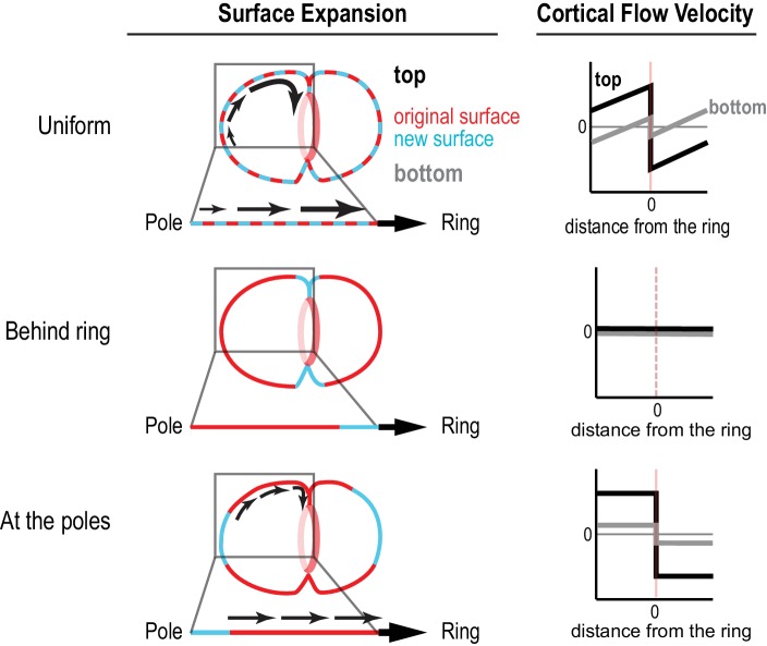
Figure 1—figure supplement 3. Different profiles of cortical surface velocity along the A-P axis are predicted for different spatial patterns of surface expansion.

Figure 1—figure supplement 3.

(top) For surface expansion behind the ring, no cortical movement is predicted on the embryo surface. (middle) For uniform surface expansion, a gradient of velocities will be observed, where the cortex immediately behind the ring moves at the speed of the ingressing furrow, and cortical velocity decreases linearly towards the cell poles. (bottom) Reproduced from Figure 1C for comparison. If surface is expansion occurs only at the poles, cortical velocity will be constant in magnitude within the flow map region with opposite direction on the two sides of the ring.

Video 1. Cortical flow imaged in a control embryo expressing myosin::GFP.

Download video file (3.8MB, mp4)
DOI: 10.7554/eLife.36073.006

Playback is 6x realtime. The video is constructed from maximum intensity projection of 3 × 0.75 µm plane z-stacks acquired at 2 s intervals. The red line marks the position of the division plane. The arrows represent the surface movement between consecutive frames at the base of the arrow. The length of the arrow is five times the magnitude of movement. The direction is also color coded according to the color wheel as shown in Figure 1B.

Video 2. Average cortical flow map calculated from time lapse imaging of the cell surface in 93 control embryos expressing myosin::GFP. (top, left).

Download video file (4.3MB, mp4)
DOI: 10.7554/eLife.36073.007

Schematic illustrates location of the cylindrical surface covered by the map. (top, right) Dynamic schematic illustrates ring size and position for each value of t/tCK. (bottom, left) The movement of each blue dot corresponds to surface movement at its location. The y-axis is the angular position relative to the initial ingression axis. The x-axis is the distance from the division plane along the anterior-posterior axis. (bottom, right) Dynamic graph plots the magnitude of the component of surface velocity aligned along the anterior-posterior axis for the top (150–180°; black) and bottom (0–30°; grey) regions of the cortex.

The 4D map allowed us to determine where cortical surface expansion occurs as the ring closes in the C. elegans embryo. Prior work monitoring the movement of surface adhered particles indicated that surface expansion occurs at the poles and immediately behind the contractile ring in sea urchin and Xenopus embryos, respectively (Bluemink and de Laat, 1973; Byers and Armstrong, 1986; Danilchik et al., 2003; Gudejko et al., 2012; Selman and Perry, 1970; Swann and Mitchison, 1958). In addition to these two patterns, we also considered the possibility that the cortex could expand uniformly, an assumption in mathematical models of cytokinesis (Turlier et al., 2014; Zumdieck et al., 2007). Each of these three patterns predicts a different profile for the Anterior-Posterior (AP) component of cortical velocity along the embryo. For uniform surface expansion, a gradient of velocities is predicted, where the velocity of the cortex immediately behind the ring is equal to the velocity of furrow ingression and the velocity decreases linearly as you move towards the cell poles. For surface expansion immediately behind the ring, no cortical movement is predicted on the observable embryo surface. If surface expansion is limited to the poles, the cortical velocity is predicted to be constant within the flow map region (Figure 1—figure supplement 3). The cortical velocity profile measured from the flow map indicated that the cortical surface at the cell poles expands as the ring constricts, whereas the cortex in the region between the poles and the division plane flows at constant velocity towards the division plane, without expansion or compression (Figure 1B). Note that the apparent velocity gradient that spans the division plane in the flow maps (Figure 1B, dashed regions on velocity curves) is a projection artifact due to the fact that the cortical surface turns to flow inwards as it approaches the furrow from either side (component of the velocity in the x-y plane decreases as the velocity vectors turn inwards). As expected, based on the asymmetric closure of the contractile ring within the division plane, the velocity of cortical flow was higher on the top of the embryo during the first half of cytokinesis when the furrow ingresses from the top (Figure 1B, black traces) and became higher on the bottom of the embryo towards the end when the furrow ingresses from the bottom (Figure 1B, grey traces; Video 2).

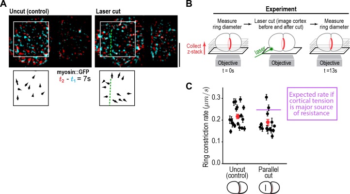
Cutting the cortex parallel to the division plane using a laser revealed that the cortex is under tension during cytokinesis (Figure 2A). To determine if cortical tension limits the constriction rate, we assayed the effect of the cortical cuts on ring closure. Cortical cuts spanning the visible area of cortex on the anterior side of the embryo (~10 µm in length) were made parallel to the division plane when the ring was at ~50% closure, and the effect on contractile ring closure rate was assessed by measuring the difference in ring sizes immediately before and 13 s after the cut. The cortical opening resulting from the ablation was approximately 35 µm2; if the cortical surface tension is the dominant force limiting the ring closure rate, this size opening would be expected to increase the constriction rate from the control rate of 0.22 ± 0.05 µm/s to ~0.25 µm/s over the 13 s interval (see Materials and methods for details). In contrast, the measured constriction rate was not increased after cutting (0.18 ± 0.03 µm/s; Figure 2B,C), indicating that cortical tension does not impose significant resistance to ring pulling. Cuts made perpendicular to the ring also had no effect on the constriction rate (0.19 ± 0.03 µm/s data not shown). Consistent with the results of the laser cutting experiments, inhibiting the Arp2/3 complex by depleting its ARX-2 subunit, which is expected to reduce effective cortical viscosity and thus cortical tension (Chaudhuri et al., 2007; Davies et al., 2014; Tseng and Wirtz, 2004), also did not alter the constriction rate (Figure 2—figure supplement 1A).

Figure 2. Cortical tension does not limit the rate of ring closure.

(A) The success of cortical cuts was assessed by comparing surface images of cortical myosin before (cyan) and after (red) the cut to monitor the movement of myosin foci away from the cut site. Representative images are shown. Scale bar is 10 µm. (B) Schematic of laser ablation experiment to determine if cortical resistance limits the rate of contractile ring closure. Contractile ring sizes were measured from z-stacks acquired before and 13 s after a cut was made across the cortex with a laser. (C) Graph plots the rates of ring closure derived from before and after ring size measurements for uncut controls (n = 19 embryos) and embryos with cuts parallel to the division plane (n = 14 embryos). Black symbols are single embryo measurements with measurement errors. Red symbols are the means; error bars are the SEM. The purple line marks expected closure rate if cortical tension is a major source of resistance.

Figure 2.

Figure 2—figure supplement 1. Arp2/3 depletion does not alter ring constriction kinetics.
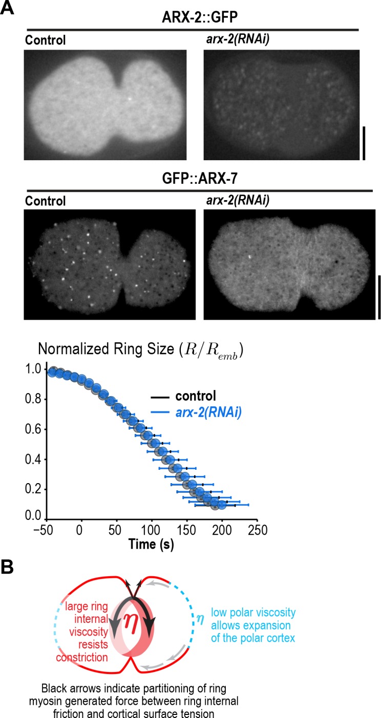
Figure 2—figure supplement 1.

(A) Images of cortical ARX-2::GFP (top) and GFP::ARX-7 (middle) in control and arx-2(RNAi) embryos confirm loss of cortical Arp2/3 complex (images are representative of 10 imaged embryos for each condition in the GFP::ARX-7 strain and 15 for control and 13 for arx-2(RNAi) in the ARX-2::GFP strain). Scale bars are 10 µm. (bottom) Graph plots average contractile ring size versus time for control (grey) and arx-2(RNAi) (blue) embryos expressing myosin::GFP (n = 93 embryos for control and 68 embryos for arx-2(RNAi)). Error bars are standard deviation. (B) Schematic illustrating the partitioning of ring myosin generated force between ring internal friction and cortical surface tension. Ring myosin generated force primarily counters ring internal friction to drive constriction. The low viscosity of the polar cortex causes it to expand when it comes under tension due to the constricting ring.

Putting the results of our flow map analysis together with our laser cutting and Arp2/3 inhibition experiments, we conclude that the cortical surface at the cell poles expands in response to tension generated by the constricting ring without providing significant resistance that would affect the rate of ring closure. In contrast, the cortex in the region between the ring and the poles flows toward the ring without expansion or compression. The differential response of the polar cortex to ring-generated tension is consistent with the idea of polar relaxation hypothesized in early conceptual models of cytokinesis (Greenspan, 1978; Swann and Mitchison, 1958; Taber, 1995; White and Borisy, 1983; Wolpert, 1960; Zinemanas and Nir, 1987, 1988), and suggests that the polar cortex has unique mechanical properties compared to the intervening cortex that does not expand (see Discussion). The fact that cortical tension does not limit the rate of ring constriction suggests that the constriction rate is instead limited by ring-internal friction. We conclude that the viscosity of the polar cortex is negligible compared to the viscosity internal to the ring; thus, ring myosin generated force primarily counters ring internal friction to drive ring constriction (Figure 2—figure supplement 1B). Ring constriction, in turn, affects cortical tension and drives expansion of the polar cortex.

Ring myosin compresses cortical surface along the axis perpendicular to the ring, pulling new cortical surface into the ring at a rate proportional to the amount of ring myosin

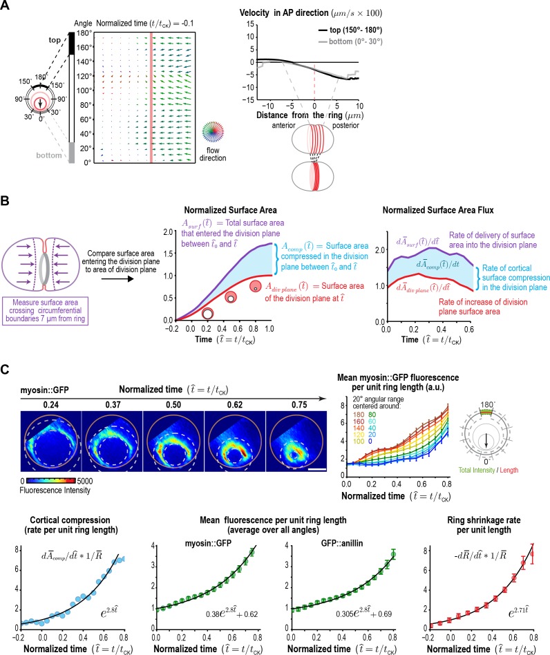
In the C. elegans embryo, as in other systems, spindle-based signaling following anaphase onset activates RhoA on the cortex in an equatorial zone (the Rho zone), leading to the recruitment of contractile ring proteins including myosin II, the septins, and anillin (Jenkins et al., 2006; Maddox et al., 2005, 2007; Mangal et al., 2018; Motegi and Sugimoto, 2006; Schonegg et al., 2007; Tse et al., 2012; Werner et al., 2007). An astral microtubule-based mechanism that clears contractile ring proteins from the polar cortex also limits contractile ring protein recruitment to a defined equatorial zone (Mangal et al., 2018; Werner et al., 2007). Prior work in the C. elegans embryo has suggested that the cortical surface within the Rho zone is compressed along the axis perpendicular to the ring as the contractile ring forms following anaphase onset, coincident with the alignment of actin filament bundles (Reymann et al., 2016). Compression, which is the reduction of cortical surface area, is detected as a gradient in the velocity of cortical surface flow. Consistent with the idea that cortical surface is compressed during contractile ring assembly, we observed a linear gradient in the velocity of cortical flow that spanned the cell equator in our flow map at early time points prior to furrow ingression (Figure 3A). The linear gradient indicated that, during contractile ring assembly, when the ring is on the embryo surface, cortical surface is uniformly compressed across a 10 µm wide region along the perpendicular-to-the-ring axis between the two relaxing poles.

Figure 3. Ring myosin compresses cortical surface along the axis perpendicular to the ring, pulling in new cortical surface at a rate proportional to the amount of ring myosin.

(A) The equatorial cortex is compressed during contractile ring assembly. Following the onset of spindle-based RhoA signaling, the initial recruitment of contractile ring proteins leads to uniform compression of cortical surface along the axis perpendicular to the forming ring across a 10 µm wide region spanning the cell equator. (left) Average flow map at (t/tCK = −0.1) immediately after the onset of spindle-based signaling (n = 93 embryos). (middle) The surface velocity profile reveals a linear velocity gradient that spans the cell equator (−5 to +5 µm), indicating a uniform zone of cortical compression. (B) Cortical compression within the ring continues during constriction. (left graph) Plot comparing the area of the forming division plane (red) with the total cortical surface area that entered the division plane from the start of cytokinesis (purple; calculated as indicated in the schematic). (right graph) Plot comparing the rate of delivery of cortical surface into the division plane (purple) with the rate of growth of the division plane (red). The difference between the two is the rate of cortical surface compression (rate of reduction of cortical surface area; cyan). (C) The per-unit-length amount of ring myosin and the rate of cortical compression increase with the same exponential kinetics, suggesting that the rate of cortical compression may be controlled by the amount of myosin in the contractile ring/Rho zone. (top left) Representative images of the division plane in embryos expressing myosin::GFP reconstructed from 40-plane z-stacks. Gold circles mark the embryo boundary and dashed circles mark the boundaries used for ring intensity measurements. Scale bar is 10 µm. (top right) Graph plots per-unit-length myosin::GFP fluorescence for the indicated angular ranges (n = 36 embryos). (bottom left) Graph plots the rate of cortical surface compression per unit ring length (n = 93 embryos). (bottom middle) Graphs plot mean per-unit-length myosin::GFP (n = 36 embryos) and GFP::anillin (n = 26 embryos) fluorescence (n = 36 embryos) in the ring. (bottom right) Graph plots the per-unit-length rate of ring closure. Black lines are fitted single exponentials. Error bars are the SEM.

Figure 3.

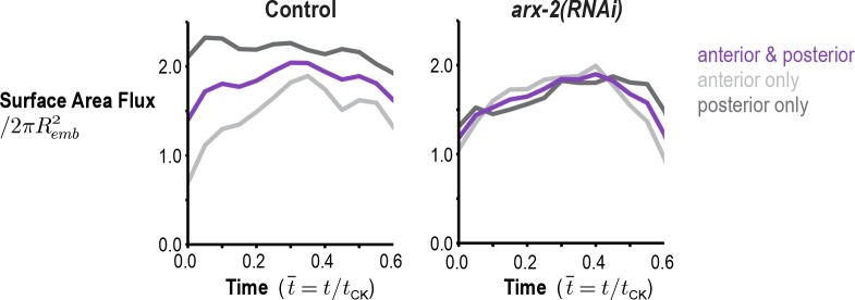
Figure 3—figure supplement 1. Arp2/3 inhibition abolishes the asymmetry in the amount of cortex entering the division plane from the anterior and posterior sides.

Figure 3—figure supplement 1.

Graphs plot the rate of cortical flux across the anterior (light grey) and posterior (dark grey) boundaries (see schematic in Figure 3B) versus the mean for the two sides (purple) for control and arx-2(RNAi) embryos. Calculated from the average flow maps for the control (n = 93 embryos) and arx-2(RNAi) (n = 68 embryos) conditions.
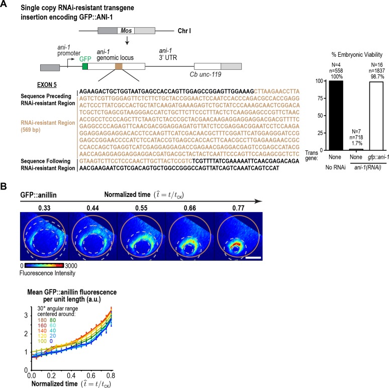
Figure 3—figure supplement 2. GFP::anillin fluorescence in the ring increases exponentially during constriction.

Figure 3—figure supplement 2.

(A) (left) Schematic of the single-copy gfp::ani-1 trangene. The transgene was re-encoded while maintaining amino acid sequence in the indicated region to render it resistant to RNAi targeting of the endogenous ani-1 gene to allow testing of the functionality of the GFP::ANI-1 fusion. (right) Graph plotting embryonic lethality demonstrates that the gfp::ani-1 transgene is functional. (B) (top) Images of the division plane in an embryo expressing GFP::anillin. Scale bar is 10 µm. (bottom) Graph plots GFP::anillin fluorescence per unit length of the ring for the indicated angular ranges. Error bars are the SEM.
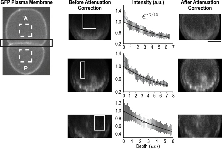
Figure 3—figure supplement 3. Correcting for signal attenuation with sample depth.

Figure 3—figure supplement 3.

Fluorescence attenuation with embryo depth was estimated from fluorescence intensity measurements made at the cell-cell boundary of two-cell embryos expressing a GFP-tagged plasma membrane marker. Cell-cell boundaries were reconstructed from 40 plane z-stacks. The intensity profile at each slice was calculated by subtracting the average background intensity estimated from dashed rectangles (left) from the cell-cell boundary region (black rectangle) at each slice and calculating the maximum intensity projection along AP axis. The effect of depth on signal was calculated from the reconstructed division planes by plotting the mean signal as a function of depth in 10 rectangular regions (white boxes) where the signal was expected to be uniform; three examples are shown here. All intensity profiles were simultaneously fitted using a single exponential. Error bars are the SD. On the right, the same cell-cell boundaries are shown after correction for depth attenuation. The scale bar is 10 µm.
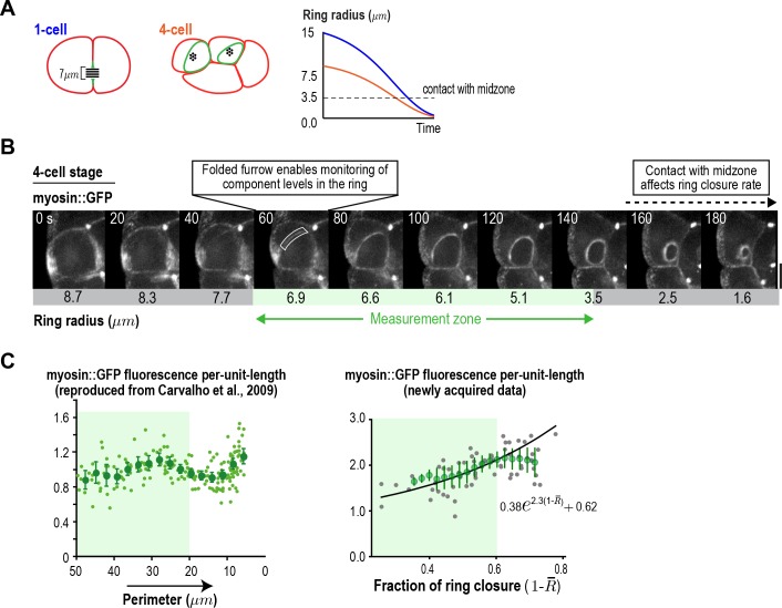
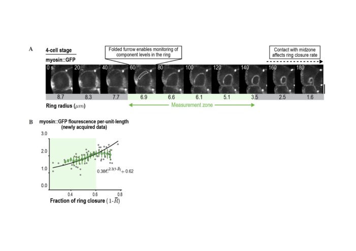
Figure 3—figure supplement 4. Ring component dynamics at the four-cell stage are consistent with exponential accumulation.

Figure 3—figure supplement 4.

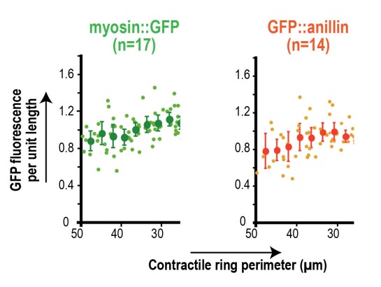
(A) (left) Schematic illustrating the relative geometries of cytokinesis in one- and four-cell stage C. elegans embryos. (right) The range of ring sizes between furrow formation and contact with the midzone, which occurs at a ring radius of about 3.5 µm (perimeter ~22 µm) in all divisions and alters constriction rate and component accumulation (Carvalho et al., 2009), is much smaller at the four-cell stage than at the one-cell stage. (B) Myosin levels in the ring can only be monitored over a limited range of ring size at the four-cell stage. Images of the division plane in a representative dividing cell at the four-cell stage reconstructed from 16 × 1 µm z-stacks of an embryo expressing myosin::GFP (n = 16 embryos imaged). The range of ring sizes between the point when the folding in of the furrow first enables monitoring of ring component levels in the end-on view, and the point when the ring contacts the spindle midzone, is indicated (green; Measurement zone). (C) Graphs plotting measured mean per-unit-length myosin::GFP fluorescence in the ring at the four-cell stage. The graph on the left is reproduced from Figure 4D of Carvalho et al. (2009) where a strain with an integrated myosin::GFP transgene under an exogenous promoter was filmed. The graph on the right is new data collected in the in situ-tagged myosin::GFP strain. The measurement zone highlighted in B (from 50 to 20 µm ring perimeter is highlighted in green for both graphs). In the graph on the right, the data for rings in the measurement zone were fit to an exponential equation with the same baseline contribution as the one-cell stage data in Figure 3C (black line). Error bars are the SEM.
© 2009 Elsevier
Figure 3—figure supplement 4C (left graph) reproduced with permission from Figure 5 of Carvalho et al. (2009).

After its assembly, the ring begins to shorten in the around-the-ring direction. Ring constriction has been proposed to be coupled to the progressive disassembly of the ring (constriction-coupled disassembly); in other words, ring components are lost in proportion to reduction in perimeter (Carvalho et al., 2009; Murrell et al., 2015; Schroeder, 1990). As the ring constricts, it pulls the cortex behind it, which leads to a flow of cortex into the division plane. We were interested in whether the compression of cortical surface area within the Rho zone/contractile ring along the perpendicular-to-the-ring axis is limited to contractile ring assembly, or whether it also continues during ring constriction. If compression stops, the constricting ring would generate the division plane by pulling the cortex behind it, and the cortical surface area entering the division plane would equal the area of the division plane. In contrast, if compression continues during constriction, the cortical surface area entering the division plane would be larger than the area of the division plane.

To distinguish between these possibilities, we used the 4D cortical flow map to measure the cortical surface area entering the division plane and compare it to the area of the division plane (accounting for the fact that two surfaces are generated-red outline in Figure 3B). This analysis revealed that the area of the cortical surface that entered the division plane during ring constriction was significantly greater than the area of the division plane (Figure 3B, middle panel). The flux of cortical area into the division plane was 1.5- to 2-fold higher than the rate of change in the area of the division plane throughout cytokinesis, suggesting that cortical surface area is continuously compressed within the Rho zone/contractile ring throughout constriction (Figure 3B, right panel). In control embryos, more cortex flowed in from the posterior side than from the anterior side, likely due to distinct mechanical properties downstream of the polarity machinery. Prior work showed that Arp2/3 inhibition impairs the recruitment of PAR-2 to the posterior cortex and makes myosin and actin dynamics on the posterior cortex more similar to those in embryo anterior (Xiong et al., 2011). Inhibiting the Arp2/3 complex by depleting ARX-2 abolished the difference between the two sides but did not change the difference between the total amount of cortex entering the division plane and the area of the plane (Figure 3—figure supplement 1; Video 3). This result suggests that cortical surface area compression along the axis perpendicular to the ring persists throughout constriction.

Video 3. Average cortical flow map calculated from time lapse imaging of the cell surface in 68 arx-2(RNAi) embryos expressing Myosin::GFP.

Download video file (4.3MB, mp4)
DOI: 10.7554/eLife.36073.015

(top, left) Schematic illustrates the location of the cylindrical surface covered by the map. (top, right) Dynamic schematic illustrates ring size and position for each value of t/tck. (bottom, left) The movement of each blue dot corresponds to surface movement at its location. The y-axis is the angular position relative to the initial ingression axis. The x-axis is the distance from the division plane along the anterior-posterior axis. (bottom, right) Dynamic graph plots the magnitude of the component of surface velocity aligned along the anterior-posterior axis for the top (150–180°; black) and bottom (0–30°; grey) regions of the cortex.

Next, we probed the relationship between the rate of cortical surface area compression along the perpendicular-to-the-ring axis and the levels of two contractile ring components, myosin, which is required for ring constriction and cortical surface compression (Reymann et al., 2016; Shelton et al., 1999), and anillin, a filament cross-linker that localizes to the ring but is not essential for constriction or compression (Maddox et al., 2005; Maddox et al., 2007; Reymann et al., 2016). To do this, we monitored in situ-tagged myosin::GFP (Dickinson et al., 2013) (Figure 3C) and GFP::anillin (Figure 3—figure supplement 2) in end-on reconstructions of the division plane. Both ring components exhibited similar behavior. Because overall measurements of ring component levels and constriction/compression rates scale with ring perimeter, all of our analyses consider measurements per unit of ring length, which capture the evolution of the material properties of the ring independent of its perimeter. Quantification of mean per-unit-length fluorescence around the ring (after attenuation correction; Figure 3—figure supplement 3) revealed a steady increase for both markers as constriction proceeded. The increase in the per-unit-length amounts of myosin and anillin began on the top of the ring, which ingresses first, and initiated later on the bottom, which ingresses after the constriction midpoint (Figure 3C, Figure 3—figure supplement 2). Comparing the per-unit-length rate of cortical compression along the perpendicular-to-the-ring axis to the per-unit-length amounts of myosin and anillin revealed that both increased with the same exponential kinetics during constriction (Figure 3C). Thus, new cortical surface is pulled into the ring due to cortical compression at a rate proportional to the amount of ring myosin. Like the rate of cortical compression along the perpendicular-to-the-ring axis, the per-unit-length constriction rate also increased in proportion to the per-unit-length amount of myosin (Figure 3D). The exponential increase in the per-unit-length constriction rate explains the observed ability of the contractile ring to close at a relatively constant rate despite its progressively decreasing perimeter (Bourdages et al., 2014; Carvalho et al., 2009; Zumdieck et al., 2007). A relatively constant overall rate of ring closure is observed over a significant portion of constriction (Figure 1A; t=50-200s) because the exponential increase in the per-unit-length constriction rate compensates for the decrease in ring perimeter.

In our prior work in four-cell stage C. elegans embryos (Carvalho et al., 2009), we showed that the per-unit-length levels of myosin, anillin and septins in the ring increase ~1.3 fold as ring perimeter decreases 2-fold (from 50 to 25 µm; see Figure 3—figure supplement 4C). At the time, we interpreted this data as suggesting that the per-unit-length amounts of ring components remain constant during ring closure. However, distinguishing between constant and exponential dynamics at the four-cell stage is challenging because the range of ring perimeters between furrow formation (perimeter ~50 µm) and contact with the midzone, which alters ring properties (perimeter ~23 µm; Carvalho et al., 2009), is too limited (Figure 3—figure supplement 4A,B). To determine if the data in four-cell stage embryos is consistent with an exponential increase in component levels, we analyzed per-unit-length myosin levels in the same strain expressing in situ-tagged myosin that we used for the one-cell experiment (right graph in Figure 3—figure supplement 4C), which yielded results very similar to our prior work employing a strain expressing myosin::GFP from an integrated transgene (left graph in Figure 3—figure supplement 4C; Carvalho et al., 2009). Although not sufficient to demonstrate exponential accumulation on their own, the four-cell data are well fit by the same exponential equation that describes myosin and anillin accumulation at the one-cell stage (Figure 3—figure supplement 4C, Figure 3C), consistent with the idea that contractile ring components accumulate via the same mechanism during both divisions.

An analytical mathematical model for the positive feedback-mediated evolution of the contractile ring

Based on the above analysis, we conclude that, per-unit-length of the ring, the amount of ring myosin and anillin, the rate of cortical compression, and the rate of ring constriction all increase with near-identical exponential kinetics as the ring closes. The fact that ring components accumulate with exponential kinetics suggests control by positive feedback. Our results further suggest that the relevant feedback driving the accumulation of ring components could be between the amount of ring myosin and the rate of cortical surface compression within the ring, since: (1) the ring compresses cortical surface along the perpendicular-to-the-ring axis at a per-unit-length rate proportional to the amount of ring myosin and, (2) the per-unit-length amount of ring myosin increases at a rate proportional to the rate at which cortical surface is compressed within the ring. To explore this idea, we developed an analytical mathematical formulation that builds on our prior work at the four-cell stage. The natural coordinate system for contractile ring dynamics has two axes, an axis parallel to ring constriction (Figure 4A, around-the-ring axis) and an axis perpendicular to the ring between the relaxing poles (Figure 4A, perpendicular-to-the-ring axis). Our prior work indicated that shortening of the ring in the around-the-ring direction is coupled to the disassembly of ring components and does not change their per-unit-length levels (Constriction-Coupled Disassembly, Figure 4A; Carvalho et al., 2009). Our model, which we call the Constriction-Coupled Disassembly with Compression Feedback model, adds compression feedback along the axis perpendicular to the ring into this framework (Figure 4A,B). Our model consists of three equations with three model parameters and can recapitulate the experimentally observed dynamics of ring components, cortical flow into the division plane, and ring constriction.

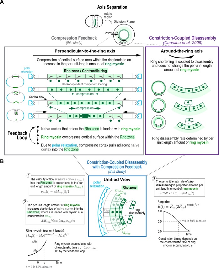
Figure 4. Constriction-coupled disassembly with compression feedback model.

Figure 4.

(A) The natural coordinate system for contractile ring dynamics has two axes, an axis parallel to ring constriction (around-the-ring axis) and an axis perpendicular to the ring (perpendicular-to-the-ring axis). Polar relaxation and filament alignment in the around-the-ring direction lead to anisotropy in behavior along the two axes, which are illustrated separately here. Our prior work analyzing component dynamics at the 4-cell stage in the C. elegans embryo has suggested that in the around-the-ring direction constriction is coupled to component disassembly (Constriction-Coupled Disassembly, right), so that ring constriction does not change the per-unit-length amount of ring components (Carvalho et al., 2009). Here, we propose that this is coupled to a feedback loop between ring myosin and compression-driven cortical flow (Compression Feedback, left) that operates along the axis perpendicular to the ring. Compression feedback would lead to an exponential increase in the per-unit-length amount of ring myosin and the per-unit-length constriction rate as the ring closes. (B) Constriction-Coupled Disassembly with Compression Feedback can be formulated as an analytical mathematical model consisting of three equations and three model parameters. (left) Equations (1) and (2) describe the feedback loop between the amount of ring myosin and the velocity of compression-driven flow of cortical surface into the ring. Solving these equations gives the expression for the per-unit-length amount of ring myosin, which accumulates exponentially as shown in the graph. (right) The feedback loop operating perpendicular to the ring controls the per-unit-length amount of ring myosin, which in turn controls the per-unit-length rate of ring constriction as described in Equation (3). Graph plots the equation for ring size resulting from solving the model equations in the time reference where t=0 is the halfway point of ring closure.

After anaphase onset, spindle-based signaling activates RhoA on the cortex in an equatorial region termed the Rho zone (Bement et al., 2006; Green et al., 2012; Jordan and Canman, 2012; Piekny et al., 2005). In our model, the Rho zone and contractile ring are interchangeable. The Rho zone occupies the central region of the perpendicular-to-the-ring axis between the poles. Within this zone, RhoA-based signaling leads to the recruitment of an ensemble of interacting contractile ring proteins, including formin-nucleated actin, myosin, anillin and the septins, to the cortex after anaphase onset (Jenkins et al., 2006; Maddox et al., 2005, 2007; Mangal et al., 2018; Motegi and Sugimoto, 2006; Schonegg et al., 2007; Tse et al., 2012; Werner et al., 2007). As our data indicate (Figure 3A), this leads to uniform compression of the cortex across this central 10 µm wide zone. We propose that, due to polar relaxation, compression of cortex in the Rho zone pulls naive cortex, defined as cortex outside the Rho zone (naive to RhoA-based signaling) into the Rho zone (Figure 4A). The new cortex that flows into the Rho zone as a result of compression is also loaded with contractile ring components and initiates compression. Thus, along the perpendicular-to-the-ring axis a feedback loop operates in which myosin in the Rho zone compresses cortical surface, which pulls naive surface into the Rho zone that is then loaded with myosin and other contractile ring components, leading to the exponential accumulation of ring components (Figure 4A, left panel). In the around-the-ring direction, ring shortening is coupled to the disassembly of ring components as we described previously (Constriction-Coupled Disassembly; Carvalho et al., 2009) and does not change per-unit-length component levels (Figure 4A, right panel).

In the mathematical formulation (Figure 4B), naive cortex flows into the Rho zone at a velocity (vflowt) proportional to the per-unit-length amount of ring myosin (Mringt; Equation (1) in Figure 4B), with α being the proportionality constant that relates the two. Ring myosin, in turn, increases at a rate proportional to this flow and the concentration of myosin that is loaded onto the cortex when it enters the rho zone (mrho; Equation (2) in Figure 4B). As a result of the positive feedback between ring myosin and compression-driven flow, ring myosin increases exponentially with a characteristic time τ:=1/αmrho  (time required for ring myosin to increase ~2.7 fold; Figure 4B, left graph). The per-unit-length rate of ring constriction (dRdt*1R) is proportional to the per-unit-length amount of ring myosin, related by the proportionality constant β (Equation (3) in Figure 4B). To avoid the difficulty of accurately assigning the exact point when cytokinesis starts, we solved these equations in the time reference where t=0 is the halfway point of ring closure (R¯(t=0)=12). In this time reference, the equation for ring size is:

R¯(t¯ )=R¯ini(2R¯ini)exp(t¯ ), (4)

where t¯:=t/τ and R¯ini is the dimensionless characteristic ring size (held fixed at a value of 1.1; see Materials and methods; Figure 4B, right graph). Other components, like anillin, that localize to the cell cortex will be delivered to the contractile ring via the same process as myosin, and would accumulate in a similar fashion, with

Cring(t¯)Cring,base=αcrhoβln(2R¯ini)et¯, (5)
Cring,base:=C0,ringln(2R¯ini)αcrhoβ, (6)

where C0,ring is the per-unit-length amount of the component at the half-way point of ring closure, Cring,base is the baseline amount of the ring component that does not increase exponentially, and crho (mrho for myosin) is the concentration of the component loaded onto naive cortex when it enters the rho zone. The velocity of cortical flow and the constriction rate are

vflow(t¯)=αβln(2R¯ini) et¯,
1R¯dR¯dt¯=ln(2R¯ini) et¯. (8)

Thus, the per-unit-length constriction rate, velocity of cortical flow, and ring component amounts would all increase exponentially, as we have observed experimentally (Figure 3C), with the characteristic time of ring myosin accumulation (τ=1/αmrho) set by the feedback loop between ring myosin and cortical flow. We conclude that an analytical mathematical formulation that combines Compression Feedback along the perpendicular-to-the-ring axis with Constriction-Coupled Disassembly along the around-the-ring axis can recapitulate the experimentally observed dynamics of ring components, cortical flow into the division plane, and ring constriction.

Fluorescence recovery after photobleaching of the division plane is consistent with constriction-coupled disassembly with compression feedback

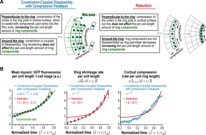
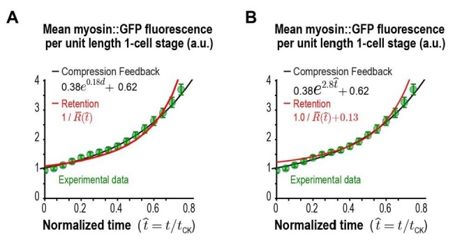
The Constriction-Coupled Disassembly with Compression Feedback model is characterized by anisotropy in the behavior of the cortex in the perpendicular-to-the-ring and around-the-ring directions that results from polar relaxation (Figure 5A, left). In the perpendicular-to-the-ring direction, cortical compression within the ring pulls in cortical surface, which is loaded with ring components and increases their per unit length amount. In contrast, in the around-the-ring direction, constriction is coupled to disassembly and does not affect the per-unit-length amount of ring components. An alternative model that could also explain the increase in the per-unit-length amount of ring components, which we will refer to as the 'Retention' model, is that the per-unit-length constriction rate accelerates due to retention of myosin and/or other ring components during ring shortening (Figure 5A, right). In the Retention model, ring components are not lost due to disassembly as ring perimeter decreases. Instead, their total amounts in the ring remain constant during constriction, resulting in an increase in per-unit-length amounts in inverse proportion to the reduction in ring perimeter (levels would increase as 1R). In the perpendicular-to-the-ring direction, compression would still pull cortical surface into the ring, as we have shown occurs experimentally, but the Retention model assumes that this flow would not deliver myosin into the ring, either because levels of myosin on the delivered cortex are insignificant relative to the amount of myosin in the ring or because the delivered myosin is lost due to disassembly. Comparison of the fits to the data for the per-unit-length amounts of ring myosin and anillin and the rates of ring shrinkage and cortical compression for the two models (Figure 5B, Figure 5—figure supplement 1) suggested that, whereas the Retention model could approximate the data, the Constriction-Coupled Disassembly with Compression Feedback model fit the data significantly better.

Figure 5. Myosin accumulation and the rates of ring constriction and cortical compression can be approximated with a retention model but are fit better by the Constriction-Coupled Disassembly with Compression Feedback model.

(A) Two models could explain the acceleration in the per-unit-length constriction rate during constriction. In the Constriction-Coupled Disassembly with Compression Feedback model, the increase in per-unit-length component amounts arises from a feedback loop between ring myosin and compression-driven cortical flow along the direction perpendicular to the ring. In the Retention model, the increase in per-unit-length component amounts arises because components are retained rather than lost due to disassembly as ring perimeter decreases. (B) Graphs show mean per-unit-length myosin::GFP fluorescence in the ring along with the per-unit-length constriction and cortical compression rates. Myosin fluorescence data is reproduced from Figure 3C to allow comparison of the best fits for the Constriction-Coupled Disassembly with Compression Feedback (blue lines) and Retention (red lines) models.

Figure 5.

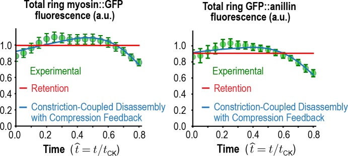
Figure 5—figure supplement 1. Total myosin::GFP and GFP::anillin in the ring.

Figure 5—figure supplement 1.

Graphs plotting mean total ring fluorescence (average over all angles; green) for myosin::GFP (n = 36 embryos) and GFP::anillin (n = 26 embryos). Error bars are the SEM. The predictions for the Constriction-Coupled Disassembly with Compression Feedback (blue lines) and Retention (red lines) models are also shown. Error bars are the SEM.

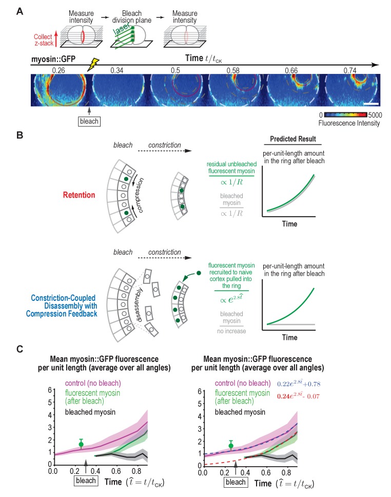
Since both models could approximate the data, we designed a photobleaching experiment to definitively distinguish between them (Figure 6A). We photobleached myosin::GFP in the division plane at ~30% closure, and then monitored the per-unit-length amount of fluorescent myosin in the bleached rings compared to control unbleached rings during constriction. The difference between the two curves is the per-unit-length amount of bleached myosin in the ring (Figure 6C). This experiment can be conceptually thought of as a ‘pulse-chase’ experiment in which we generate a population of bleached subunits in the ring and then follow their fate as the ring constricts. In the Retention model, all ring components are recruited as the ring assembles; as the ring constricts, these components are not lost, but instead become progressively more concentrated. In our experiment, we bleached all the myosin in the ring at the point when the ring was 30% closed. If we successfully bleached all the fluorescent myosin in the ring, the Retention model would predict that there would be no recruitment of additional components and therefore no new fluorescent myosin would appear as the ring decreased in size. However, we also need to consider the likely possibility that our bleach might not be perfect, and some fluorescent subunits, initially below the level of detection, remain in the ring. In this case, the per unit length amount of both the bleached and the unbleached subunits would be expected to increase in inverse proportion to the reduction in ring perimeter (as 1R, Figure 6B, top panels). This is not what we observed. Instead, the per-unit-length amount of bleached subunits (subunits present in the ring when it was 30% closed) did not increase as 1R, but instead remained constant (Figure 6C, black curves). From this, we conclude that the bleached subunits are not retained but are lost in proportion to the reduction in ring perimeter as the ring constricts as expected for the Constriction-Coupled Disassembly with Compression Feedback model (Figure 6B, lower panels).

Figure 6. Fluorescence recovery after photobleaching of the division plane at the one-cell stage rules out Retention and is consistent with Constriction-Coupled Disassembly with Compression Feedback.

Figure 6.

(A) (top) Schematic of the photobleaching experiment. (bottom) Images of the division plane reconstructed from 30 × 1 μm z-stacks of an embryo expressing myosin::GFP whose division plane was bleached at t/tCK ~0.3. Red circle marks the contractile ring and dashed circles mark the boundaries used for ring intensity measurements. Image series is representative of eight imaged embryos. (B) Schematics illustrate the expected results predicted by the Retention and Constriction-Coupled Disassembly with Compression Feedback models. (C) (left) Graph plotting the mean per-unit-length amounts of fluorescent myosin::GFP in the ring for control embryos (pink, n = 24 embryos) and embryos in which the division plane was bleached at the indicated time (green, n = 8 embryos). The amount of bleached myosin::GFP in the ring (black) was calculated as the difference between the control and after bleach curves. Solid continuous lines are the average curves with errors shown as shaded regions. (right) Same as the graph on the left with dashed lines, representing exponential fits to the data, added. Errors for the control and after bleach data are SD and errors for the difference are SEM. Scale bar is 10 µm.

Our ‘pulse chase’ experiment indicated that the myosin in the ring when it was 30% closed is subsequently lost in proportion to the reduction in ring perimeter as the ring constricts. In other words, the contribution of the population of myosin that was in the ring at the 30% closed stage to the total per-unit-length amount of ring myosin neither increases nor decreases as the ring constricts further, but instead remains constant. Thus, since the per-unit-length amount of myosin in the ring increases exponentially as the ring closes, this exponential increase must be due to addition of new myosin to the ring. Our data also revealed that the per-unit-length amount of fluorescent myosin in the bleached rings was equivalent to the per-unit-length amount of myosin in control rings minus the per-unit-length amount of myosin in the ring at the time when it was bleached. The fact that fluorescent myosin is added to bleached rings at the same rate as it is added to control rings suggests that there is very little exchange of the bleached myosin subunits with fluorescent myosin from the cytoplasm. If there was significant exchange, we would expect the rate of increase of the per unit length amount of fluorescent myosin to be higher in the bleached rings than in the controls (equal to new addition plus exchange). In summary, our division plane photobleaching experiment at the one-cell stage is consistent with the Constriction-Coupled Disassembly with Compression Feedback model.

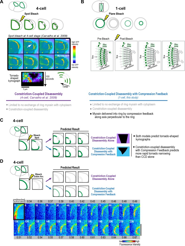
Finally, we wanted to assess whether the results of our division plane bleaching experiment at the one-cell stage are consistent with our prior work bleaching a spot on the contractile ring in embryos expressing fluorescent myosin at the four-cell stage. In this prior work, kymographs of a fixed length region along the constricting arc showed that the bleached spot narrowed as the ring constricted, yielding a tornado shape (Figure 7A; Carvalho et al., 2009). These experiments suggested that myosin in the ring disassembles in a constriction-coupled fashion, which is in agreement with our findings in the division plane bleach experiments at the one-cell stage (black curves in Figure 6C). In addition to Constriction-Coupled Disassembly, which does not alter the per-unit-length amount of ring myosin, the division plane bleaching experiments, along with our cortical flow analysis and modeling at the one-cell stage, suggested that new myosin is delivered into the ring by compression feedback along the axis perpendicular to the ring (illustrated in Figure 7B). We were interested in whether compression feedback could also drive component accumulation and constriction rate acceleration at the four-cell stage. We note that in our spot bleaching experiments, we had noticed that rather than closing ~50% over 95 s as predicted by Constriction-Coupled Disassembly alone, the tornados thinned more rapidly than expected (Carvalho et al., 2009), which is a prediction of the Constriction-Coupled Disassembly with Compression Feedback model (Figure 7C). As a more definitive test of whether myosin is delivered into the ring during constriction at the four-cell stage, like it is at the one-cell stage, we bleached the entire visible arc in cells at the four-cell stage and then monitored fluorescent myosin as they constricted. New myosin appeared in the bleached four-cell stage arcs in a fashion very similar to the bleached rings at the one-cell stage, suggesting that a similar mechanism delivers new myosin into the ring to drive the increase in the per-unit-length constriction rate at the one- and four-cell stages.

Figure 7. Fluorescence recovery after photobleaching of the division plane at the four-cell stage is consistent with Constriction-Coupled Disassembly with Compression Feedback.

Figure 7.

(A) (top) Schematics of an experiment that we performed previously in which a spot was bleached in contractile rings at the four-cell stage (Carvalho et al., 2009). In kymographs of a fixed length region of the arc, the bleached spot narrowed as the ring constricted, yielding a tornado shape. (middle) Image panels and kymograph reproduced from Figure 5 of Carvalho et al. (2009). The top set of panels show a four-cell stage spot bleaching experiment. The region indicated by the arrow was bleached. Time is in seconds after photobleaching. Stills are in pseudocolor with hot-to-cold colors representing high-to-low GFP intensity. Scale bar, 5 μm. Below the images is a representative tornado-shaped kymograph. The time interval between each row of pixels is 5 s. The top row corresponds to the time point before photobleaching. Pixel dimensions are 0.27 × 0.27 μm. These experiments suggested that after its incorporation into the ring, ring myosin does not exchange with myosin in the cytoplasm, and that the ring disassembles in constriction-coupled fashion. (B) In addition to Constriction-Coupled Disassembly, which does not alter the per-unit-length amount of ring myosin, the division plane bleaching experiments that we performed at the one-cell stage suggest that new myosin is delivered into the ring by compression feedback along the axis perpendicular to the ring. (C, D) Schematics in C and D show the predictions of Constriction-Coupled Disassembly Alone and Constriction-Coupled Disassembly with Compression Feedback for spot (C) and division plane (D) bleaching experiments at the four-cell stage. Rather than closing by 50% over 95 s as predicted by Constriction-Coupled Disassembly Alone, the tornados thinned more rapidly, consistent with the prediction of the Constriction-Coupled Disassembly with Compression Feedback model (Carvalho et al., 2009). (D) To test whether Compression Feedback delivers components to the ring at the four-cell stage as well as at the one-cell stage, we monitored recovery after photobleaching the entire contractile arc. Images show a representative bleached embryo (n = 10). The observed recovery pattern was similar to what we observed at the one-cell stage, consistent with the idea that compression feedback contributes to ring component accumulation at both the one- and four-cell stages. Scale bar is 10 µm.

© 2009 Elsevier

Figure 7A (spot bleach at four-cell stage) reproduced with permission from Figure 5 of Carvalho et al. (2009).

Discussion

The near-constant overall rate of cytokinetic ring closure, despite the decreasing size of the ring, ensures that the partitioning of the contents of a mother cell occurs in a timely manner. Here, we provide evidence for a mechanism that explains this conserved property of the cytokinetic contractile ring. Our proposal arises from the near-identical exponential kinetics with which the amount of ring components, constriction rate, and rate of cortical compression (all measured per unit length of the ring) increase as the ring closes. This similarity led us to propose and quantitatively model positive feedback between ring myosin and compression-driven flow of cortex into the ring as the basis for the constant closure of the ring. Below we discuss this proposal in light of prior observations and models for cytokinetic ring closure.

The constriction rate increase during ring closure is accompanied by an increase in the amount of myosin and other contractile ring components

In prior work (Carvalho et al., 2009), we found that once components are incorporated into the contractile ring, they did not exchange with subunits in the cytoplasm, but were instead lost via constriction-coupled disassembly. Our new experiments monitoring component levels at the one-cell stage and division plane photobleaching experiments at the one- and four-cell stages, support these conclusions. However, they also suggest the existence of a new feature that was not appreciated in the prior study due to the technical limitation that, at the four-cell stage, ring component levels could only be analyzed over a limited range of ring perimeters. This new feature is a feedback-based mechanism that operates along the axis perpendicular to the ring to drive the exponential accumulation of contractile ring proteins. We refer to the model that incorporates this feedback mechanism as Constriction-Coupled Disassembly with Compression Feedback (Figure 8).

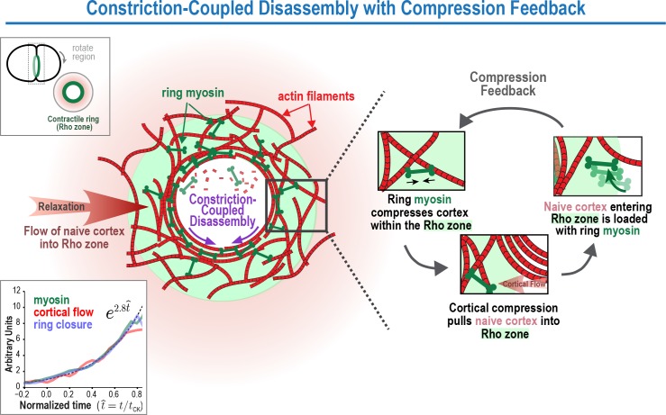
Figure 8. The Constriction-Coupled Disassembly with Compression Feedback model for contractile ring dynamics during cytokinesis.

Figure 8.

Polar relaxation allows ring myosin to compress cortical surface along the axis perpendicular to the ring. Compression pulls naive cortex not previously exposed to RhoA-based signaling into the contractile ring/Rho zone, where it is loaded with myosin and other contractile ring components. Feedback between ring myosin and compression-driven cortical flow leads to an exponential increase in the per-unit-length amount of ring myosin that maintains the high overall closure rate as ring perimeter decreases.

The presence of compression feedback has important implications for understanding the mechanism that underlies the acceleration of the per-unit-length constriction rate during ring closure. In contrast to the prior view that the per-unit-length amount of ring components remains constant as ring perimeter decreases (Carvalho et al., 2009), our one-cell embryo data show that the per-unit-length amount of ring components increases exponentially in the same manner as the per-unit-length constriction rate. This finding suggests that the per-unit-length increase in the concentration of myosin and other contractile ring components underlies the acceleration of the constriction rate, and argues against the contractile unit model, which we had previously proposed to explain how the per-unit-length constriction rate might increase in the absence of a proportional increase in component levels (Carvalho et al., 2009).

Constriction-Coupled Disassembly with Compression Feedback: a new explanation for acceleration of the per-unit-length constriction rate during cytokinesis

Our data suggest that the constriction rate acceleration is due to an increase in the per-unit-length amount of myosin and other contractile ring components. Our division plane photobleaching data rule out the idea that the increase in the per-unit-length amount of ring components is due to component retention and suggest instead that it is due to the action of a feedback loop that delivers myosin and other components into the ring. Our cortical flow analysis and modeling suggest that the feedback could be between ring myosin and compression-driven cortical flow in the perpendicular-to-the-ring direction (Figure 8). In our model, polar relaxation allows ring myosin to compress cortical surface along the pole-to-pole axis perpendicular to the ring, thereby pulling new cortex into the contractile ring/Rho zone that is loaded with myosin and other contractile ring components. An increase in the per-unit-length amount of ring myosin, in turn, would lead to increased cortical compression, resulting in a feedback loop that drives an exponential increase in the per-unit-length amount of ring components. In this model, the overall amounts of myosin, anillin (and presumably other components) in the ring would remain relatively constant as the ring constricts, as is experimentally observed (Figure 5—figure supplement 1), due to a balance between loss resulting from disassembly-coupled ring shortening and accumulation due to the feedback loop in the perpendicular-to-the-ring direction. Thus, the relatively constant overall levels of ring components would mask a dramatic restructuring of the ring that is occurring during closure.

We note that the Compression Feedback component of the model we propose here is reminiscent of early conceptual models of cytokinesis, which hypothesized that polar relaxation coupled to a global upregulation of surface tension could trigger a flow of tension-generating elements toward the equator that would compress into a circular band and initiate a feedback loop (Greenspan, 1978; Swann and Mitchison, 1958; Taber, 1995; White and Borisy, 1983; Wolpert, 1960; Zinemanas and Nir, 1987, 1988). However, in contrast to these early models, which proposed that polar relaxation would be sufficient for ring assembly, the feedback loop that we propose requires both polar relaxation and continuous loading of contractile ring components onto the cortex pulled into the equatorial Rho zone by compression. Thus, although polar relaxation allows the cortex within the ring to compress along the axis perpendicular to the ring, the ring is locally fed by RhoA-based signaling acting on the equatorial cortex. We note that this mechanism would also explain why the furrow would slide or regress and reform following repositioning of the spindle (Rappaport, 1985). Displacing the spindle to one side of the ring would reposition the Rho zone, causing components to be loaded onto the cortex entering the ring on one side but not the other, generating a force imbalance that would slide the ring over to align it with the spindle.

In addition to maintaining a high overall constriction rate to facilitate timely cell content partitioning, a second advantage of the feedback-based mechanism that we propose is that it would render the ring robust to internal or external mechanical challenges, such as cell-cell contacts, obstacles in the crowded cell interior, or defects in the cytokinesis machinery. If the ring stalled due to any of these causes, the feedback loop between ring myosin and compression-based flow along the direction perpendicular to constriction would lead to the progressive build-up of contractile ring components until they reached a level where the obstacle could be overcome and constriction would again be able to proceed. Concentrating components via retention in the around-the-ring direction would not have this property, since successful constriction would be required for an increase in component levels. We note that ring-directed cortical flows similar to the ones we document here have also been observed in the context of wound healing (Mandato and Bement, 2003), where they could potentially serve a similar function in allowing the cell to ramp up contractile force and achieve wound closure.

The experimental basis for our model is our analysis of cortical dynamics, which indicates that the compression of cortical surface that initiates during contractile ring assembly (Figure 3; Reymann et al., 2016), persists throughout constriction, resulting in a continuous flow of cortical surface into the ring. A second key finding is that the per-unit-length amount of ring myosin and anillin and the per-unit-length rates of cortical compression and ring constriction increase with the same exponential kinetics, suggesting control by positive feedback. We note that it remains possible that there is a distinct source of positive feedback (other than between ring myosin and cortical compression as we propose) that controls myosin recruitment, and that myosin levels in turn control the rates of constriction and cortical compression. However, since our data indicate that cortical surface is compressed within the ring, such a model would need to invoke an as yet uncharacterized process to explain why compression of the cortex within the ring does not increase the concentration of ring components, as well as detail the nature of the alternative positive feedback loop that controls ring component accumulation. We note that compression within the ring along the direction perpendicular to the ring is also consistent with work in S. pombe, which has shown that contractile ring assembly occurs via a similar acto-myosin based compression of an equatorial band of nodes into a compact ring along the long axis of the cell (Vavylonis et al., 2008; Wu et al., 2006). However, in contrast to S. pombe where ring assembly and constriction occur in distinct phases, our model predicts that in animal cells, the accumulation of ring components due to compression along the direction perpendicular to the ring is ongoing and serves to accelerate the per-unit-length constriction rate as the ring closes.

Polar relaxation enables cortical compression within the ring along the axis perpendicular to the ring

Monitoring cortical dynamics in combination with laser ablation experiments indicates that the polar cortex is distinct from the cortex in the intervening region between the contractile ring and the poles. The polar cortex expands in response to tension generated by the constricting ring, whereas the intervening cortex flows toward the ring without expanding. One possibility is that the polar cortex is less stiff than the rest of the cortex, causing it to stretch and thin in response to ring constriction-induced tension. Alternatively, the polar cortex could turnover more rapidly, leading to a higher rate of surface renewal after stretching. A third possibility is that the polar cortex is more prone to rupture, repair of which would locally increase cortical surface. Consistent with this last idea, blebs have been reported at the cell poles in cultured vertebrate and Drosophila cells, where they have been proposed to release tension at the poles (Hickson et al., 2006; Sedzinski et al., 2011). The distinct mechanical properties of the polar cortex suggest that its composition could be different from that of the adjacent cortex. This idea is consistent with both older work suggesting the existence of mechanisms that clear contractile ring proteins from the poles (Bement et al., 2005; Chen et al., 2008; Foe and von Dassow, 2008; Murthy and Wadsworth, 2008; von Dassow, 2009; Werner et al., 2007; Zanin et al., 2013) and recent studies that have begun to uncover molecular mechanisms that may drive clearing. Work in C. elegans has demonstrated the existence of a mechanism in which Aurora A, localized to astral microtubules by association with its activator TPXL-1, actively clears contractile ring proteins from the polar cortex (Mangal et al., 2018). A reduction in f-actin intensity at the cell poles due to delivery of a phosphatase by segregating chromosomes has also been reported in Drosophila cells (Rodrigues et al., 2015). Understanding how the polar cortex is different in molecular and mechanical terms, and the mechanisms that generate these differences are important goals for future work.

Cleaving sea urchin embryos exhibit constriction kinetics essentially identical to those during the first division of the C. elegans embryo (Mabuchi, 1994). Pioneering work measuring the distance between surface-adhered particles and the behavior of pigmented cortex-associated granules (Dan, 1954; Dan and Dan, 1940; Dan et al., 1938), indicated that sea urchin embryos also exhibit a similar pattern of cortical expansion during ring constriction, in this case, a wave of cortical expansion that initiates at the poles and propagates through to the region adjacent to the furrow (Dan et al., 1938; Dan and Ono, 1954; Dan et al., 1937; Gudejko et al., 2012; Swann and Mitchison, 1958). Cortical compression and expansion have not been mapped in vertebrate cells; however, monitoring of fluorescent latex spheres adhered to cell surface proteins (Fishkind et al., 1996; Wang et al., 1994), injected stabilized fluorescent actin filaments (Cao and Wang, 1990), and fluorescently labeled myosin II (DeBiasio et al., 1996) all revealed concerted cortical flow towards the division plane in the equatorial region of the cell that contrasted with random surface movements at the cell poles. These observations suggest that feedback in which relaxation enables compression-driven cortical flow may be a conserved feature of animal cell cytokinesis.

It is worth noting that the model we propose suggests a resolution to a long-standing debate as to whether contractile ring components are delivered into the ring via cortical flow (Cao and Wang, 1990; DeBiasio et al., 1996; Fishkind et al., 1996; Wang et al., 1994) or recruited de novo from the cytoplasm downstream of RhoA-based signaling (Vale et al., 2009; Yumura, 2001; Zhou and Wang, 2008). In our model, following anaphase onset contractile ring components are initially recruited to the equatorial cortex de novo, as has been observed (Vale et al., 2009; Yumura, 2001; Zhou and Wang, 2008), but then component levels are amplified by a feedback loop that involves flow and compression of cortical surface as well as de novo component loading. Thus, in our model, both de novo loading of components by Rho-based signaling and compression-driven flow contribute to the change in component levels in the ring during constriction.

The Constriction-Coupled Disassembly with Compression Feedback model as a tool to describe the evolution of the contractile ring

To quantitatively explore the idea that a feedback loop between the amount of ring myosin and compression-driven flow of cortical surface into the ring drives component accumulation during constriction, we developed an analytical mathematical framework. The model consists of three equations with three model parameters that describe this feedback and can recapitulate our experimental results. In addition to describing the processes underlying the evolution of the contractile ring, the Constriction-Coupled Disassembly with Compression Feedback model provides a simple framework that can be used to analyze the consequences of molecular perturbations. An additional interesting future direction will be to use parameter changes derived from the model as input for a finite-element model (similar to Turlier et al., 2014) in order to predict the evolution of cell shape given an a priori knowledge of cortical and contractile ring dynamics.

Materials and methods

Key resources table.

Strain name Genotype Reference
OD821 ltSi200[pOD1997; Pnmy-2::nmy-2::gfp; cb-unc-119(+)] II; unc-119(ed3) III This study
OD857 ltSi200[pOD1997; Pnmy-2::nmy-2::gfp; cb-unc-119(+)] II; unc-119(ed3);
ruIs32[pAZ132; pie-1/GFP::histone H2B] III
This study
OD858 ltSi803[pOD1998; Parx-7::GFP::arx-7; cb-unc-119(+)] II; unc-119(ed3) III; This study
LP162 nmy-2(cp13[nmy-2::gfp + LoxP]) I Dickinson et al., 2013
OD95 unc-119(ed3) III; ltIs37 [pAA64; Ppie-1::mCherry::his-58; unc-119(+)] IV;
ltIs38 [pAA1; Ppie-1::GFP::PH(PLC1delta1); unc-119 (+)]
Essex et al., 2009
OD3011 ltSi1123[pSG017; Pani-1::GFP::ani-1 RE-encoded-exon5::ani-1 3'-UTR;
cb unc-119(+)]II;unc-119(ed3)III
This study
GOU2047 cas607[arx‐2::gfp knock‐in] V Zhu et al., 2016

C. elegans strains used in this study

The C. elegans strains listed in the table were maintained at 20°C using standard methods. OD821, OD3011, and OD858, expressing NMY-2::GFP, GFP::anillin, and GFP::ARX-7, respectively, were generated using a transposon-based strategy (MosSCI; [Frøkjaer-Jensen et al., 2008]). Genomic regions encoding nmy-2 (including 2079 bp and 1317 bp up and downstream of the stop codon, respectively), ani-1 (including 2015 bp and 1215 bp up and downstream of the stop codon), and arx-7 (including 3056 bp and 634 bp up and downstream of the stop codon) were cloned into pCFJ151 and sequences encoding GFP were inserted either just before (nmy-2) or after (arx-7 and ani-1) the start codon. The single copy nmy-2 transgene was generated by injecting a mixture of repairing plasmid (pOD1997, 50 ng/µL), transposase plasmid (pJL43.1, Pglh-2::Mos2 transposase, 50 ng/µL), and fluorescence selection markers (pGH8, Prab-3::mCherry neuronal, 10 ng/µL; pCFJ90, Pmyo-2::mCherry pharyngeal, 2.5 ng/µL; pCFJ104, Pmyo-3::mCherry body wall, 5 ng/µL) into EG6429 (ttTi5605, Chr II). Single copy ani-1 and arx-7 transgenes were generated by injecting a mixture of repairing plasmid (pSG017 (ani-1) or pOD1998 (arx-7), 50 ng/µL), transposase plasmid (CFJ601, Peft-3::Mos1 transposase, 50 ng/µL), selection markers (same as for nmy-2 strain) and an additional negative selection marker (pMA122; Phsp-16.41::peel-1, 10 ng/µL) into EG6429 (ttTi5605, Chr II). After 1 week, progeny of injected worms were heat-shocked at 34°C for 2–4 hr to induce PEEL-1 expression and kill extra chromosomal array containing worms (Seidel et al., 2011). Moving worms without fluorescent markers were identified and transgene integration was confirmed in their progeny by PCR spanning both homology regions in all strains.

C. elegans RNA-mediated interference

Double stranded RNA (dsRNA) targeting arx-2 (K07C5.1) at a concentration of 1.7 mg/ml was generated by synthesizing single-stranded RNAs in 50 µL T3 and T7 reactions (MEGAscript, Invitrogen, Carlsbad, CA) using cleaned DNA template generated by PCR from N2 DNA using the oligos (TAATACGACTCACTATAGGTCAGCTTCGTCAAATGCTTG and AATTAACCCTCACTAAAGGTGCAATACGCGATCCAAATA). Reactions were cleaned using the MEGAclear kit (Invitrogen, Carlsbad, CA), and the 50 µL T3 and T7 reactions were mixed with 50 µL of 3 × soaking buffer (32.7 mM Na2HPO4, 16.5 mM KH2PO4, 6.3 mM NaCl, 14.1 mM NH4Cl), denatured at 68°C for 10 min, and then annealed at 37°C for 30 min to generate dsRNA. L4 hermaphrodite worms were injected with dsRNA and allowed to recover at 16°C for 44–50 hr prior to imaging.

Generating a 4D map of cortical flow

Cortical flow was monitored in embryos expressing myosin::GFP obtained from adult hermaphrodites by dissection. Embryos were mounted followed by sealing with a coverslip on double thick (1 mm) low percentage agarose (0.5%) pads to prevent compression that biases the initial angle of furrow ingression (Figure 1—figure supplement 1B). Images were acquired on an inverted microscope (Axio Observer.Z1; Carl Zeiss) equipped with a spinning-disk confocal head (CSU-X1; Yokogawa) and a 63 × 1.40 NA Plan Apochromat lens (Zeiss) using a Hamamatsu Orca-ER digital camera (Model C4742-95-12ERG, Hamamatsu photonics). Images were collected using custom software, written in Python, that utilizes the Micro-Manager (open source software, Edelstein et al., 2014) microscope control library. A 3 × 0.75 µm z-series was collected (400 ms exposure, 10–20% laser power) every 2 s. After 15 time points, a 15 × 1 µm z-stack, offset by 3 µm from the cortical surface, was imaged to monitor the position of the closing contractile ring. The entire imaging series was repeated every 36 s until the end of cytokinesis. Cortical flow was measured in maximum intensity projections of the 3 × 0.75 µm z-stacks of the cortical surface, after orientation of the images to place the embryo anterior at the top and the posterior at the bottom, by correlating myosin fluorescence between consecutive images using Gunnar Farnebäck’s algorithm (Farnebäck, 2003) implemented within the openCV library with a 30-pixel window size. The threshold was calculated for every image by maximizing the ratio of total intensity inside a 200 × 350 pixel box positioned in the center of the embryo to the total intensity outside that box.

Measurement of contractile ring position and size

Automated methods were employed to identify the edges of the embryo, determine the position of the contractile ring, and reconstruct the rings for each time point in an end-on view to determine the initial ingression axis (Figure 1—figure supplement 2). Ring size and position were determined using custom Python software that: (1) identifies the orientation of the anterior-posterior (AP) axis and rotates the embryo to place the embryo anterior at the top and the embryo posterior at the bottom, (2) finds the embryo center in different x-z planes along the AP axis and calculates embryo radius, and (3) calculates the radius of the contractile ring and determines its position within the division plane. Details of each step are outlined below.

Orienting embryos with their anterior end to the top

Acquired z-plane images were convolved with a 10-pixel Gaussian kernel to reduce noise. An optimal signal threshold that partitioned the embryo interior from exterior was identified by finding a local minimum in the intensity histogram that produced a binary mask with the expected area (~120,000 ± 50,000 pixel2). The orientation of the AP axis was identified by fitting an ellipse to the thresholded area in the middle plane of the z-stack. The anterior side was identified by higher cortical myosin fluorescence and all images were rotated to place the embryo anterior at the top of the image and the embryo posterior at the bottom.

Defining the central axis of embryo and determining embryo width

The central axis of the embryo was defined by drawing a horizontal line across the oriented embryo at the midpoint between its anterior and posterior ends and identifying the first and last points along this line with signal above the threshold for each z-plane. The identified pixels were virtually projected in an end-on (x-z) view and fit to a circle by minimizing residuals. To account for fluctuations in the embryo boundary due to noise and fluorescence variation, the procedure was repeated nine more times after shifting the position of the horizontal line toward the anterior pole by 10 pixels, covering approximately 1/5 of the embryo length (500 pixels). The position of the AP axis and the radius of the embryo were determined by averaging the 10 measurements.

Measuring contractile ring size and position

As illustrated for the central plane images shown in Figure 1—figure supplement 2, the position of the contractile ring was determined by identifying pairs of points with the highest myosin fluorescence intensity on the opposite edges of the embryo in each z-plane that were not more than 20 pixels apart in the horizontal direction and were located at a y-axis position near the embryo middle. Contractile ring radius and position were determined by projecting the points to generate an end-on (x-z) view and fitting the data with a circle. The ring fit was iteratively improved by calculating predicted positions of myosin fluorescence at the ring in each z-plane using initially fitted parameters. Intensity maxima within 5 pixels of the predicted location were identified and the ring was refit. The initial guesses for the contractile ring size and position at the next time point were estimated from the previously calculated ring values. The algorithm restricted ring position fluctuations to 20 pixels along anterior-posterior axis and the size was estimated assuming constant rate of ring constriction. The automatic ring measurements were manually confirmed for each embryo. The initial ingression axis was determined as illustrated (Figure 1—figure supplement 2) by fitting a line through the centers of the rings with a normalized ring size (R¯:=R/Remb > 0.3).

Embryo time alignment for averaging

Sequences from individual embryos were time aligned by defining zero time (t0) and the total time of cytokinesis (tCK) for each embryo, and normalizing time by tCK prior to averaging, t^:=tt0tCK. An initial determination of t0 and tCK was made by fitting a line to the plot of normalized ring size (R¯(t):=R/Remb) versus time between 30% and 80% closure for each embryo as outlined in Figure 1A. Extrapolation of this line for each embryo defined t0 as the time where the fitted line intersects 1, and the time of cytokinesis, tCK as the time where the fitted line intersects 0. Due to the small number of measurements from each embryo available for fitting (3-5 values where 0.8 > R¯ > 0.3), the values of t0 and tCK were refined by fitting R¯(t^) for each embryo to the average dimensionless ring size, < R¯ > (t^). Calculation of the average dimensionless ring size was performed in iterative manner. The time for each embryo was aligned by t0 and normalized by tCK using estimates from the fitted line in the first iteration. The average dimensionless ring size (< R¯ > (t^)) was calculated by averaging normalized ring sizes of all embryos at corresponding normalized time. Contractile ring size was approximated for intermediate time points by linear interpolation. In further iterations, t0 and tCK were refined for every embryo by minimizing the residuals between its normalized ring size, R¯(t^), and the average dimensionless ring size, < R¯ > (t^), throughout the entire timecourse of cytokinesis, thus increasing the number of time points available for fitting t0 and tCK (6-10 values per embryo). After refining time alignment and normalization for each embryo, average dimensionless ring size was re-calculated and t0 and tCK were refined for each embryo again. The refinement process was repeated until changes in average dimensionless ring size, < R¯ > (t^), were smaller than 0.001 on average (achieved within a few iterations). The collective fitting of all t0 and tCK at every iteration was performed under restriction that the line fit through < R¯ > (t^) between 0.8 and 0.3 intercepted 0 at t^= 0 and 1 at t^= 1. This restriction ensured that t0 and tCK determined from fits of individual embryos to the average ring size would be consistent with their original definition. The dimensional ring kinetics, < R > (t), can be recovered using the following equation

<R>(t)= <Remb><R¯>(t^<tCK>), (9)

where < Remb > =14.7±0.7 μm and < tCK > =200 ±30 s are average embryo radius and time of cytokinesis accordingly.

Cortical flow averaging

Cortical flow averaging was performed after spatial and temporal alignment of data collected in different embryos (n=93 embryos from 93 worms filmed over the course of 5 days for control, Video 2; n=68 embryos from 68 worms filmed over the course of 4 days for arx-2(RNAi), Video 3). The number of embryos was chosen to achieve at least 10-fold coverage for all areas of the cortical map for controls and 5-fold coverage for arx-2(RNAi). Linear interpolation was used to approximate the flow between consecutive time points. Because our imaging regime required periodic z-stack acquisition to determine the trajectory of ring closure, no flow approximation was done during those time periods (~6 s gap every 30 s). The flow data for each time point was represented as a set of vectors with direction and magnitude corresponding to the direction and magnitude of the cortical flow at the base of the vector. The base of each vector had two spatial coordinates: x, the position along the anterior-posterior axis (where the position of the contractile ring was defined as 0), and θ, the angular position relative to the initial ingression axis (defined as described in Figure 1A and Figure 1—figure supplement 2). We note that mitotic exit is accompanied by a brief (~50-60s) period of rotational flow ([Naganathan et al., 2014; Schonegg et al., 2014]; see Video 1), which dissipates soon after initiation of cytokinesis (~t^=0.2-0.3). As this rotational contribution is not relevant here, we removed it by averaging the data from the right and left halves of the embryo (in an end-on view), allowing us to focus on rotation-independent flows. Thus, the flow with angular positions greater than 180 degrees was mirrored in angular direction

fθ(t^, x, θ> 180)  fθ(t^,x, 360θ), (10)

fθ is the angular component of the flow vector f. The flows were normalized by the embryo size and cytokinesis rate f¯(t^,x, θ):=tCKRembf(t^,x, θ) and averaged according to its position and time

< f¯> (t,x, θ)=embf¯(t^,x, θ)Nemb. (11)

Calculation of expected cortical surface flow profiles

To aid in the interpretation of experimental results, expected profiles for cortical surface movement were calculated for defined patterns of cortical surface increase and plotted (Figure 1B and Figure 1—figure supplement 3). The general form of surface movement velocity is given by the following equation

v(x)= 0xg(x)dx+u, (12)

where gx is the amount of cortical surface gain and u is the velocity of asymmetric ring movement, which could be positive or negative, depending on whether the ring is moving towards or away from the surface. From equation (12) we obtain the following predictions

Uniform surface increase: vx=Cx+u;

Polar surface increase: vx=C+u;

Behind the ring surface increase: vx=u (if the asymmetry of cytokinetic furrowing arises due to global surface movement) or vx=0 (if the asymmetry in surface increase is related to the asymmetric furrowing).

Cortical laser ablation

Cortical laser ablations, presented in Figure 2, were performed using a robotic laser microscope system (RoboLase; Botvinick and Berns, 2005). Embryos expressing myosin::GFP were mounted using standard procedures. A cortical cut, approximately 10 µm long, was made on the anterior side of the embryo when the ring was at ~50% closure (7 µm radius). The cut was confirmed by comparison of cortical fluorescence images before and after the cut and was considered successful if the foci moved away from the cut area (~3.5 µm distance), indicating cortical tension release. Contractile ring closure rate was calculated by measuring the difference in ring sizes before and after the cut, assessed from two 4 × 2 µm z-stacks acquired immediately before the cut and 13 s later. Errors in measuring the radius at the two timepoints were determined from the procedure used to fit the data to a circle and were propagated to determine the errors in the constriction rate measurements for individual embryos; mean errors are S.E.M. The cortical opening after ablation was approximately 35 μm2 and, at the time of the cut, the rings were approximately 14 μm in diameter. If the ring size is determined by the available cell surface, then this gain in surface area should result in additional area increase of the division plane. Assuming that the surface area gain resulting from the lesion is distributed between the two cells it would result in ~18 μm2 increase in the division plane, which corresponds to an additional ~0.4 μm reduction in ring radius. This reduction in size would lead to an increase in constriction rate of ~0.03 μm/s, during the 13 s interval between image acquisitions. The experiment was repeated 19 times for no cut condition, 14 times for parallel cut, and 15 times for perpendicular cut. All imaging was performed over the course of 5 days. The number of embryos was chosen to achieve sufficient accuracy in the determination of mean ring closure rates to assess whether it was altered by the cuts.

Calculation of the surface area flowing into the division plane

We calculated the amount of surface area flowing into the division plane from flow measurements made 7 µm away from the position of the furrow on the anterior and posterior sides (as illustrated in Figure 3B). The rate of the surface flow is

dAsurfdt^(t^)=2Remb0  π<f>(t^,x0,θ)dθ, (13)

where x0 is -7 µm and 7 µm for the rate of flow from the anterior or the posterior sides, respectively. The total amount of surface area that entered the division plane from any time t^0 to t^ is obtained by integrating equation (13) over time

Asurf(t^)= t^0t^dAsurfdt^|ant+dAsurfdt^|post(t)dt. (14)

The increase in area of the division plane was calculated as following

Adiv plane(t^)=2π(<R>2(t^0)<R>2(t^)). (15)

In Figure 3B, we used t^0=0.2. The cortical surface area compressed in the ring can be inferred from the difference between the surface area entering the division plane and the area of the division plane

Acomp(t^):=Asurf(t^)Adiv plane(t^). (16)

Division plane imaging

For quantification of myosin::GFP and GFP::anillin amounts in the contractile ring, adult worm dissection and one-cell stage embryos imaging was performed in a custom microdevice (Carvalho et al., 2011). The device was mounted on an inverted microscope (Axio Observer.Z1; Carl Zeiss) and embryos were imaged with a 63 × 1.4 NA Plan Apochromat objective using an electron-multiplying charge-coupled device camera (QuantEM:512SC, Photometrics; 100 ms exposure, EM gain set to 500, 10% laser power). Division planes were reconstructed from 40 × 0.5 µm z-stacks collected every 30 s after background subtraction and attenuation correction. All imaging was done at 20°C.

Contractile ring photo-bleaching and imaging

One-cell stage embryos were mounted in microdevices as for division plane imaging and four-cell stage embryos were mounted on slides with 2% agarose pads. Embryos were imaged on a Nikon TE2000-E inverted microscope equipped with a 60 × 1.40 NA objective, an EM-CCD camera (iXon; Andor Technology; EM-Gain = 220, Exposure = 100 ms), and a krypton-argon 2.5 W water-cooled laser. For one-cell stage embryos, division planes were reconstructed from 30 × 1 µm stacks acquired every 20 s with 20% laser power and photo-bleaching was performed by 2 sweeps of a 488 nm laser with 100% power and 500 µs dwell time. For four-cell stage embryos, division planes were reconstructed from 16 × 1 µm stacks acquired every 10 s with 50% laser power and photo-bleaching was performed by 2 sweeps of a 488 nm laser with 100% power and 100 µs dwell time. For four-cell stage embryos, the time between the prebleached and first postbleached images was 6 s.

Estimation of depth attenuation

To estimate depth attenuation within the division plane, we quantified the intensity of the division plane in two cell embryos expressing a GFP-tagged probe expected to be uniformly present on the plasma membrane. From each image, we subtracted a background intensity calculated as the average value inside two 11 × 11 µm rectangles positioned 2 µm away from the division plane inside the anterior and posterior cells (Figure 3—figure supplement 3). The division plane intensity profile was obtained by performing a 30-pixel maximum intensity projection along the AP axis, with the division plane positioned approximately in the middle (Figure 3—figure supplement 3). The intensity profiles in z from 13 embryos were fitted to an exponential using the same characteristic attenuation depth for all embryos

I=I0e-z/zatt, (17)

which yielded a characteristic depth of attenuation, zatt, of 15 µm.

Quantification of myosin and anillin intensity in the contractile ring and on the cortex

For embryos at the one-cell stage, myosin::GFP and GFP::anillin intensities in the contractile ring and on the cortex were quantified in 40 x 0.5 µm z-stacks containing the ring after correction for depth attenuation and subtraction of background fluorescence. Average intensity along the ring was calculated across a set of embryos in 30 degree arcs (for myosin::GFP, n=36 embryos from 18 worms filmed over 5 days; for anillin::GFP, n= 26 embryos from 14 worms filmed over 4 days). The number of embryos was chosen to determine mean fluorescence with sufficient accuracy to derive appropriate conclusions. Positions along the ring were referenced based on the angle between the line from the position on the ring to the ring center and the initial ingression axis. Linear interpolation in time was used for every embryo to estimate intensity in the intermediate time points to perform averaging. Measured intensities were divided by arc length and averaged between different embryos to obtain mean GFP fluorescence per-unit-length for different angular ranges and the average for all angles. Total ring GFP fluorescence was calculated by integrating over ring perimeter. Cortical intensities were quantified by choosing the time point with the ring size closest to R¯=0.8 and measuring total fluorescence in the 15th plane after correction for depth attenuation and subtraction of background fluorescence.

Measurements of myosin::GFP fluorescence in the ring at the four-cell stage were performed as described in Carvalho et al. (2009). However, background fluorescence was determined as the mean fluorescence within a variable size circle at least 10 pixels in diameter, instead of fixed at 10 pixels, to improve measurement quality.

Derivation of the constriction-coupled disassembly with compression feedback model for cytokinesis

The Constriction-Coupled Disassembly with Compression Feedback model formalizes the following conceptual view of cytokinesis: After anaphase onset, spindle-based signaling patterns the cortex, generating an equatorial zone where RhoA promotes the recruitment of contractile ring components (the Rho zone). Within the Rho zone, myosin engages with actin to exert an isotropic force that compresses the cortical surface, resulting in uniform compression across this region, as is observed experimentally (Figure 3A). Due to polar relaxation, the compressing cortex pulls naive cortex (defined as cortex outside the Rho zone) into the Rho zone. We propose that the new cortical surface that flows into the Rho zone as a result of compression is also loaded with contractile ring components. Thus, a feedback loop is established along the direction perpendicular to the ring, in which myosin in the ring compresses cortical surface, which pulls more surface that is loaded with myosin into the ring. Consistent with our division plane photobleaching experiment at the one-cell stage (Figure 6C) and our prior work (Carvalho et al., 2009), we propose that in the around-the-ring direction constriction-coupled disassembly leads to a reduction in ring components in proportion to the reduction in length. Thus, constriction does not alter the per-unit-length amount of ring components. Changes in myosin levels are therefore determined solely by the rate of flow of naive cortex into the Rho zone along the direction perpendicular to the ring, which can be solved as a one-dimensional problem. We assume that the rate of compression of cortical surface (between x and x+dx) is proportional to local myosin concentration, m(x,t), which exerts stress onto the actin network resulting in

 δε δt(x,t)=αm(x,t), (18)

where ε is the cortical strain (i.e. change in length of cortical surface per-unit-length) and α is a proportionality constant that reflects the ability of the cortex to be compressed by ring myosin. The velocity of cortical surface movement is obtained from the following relationship (see also equation (12)).

v(x,t)=0x δε δt(x,t)dx. (19)

The conservation of mass for myosin flow results in the following

 m t(x,t)=  x(m(x,t)v(x,t))=  x(m(x,t)0x αm(x,t)dx). (20)

If we integrate equation (20) over x on (-w, w) domain we obtain

dMring(t)/dt=αmrhoMring(t), (21)

where Mring(t) :=wwm(x,t)dx is the total per-unit-length amount of ring myosin engaged in compression, 2w is the width of the contractile ring/Rho zone where myosin is engaged and compressing cortex and mrho:=m(w,t) is the concentraton of myosin loaded onto the cortex when it enters the rho zone. The velocity of flow of naive cortex into the rho zone is

vflow(t)=αMring(t)/2, (22)

The one half is included to account for the fact that flow comes in from both sides. The solution of Equation (21) is

Mring(t)=M0 ring et/τ, (23)

where we define the characteristic time of myosin accumulation, τ, as 1αmrho. Note that the total amount of myosin in the ring will be the amount of engaged ring myosin plus an added baseline that would include any myosin not involved in compression (see Equation 5). We assume the per-unit-length rate of ring shrinkage is proportional to the amount of ring myosin, as observed in our data,

1RdRdt=-βMring(t), (24)

where β is a proportionality coefficient that reflects the ability of the ring to be constricted by ring myosin. Using Equations (23) and (24), we obtain the dynamics of contractile ring size over time

R¯(t)=R¯inieβτM0ringexp(t/τ), (25)

where R¯ini is the dimensionless characteristic size of the ring; essentially the radius at minus infinity if the same exponential process controlling contractile ring assembly extended back in time infinitely. Instead, in vivo cytokinesis initiates when spindle-based signaling activates RhoA on the equatorial cortex leading to the abrupt recruitment of contractile ring components. If the time frame of reference is chosen so that t=0 is cytokinesis onset immediately following the initial patterning of the cortex by RhoA, M0ring is the amount of ring myosin immediately following this event and the initial size of the ring is

R¯0(t)=R¯inieβτM0 ring. (26)

To facilitate future use of our model for analysis of contractile ring closure data, we use the time frame of reference wheret=0 is the point of 50% closure (i.e. R¯(t=0)=12), an easily identifiable time point that does not rely on exact assessment of the precise onset of cytokinesis. In this reference, M0 ring=ln(R¯ini)βτ, and by defining dimensionless velocity as v¯:=τv, we obtain equations (4-8). Note that equation (4) can be rewritten in the following way

R¯(t¯)=R¯inie1R¯dR¯dt¯, (27)

where t¯:=t/τ. This relationship implies that in this dimensionless time, where R¯(t¯=0)=12, any two rings of the same size have the same dimensionless constriction rate.

Data availability

Key source data is available from the Dryad repository.

Code availability

The custom computer code used in this study is freely available from GitHub (Khaliullin, 2018; copy archived at https://github.com/elifesciences-publications/cytokinesis).

Acknowledgements

This work was supported by a fellowship from the Jane Coffin Childs Memorial Fund to RNK and grants to MWB from AFOSR (FA9550-08-1-0284) and the Beckman Laser Institute Foundation. JSG-C was supported by the University of California, San Diego Cancer Cell Biology Training Program (T32 CA067754). AD and KO receive salary and other support from the Ludwig Institute for Cancer Research. We would also like to thank Michael Glotzer for discussions that helped us align our model with current thinking about the Rho zone.

Funding Statement

The funders had no role in study design, data collection and interpretation, or the decision to submit the work for publication.

Contributor Information

Renat N Khaliullin, Email: renatkh@gmail.com.

Karen Oegema, Email: koegema@ucsd.edu.

Mohan K Balasubramanian, University of Warwick, United Kingdom.

Anna Akhmanova, Utrecht University, Netherlands.

Funding Information

This paper was supported by the following grants:

  • Ludwig Institute for Cancer Research to Arshad Desai, Karen Oegema.

  • Beckman Laser Institute and Medical Clinic to Michael W Berns.

  • Air Force Office of Scientific Research FA9550-08-1-0284 to Michael W Berns.

  • Jane Coffin Childs Memorial Fund for Medical Research to Renat N Khaliullin.

  • National Institutes of Health T32 CA067754 to J Sebastian Gomez-Cavazo.

Additional information

Competing interests

No competing interests declared.

Author contributions

Conceptualization, Data curation, Software, Formal analysis, Investigation, Visualization, Methodology, Writing—original draft, Writing—review and editing.

Visualization, Writing—original draft, Writing—review and editing.

Investigation, Methodology.

Investigation, Constructed the C. elegans strain expressing GFP::Anillin that was used to monitor accumulation of the contractile ring component anillin in this study.

Conceptualization, Resources, Supervision, Funding acquisition, Methodology.

Conceptualization, Supervision, Writing—original draft, Writing—review and editing.

Conceptualization, Resources, Supervision, Funding acquisition, Writing—original draft, Project administration, Writing—review and editing.

Additional files

Transparent reporting form
DOI: 10.7554/eLife.36073.022

Data availability

All data generated during this study are included in the manuscript and supporting files.

References

  1. Bement WM, Benink HA, von Dassow G. A microtubule-dependent zone of active RhoA during cleavage plane specification. The Journal of Cell Biology. 2005;170:91–101. doi: 10.1083/jcb.200501131. [DOI] [PMC free article] [PubMed] [Google Scholar]
  2. Bement WM, Miller AL, von Dassow G. Rho GTPase activity zones and transient contractile arrays. BioEssays. 2006;28:983–993. doi: 10.1002/bies.20477. [DOI] [PMC free article] [PubMed] [Google Scholar]
  3. Biron D, Libros P, Sagi D, Mirelman D, Moses E. Biphasic cytokinesis and cooperative single cell re-production. In: Skejeltorp A. T, Belushkin A. V, editors. Forces, Growth and Form in Soft Condensed Matter: At the Interface Between Physics and Biology. Berlin: Springer; 2004. pp. 217–234. [Google Scholar]
  4. Bluemink JG, de Laat SW. New membrane formation during cytokinesis in normal and cytochalasin B-treated eggs of Xenopus laevis. I. Electron microscope observations. The Journal of Cell Biology. 1973;59:89–108. doi: 10.1083/jcb.59.1.89. [DOI] [PMC free article] [PubMed] [Google Scholar]
  5. Botvinick EL, Berns MW. Internet-based robotic laser scissors and tweezers microscopy. Microscopy Research and Technique. 2005;68:65–74. doi: 10.1002/jemt.20216. [DOI] [PubMed] [Google Scholar]
  6. Bourdages KG, Lacroix B, Dorn JF, Descovich CP, Maddox AS. Quantitative analysis of cytokinesis in situ during C. elegans postembryonic development. PLoS One. 2014;9:e110689. doi: 10.1371/journal.pone.0110689. [DOI] [PMC free article] [PubMed] [Google Scholar]
  7. Bridges AA, Gladfelter AS. Septin form and function at the cell cortex. Journal of Biological Chemistry. 2015;290:17173–17180. doi: 10.1074/jbc.R114.634444. [DOI] [PMC free article] [PubMed] [Google Scholar]
  8. Byers TJ, Armstrong PB. Membrane protein redistribution during Xenopus first cleavage. The Journal of Cell Biology. 1986;102:2176–2184. doi: 10.1083/jcb.102.6.2176. [DOI] [PMC free article] [PubMed] [Google Scholar]
  9. Calvert ME, Wright GD, Leong FY, Chiam KH, Chen Y, Jedd G, Balasubramanian MK. Myosin concentration underlies cell size-dependent scalability of actomyosin ring constriction. The Journal of Cell Biology. 2011;195:799–813. doi: 10.1083/jcb.201101055. [DOI] [PMC free article] [PubMed] [Google Scholar]
  10. Canman JC, Hoffman DB, Salmon ED. The role of pre- and post-anaphase microtubules in the cytokinesis phase of the cell cycle. Current Biology. 2000;10:611–614. doi: 10.1016/S0960-9822(00)00490-5. [DOI] [PubMed] [Google Scholar]
  11. Cao LG, Wang YL. Mechanism of the formation of contractile ring in dividing cultured animal cells. II. Cortical movement of microinjected actin filaments. The Journal of Cell Biology. 1990;111:1905–1911. doi: 10.1083/jcb.111.5.1905. [DOI] [PMC free article] [PubMed] [Google Scholar]
  12. Carvalho A, Desai A, Oegema K. Structural memory in the contractile ring makes the duration of cytokinesis independent of cell size. Cell. 2009;137:926–937. doi: 10.1016/j.cell.2009.03.021. [DOI] [PubMed] [Google Scholar]
  13. Carvalho A, Olson SK, Gutierrez E, Zhang K, Noble LB, Zanin E, Desai A, Groisman A, Oegema K. Acute drug treatment in the early C. elegans embryo. PLoS One. 2011;6:e24656. doi: 10.1371/journal.pone.0024656. [DOI] [PMC free article] [PubMed] [Google Scholar]
  14. Chaudhuri O, Parekh SH, Fletcher DA. Reversible stress softening of actin networks. Nature. 2007;445:295–298. doi: 10.1038/nature05459. [DOI] [PMC free article] [PubMed] [Google Scholar]
  15. Chen W, Foss M, Tseng KF, Zhang D. Redundant mechanisms recruit actin into the contractile ring in silkworm spermatocytes. PLoS Biology. 2008;6:e209. doi: 10.1371/journal.pbio.0060209. [DOI] [PMC free article] [PubMed] [Google Scholar]
  16. D'Avino PP. How to scaffold the contractile ring for a safe cytokinesis - lessons from Anillin-related proteins. Journal of Cell Science. 2009;122:1071–1079. doi: 10.1242/jcs.034785. [DOI] [PubMed] [Google Scholar]
  17. Dan K, Dan JC, Yanagita T. Behaviour of the cell surface during cleavage. II. Cytologia. 1938;8:521–531. doi: 10.1508/cytologia.8.521. [DOI] [Google Scholar]
  18. Dan K, Dan JC. Behavior of the cell surface during cleavage: iii. on the formation of new surface in the eggs of Strongylocentrotus pulcherrimus. The Biological Bulletin. 1940;78:486–501. [Google Scholar]
  19. Dan K, Ono T. A method of computation of the surface area of the cell. Development, Growth and Differentiation. 1954;2:87–99. doi: 10.1111/j.1440-169X.1954.tb00043.x. [DOI] [Google Scholar]
  20. Dan K, Yanagita T, Sugiyama M. Behavior of the cell surface during cleavage. I. Protoplasma. 1937;28:66–81. doi: 10.1007/BF01624958. [DOI] [Google Scholar]
  21. Dan K. The cortical movement in Arbacia punctulata eggs through cleavage cycles*. Development, Growth and Differentiation. 1954;2:115–122. doi: 10.1111/j.1440-169X.1954.tb00045.x. [DOI] [Google Scholar]
  22. Danilchik MV, Bedrick SD, Brown EE, Ray K. Furrow microtubules and localized exocytosis in cleaving Xenopus laevis embryos. Journal of Cell Science. 2003;116:273–283. doi: 10.1242/jcs.00217. [DOI] [PubMed] [Google Scholar]
  23. Davies T, Jordan SN, Chand V, Sees JA, Laband K, Carvalho AX, Shirasu-Hiza M, Kovar DR, Dumont J, Canman JC. High-resolution temporal analysis reveals a functional timeline for the molecular regulation of cytokinesis. Developmental Cell. 2014;30:209–223. doi: 10.1016/j.devcel.2014.05.009. [DOI] [PMC free article] [PubMed] [Google Scholar]
  24. DeBiasio RL, LaRocca GM, Post PL, Taylor DL. Myosin II transport, organization, and phosphorylation: evidence for cortical flow/solation-contraction coupling during cytokinesis and cell locomotion. Molecular Biology of the Cell. 1996;7:1259–1282. doi: 10.1091/mbc.7.8.1259. [DOI] [PMC free article] [PubMed] [Google Scholar]
  25. Dickinson DJ, Ward JD, Reiner DJ, Goldstein B. Engineering the Caenorhabditis elegans genome using Cas9-triggered homologous recombination. Nature Methods. 2013;10:1028–1034. doi: 10.1038/nmeth.2641. [DOI] [PMC free article] [PubMed] [Google Scholar]
  26. Edelstein AD, Tsuchida MA, Amodaj N, Pinkard H, Vale RD, Stuurman N. Advanced methods of microscope control using μmanager software. Journal of Biological Methods. 2014;1:10. doi: 10.14440/jbm.2014.36. [DOI] [PMC free article] [PubMed] [Google Scholar]
  27. Essex A, Dammermann A, Lewellyn L, Oegema K, Desai A. Systematic analysis in Caenorhabditis elegans reveals that the spindle checkpoint is composed of two largely independent branches. Molecular Biology of the Cell. 2009;20:1252–1267. doi: 10.1091/mbc.e08-10-1047. [DOI] [PMC free article] [PubMed] [Google Scholar]
  28. Farnebäck G. Two-Frame Motion Estimation Based on Polynomial Expansion Lecture Notes in Computer Science. Springer Science and Buisness Media; 2003. pp. 363–370. [Google Scholar]
  29. Fededa JP, Gerlich DW. Molecular control of animal cell cytokinesis. Nature Cell Biology. 2012;14:440–447. doi: 10.1038/ncb2482. [DOI] [PubMed] [Google Scholar]
  30. Fishkind DJ, Silverman JD, Wang YL. Function of spindle microtubules in directing cortical movement and actin filament organization in dividing cultured cells. Journal of Cell Science. 1996;109:2041–2051. doi: 10.1242/jcs.109.8.2041. [DOI] [PubMed] [Google Scholar]
  31. Foe VE, von Dassow G. Stable and dynamic microtubules coordinately shape the myosin activation zone during cytokinetic furrow formation. The Journal of Cell Biology. 2008;183:457–470. doi: 10.1083/jcb.200807128. [DOI] [PMC free article] [PubMed] [Google Scholar]
  32. Frøkjaer-Jensen C, Davis MW, Hopkins CE, Newman BJ, Thummel JM, Olesen SP, Grunnet M, Jorgensen EM. Single-copy insertion of transgenes in Caenorhabditis elegans. Nature Genetics. 2008;40:1375–1383. doi: 10.1038/ng.248. [DOI] [PMC free article] [PubMed] [Google Scholar]
  33. Green RA, Paluch E, Oegema K. Cytokinesis in animal cells. Annual Review of Cell and Developmental Biology. 2012;28:29–58. doi: 10.1146/annurev-cellbio-101011-155718. [DOI] [PubMed] [Google Scholar]
  34. Greenspan HP. On fluid-mechanical simulations of cell division and movement. Journal of Theoretical Biology. 1978;70:125–134. doi: 10.1016/0022-5193(78)90305-3. [DOI] [PubMed] [Google Scholar]
  35. Gudejko HF, Alford LM, Burgess DR. Polar expansion during cytokinesis. Cytoskeleton. 2012;69:1000–1009. doi: 10.1002/cm.21078. [DOI] [PMC free article] [PubMed] [Google Scholar]
  36. Hickson GR, Echard A, O'Farrell PH. Rho-kinase controls cell shape changes during cytokinesis. Current Biology. 2006;16:359–370. doi: 10.1016/j.cub.2005.12.043. [DOI] [PMC free article] [PubMed] [Google Scholar]
  37. Hird SN, White JG. Cortical and cytoplasmic flow polarity in early embryonic cells of Caenorhabditis elegans. The Journal of Cell Biology. 1993;121:1343–1355. doi: 10.1083/jcb.121.6.1343. [DOI] [PMC free article] [PubMed] [Google Scholar]
  38. Jenkins N, Saam JR, Mango SE. CYK-4/GAP provides a localized cue to initiate anteroposterior polarity upon fertilization. Science. 2006;313:1298–1301. doi: 10.1126/science.1130291. [DOI] [PubMed] [Google Scholar]
  39. Jordan SN, Canman JC. Rho GTPases in animal cell cytokinesis: an occupation by the one percent. Cytoskeleton. 2012;69:919–930. doi: 10.1002/cm.21071. [DOI] [PMC free article] [PubMed] [Google Scholar]
  40. Khaliullin R. cytokinesis. cac9823Github. 2018 https://github.com/renatkh/cytokinesis
  41. Ma X, Kovács M, Conti MA, Wang A, Zhang Y, Sellers JR, Adelstein RS. Nonmuscle myosin II exerts tension but does not translocate actin in vertebrate cytokinesis. PNAS. 2012;109:4509–4514. doi: 10.1073/pnas.1116268109. [DOI] [PMC free article] [PubMed] [Google Scholar]
  42. Mabuchi I. Cleavage furrow: timing of emergence of contractile ring actin filaments and establishment of the contractile ring by filament bundling in sea urchin eggs. Journal of Cell Science. 1994;107:1853–1862. doi: 10.1242/jcs.107.7.1853. [DOI] [PubMed] [Google Scholar]
  43. Maddox AS, Habermann B, Desai A, Oegema K. Distinct roles for two C. elegans anillins in the gonad and early embryo. Development. 2005;132:2837–2848. doi: 10.1242/dev.01828. [DOI] [PubMed] [Google Scholar]
  44. Maddox AS, Lewellyn L, Desai A, Oegema K. Anillin and the septins promote asymmetric ingression of the cytokinetic furrow. Developmental Cell. 2007;12:827–835. doi: 10.1016/j.devcel.2007.02.018. [DOI] [PubMed] [Google Scholar]
  45. Mandato CA, Bement WM. Actomyosin transports microtubules and microtubules control actomyosin recruitment during Xenopus oocyte wound healing. Current Biology. 2003;13:1096–1105. doi: 10.1016/S0960-9822(03)00420-2. [DOI] [PubMed] [Google Scholar]
  46. Mangal S, Sacher J, Kim T, Osório DS, Motegi F, Carvalho AX, Oegema K, Zanin E. TPXL-1 activates Aurora A to clear contractile ring components from the polar cortex during cytokinesis. The Journal of Cell Biology. 2018;217:837–848. doi: 10.1083/jcb.201706021. [DOI] [PMC free article] [PubMed] [Google Scholar]
  47. Martineau SN, Andreassen PR, Margolis RL. Delay of HeLa cell cleavage into interphase using dihydrocytochalasin B: retention of a postmitotic spindle and telophase disc correlates with synchronous cleavage recovery. The Journal of Cell Biology. 1995;131:191–205. doi: 10.1083/jcb.131.1.191. [DOI] [PMC free article] [PubMed] [Google Scholar]
  48. Matsumura F, Yamakita Y, Yamashiro S. Myosin light chain kinases and phosphatase in mitosis and cytokinesis. Archives of Biochemistry and Biophysics. 2011;510:76–82. doi: 10.1016/j.abb.2011.03.002. [DOI] [PMC free article] [PubMed] [Google Scholar]
  49. Motegi F, Sugimoto A. Sequential functioning of the ECT-2 RhoGEF, RHO-1 and CDC-42 establishes cell polarity in Caenorhabditis elegans embryos. Nature Cell Biology. 2006;8:978–985. doi: 10.1038/ncb1459. [DOI] [PubMed] [Google Scholar]
  50. Murrell M, Oakes PW, Lenz M, Gardel ML. Forcing cells into shape: the mechanics of actomyosin contractility. Nature Reviews Molecular Cell Biology. 2015;16:486–498. doi: 10.1038/nrm4012. [DOI] [PMC free article] [PubMed] [Google Scholar]
  51. Murthy K, Wadsworth P. Dual role for microtubules in regulating cortical contractility during cytokinesis. Journal of Cell Science. 2008;121:2350–2359. doi: 10.1242/jcs.027052. [DOI] [PMC free article] [PubMed] [Google Scholar]
  52. Naganathan SR, Fürthauer S, Nishikawa M, Jülicher F, Grill SW. Active torque generation by the actomyosin cell cortex drives left-right symmetry breaking. eLife. 2014;3:e04165. doi: 10.7554/eLife.04165. [DOI] [PMC free article] [PubMed] [Google Scholar]
  53. Otomo T, Otomo C, Tomchick DR, Machius M, Rosen MK. Structural basis of Rho GTPase-mediated activation of the formin mDia1. Molecular Cell. 2005;18:273–281. doi: 10.1016/j.molcel.2005.04.002. [DOI] [PubMed] [Google Scholar]
  54. Pelham RJ, Chang F. Actin dynamics in the contractile ring during cytokinesis in fission yeast. Nature. 2002;419:82–86. doi: 10.1038/nature00999. [DOI] [PubMed] [Google Scholar]
  55. Piekny A, Werner M, Glotzer M. Cytokinesis: welcome to the Rho zone. Trends in Cell Biology. 2005;15:651–658. doi: 10.1016/j.tcb.2005.10.006. [DOI] [PubMed] [Google Scholar]
  56. Piekny AJ, Maddox AS. The myriad roles of Anillin during cytokinesis. Seminars in Cell & Developmental Biology. 2010;21:881–891. doi: 10.1016/j.semcdb.2010.08.002. [DOI] [PubMed] [Google Scholar]
  57. Rappaport R. Repeated furrow formation from a single mitotic apparatus in cylindrical sand dollar eggs. Journal of Experimental Zoology. 1985;234:167–171. doi: 10.1002/jez.1402340120. [DOI] [PubMed] [Google Scholar]
  58. Reymann AC, Staniscia F, Erzberger A, Salbreux G, Grill SW. Cortical flow aligns actin filaments to form a furrow. eLife. 2016;5:e17807. doi: 10.7554/eLife.17807. [DOI] [PMC free article] [PubMed] [Google Scholar]
  59. Rodrigues NT, Lekomtsev S, Jananji S, Kriston-Vizi J, Hickson GR, Baum B. Kinetochore-localized PP1-Sds22 couples chromosome segregation to polar relaxation. Nature. 2015;524:489–492. doi: 10.1038/nature14496. [DOI] [PubMed] [Google Scholar]
  60. Schonegg S, Constantinescu AT, Hoege C, Hyman AA. The Rho GTPase-activating proteins RGA-3 and RGA-4 are required to set the initial size of PAR domains in Caenorhabditis elegans one-cell embryos. PNAS. 2007;104:14976–14981. doi: 10.1073/pnas.0706941104. [DOI] [PMC free article] [PubMed] [Google Scholar]
  61. Schonegg S, Hyman AA, Wood WB. Timing and mechanism of the initial cue establishing handed left–right asymmetry in Caenorhabditis elegans embryos. Genesis. 2014;52:572–580. doi: 10.1002/dvg.22749. [DOI] [PubMed] [Google Scholar]
  62. Schroeder TE. The contractile ring and furrowing in dividing cells. Annals of the New York Academy of Sciences. 1990;582:78–87. doi: 10.1111/j.1749-6632.1990.tb21669.x. [DOI] [PubMed] [Google Scholar]
  63. Sedzinski J, Biro M, Oswald A, Tinevez JY, Salbreux G, Paluch E. Polar actomyosin contractility destabilizes the position of the cytokinetic furrow. Nature. 2011;476:462–466. doi: 10.1038/nature10286. [DOI] [PubMed] [Google Scholar]
  64. Seidel HS, Ailion M, Li J, van Oudenaarden A, Rockman MV, Kruglyak L. A novel sperm-delivered toxin causes late-stage embryo lethality and transmission ratio distortion in C. elegans. PLoS Biology. 2011;9:e1001115. doi: 10.1371/journal.pbio.1001115. [DOI] [PMC free article] [PubMed] [Google Scholar]
  65. Selman GG, Perry MM. Ultrastructural changes in the surface layers of the newt's egg in relation to the mechanism of its cleavage. Journal of Cell Science. 1970;6:207–227. doi: 10.1242/jcs.6.1.207. [DOI] [PubMed] [Google Scholar]
  66. Shelton CA, Carter JC, Ellis GC, Bowerman B. The nonmuscle myosin regulatory light chain gene mlc-4 is required for cytokinesis, anterior-posterior polarity, and body morphology during Caenorhabditis elegans embryogenesis. The Journal of Cell Biology. 1999;146:439–451. doi: 10.1083/jcb.146.2.439. [DOI] [PMC free article] [PubMed] [Google Scholar]
  67. Straight AF, Cheung A, Limouze J, Chen I, Westwood NJ, Sellers JR, Mitchison TJ. Dissecting temporal and spatial control of cytokinesis with a myosin II Inhibitor. Science. 2003;299:1743–1747. doi: 10.1126/science.1081412. [DOI] [PubMed] [Google Scholar]
  68. Swann MM, Mitchison JM. Cleavage in animal cells. Biological Reviews. 1958;33:103–135. doi: 10.1111/j.1469-185X.1958.tb01409.x. [DOI] [Google Scholar]
  69. Taber LA. Biomechanics of growth, remodeling, and morphogenesis. Applied Mechanics Reviews. 1995;48:487–545. doi: 10.1115/1.3005109. [DOI] [Google Scholar]
  70. Tse YC, Werner M, Longhini KM, Labbe JC, Goldstein B, Glotzer M. RhoA activation during polarization and cytokinesis of the early Caenorhabditis elegans embryo is differentially dependent on NOP-1 and CYK-4. Molecular Biology of the Cell. 2012;23:4020–4031. doi: 10.1091/mbc.e12-04-0268. [DOI] [PMC free article] [PubMed] [Google Scholar]
  71. Tseng Y, Wirtz D. Dendritic branching and homogenization of actin networks mediated by arp2/3 complex. Physical Review Letters. 2004;93:258104. doi: 10.1103/PhysRevLett.93.258104. [DOI] [PubMed] [Google Scholar]
  72. Turlier H, Audoly B, Prost J, Joanny JF. Furrow constriction in animal cell cytokinesis. Biophysical Journal. 2014;106:114–123. doi: 10.1016/j.bpj.2013.11.014. [DOI] [PMC free article] [PubMed] [Google Scholar]
  73. Vale RD, Spudich JA, Griffis ER. Dynamics of myosin, microtubules, and Kinesin-6 at the cortex during cytokinesis in Drosophila S2 cells. The Journal of Cell Biology. 2009;186:727–738. doi: 10.1083/jcb.200902083. [DOI] [PMC free article] [PubMed] [Google Scholar]
  74. Vavylonis D, Wu JQ, Hao S, O'Shaughnessy B, Pollard TD. Assembly mechanism of the contractile ring for cytokinesis by fission yeast. Science. 2008;319:97–100. doi: 10.1126/science.1151086. [DOI] [PubMed] [Google Scholar]
  75. von Dassow G. Concurrent cues for cytokinetic furrow induction in animal cells. Trends in Cell Biology. 2009;19:165–173. doi: 10.1016/j.tcb.2009.01.008. [DOI] [PubMed] [Google Scholar]
  76. Wang YL, Silverman JD, Cao LG. Single particle tracking of surface receptor movement during cell division. The Journal of Cell Biology. 1994;127:963–971. doi: 10.1083/jcb.127.4.963. [DOI] [PMC free article] [PubMed] [Google Scholar]
  77. Werner M, Munro E, Glotzer M. Astral signals spatially bias cortical myosin recruitment to break symmetry and promote cytokinesis. Current Biology. 2007;17:1286–1297. doi: 10.1016/j.cub.2007.06.070. [DOI] [PMC free article] [PubMed] [Google Scholar]
  78. White JG, Borisy GG. On the mechanisms of cytokinesis in animal cells. Journal of Theoretical Biology. 1983;101:289–316. doi: 10.1016/0022-5193(83)90342-9. [DOI] [PubMed] [Google Scholar]
  79. Wolpert L. The mechanics and mechanism of cleavage. International Review of Cytology. 1960;10:163–216. [Google Scholar]
  80. Wu JQ, Sirotkin V, Kovar DR, Lord M, Beltzner CC, Kuhn JR, Pollard TD. Assembly of the cytokinetic contractile ring from a broad band of nodes in fission yeast. The Journal of Cell Biology. 2006;174:391–402. doi: 10.1083/jcb.200602032. [DOI] [PMC free article] [PubMed] [Google Scholar]
  81. Xiong H, Mohler WA, Soto MC. The branched actin nucleator Arp2/3 promotes nuclear migrations and cell polarity in the C. elegans zygote. Developmental Biology. 2011;357:356–369. doi: 10.1016/j.ydbio.2011.07.008. [DOI] [PMC free article] [PubMed] [Google Scholar]
  82. Yumura S. Myosin II dynamics and cortical flow during contractile ring formation in Dictyostelium cells. The Journal of Cell Biology. 2001;154:137–146. doi: 10.1083/jcb.200011013. [DOI] [PMC free article] [PubMed] [Google Scholar]
  83. Zanin E, Desai A, Poser I, Toyoda Y, Andree C, Moebius C, Bickle M, Conradt B, Piekny A, Oegema K. A conserved RhoGAP limits M phase contractility and coordinates with microtubule asters to confine RhoA during cytokinesis. Developmental Cell. 2013;26:496–510. doi: 10.1016/j.devcel.2013.08.005. [DOI] [PMC free article] [PubMed] [Google Scholar]
  84. Zhou M, Wang YL. Distinct pathways for the early recruitment of myosin II and actin to the cytokinetic furrow. Molecular Biology of the Cell. 2008;19:318–326. doi: 10.1091/mbc.e07-08-0783. [DOI] [PMC free article] [PubMed] [Google Scholar]
  85. Zhu Z, Chai Y, Jiang Y, Li W, Hu H, Li W, Wu JW, Wang ZX, Huang S, Ou G. Functional coordination of WAVE and WASP in C. elegans neuroblast migration. Developmental Cell. 2016;39:224–238. doi: 10.1016/j.devcel.2016.09.029. [DOI] [PubMed] [Google Scholar]
  86. Zinemanas D, Nir A. Fluid mechanical simulations of cell furrowing due to anisotropic surface forces. In: Akkas N, editor. Biomechanics of Cell Division. New York: Plenum Press; 1987. pp. 281–305. [Google Scholar]
  87. Zinemanas D, Nir A. On the viscous deformation of biological cells under anisotropic surface tension. Journal of Fluid Mechanics. 1988;193:217–241. doi: 10.1017/S0022112088002125. [DOI] [Google Scholar]
  88. Zumdieck A, Kruse K, Bringmann H, Hyman AA, Jülicher F. Stress generation and filament turnover during actin ring constriction. PLoS One. 2007;2:e696. doi: 10.1371/journal.pone.0000696. [DOI] [PMC free article] [PubMed] [Google Scholar]

Decision letter

Editor: Mohan K Balasubramanian1

In the interests of transparency, eLife includes the editorial decision letter and accompanying author responses. A lightly edited version of the letter sent to the authors after peer review is shown, indicating the most substantive concerns; minor comments are not usually included.

[Editors’ note: a previous version of this study was rejected after peer review, but the authors submitted for reconsideration. The first decision letter after peer review is shown below.]

Thank you for submitting your work entitled "Positive feedback between contractile ring myosin and ring-directed cortical flow drives cytokinesis" for consideration by eLife. Your article has been reviewed by three peer reviewers, and the evaluation has been overseen by a Reviewing Editor and a Senior Editor. The following individual involved in review of your submission has agreed to reveal their identity: Michael Glotzer (Reviewer #2).

Our decision has been reached after consultation between the reviewers. Based on these discussions and the individual reviews below, we regret to inform you that your work will not be considered further for publication in eLife at present.

The reviewers appreciated the novel imaging and computational approaches you are using to understand mechanisms of cytokinetic force generation. The reviewers also found the premise that cortical flow of myosin together with existing myosin in the furrow regulates the rate of furrow contraction interesting. However, a number of issues were raised. These include 1) inconsistencies with what is known in the literature (some experiments with Rho pathway and Nop1 mutants can address these), 2) issues with the theoretical framework and terminologies used (raised by reviewers 1 and 2, which may need experiments and rewriting), 3) aspects of the data fitting and conclusions regarding cortical flow (reviewer 1; may require reanalysis / reconciliation), and 4) referencing (reviewers 2 and 3; should be easily fixed).

If you feel you can address the issues raised with experiments and rewriting / analysis, we will be happy to consider a revised version or as a new submission. In both cases, we will send the paper to the same reviewers.

Reviewer #1:

Cleavage furrow ingression during animal cytokinesis is driven by constriction of the actomyosin contractile ring. While major components of the contractile ring and key regulators for its assembly and constriction have been identified, our mechanistic understanding on how it generates force is limited. In their previous publication, based on the observation that the rate of the ring contraction is largely constant and proportional to the initial size of the ring, the Oegema group proposed a 'contractile unit' model, which assumes a presence of a contractile unit with a fixed initial length, which retains myosin while it shortens.

In this manuscript, the Oegema group studied further details of the mechanism of the ring constriction by precisely measuring the flow and total amount of cortical myosin as well as the myosin in the ring during the ring constriction. Based on these analyses as well as the results of laser micro-surgery and genetic manipulations, they propose a mathematical model with three parameters, which was demonstrated to be useful to explain the effects of a perturbation (depletion of Rho-kinase). The quality of image analysis, especially the 4D mapping of the cortical flow, is extremely high with striking number of video data analyzed. However, there are major problems to be addressed before publication.

1) Logic to choose the feedback model instead of the retention model.

The authors' logic for throwing away the retention model is not clear. The similarity between the time courses of the ring-directed cortical flow and the mean ring myosin or anillin per unit length, and the behavior of the ring myosin after FRAP are the observations on which they were based. Good fitting to exponential curves with a common time constant is interesting and is consistent with the cortical flow feedback model. However, the data are not strong enough to tell exponential from hyperbolic curves. The retention model predicts that the ring myosin per unit ring length is proportional to the inverse of the ring radius (∝1/R) (Figure 4C). Under the constant rate of ring constriction, this means that the ring myosin per unit ring length is proportional to 1/(Runit – v∙t) (t: time, v: rate of ring constriction measured by radius), a hyperbola. With the value range used for Figure 3 and 4 (3 to 4-fold increase), it is almost impossible to distinguish an exponential curve from a hyperbolic one. Indeed, as a simplest example, a set of (x, y) calculated by y=1/(1-x) can be fitted with an exponential curve very well (as we can see by an R script below). The authors should explain why the data support exponential increase better than hyperbolic one.

What data tell us are the largely fixed total amount of ring myosin (with limited exchange with flanking cortex or cytoplasm) and the constant overall cortical flow into the division plane. These two are balanced by some events in the division plane or in the ring (this remains a big black box mainly due to missing direct observation of the cortical flow in the division plane). I appreciate that it was possible to make a mathematical model based on the positive feedback and that it could be fitted to two different conditions (control vs. rho kinase depletion). However, considering that the data don't indicate increase of the total amount of ring myosin, it is difficult for me to understand the necessity of the positive feedback.

2) Logic to disregard accumulation in the division plane, loss by disassembly and turnover with the cytoplasmic pool.

As far as I understand, the only reasoning for disregarding "accumulation in the division plane" and "loss due to disassembly" (Figure 3C) is the similarity between the time course of the ring-directed cortical flow and the time course of ring myosin and anillin per unit length. However, this argument assumes that there is no turnover of myosin with the cytoplasmic pool at the division plane and the ring and that the flow within the division plane is uniform and constant (no accumulation). The authors claim that the result of FRAP excludes the exchange of the ring myosin with the cytoplasmic pool. However, their argument "If ring myosin were turning over due to exchange with cytoplasmic myosin, we would expect the FRAP curve to approach the control curve and the difference between the FRAP and control curves to disappear" is true only if all the ring myosin is exchangeable with the cytoplasmic pool and the exchange occurs rapid enough. This assumption is too strong (a kind of straw man argument). The absence of complete recovery simply tells us that there is a non-exchangeable population in the ring myosin. Moreover, indeed, the difference is getting smaller. The basis for the constant cortical flow in the division plane is unclear.

3) Laser dissection.

The authors performed laser dissection experiments (Figure 2) to assess the influence of the cortical resistance on the rate of ring closure. However, it doesn't seem to be sufficient/complete to draw any firm conclusion. It is not clear whether they are comparing the parallel and perpendicular cuts on their effects on the ring constriction. Only the absence of the effect of the parallel cut was mentioned. The cortical flow is not spatially uniform (Figure 1), implying that the cortical tension is also non-uniform. Then, don't we need to consider the relative positioning (angle and distance) between the ring and the cut? Is the lesion big enough? Even after the cut the ring can still be connected to the polar cortex via the unaffected zone of cortex. What would happen if a whole polar cortex is completely separated from the other part of the cortex and the ring? The rationale for the purple line in the Figure 2C "Expected rate if […]" described in the Materials and methods "The cortical opening after ablation was approximately 35μm2; this translates into an additional reduction in ring radius by ~0.8μm, if the cortical surface tension dominates the ring closure rate." needs more detailed explanation.

(R script to demonstrate the difficulty in distinguishing exponential curve and hyperbolic curve)x <- (0:16)/20y <- 1/(1-x)model <- nls(y~a+b*exp(c*x), start=list(a=1, b=1, c=1))xx <- (0:80)/100z <- predict(model, newdata=list(x=xx))plot(x, y, ylim=c(0,5))lines(xx,z)coef <- summary(model)$coefficients[,1]fit = sprintf("y=%.4f+%.4f*exp(%.4f*x)", coef[1], coef[2], coef[3])legend("topleft", legend=c("y=1/(1-x)", fit), pch=1, lty=c(0,1), pt.cex=c(1,0))

Reviewer #2:

This manuscript describes the kinematic analysis and mathematical modeling of the behavior of contractile ring components during cytokinesis. The basic assertion is that cortical myosin flows into the furrow region during ingression and these flows lead to increased accumulation which drives more rapid ingression as furrowing proceeds. Within its technical limitations, this work is carefully performed. It makes its underlying assumptions clear, and is supported by a model that appears consistent with these assumptions. However, there are fundamental problems with these assumptions and significant technical limitations. Ultimately, I do not find the central point of the manuscript convincingly demonstrated.

Concerning the technical weakness: The majority of the data in the manuscript is based on measurements of cortical components on the flatter parts of the embryo. "Cortical flow could not be monitored in the division plane or at the cell poles, due to their high curvature." Thus the part of the cleavage furrow where contractile ring components accumulate to the greatest extent are not detected with the same spatial or temporal resolution. Thus there is an intrinsic limit to the authors ability to track flow of cortical components from the surface and account for its accumulation at the furrow tip. This could be addressed by analyzing cell division of blastomeres that furrow in the imaging plane.

The manuscript extensively discusses a concept they call cortical surface area. This is a mixed metaphor that appears to include aspects of the cortex and the plasma membrane. The authors state, "New cortical surface could be gained uniformly, immediately behind the contractile ring, or at the cell poles" and cite a number of publications that assess the behavior of the plasma membrane during cytokinesis. However, this concept is flawed. As cytokinesis proceeds, the membrane surface area must expand in order for the cell to retain integrity. While the plasma membrane is by definition continuous and uninterrupted, the cell cortex is different, it need not cover the entire membrane to a constant "depth". Furthermore, fluorescence imaging reveals large inhomogeneities in the cortex and, unlike membrane lipids, cortical components can associate and dissociate.

Another major, central weakness is that the manuscript primarily considers two models for accumulation of myosin in the furrow: retention and ring-directed flow. While these mechanisms likely contribute, they represent an incomplete view of the mechanisms by which components accumulate in the furrow. The authors appear to assume that contractile ring components are recruited at a specific time and then reorganized on the cortex. Indeed, the text states, "cytokinesis initiates when spindle-based signaling activates RhoA on the equatorial cortex leading to the abrupt recruitment of contractile ring components." Presumably, they also imagine that additional components are recruited at poles to replace the material that flows toward the furrow. However, this view is inconsistent with extensive analysis of the mechanism of Rho-dependent assembly of the contractile ring. RhoA is active throughout cytokinesis, as indicated by continual association of a RhoA biosensor, which is most concentrated at the furrow. Unfortunately, this key region largely falls outside of the part of the embryo the authors image at high spatial and temporal resolution. Given what is known about RhoA and its effectors, there is no reason to posit that during the course of furrow ingression, active RhoA does not continually activate its effectors and induce recruitment of contractile ring components throughout the progression of cytokinesis. Indeed, the observed increases in ring components may follow the increase in the concentration of active RhoA at the furrow.

The authors suggest that their data excludes recruitment of myosin from the cytoplasm, but the evidence is unconvincing (Figure 4C, subsection “Component levels and fluorescence recovery after photobleaching of the division plane support constriction rate acceleration due to ring-directed flow versus component retention”, second paragraph). For example, the FRAP data is consistent with the furrow tip containing a pool of myosin that has lower mobility. The "recovery" after bleaching could reflect de novo recruitment on top of the slowly exchanging bleached myosin. In addition, there is little evidence of flow of unbleached myosin into the ring, which would be predicted from the author's model.

The authors find a correlation between the rate accumulation of myosin in the ring, and the rate of constriction. There is no evidence that this correlation reflects a causative relationship.

Consistent with the previous point, evidence in the literature contradicts the authors explanation that cortical flows of myosin from regions flanking the furrow are required for the proposed exponential increase in contractile ring myosin that speeds up the rate of ingression. Specifically, C. elegans embryos deficient in NOP-1 are significantly depleted of cortical accumulation of contractile ring components outside the of the equatorial/furrow region. However, these furrows ingress with near wild-type kinetics, indicating that efficient furrow ingression does not require these major flows of contractile ring components. Rather, it suggests the existence of an alternative mechanism that provides for a time-dependent increase in contractile ring components.

Conversely, embryos that are defective in centralspindlin-directed RhoA activation, do contain cortical myosin that appears to flow in the proposed manner, yet such embryos ingress partially and slowly, suggesting that flow-mediated concentration of contractile ring components is insufficient to generate the proposed behavior of the ring.

The authors state "The broad conservation of this property, which allows cytokinesis to complete in a temporally restricted cell cycle window, suggests that feedback between contractile ring myosin and ring-directed cortical flow will be a broadly conserved property of contractile rings in animal cells." This would imply that cells that lack ring-directed cortical flow will exhibit aberrant timing of ring closure. However as mentioned above, analysis of NOP-1 deficient embryos violates this conjecture.

The authors discuss the concept of astral relaxation: "This differential response of the polar cortex to ring-generated tension, which results in a flow of myosin and other cortical components towards the cell equator, is consistent with the idea of polar relaxation hypothesized in early conceptual models of cytokinesis." They fail to mention or cite that astral relaxation has been experimentally documented in the early C. elegans embryo. Indeed it has been demonstrated that a posterior directed spindle directs anterior-directed flow of cortical components that self organize into a furrow (PMID 17669650). Importantly, this anterior furrow is entirely dependent upon the protein NOP-1 (PMID 22918944).

Reviewer #3:

The manuscript from Khaliulin et al. investigated the contribution of cortical flow in maintaining constant rate of ring constriction during cytokinesis in worm embryos. Both de novo actin/myosin assembly at the division site and cortical flow of components to the cleavage furrow are involved in cytokinesis. However, it remains controversial about the relative importance of each pathway. But I think the paper still need some minor revisions to be acceptable for publication.

1. Due to no cell cortex in yeasts but the ring constricts at a constant rate (Pelham and Chang, Nature, 2002), the references of Wu and Pollard 2005 paper in the Introduction and Discussion are somehow misleading. In fission yeast, myosin-IIs mostly retain in the ring during its constriction, they are also highly dynamic by exchanging between the ring and cytoplasm. The similarity and difference between Wu and Pollard, Carvalho et al, and the current work should be discussed.

2. In Zhou, M., & Wang, Y. L. (2008), "Distinct pathways for the early recruitment of myosin II and actin to the cytokinetic furrow" (Mol Biol Cell, 19(1), 318-326), it is found that myosin-II is recruited to the furrow mainly by de novo assembly, but not cortical flow, during early cytokinesis in mammalian cells. This and other similar works on cortical flow and de novo assembly should be cited and discussed.

3. A figure supplement showing the cortical flow map at cell poles before and during ring constriction will be useful.

4. The terms "cortex surface" and "surface gain" are confusing. Because the plasma membrane was not directly tracked, it should be make clear what you mean here. Otherwise, casual readers may think the plasma membrane is inserted at cell pole, which is likely, but not tested here.

5. The Materials and methods section is not clear regarding how background fluorescence was subtracted. Which region was used as the background?

6. The reference citations are not consistent, sometimes multiple authors from the same paper are listed.

[Editors’ note: what now follows is the decision letter after the authors submitted for further consideration.]

Thank you for resubmitting your work entitled "A positive feedback-based mechanism for constriction rate acceleration during cytokinesis in C. elegans" for further consideration at eLife. Your revised article has been favorably evaluated by Anna Akhmanova as the Senior Editor, Mohan Balasubramanian as the Reviewing Editor, and three reviewers (including Michael Glotzer).

The manuscript has been improved but there are several remaining issues that need to be addressed before acceptance, as outlined below.

The referees have returned their comments and I have discussed the comments with the Senior Editor and we have compiled this decision letter with all substantive points raised by the three referees.

In particular, I have taken the step of providing the full list of comments so that the counter arguments to your model for mechanism of acceleration of cytokinetic ring contraction and the role of myosin II accumulation via cortical flow are fully captured.

In light of the fact that all three referees have concurred that the imaging data are among the best in the field of worm cytokinesis and that I believe you are proposing a striking and provocative new model for aspects of cytokinesis, we are interested in publishing your work, and invite you to submit a revision. In the discussions between referees, it was highlighted that the paper was worthy of publication, but that the models need to be considered more critically.

We would like you to rewrite the paper significantly, based on these comments, as well as perform a straight-forward experiment.

One of the referees has raised the unsatisfactory resolution of the current study vis-à-vis the previous work from your laboratory (Carvalho et al., 2009) (Points 10 and importantly 11). The Senior Editor and I concur with the points the referee has raised. We believe a clear statement of your current position is consolidating the Carvalho paper and the current study will be very valuable for the field and will prevent any confusion in the field.

Also, I believe one experiment, of treating 1 and 4 cell embryos with LatA, as a test of your model is required (point 11).

Point 9 below also requires special attention. The exponential vs. hyperbolic accumulation of components in the ring cannot be easily distinguished and the referee has detailed their concern. Please clarify the limitations of your analysis.

Please also pay attention to the use of terminology, which have been raised by all referees (cortical surface area, cortical area, naive cortex), raised by the referees throughout the comments.

Below, I mention what needs to be done for each point.

Although I think I can decide on any submitted revision, I might call upon one of the referees, if required.

Please find below the consolidated comments of the three referees and my recommendations.

1) The revisions to the manuscript have clarified their model so that readers can better understand what the authors claim to demonstrate. I remain unconvinced of the authors model. My concerns are due to the fact that the manuscript is largely based on an inference of cortical compression.

I want to first re-state that the data shown are carefully obtained. The measurements of cortical flow are of interest. The authors provide evidence that the rate of furrow formation is limited by internal viscosity in the contractile ring, which is a novel insight and an important point.

Overall, I remain unconvinced by the author's interpretation of their results. There is some value in their quantitative model, though (1) it is not well constrained, there is no reason why the flow has to be the source of positive feedback and (2) it has not been extensively tested experimentally. However, if the authors want to publish their interpretation, I have no strong objection, particularly in eLife where readers can readily access the reviews that indicate that experts in the field do not subscribe to their interpretation of their results. Notably, the authors and I communicated following the first version. This communication has lead to a more clear description of their model. Yet during that process I communicated the gist of the comments below, yet they remain unresolved in this version. Specifically, in the previous version, the manuscript gave the impression that cortical flows delivered myosin into the furrow region, thereby accelerating ingression, flows, and myosin accumulation through a form of positive feedback. Now, the authors have clarified their interpretation as follows:

a) The amount of cortical surface area that flows into the furrow region exceeds the surface area of the division plane.b) As a consequence of (1) and an unstated assumption that cortical surface area is not disassembled, the authors infer that cortical surface area is compressed. There is no direct evidence for cortical compression except at the very earliest stages of furrow formation. Interestingly the rate of flow at the stage where compression is observed is 2-3x lower than that during furrow ingression (Figure 1B vs. 3A).c) The total cortical surface area is inferred to increase exponentially and it parallels the increase in ring components.d) Yet, the myosin that flows into the furrow region is not a major contributor to ring myosin, rather it largely disassembles. Indeed, in en face views of the division plane, there is no detectable flow of myosin from the "exposed" cortex to the ring.e) However the flow of cortex is proposed to provide additional, initially "naive", cortical surface area that is then patterned by RhoA (Figure 7), and it is this exponentially increasing cortical surface area that leads to exponentially increasing levels of ring myosin. This begs the question, what is "naive" cortex?

(Editors’ recommendation: rewrite significantly for clarity)

2) At the core of the issue with this model is inference of cortical surface area compression. First, the authors claim that cortical compression can be readily inferred from the difference between the amount of cortical flow into the division plane and the area of the division plane. Yet, the actomyosin cortex is dynamic, in addition to compressing and expanding, it can assemble and disassemble. Indeed, the manuscript shows that at the poles, cortical surface area is created as the cortex flows into the furrow region. And, myosin – a key component of the cortex – is largely assumed to be lost as the furrow flows into the division plane (see point 3). Thus, while cortical compression is possible, cortical disassembly is another possibility, which is not given sufficient consideration. In fact it is a strong possibility given that there is loss of a key component of the cortex, myosin.

(Editors’ recommendation: rewrite significantly for clarity)

3) Furthermore, the authors have not explained why "naive cortex" would be required for the zone of active RhoA to drive an increase in myosin accumulation?

(Editors’ recommendation: rewrite significantly for clarity)

4) Given that ARP-2/3 nucleated actin is likely a nucleator of some of the actin in the cell cortex, it is notable that its depletion does not dramatically affect the rate or extent of furrow ingression in otherwise WT embryos, as has been shown previously (PMID 22226748). This raises the follow-up question: what is "naive" cortex in ARP-2/3 depleted embryos?

(Editors’ recommendation: rewrite significantly for clarity)

5) In the author's rebuttal letter (reviewer 2, fourth response) the authors state, "The reviewer would propose that there could be another source of positive feedback that controls myosin accumulation (for example some type of ring intrinsic feedback loop involving Rho-based signaling), and that exponential accumulation of myosin arising from this as yet un-described feedback loop could, in turn, control the constriction rate and the rate of cortical compression. We do not disagree that this could be the case." Positive feedback in RhoA signaling during cytokinetic processes has been demonstrated, (PMID 26479320), and there is evidence for a mechanism in which RhoA might generate positive feedback through the recruitment of centralspindlin and its activation of the RhoGEF ECT-2 (PMID 26252513).

(Editors’ recommendation: rewrite significantly for clarity)

6) Given the topic of this manuscript, it is surprising that the authors do not mention that local RhoA activation is sufficient to induce furrow formation (PMID 27298323) and all of the literature concerning the mechanism of RhoA activation during cytokinesis.

(Editors’ recommendation: consider discussing this paper)

7) The authors state, "We propose that, due to polar relaxation, the compressing cortex pulls naive cortex not patterned by the initial round of RhoA signaling, into the Rho zone." Here the authors are generating confusion between terms that have a different historical meaning. Polar relaxation was used to describe a mechanism by which astral microtubules might induce a net increase in equatorial contractility by the local inhibition of contractility at the poles (polar relaxation). Here, they are discussing how existing equatorial contractility induces flow of cortex away from the poles. These terms are already sufficiently confused in the literature, it would be better to avoid adding to it.

(Editors’ recommendation: rewrite significantly for clarity)

8) One paper published during their revision is quite relevant: PMID: 29146911. DOI: 10.1038/s41467-017-01231-x. I suggest that the authors cite and briefly discuss the paper in their final manuscript.

(Editors’ recommendation: consider discussing this paper)

9) Exponential/hyperbolic accumulation.

In Figure 5, the authors compare exponential curves and hyperbolas for fitting with the experimental data and conclude that exponential curves fit better. However, it is unclear whether their comparison is fair. For fitting with the data of mean fluorescence per unit length with an exponential curve (Compression feedback), three parameters, i.e., the amplitude, the time constant and baseline can be adjusted. On the other hand, for fitting with a hyperbola (Retention model), it is unclear what the formula for R(t) looks like and what degree of freedom was allowed.

As I pointed out in the previous reviewer comments, clear distinction between the exponential curve and hyperbola is not trivial. The authors' own data and interpretation demonstrate this difficulty. First, in Figure 3C, "Cortical compression (rate per unit ring length)" is fitted with an exponential function. However, this quantity dAcompdt1R should follow a hyperbolic increase in time since the first term, dAcompdt, is largely constant (Figure 3B 'Normalized Surface Area Flux') and the second term, 1/R, is an inverse of a linearly decreasing function of time (Figure 1A). Second, in the same panel, "Ring shrinkage rate per unit length (-dRdt1R)" is also fitted with an exponential curve. However, this quantity should also be hyperbolic for the same reason (-dRdtis constant most of the time during furrow ingression Figure 1A). These examples nicely demonstrate the difficulty in distinguishing between exponential and hyperbolic changes by curve fitting with a set of data that are not really suitable. The authors' approach doesn't have sufficient power to discriminate between possible theories.

(Editors’ recommendation: rewrite significantly for clarity as well as discuss the limitation of the curve fitting approaches you have taken)

10) Exchange of ring myosin with cytoplasmic pool.

I am really confused with what the authors are actually thinking about the exchange of myosin with the cytoplasmic and cortical pools. Based on the whole division plane FRAP experiment in Figure 6, they exclude the exchange of the ring myosin with cytoplasmic myosin. On the other hand, in the schematic in Figure 4, the recruitment of cytoplasmic myosin to the Rho zone is depicted as the major source of the increase of the cortical myosin in the Rho zone. Although it was not explicitly stated in mathematical modeling, myosin on the cortical flow within the Rho zone and myosin accumulated at the contractile ring behave differently as to new recruitment and removal by disassembly. In the FRAP in Figure 6, both of these myosins, as well as myosin on the 'naive cortex' in the division plane, were photobleached. Recovery seems to have started at the contractile ring instead of the flowing cortex outside of the ring. The simplest explanation would be that there is an exchange of myosin at the ring.

A constant level of per-unit-length bleached myosin is a basis for their compression feedback model. However, bleached myosin calculated by the two formulas made by exponential fitting is not constant. Contrary to their description (subsection “Fluorescence recovery after photobleaching of the division plane is consistent with the Compression Feedback model”, last paragraph), the two curves are getting closer (see graphs that can be generated by running an R script at the bottom). This point was clearer in Figure 4C of the original submission. If we apply the same logic as later in the aforementioned paragraph, the data indicate that the recovery is at least partially due to the exchange with cytoplasmic myosin. I don't understand why they could assert "We also note that, consistent with our prior observations at the 4-cell stage (Carvalho et al., 2009) we did not observe evidence of turnover of ring myosin due to exchange with myosin in the cytoplasm."

(Editors’ recommendation: rewrite significantly for clarity and explain limitations)

11) Consistency with Carvalho 2009.

The authors' argument in Figure 6B is valid to exclude the retention model without any exchange at the ring. However, the same logic also strongly argues against the model proposed by Carvalho (2009), which excludes both the exchange of the ring myosin with cytoplasmic myosin and with the nearby cortex. The tornado-shaped non-recovery zones in the kymographs were explained by the closure of the ring and slow exchange within the ring in the absence of the exchange with the cytoplasm nor delivery by flow from the flanking cortexes. However, if the current model is correct, the flow from the flanking cortex should cause a gradual recovery in the tornado-shaped zones in the kymographs. In other words, the current model is not consistent with the data by Carvalho (2009).

In Figure 3—figure supplement 4, the authors quantified the per-unit-length amount of myosin in 4-cell stage division. This should be essentially equivalent to Figure 4D in Carvalho (2009), from which they had concluded that the per-unit-length amount of myosin in the ring is constant (note: this is reproduced as Author Response Image 2 in their reviewer response, hiding the latter half of the time scale where the drop was observed before the sudden 1.3-fold increase at 10 µm perimeter. In addition "In contrast, we observed a ~1.3 fold increase for all three components." is also misleading as they mentioned the 1.3-fold increase only in the last few µm, sticking to the constant per-unit-length level. Additionally, the point of transition is 18 µm in Figure 4D of Carvalho (2009) but 25 µm in their rebuttal. It is not clear why at a glance they look different (or the author could conclude differently). Are they based on the same set of image data? Or, was the recording newly performed? How do they look if they are plotted with the same x-axis (the perimeter of the ring or fraction of ring closure)?

Simply speaking, the major conclusions in Carvalho (2009) are inconsistent with those in current manuscript. There are 4 possibilities:a) Divisions in 1-cell stage and in 4-cell stage are different.b) There is no such difference. The data or interpretation in Carvalho (2009) was wrong. The current model is correct.c) There is no such difference. The current model is wrong. The old model was correct.d) There is no such difference. Both the old and new models are wrong

The authors should clarify which is the case. If b) is the case, detailed point-by-point explanations will be essential as to which data/interpretation in the previous paper still stand or don't stand anymore.

To distinguish between these possibilities, repeating latrunculin A treatment during furrow constriction at 1-cell and 4-cell stages using eggshell permeabilization by perm-1(RNAi) would be highly informative. In Carvalho (2009), insensitivity of the 4-cell division to latrunculin A added during furrow ingression provided a support for disregarding the importance of turnover.

(Editors’’ recommendation: Perform the LatA treatment experiment as well as clearly state what exactly your position is in terms of this manuscript vs. the Carvalho manuscript)

12) Inconsistency between the scheme in Figure 4 and their own observation (Naive cortex?).

The model doesn't match with what was observed by the authors. It is ambiguous what the 'naive cortex' is. In the scheme in Figure 4A left box, it is placed between the equatorial Rho zone and the polar cortexes and treated as empty boxes without myosin. The only route of recruitment of myosin to the cortex is the direct recruitment of myosin II from the cytoplasm in the Rho zone. However, this picture is not consistent with their own observations.

In Figures 1 and 3, they observed the flow of myosin II in the cortical regions at the surface of the embryos, which largely correspond to the regions marked in grey in Figure 4 (and the green Rho zone before furrow ingression starts). Myosin DOES exist in these regions of "naive cortex". The authors may argue that myosin in these regions is inactive. However, at the beginning of furrow ingression (Figure 3, t/ttck -0.1 and Figure 1t/tck 0.2 'bottom'), the gradients are observed in regions wider than the ~10 µm wide 'Rho zone'. In later stages, the authors attribute the velocity gradients in the regions flanking the furrow to a projection artifact (dotted segments in Figure1) although it is not very clear how convincing this interpretation is. In the regions where the flow is represented by a solid line, the velocity is largely constant. However, an absence of compression doesn't necessarily mean that myosin is inactive. The tension generated by myosin might just be balanced. Indeed, laser ablation caused outward flows in all the directions (not limited to the direction towards the furrow), indicating that the 'naive cortex' is under active tension although it would be lower than that in the Rho zone.

(Editors’ recommendation: rewrite significantly for clarity)

13) Inconsistency between the mathematical model and the scheme in Figure 4

In the schematic in Figure 4, recruitment of myosin from the cytoplasm is depicted as the major route of the increase in the total amount of myosin in the Rho zone. However, in the mathematical model, a key assumption is that the mass of cortical myosin is conserved while it flows (formula 20). There is no term that corresponds to the recruitment from the cytoplasm.

(Editors’ recommendation: rewrite significantly for clarity)

14) Lack of the effect of geometry change by furrow constriction, or the mechanism for coupling removal of myosin with furrow constriction.

Simply due to the geometry change, even without new recruitment of myosin either via cortical flow or from the cytoplasm, furrow constriction results in an increase of the per-length-amount of myosin if there is no removal of myosin coupled with the disassembly of the contractile units. However, this fundamental fact hasn't been properly incorporated in their mathematical model.

The consequence of this process is mentioned and depicted in the right box of the Figure 4A "Ring shortening is coupled to disassembly and does not change the per unit length amount of ring myosin". In Figure 4B, in box 3, under the lead "The per unit length rate of ring disassembly is proportional to the per unit length amount of ring myosin", the formula (24) is shown. However, this formula is about the relationship between the per unit length amount of ring myosin and the per unit length rate of ring constriction (instead of the per unit length rate of ring disassembly). The caption can be true only when the ring disassembly is proportional to the ring constriction. In their model, this is achieved by neglecting the effect of geometry change due to furrow constriction while they are discussing formula 18 to 23, and later on by using Mring as equivalent to the per unit length amount of ring myosin without properly explaining that their model doesn't include a mechanism for coupling constriction and disassembly, which is not trivial and was a key discovery in Carvalho (2009).

(Editors’ recommendation: rewrite significantly for clarity)

15) The velocity of flow of naive cortex into the Rho zone.

The authors' theory predicts a linear relationship between vflow, the velocity of flow of naive cortex into the Rho zone, and Mring, the total per-unit-length amount of ring myosin (formula 22). Although, mathematically, vflow(t) is the speed of the flow at the boundary of the Rho zone, considering the continuity of the flow at the boundary of the Rho zone and largely uniform flow, it is reasonable to interpret vflow(t) as the velocity/speed of flow of naive cortex. In Figure 4, this is indicated by growing arrows labeled "Cortical flow" on the 'naive cortex'. However, the data in Figure 1 and 3 don't show such behavior. Instead, the flow seems to show rapid increase around t/tck~0 and gradually slows down between t/tck>0.2. It will be informative if the top and bottom speeds of cortical flow are plotted against time. Anyway, this pattern is inconsistent with the theoretical prediction. In this case, their favourite trick to convert a constant quantity into an exponentially increasing one by dividing by the ring radius wouldn't work well since the radius that can be used here is the radius at the boundary of the Rho zone, which only decreases towards 5 µm, instead of the ring radius, which decreases towards 0 µm.

(Editors’ recommendation: rewrite significantly for clarity)

16) 'Rho zone'.

It is not clear what exactly the 'Rho zone' is especially after the furrow has deepened (the distance from the embryo surface to the contractile ring is larger than 5 µm). In the mathematical model, they assume that w is a constant. However, this is not realistic. The actual width of the distribution of active Rho in the cell is likely to be broader in the beginning and become narrower. The word "ring" stands for the contractile ring in most places while in some places it refers to a broader zone used for quantifying myosin (e.g. Figure 6A, the zone between the two boundaries marked with dotted lines). In the latter case, the 'ring' largely overlaps with the 'Rho zone' but not in the former case.

(Editors’ recommendation: rewrite significantly for clarity)

17) Feedback?

The authors argue that exponential increase suggests a positive feedback. However, this is not necessarily true (even if their interpretation of exponential increase were true). For example, under an optimal condition, bacteria grow exponentially. Usually, this is not explained by a positive feedback loop. To confirm a feedback loop, an experiment to perturb a key step in the loop should be performed.

(Editors’ recommendation: rewrite significantly for clarity or perform an experiment)

18) "Cortical surface".

I support reviewer #2's original point about the terms "cortical surface" or "cortical surface area". The authors' rebuttal is not convincing. In 50's, the current concepts of "plasma membrane/cell membrane" and "cell cortex" were not established yet. The fluid mosaic model was established in 70's. The "membrane" in Swann and Mitchision (1958) refers to a combination of the lipid bilayer and the underlining cytoskeletal network.

The markers listed are all added from the medium and attached on the cell surface (except for Dan's pigmented granules). The relations between these surface markers and the cortical cytoskeletal network haven't been clarified and can be variable. The expansion (an increase of the distance between the markers) can be caused between the markers that are not anchored to the cortical cytoskeleton by insertion of new membrane lipid bilayer. If markers are somehow anchored to the cortical cytoskeleton, the expansion can also be caused by relaxation of the cytoskeletal network (or radial pull by the neighboring cytoskeletons).

The terms "cortical surface" and "cortical surface area" are confusing. The latter has been widely used to describe the geometry of brains. The usage of it in the context of cytokinesis seems to be a recent invention by the authors. At least, this word doesn't appear in Dan (1954) nor in Swann and Mitchision (1958) although they used "cortical layer", "cortical gel" etc.

In most of the cases in this manuscript, just "cell cortex" instead of "cortical surface" seems to be appropriate.

# an R script to plot unbleached myosin based on the fitted formulas in Figure 6Ct <- (0:100)/100y1 <- 0.22*exp(2.8*t)+0.78y2 <- 0.24*exp(2.8*t)-0.07y3 <- y1-y2quartz(width=4, height=4.5)plot(t, y3)plot(0,0, xlim=c(0,1), ylim=c(0,4.5), type="n", xlab="time", ylab="signal per length", xaxs="i", yaxs="i")lines(t, y1, col='magenta')lines(t, y2, col='green')lines(t, y3)

(Editors’ recommendation: rewrite significantly for clarity)

eLife. 2018 Jul 2;7:e36073. doi: 10.7554/eLife.36073.031

Author response


[Editors’ note: the author responses to the first round of peer review follow.]

Our decision has been reached after consultation between the reviewers. Based on these discussions and the individual reviews below, we regret to inform you that your work will not be considered further for publication in eLife at present.

The reviewers appreciated the novel imaging and computational approaches you are using to understand mechanisms of cytokinetic force generation. The reviewers also found the premise that cortical flow of myosin together with existing myosin in the furrow regulates the rate of furrow contraction interesting. However, a number of issues were raised. These include 1) inconsistencies with what is known in the literature (some experiments with Rho pathway and Nop1 mutants can address these), 2) issues with the theoretical framework and terminologies used (raised by reviewers 1 and 2, which may need experiments and rewriting), 3) aspects of the data fitting and conclusions regarding cortical flow (reviewer 1; may require reanalysis / reconciliation), and 4) referencing (reviewers 2 and 3; should be easily fixed).

If you feel you can address the issues raised with experiments and rewriting / analysis, we will be happy to consider a revised version or as a new submission. In both cases, we will send the paper to the same reviewers.

Summary of major changes in the revised manuscript:

The comments from reviewers on our initial submission focused on the following major requests:

1) Re-write the text to clarify and provide more context for the nature of our cortical dynamics measurements and the conclusions that can be drawn from them.

2) Revise our model to be integrated with the current concept of RhoA-based signaling.

Examine cortical flows in NOP-1-depleted embryos to exclude that nop-1 inhibition presents a counter example to the model we propose.

4) Expand the presentation to clarify why our data support the exponential accumulation of contractile ring components due to positive feedback versus a hyperbolic increase (1/R) due to component retention.

5) Improve the presentation of our division plane photobleaching experiment.

6) Include a discussion of how our model relates to the long-running debate in the field as to whether contractile ring components are delivered into the ring by cortical flow or recruited de novo downstream of RhoA-based signaling.

To achieve these 6 goals, as well as address other reviewer comments, we decided to remove the analysis of Rho kinase depleted embryos (originally presented in Figures 6 and 7) from the manuscript, and replace them with 4 new figures that explain our revised model and the experiments we performed to validate its predictions in greater detail (new Figures 4-7 added to the revision). The text has also been largely re-written to address all of the reviewer-highlighted issues.

As requested by reviewer #2, we also performed an analysis of cortical flow in nop-1(RNAi) embryos and found that neither the amount of myosin in the cortical puncta that we use as fiduciary marks for monitoring cortical dynamics nor the rates of cortical flow during constriction are detectably altered by NOP-1 depletion. Thus, cortical dynamics in NOP-1 depleted embryos are not in conflict with our model.

Note that in our response we will address the concerns of reviewer #2 first, because the discussion of the revisions we made to our model in response to his comments provides useful context for discussing how we addressed the concerns of reviewers #1 and #3.

Reviewer #2:

[…] The manuscript extensively discusses a concept they call cortical surface area. This is a mixed metaphor that appears to include aspects of the cortex and the plasma membrane. The authors state, "New cortical surface could be gained uniformly, immediately behind the contractile ring, or at the cell poles" and cite a number of publications that assess the behavior of the plasma membrane during cytokinesis. However, this concept is flawed. As cytokinesis proceeds, the membrane surface area must expand in order for the cell to retain integrity. While the plasma membrane is by definition continuous and uninterrupted, the cell cortex is different, it need not cover the entire membrane to a constant "depth". Furthermore, fluorescence imaging reveals large inhomogeneities in the cortex and, unlike membrane lipids, cortical components can associate and dissociate.

The property we are measuring, cortical surface area, is clearly defined, and we are very careful not to conflate cortical expansion with membrane expansion. Cortical surface area is not a concept that we invent here, it is the physical property that is measured by our method. Our experiments are an updated quantitative version of the classic experiments performed by Katsuma Dan, who tracked the movement of kaolin particles adhered to the surface of sea urchin embryos during cleavage (Dan et al., 1937; Dan et al., 1938; Dan and Dan, 1940; Dan, 1954; Dan and Ono, 1954; summarized by Swann and Mitchison, 1958). It is important to note that work in multiple systems has shown that the entire cell surface, from cortex-associated granules in the cytoplasm to cell surface receptors, moves in a coordinated fashion during cytokinesis (Cao and Wang, 1990; Dan, 1954; Dan and Dan, 1940; Dan, Dan, and Yanagita, 1938; DeBiasio, LaRocca, Post, and Taylor, 1996; Fishkind, Silverman, and Wang, 1996; Hird and White, 1993; Reymann et al., 2016; Swann and Mitchison, 1958; Wang, Silverman, and Cao, 1994). In the C. elegans embryo, myosin II exhibits a RhoA-dependent enrichment in the contractile ring. However, it is also present in small puncta, distributed over the entire cortex, that we use as fiduciary marks akin to the kaolin particles in the Dan experiment. Consistent with prior work, we also show the movement of the myosin foci that we track mirrors that of cortical actin filaments in our system (Figure 1—figure supplement 1C). Our method allows us to monitor movement of the cortical surface as well as cortical surface compression and expansion (which are detected as gradients in cortical velocity).

Our measurements do not have anything to do with the plasma membrane or how its deposition is patterned. Membrane deposition could be patterned in a similar way to cortical expansion or it could exhibit a distinct spatial distribution. We do not monitor membrane deposition and make no claims about this in the manuscript. The depth of the cortex whose flow we are monitoring and whether or not the cortex is homogeneous also do not impact our conclusions. We completely agree with the idea that the cortex is likely to be heterogeneous – for example, less dense at the poles than at the equator – and this is an interesting topic, but this does not bear on our conclusions.

With respect to discussion of prior work when we present our analysis of where cortical surface expansion occurs during cytokinesis to determine whether it: (i) is limited to the poles, ii) occurs uniformly over the cortex, or iii) occurs in the division plane behind the ingressing ring (illustrated in Figure 1—figure supplement 3), we cite references where people have observed or assumed each pattern. In Xenopus, it is usually stated that new differentially pigmented “membrane” is deposited behind the ingressing ring. Membrane may indeed be deposited here as localized exocytosis has been observed in this location (Danilchik et al., 2003). However, in this system, analysis of the movement of iron oxide (Bluemink and de Laat, 1973) and carbon particles (Byers and Armstrong, 1986; Danilchik et al., 2003) adhered to cell surface receptors integrated into the cortex, and of radiolabeled (Byers and Armstrong, 1986) or lectin-labeled cell surface proteins (Danilchik et al., 2003), was also used to monitor cortical surface expansion/compression, analogous to the original experiments by Dan. The results of these experiments indicated that cortical surface expanded in the region behind the furrow in addition to any membrane that may be deposited there. That cortical surface expansion can occur at the cell poles was similarly demonstrated by Dan, who measured the distance between surface-adhered particles and the distribution of pigmented cortex-associated granules (Dan et al., 1937; Dan et al., 1938; Dan and Dan, 1940; Dan, 1954; Dan and Ono, 1954; Swann and Mitchison, 1958). We also chose uniform cell surface expansion as one of our test patterns because this is an assumption made in modeling papers (the examples we cite are Turlier et al., 2014 and Zumdieck et al., 2007).

Because of the confusion in the literature that the reviewer highlights, and to clarify the history, nature, and significance of the measurement we are making, we have rewritten the section of the manuscript describing the generation and analysis of our cortical flow map. The first section of which is included below:

“During the first division of the C. elegans embryo, the surface area of the cell increases by ~40% to accommodate the shape change that generates the daughter cells. […] How deposition of plasma membrane, the fluid lipid layer that overlies the cortex, is controlled and where it occurs are distinct questions that we will not discuss here.”

Concerning the technical weakness: The majority of the data in the manuscript is based on measurements of cortical components on the flatter parts of the embryo. "Cortical flow could not be monitored in the division plane or at the cell poles, due to their high curvature." Thus the part of the cleavage furrow where contractile ring components accumulate to the greatest extent are not detected with the same spatial or temporal resolution. Thus there is an intrinsic limit to the authors ability to track flow of cortical components from the surface and account for its accumulation at the furrow tip. This could be addressed by analyzing cell division of blastomeres that furrow in the imaging plane.

In addressing the reviewer’s concern, we realized that the misconception that we are proposing “long distance transport” of myosin from the embryo surface into the ring was arising from the schematics (in Figure3G and 5B of the original submission) that we were using to illustrate our model, which we have removed and replaced with a completely reworked figure in the revised manuscript (new Figure 4). We describe these revisions in detail in response to the reviewer’s next point below. We note that, despite the turnover of its components, the cortical surface as a whole is a persistently interconnected entity. Thus, monitoring the movement of the cortex on the embryo surface allows us to accurately measure the area of the cortical surface flowing into the division plane, which we can compare to the area of the division plane to measure the amount of cortical surface compression.

The reviewer also asks whether we could monitor cortical flow in the division plane by imaging at the four-cell stage when this division provides an end-on view of the division plane. These divisions have a number of limitations (some of which are detailed in Figure 3—figure supplement 4 in the revision). However, the major limitation is that the division plane, while oriented end-on, is relatively deep within the embryo (~15 µm from the surface) and is not in a single focal plane. We visualized the division plane at the four-cell stage in maximum intensity projections of 16 z-planes collected at 1 µm intervals. The combination of the imaging depth and the number of z-planes required to capture the division plane make it impossible for us to monitor cortical flow with sufficient spatial and temporal resolution for detailed quantitative analysis.

Another major, central weakness is that the manuscript primarily considers two models for accumulation of myosin in the furrow: retention and ring-directed flow. While these mechanisms likely contribute, they represent an incomplete view of the mechanisms by which components accumulate in the furrow. The authors appear to assume that contractile ring components are recruited at a specific time and then reorganized on the cortex. Indeed, the text states, "cytokinesis initiates when spindle-based signaling activates RhoA on the equatorial cortex leading to the abrupt recruitment of contractile ring components." Presumably, they also imagine that additional components are recruited at poles to replace the material that flows toward the furrow. However, this view is inconsistent with extensive analysis of the mechanism of Rho-dependent assembly of the contractile ring. RhoA is active throughout cytokinesis, as indicated by continual association of a RhoA biosensor, which is most concentrated at the furrow. Unfortunately, this key region largely falls outside of the part of the embryo the authors image at high spatial and temporal resolution. Given what is known about RhoA and its effectors, there is no reason to posit that during the course of furrow ingression, active RhoA does not continually activate its effectors and induce recruitment of contractile ring components throughout the progression of cytokinesis. Indeed, the observed increases in ring components may follow the increase in the concentration of active RhoA at the furrow.

We are grateful for the useful feedback, which has helped us to significantly improve how we present our model. We would like to stress that, in contrast to the reviewer’s suggestion, we do not think that the cortex is patterned and then the initially recruited components are subsequently rearranged as the process proceeds, or that contractile ring components are recruited at the poles and then delivered to the ring. Our model is not that different than what is suggested by the Reviewer. In our revised model, we have made it clear that we think that RhoA-based signaling loads contractile ring proteins onto the cortex as it is pulled into the Rho zone by the compression of cortical surface within the ring. In the model in our original manuscript, this concept was embedded into the parameter mcort, which was defined as the concentration of myosin on the cortex at the point where it enters the Rho zone. Motivated by the reviewer’s comments we have renamed this constant mrho and have clarified that this most likely corresponds the concentration of myosin that is loaded onto the cortex by RhoA-based signaling after the cortex is pulled into the Rho zone due to cortical compression within the ring. To clarify our vision for how the feedback loop that we propose between ring myosin and compression-driven cortical flow is integrated with RhoA-based signaling, we have completely reworked the presentation of our model (presented in anew Figure 4). The description of our revised model that appears in the text is:

“The natural coordinate system for contractile ring dynamics has two axes, an axis parallel to ring constriction (Figure 4A, around-the-ring axis) and an axis perpendicular to the ring (Figure 4A, perpendicular-to-the-ring axis). […] Thus, unlike the feedback loop operating along the perpendicular-to-the-ring axis, which would lead to an exponential increase in the per-unit-length levels of ring components, ring shortening would be coupled to disassembly and would not alter the per-unit-length amount of ring components.”

The authors suggest that their data excludes recruitment of myosin from the cytoplasm, but the evidence is unconvincing (Figure 4C, subsection “Component levels and fluorescence recovery after photobleaching of the division plane support constriction rate acceleration due to ring-directed flow versus component retention”, second paragraph). For example, the FRAP data is consistent with the furrow tip containing a pool of myosin that has lower mobility. The "recovery" after bleaching could reflect de novo recruitment on top of the slowly exchanging bleached myosin. In addition, there is little evidence of flow of unbleached myosin into the ring, which would be predicted from the author's model.

The model that we present in the revision for how unbleached myosin is delivered to the ring following bleaching of the division plane (new Figure 6B) is quite similar to what we would understand the reviewer’s proposed model to be. Both would presume, as the FRAP data suggests, that the bleached myosin in the ring is not significantly exchanging with myosin in the cytoplasm and is disassembled rather than retained in the ring as it constricts. Both our revised model and the reviewer’s model also propose that there is a mechanism that leads to the exponential de novo accumulation of unbleached myosin in the ring as indicated by our FRAP data. The fact that this accumulation is exponential suggests that it is controlled by a positive feedback loop. The difference between the model we propose and the reviewer’s model is in what this feedback loop might be. Our quantitative imaging data show that the per-unit-length amounts of myosin and the rate of cortical compression increase with the same exponential kinetics during ring closure. In the modeling portion of our paper, we therefore propose that the relevant feedback driving myosin accumulation could be between the amount of ring myosin and the rate of cortical surface compression within the ring. In our model myosin and other contractile ring components are loaded de novo on cortex that is pulled into the Rho zone by cortical compression. We develop an analytical mathematical formulation that describes this feedback, and show that it can recapitulate our experimental results (Figure 4). The reviewer would propose that there could be another source of positive feedback that controls myosin accumulation (for example some type of ring intrinsic feedback loop involving Rho-based signaling), and that exponential accumulation of myosin arising from this as yet undescribed feedback loop could, in turn, control the constriction rate and the rate of cortical compression. We do not disagree that this could be the case. However, since our experimental data indicate that cortical surface is compressed within the ring, such a model would also need to invoke a second as-yet-uncharacterized disassembly process to explain why compression of the cortex within the ring would not increase the concentration of ring components. In our view, it makes sense to propose the simpler model of feedback between ring myosin and compression driven flow because all of the necessary elements have been shown to occur, and the model can explain available data without needing to assume processes that have not yet been identified.

In our view, the reviewer’s criticism stems in part from the philosophical divide between the expectations of experimental science and modeling. In our view, models should be constructed based on reasonable assumptions and be able to explain available experimental data. However, to propose a model, it is not necessary to experimentally rule out all other possible models that might emerge in response to new data in the future.

However, to address the reviewer’s criticism since we hope that this paper will be read by both an experimental and modeling audience, we have re-written the manuscript to make the line between what we have shown experimentally and the model that we are proposing more clear. For example, see the transition in the paragraph below that leads into our modeling section in the revision.

“From our experimental work we conclude that: (1) the ring compresses cortical surface along the axis perpendicular to ring constriction throughout cytokinesis at a rate proportional to the amount of ring myosin and, (2) the amount of ring myosin and anillin increase at a rate proportional to the rate at which cortical surface is compressed into the ring, (3) the per-unit length amounts of ring myosin and anillin and the per-unit-length rates of cortical compression and ring constriction increase with the same exponential kinetics as the ring closes. […]To explore this idea, we developed an analytical mathematical formulation, which we call the Compression Feedback model, consisting of three equations with three model parameters, that describes this feedback and can recapitulate our experimental results (Figure 4A, B).”

We have also added a new paragraph to our Discussion that highlights this boundary, and the fact that the model proposed by the reviewer has not been ruled out.

“The experimental basis for our model is our analysis of cortical dynamics, which indicates that the compression of cortical surface within the ring along the axis between the relaxing poles that initiates during contractile ring assembly (Figure 3; (Reymann et al., 2016)), persists throughout constriction, resulting in a continuous flow of cortical surface into the ring. […] However, since our data indicate that cortical surface is compressed within the ring, such a model would need to invoke an as-yet-uncharacterized process to explain why compression of the cortex within the ring would not increase the concentration of ring components.”

The authors find a correlation between the rate accumulation of myosin in the ring, and the rate of constriction. There is no evidence that this correlation reflects a causative relationship.

Here we show that the per-unit-length rate of ring constriction is proportional to the amount of ring myosin, which, as the reviewer points out, is a correlation. In our mathematical model, we propose that myosin levels control constriction rate. We believe this is a reasonable proposal because it is consistent with prior experimental data from our lab, which showed that inhibition of Rho kinase, which reduces the concentration of cortical myosin, reduces the constriction rate (Piekny and Mains, 2002, Maddox, 2007), from the Canman lab analyzing the immediate effect on ring constriction of temperature upshift during cytokinesis of embryos expressing a temperature-sensitive myosin heavy chain mutant predicted to disrupt myosin dimerization and head-head coupling (Davies et al., 2014; PMID: 25073157), and from the Maddox lab showing that partial depletion of myosin reduces the constriction rate in the one cell stage C. elegans embryo (Descovich et al., 2017, PMID: 29282285).

Consistent with the previous point, evidence in the literature contradicts the authors explanation that cortical flows of myosin from regions flanking the furrow are required for the proposed exponential increase in contractile ring myosin that speeds up the rate of ingression. Specifically, C. elegans embryos deficient in NOP-1 are significantly depleted of cortical accumulation of contractile ring components outside the of the equatorial/furrow region. However, these furrows ingress with near wild-type kinetics, indicating that efficient furrow ingression does not require these major flows of contractile ring components. Rather, it suggests the existence of an alternative mechanism that provides for a time-dependent increase in contractile ring components.

The authors state "The broad conservation of this property, which allows cytokinesis to complete in a temporally restricted cell cycle window, suggests that feedback between contractile ring myosin and ring-directed cortical flow will be a broadly conserved property of contractile rings in animal cells." would imply that cells that lack ring-directed cortical flow will exhibit aberrant timing of ring closure. However as mentioned above, analysis of NOP-1 deficient embryos violates this conjecture.

Cortical flow has not been previously measured during cytokinesis in NOP-1-depleted embryos. Thus, to address the reviewer’s comment, we imaged nop-1(RNAi) embryos expressing myosin::GFP and monitored cortical flow (n=16 embryos; Author response image 1). Prior work has shown that NOP-1-depleted embryos fail pseudocleavage, an incomplete cytokinesis-like event that occurs prior to mitosis during the establishment of embryonic polarity. NOP-1 depleted embryos are viable because pseudocleavage is not an essential event. To confirm that NOP-1 was depleted under our RNAi conditions, we filmed embryos by DIC microscopy prior to mitosis to ensure that pseudocleavage failed in the embryos in which we imaged cortical flow during cytokinesis (Author response image 1A). The myosin foci that we use as fiduciary marks to monitor cortical dynamics appeared normal in NOP-1-depleted embryos (Author response image 1B). Plotting cortical velocity in the AP direction versus distance from the ring at different times during cytokinesis revealed that cortical flows during cytokinesis were also not detectably altered by NOP-1 depletion (Author response image 1C). We conclude that NOP-1-depleted embryos exhibit cortical flows similar to controls during cytokinesis, and thus NOP-1 depletion does not invalidate our model.

Author response image 1. Cortical flows during cytokinesis are not altered in NOP-1-depleted embryos.

Author response image 1.

(A) nop-1(RNAi) embryos were monitored prior to the first mitotic division to confirm that pseudocleavage failed indicating successful NOP-1 depletion. (B) Fluorescence confocal images showing that the myosin::GFP foci that we use as fiduciary marks to monitor cortical flow were not altered by NOP-1 depletion. (C) Graphs plotting cortical velocity in the AP direction versus distance from the ring at different times during cytokinesis. Two examples of control (black) and nop-1(RNAi) (red) embryos are shown.

Conversely, embryos that are defective in centralspindlin-directed RhoA activation, do contain cortical myosin that appears to flow in the proposed manner, yet such embryos ingress partially and slowly, suggesting that flow-mediated concentration of contractile ring components is insufficient to generate the proposed behavior of the ring.

Again, there is no data on cortical flows in centralspindlin-inhibited embryos during cytokinesis. Based on work to date on centralspindlin, we anticipate that the loading of components onto the cortex that is pulled into the Rho zone by cortical compression (Figure 4A) would be compromised, short-circuiting the feedback loop driving the accumulation of contractile ring components.

The authors discuss the concept of astral relaxation: "This differential response of the polar cortex to ring-generated tension, which results in a flow of myosin and other cortical components towards the cell equator, is consistent with the idea of polar relaxation hypothesized in early conceptual models of cytokinesis." They fail to mention or cite that astral relaxation has been experimentally documented in the early C. elegans embryo. Indeed it has been demonstrated that a posterior directed spindle directs anterior-directed flow of cortical components that self organize into a furrow (PMID 17669650). Importantly, this anterior furrow is entirely dependent upon the protein NOP-1 (PMID 22918944).

We now cite the indicated reference.

Reviewer #1:

Cleavage furrow ingression during animal cytokinesis is driven by constriction of the actomyosin contractile ring. While major components of the contractile ring and key regulators for its assembly and constriction have been identified, our mechanistic understanding on how it generates force is limited. In their previous publication, based on the observation that the rate of the ring contraction is largely constant and proportional to the initial size of the ring, the Oegema group proposed a 'contractile unit' model, which assumes a presence of a contractile unit with a fixed initial length, which retains myosin while it shortens.

The reviewer is incorrect. In our 2009 paper, we argued against myosin retention. We had measured the per-unit-length amount of three contractile ring components, including myosin::GFP and GFP::anillin (Author response Image 2, reproduced with permission from Figure 4D in Carvalho et al., 2009, PMID:19490897), as the ring decreased from 50 µm to 25 µm in perimeter at the four-cell stage. We also showed that when rings of all sizes reach a perimeter of 25 µm (radius ~3.5 µm), they undergo a transition due to contact with the midzone that alters the rate of ring constriction and component accumulation. The retention model predicts that component levels will double as contractile ring perimeter decreases 2-fold. In contrast, we observed an ~1.3 fold increase for all three components. This, combined with FRAP analysis we performed, suggested that components in the ring are not exchanging with components in the cytoplasm, which led us to propose that ring components are lost due to disassembly in proportion to the reduction in ring length during constriction. The “contractile unit model” that the reviewer refers to was a speculation that we made to explain how the per-unit-length constriction rate could increase during constriction despite the apparent lack of ring component retention. We hypothesized that if actin filaments shortened by depolymerizing from their ends during constriction, retention of the overall number of filament ends in the ring might explain how a high constriction rate is maintained. To make our prior model clearer, we have now included the following sentence in the Introduction.

Author Response Image 2. Amounts of myosin::GFP and GFP:anillin per unit ring length measured at the 4-cell stage.

Author Response Image 2.

© 2009 Elsevier

Author response image 2 reproduced with permission from Carvalho et al., 2009.

“Alternatively, it has been proposed that the number of actin filaments could be retained. If actin filaments shorten from their ends during constriction, the overall amount of actin polymer could decrease in proportion to the reduction in perimeter while the number of filament ends remains constant, perhaps leading to observed increase in the per-unit-length constriction rate (Carvalho et al., 2009).”

At the four-cell stage, the range of ring sizes for which it is possible to monitor component levels is limited (see Figure 3—figure supplement 4 of the revision) and the fact that cells are in contact on 2 sides with neighboring cells further complicates analysis, making it difficult to study the basis for constriction rate acceleration. These difficulties motivated us to develop methodologies for computational reconstruction of cortical flow and correction for attenuation with depth in 1-cell stage embryos. Our analysis and modeling lead us to propose that a feedback loop between ring myosin and compression-driven cortical flow along the axis perpendicular to the ring leads to an exponential increase in the per-unit-length amount of myosin and other contractile ring components that balances the decrease in ring size to allow the ring to close at a relatively constant overall rate during a significant portion of constriction. We missed this exponential accumulation in the prior work because it is difficult to appreciate at the four-cell stage due to the limited fraction of ingression over which component levels can be analyzed. In the revision, the relationship between the results we present here and our prior work at the four-cell stage are explained in Figure 3—figure supplement 4.

In this manuscript, the Oegema group studied further details of the mechanism of the ring constriction by precisely measuring the flow and total amount of cortical myosin as well as the myosin in the ring during the ring constriction. Based on these analyses as well as the results of laser micro-surgery and genetic manipulations, they propose a mathematical model with three parameters, which was demonstrated to be useful to explain the effects of a perturbation (depletion of Rho-kinase). The quality of image analysis, especially the 4D mapping of the cortical flow, is extremely high with striking number of video data analyzed. However, there are major problems to be addressed before publication.

1) Logic to choose the feedback model instead of the retention model.

The authors' logic for throwing away the retention model is not clear. The similarity between the time courses of the ring-directed cortical flow and the mean ring myosin or anillin per unit length, and the behavior of the ring myosin after FRAP are the observations on which they were based. Good fitting to exponential curves with a common time constant is interesting and is consistent with the cortical flow feedback model. However, the data are not strong enough to tell exponential from hyperbolic curves. The retention model predicts that the ring myosin per unit ring length is proportional to the inverse of the ring radius (∝1/R) (Figure 4C). Under the constant rate of ring constriction, this means that the ring myosin per unit ring length is proportional to 1/(Runit – v∙t) (t: time, v: rate of ring constriction measured by radius), a hyperbola. With the value range used for Figure 3 and 4 (3 to 4-fold increase), it is almost impossible to distinguish an exponential curve from a hyperbolic one. Indeed, as a simplest example, a set of (x, y) calculated by y=1/(1-x) can be fitted with an exponential curve very well (as we can see by an R script below). The authors should explain why the data support exponential increase better than hyperbolic one.

What data tell us are the largely fixed total amount of ring myosin (with limited exchange with flanking cortex or cytoplasm) and the constant overall cortical flow into the division plane. These two are balanced by some events in the division plane or in the ring (this remains a big black box mainly due to missing direct observation of the cortical flow in the division plane). I appreciate that it was possible to make a mathematical model based on the positive feedback and that it could be fitted to two different conditions (control vs. rho kinase depletion). However, considering that the data don't indicate increase of the total amount of ring myosin, it is difficult for me to understand the necessity of the positive feedback.

We have taken the reviewer’s concern to heart, and have addressed this point by restructuring the manuscript to include two new figures (new Figures 5 and 6) and a new accompanying text section that explains why our data support an exponential increase due to positive feedback rather than the hyperbolic increase (1/R) predicted by component Retention. Please note that the new figures also incorporate comments from the other reviewers, particularly reviewer 2 who indicated the need to integrate our observations with the established concept of the Rho zone during cytokinesis.

We agree with the reviewer that our measurements of per-unit-length myosin (and anillin) fluorescence do not distinguish between an exponential increase due to positive feedback between ring myosin and compression-driven cortical flow (which we refer to as the Compression Feedback model) and a hyperbolic increase (1/R) as would be predicted by component Retention (Retention model). We had indicated this in the original submission in Figure 4B (which showed total myosin levels with the fits for the Retention and Compression Feedback models), and used that to motivate the division plane photobleaching experiment that does rule out the Retention model. However, our presentation was not clear and we have now both better delineated the models and split the fitting analysis from the photobleaching.

In our new Figure 5, we show that the measurements of the per-unit-length rates of constriction and compression are significantly better fit by the Compression Feedback model than the Retention model (see new Figure 5). More importantly, the photobleaching analysis (now described in a new Figure 6) strongly argues against the Retention model and is consistent with the Compression Feedback model. In addition to this new figure structure, we have completely reworked the text section that describes these results and believe this has greatly improved this aspect of the manuscript.

2) Logic to disregard accumulation in the division plane, loss by disassembly and turnover with the cytoplasmic pool.

This criticism arose from the schematics and text in our original submission, which implied “long distance transport” of myosin foci from the cortex on the surface of the embryo into the ring. As reviewer 2 correctly pointed out, this presentation was misleading. In response, we have completely reworked the presentation of the model and significantly restructured the manuscript (see detailed third response to reviewer #2). In this reworked presentation, this criticism is no longer relevant. Please note that the experimental observations – generation of the cortical flow map, definition of the similar exponential behavior of rate of ring component accumulation, rate of cortical compression, and rate of ring constriction, and the exclusion of Retention by the photobleaching experiment – as well as the mathematical model built on them are unchanged in the revision.

As far as I understand, the only reasoning for disregarding "accumulation in the division plane" and "loss due to disassembly" (Figure 3C) is the similarity between the time course of the ring-directed cortical flow and the time course of ring myosin and anillin per unit length. However, this argument assumes that there is no turnover of myosin with the cytoplasmic pool at the division plane and the ring and that the flow within the division plane is uniform and constant (no accumulation). The authors claim that the result of FRAP excludes the exchange of the ring myosin with the cytoplasmic pool. However, their argument "If ring myosin were turning over due to exchange with cytoplasmic myosin, we would expect the FRAP curve to approach the control curve and the difference between the FRAP and control curves to disappear" is true only if all the ring myosin is exchangeable with the cytoplasmic pool and the exchange occurs rapid enough. This assumption is too strong (a kind of straw man argument). The absence of complete recovery simply tells us that there is a non-exchangeable population in the ring myosin. Moreover, indeed, the difference is getting smaller. The basis for the constant cortical flow in the division plane is unclear.

The reviewer asks whether the data from our photobleaching allows us to exclude the possibility that myosin that has already been incorporated into the ring can exchange with the cytoplasmic pool. The data in question is produced in Figure 6C. As we explain in the manuscript, if ring myosin turns over due to exchange with cytoplasmic myosin, we would expect the curve for fluorescence in the ring after the bleach to approach the control curve, and the difference between the two curves (black points in the graph in Figure 6C) to decrease exponentially. In contrast to this prediction, the difference between the two curves is about as constant as can be expected for experimental data. Although the experimental data do not completely rule out the possibility of any exchange, they suggest that exchange is minimal.

We are not sure what the reviewer means by “the basis for the constant cortical flow in the division plane is unclear”. We assume that the reviewer is referring to the right graph shown in Figure 3B in the revision which shows that the rate of surface area delivery into the division plane is roughly constant during the majority of constriction. We think that the reason for this is the same as why the overall constriction rate of the ring remains roughly constant despite its decreasing perimeter. As the ring closes, the exponential increase in the per-unit-length amount of ring myosin leads to a corresponding exponential increase in the per-unit-length rate of cortical surface area compression that balances the decrease in ring size leading to a roughly constant rate of cortical surface area delivery into the division plane throughout the majority of constriction.

3) Laser dissection.

The authors performed laser dissection experiments (Figure 2) to assess the influence of the cortical resistance on the rate of ring closure. However, it doesn't seem to be sufficient/complete to draw any firm conclusion. It is not clear whether they are comparing the parallel and perpendicular cuts on their effects on the ring constriction. Only the absence of the effect of the parallel cut was mentioned.

We performed perpendicular cuts as a second control, which would have been useful if the parallel cuts had resulted in an effect on the constriction rate. To simplify the presentation, we have now removed the data for the perpendicular cuts from Figure 2C in the revision, and mention that perpendicular cuts also did not have an effect as “data not shown”.

The cortical flow is not spatially uniform (Figure 1), implying that the cortical tension is also non-uniform. Then, don't we need to consider the relative positioning (angle and distance) between the ring and the cut?

The reviewer is correct. Due to variable orientation of the embryos we expect variable amount of tension release. This variability would introduce variable degrees of acceleration, but in all cases the effect should have been positive, driving the mean in the same direction. In addition, the embryos presented in the figure all showed opening of the cuts to a similar degree. Those that did not open to a similar extent were not considered. This validation step additionally ensured that only the embryos with a high tension area facing the objective were analyzed. We also note that the embryos were under compression in our laser ablation setup and, as shown in Figure 1—figure supplement 1B, these embryos tend to be relatively uniformly oriented with their initial ingression axis 90 degrees to the objective axis.

Is the lesion big enough? Even after the cut the ring can still be connected to the polar cortex via the unaffected zone of cortex. What would happen if a whole polar cortex is completely separated from the other part of the cortex and the ring? The rationale for the purple line in the Figure 2C "Expected rate if […]" described in the Materials and methods "The cortical opening after ablation was approximately 35μm2; this translates into an additional reduction in ring radius by ~0.8μm, if the cortical surface tension dominates the ring closure rate." needs more detailed explanation.

(R script to demonstrate the difficulty in distinguishing exponential curve and hyperbolic curve)x <- (0:16)/20y <- 1/(1-x)model <- nls(y~a+b*exp(c*x), start=list(a=1, b=1, c=1))xx <- (0:80)/100z <- predict(model, newdata=list(x=xx))plot(x, y, ylim=c(0,5))lines(xx,z)coef <- summary(model)$coefficients[,1]fit = sprintf("y=%.4f+%.4f*exp(%.4f*x)", coef[1], coef[2], coef[3])legend("topleft", legend=c("y=1/(1-x)", fit), pch=1, lty=c(0,1), pt.cex=c(1,0))

The lesions that were made with the laser across the cortex were as large as possible, spanning the entire visible area of cortex on the anterior side of the embryo (~10um in length). We believe the lesions are large enough to result in a detectable change in rate if cortical surface tension limits ring constriction. The cortical opening after ablation was approximately 35 µm2 and, at the time of the cut, the rings were approximately 14 µm in diameter. If the ring size is determined by the available cell surface, then this gain in the surface area should result in additional area increase of the division plane. Assuming that the surface area gain resulting from the lesion is distributed between the two cells it would result in ~18µm2 increase in the division plane, which corresponds to an additional ~0.4 µm reduction in ring radius. This reduction in size would lead to an increase in constriction rate of ~0.03µm/s, during the 13s interval between image acquisitions. This expected increase is marked with the purple line in Figure 2C. We have improved the clarity of this section in the revision and now refer the readers to the supplement where the detailed calculation is presented. The laser cutting result is also supported by our experiments inhibiting the Arp2/3 complex, which is expected to reduce effective cortical viscosity and thus cortical tension. This perturbation also did not alter the constriction rate, supporting the conclusion that cortical tension does not limit the rate of ring constriction.

While it would be interesting to see what would happen if the whole polar cortex could be completely separated from the other part of the cortex and the ring” this would be very technically challenging given the geometry and is beyond the scope of the current work.

Reviewer #3:

[…] 1. Due to no cell cortex in yeasts but the ring constricts at a constant rate (Pelham and Chang, Nature, 2002), the references of Wu and Pollard 2005 paper in the Introduction and Discussion are somehow misleading. In fission yeast, myosin-IIs mostly retain in the ring during its constriction, they are also highly dynamic by exchanging between the ring and cytoplasm. The similarity and difference between Wu and Pollard, Carvalho et al, and the current work should be discussed.

We agree that a thorough comparison of our work with what is known about ring dynamics in fungi, particularly S. pombe, is of importance. However, in this paper we already have had a challenging time clarifying our measurements, the analytical model and placing the findings in the context of what is known about cortical and contractile ring dynamics in animal cell systems, We believe that a comparative discussion including fungi would much better suited to a collaborative review format, which we would be excited to work on in the coming months after publication of this work. We also agree with the point that the reference to the Wu and Pollard 2005 paper in the introduction and discussion is somewhat misleading because of these differences, so we have now removed this reference.

2. In Zhou, M., & Wang, Y. L. (2008), "Distinct pathways for the early recruitment of myosin II and actin to the cytokinetic furrow" (Mol Biol Cell, 19(1), 318-326), it is found that myosin-II is recruited to the furrow mainly by de novo assembly, but not cortical flow, during early cytokinesis in mammalian cells. This and other similar works on cortical flow and de novo assembly should be cited and discussed.

In this revision, reviewer 2 persuaded us to place our model in the context of Rho-based signaling, which we have done. Thinking hard about this made us realize that we are proposing a hybrid model that sits halfway between de novo recruitment downstream or Rho-based signaling and recruitment by cortical flow. As reviewer 2 pointed out, recent work, including our own (Mangal et al., 2018; PMID: 29311228), indicates that contractile ring proteins are recruited within the Rho zone and are cleared from the cortex outside the Rho zone. Incorporating this into our model, where the relevant concentration of components on the cortex is at the point where the cortex enters the rho zone/contractile ring, we propose that the compression of cortical surface in the Rho zone pulls new cortex into the Rho zone that is then loaded with contractile ring components triggering the initiation of compression. For a detailed description of the revised model see Figure 4 and the response to reviewer 2 (point 3) above. In the revised model, the flow of new cortex into the ring is required for the de novo loading of ring components. In other words, if our model is correct, both cortical flow and de novo loading contribute to the evolution of the component levels in the ring during constriction.

As requested by the reviewer, we have added a paragraph into the Discussion, that discusses the relationship between our model and this prior work. “It is worth noting that our proposed model represents an interesting twist on an ongoing debate in the cytokinesis field as to whether contractile ring components are recruited via cortical flow into the ring (Cao and Wang, 1990; DeBiasio et al., 1996; Fishkind et al., 1996; Wang et al., 1994) or de novo from the cytoplasm downstream of RhoA-based signaling (Vale et al., 2009; Yumura, 2001; Zhou and Wang, 2008). […] We would therefore propose that both the de novo loading of components by Rho-based signaling and compression-driven flow contribute to the evolution of the component levels in the ring during constriction.”

3. A figure supplement showing the cortical flow map at cell poles before and during ring constriction will be useful.

We have not been able to monitor cortical flow at the poles due to the high surface curvature in this region. Even in attempts we have made to image embryos “on end”, the observable surface area of the polar region is very small (see Author Response Image 3). The size of the imageable region is comparable to the size of the flow averaging window used for correlation between consecutive slides, which makes flow analysis uninterpretable. The area can be increased by increasing the number of z planes used in the maximum intensity projection (Author Response Image 3), however, each additional plane increases the area by a small amount due to high curvature and the plane would be collected with a larger time delay from the first plane decreasing our temporal resolution. Finally, the region between the reconstructed polar region and the flow map presented in this paper would prevent us from connecting it with the remainder of our flow map, limiting its utility. In conclusion, although we would like to be able directly observe cortical flow at the poles, due to the technical limitations outlined above, this is not currently a viable approach with our imaging setup.

Author response image 3. Technical limitations prevent imaging of cortical flow at the cell poles.

Author response image 3.

4. The terms "cortex surface" and "surface gain" are confusing. Because the plasma membrane was not directly tracked, it should be make clear what you mean here. Otherwise, casual readers may think the plasma membrane is inserted at cell pole, which is likely, but not tested here.

Yes, this is indeed a problem. To eliminate these sources of possible confusion, we have included a new introductory section (below) that clarifies this issue. We have also edited the text to consistently use the terms cortical surface area, cortical surface compression, and cortical surface expansion, which are what we measure using our method.

“During the first division of the C. elegans embryo, the surface area of the cell increases by ~40% to accommodate the shape change that generates the daughter cells. […] How deposition of plasma membrane, the fluid lipid layer that overlies the cortex, is controlled and where it occurs are distinct questions that we will not discuss here.”

5. The Materials and methods section is not clear regarding how background fluorescence was subtracted. Which region was used as the background?

This was not clear because we were missing a reference to Figure 3—figure supplement 3 where we illustrate the region used for the background intensity measurements. The background was calculated as the average value inside two 11x11µm rectangles positioned 2µm away from the division plane inside the anterior and posterior cells as illustrated in this figure.

6. The reference citations are not consistent, sometimes multiple authors from the same paper are listed.

This problem has been corrected.

[Editors' note: the author responses to the re-review follow.]

[…] Below, I mention what needs to be done for each point. Although I think I can decide on any submitted revision, I might call upon one of the referees, if required.

1) The revisions to the manuscript have clarified their model so that readers can better understand what the authors claim to demonstrate. I remain unconvinced of the authors model. My concerns are due to the fact that the manuscript is largely based on an inference of cortical compression.

I want to first re-state that the data shown are carefully obtained. The measurements of cortical flow are of interest. The authors provide evidence that the rate of furrow formation is limited by internal viscosity in the contractile ring, which is a novel insight and an important point.

Notably, the authors and I communicated following the first version. This communication has lead to a more clear description of their model. Yet during that process I communicated the gist of the comments below, yet they remain unresolved in this version. Specifically, in the previous version, the manuscript gave the impression that cortical flows delivered myosin into the furrow region, thereby accelerating ingression, flows, and myosin accumulation through a form of positive feedback. Now, the authors have clarified their interpretation as follows:

a) The amount of cortical surface area that flows into the furrow region exceeds the surface area of the division plane.b) As a consequence of (1) and an unstated assumption that cortical surface area is not disassembled, the authors infer that cortical surface area is compressed. There is no direct evidence for cortical compression except at the very earliest stages of furrow formation. Interestingly the rate of flow at the stage where compression is observed is 2-3x lower than that during furrow ingression (Figure 1B vs. 3A).

The reviewer states that our model is based on “inference” of cortical surface area compression, which is not the case. In fact, we are directly measuring this property. We suspect that the reviewer’s confusion stems from a misunderstanding with respect to the terminology we are using. In mechanics, “compression” describes a reduction in material size, thus the rate of “cortical surface area compression” is simply defined as the rate of reduction in cortical surface area. This reduction could be accompanied by (1) concentration of cortical components or (2) cortical component disassembly. We suspect the reviewer may be confusing the mechanics definition of “compression” with the lay use of the word, which often corresponds only to the first possibility. We have added text to clarify the definition of this term within the body of the text to avoid further confusion.

Our measurements allow us to conclude that cortical surface area is compressed throughout cytokinesis, both in the very early stages of furrowing when the ring is still on the surface (t/tck=0.1, shown in Figure 3A) and after the ring has ingressed from the surface (Figures 3B and 3C). Measuring cortical surface area compression as a gradient in cortical velocity (as we do in Figure 3A) is equivalent to measuring it based on the difference between the flow of cortex into the division plane and the area of the division plane (as we do in Figure 3B and C).

Again, we emphasize that this conclusion is agnostic to the mechanism by which this reduction occurs. Cortical surface area compression could be accompanied by actin-myosin disassembly, in which case components would not accumulate as cortical surface area decreases. Alternatively, in the absence of a coupled mechanism for acto-myosin disassembly, a reduction in cortical surface area would lead to an increase in the concentration of cortical components.

Overall, I remain unconvinced by the author's interpretation of their results. There is some value in their quantitative model, though (i) it is not well constrained, there is no reason why the flow has to be the source of positive feedback and (2) it has not been extensively tested experimentally. However, if the authors want to publish their interpretation, I have no strong objection, particularly in eLife where readers can readily access the reviews that indicate that experts in the field do not subscribe to their interpretation of their results.

The reviewer does not take issue with our experimental findings. He agrees that our results show that there is a on ongoing reduction of cortical surface area (cortical surface area compression) within the division plane and that the accumulation of contractile ring components exhibits exponential kinetics suggesting positive feedback. Where the reviewer has difficulty is in how we translate these findings into the assumptions that underlie our analytical mathematical model for contractile ring dynamics.

Based on the fact that cortical compression within the ring and the accumulation of contractile ring components occur with parallel exponential kinetics, we propose that cortical compression is part of a feedback loop that drives component accumulation. This is a natural proposal since cortical compression would drive component accumulation unless there is a mechanism to disassemble components as the cortex is compressed that prevents this accumulation.

In contrast to our proposal, discussions with the reviewer on this point have revealed that he prefers the idea that there is a different positive feedback loop that drives contractile ring protein accumulation, whose mechanism is as yet unknown. In this model, the rates of cortical compression and ring constriction would follow, but not contribute to component accumulation. For the reviewer’s model to work, one needs to assume that cortical surface compression does not concentrate contractile ring components. Thus, this alternative model requires two new additional mechanisms, for which there is no experimental evidence. In contrast, for our model, all of the necessary elements have been demonstrated to occur.

We have tried to draw a clean line between our experimental results and the modeling portion of the paper and have strengthened this separation in the revision. Philosophically, we also strongly disagree with the reviewer about the purpose of modeling in the context of scientific exploration. We do not think that it is possible to prove a model (only to disprove one), or that a model should be viewed as an “interpretation” of experimental results. Modeling is a tool to quantitatively explore whether a proposal would/could work. Our model represents a simple, reasonable framework that can explain all of the available data. The point of putting forward a model is that it constitutes a concrete framework that can be challenged by future work advance our understanding of a process. To propose a model, it is not necessary to rule out every possible alternative model that could arise as new data emerges.

c) The total cortical surface area is inferred to increase exponentially and it parallels the increase in ring components.

To clarify, what we show is that there is an exponential increase in the amount of cortex that is compressed per unit length of the ring during constriction. This increase is offset by the decrease in ring perimeter during constriction so that the overall amount of cortex compressed by the ring and the overall rate of ring constriction remain relatively constant during a large portion of constriction.

d) Yet, the myosin that flows into the furrow region is not a major contributor to ring myosin, rather it largely disassembles. Indeed, in en face views of the division plane, there is no detectable flow of myosin from the "exposed" cortex to the ring.e) However the flow of cortex is proposed to provide additional, initially "naive", cortical surface area that is then patterned by RhoA (Figure 7), and it is this exponentially increasing cortical surface area that leads to exponentially increasing levels of ring myosin. This begs the question, what is "naive" cortex?

(Editors’ recommendation: rewrite significantly for clarity)

Once cortex enters the Rho zone, RhoA-based signaling recruits an ensemble of proteins to generate a highly-cross linked matrix. In addition to formin-nucleated actin, anillin, the septins, and myosin are also concentrated in the Rho zone/contractile ring (which are equivalent in our model). Assembly of this cross-linked cortical meshwork likely explains why whereas myosin on the cortex turns over with a t1/2 of ~30s (PMID:20852613), whereas myosin in the contractile ring exchanges slowly or not at all. In our model, naive cortex is cortex outside the Rho zone (naive to RhoA-based signaling). Although this cortex is not loaded with high levels of contractile ring components, it is decorated with myosin puncta that we use as fiduciary marks to track cortical movements. One consequence of cortical compression of the cortex within the Rho zone/contractile ring is that it will, by definition, pull naive cortex into the Rho zone.

2) At the core of the issue with this model is inference of cortical surface area compression. First, the authors claim that cortical compression can be readily inferred from the difference between the amount of cortical flow into the division plane and the area of the division plane.

As discussed above, this issue arises from a misperception by the reviewer of the definition of cortical surface area “compression”. Cortical surface area compression is defined as a reduction in cortical surface area; this property is directly measured by our methods. The reviewer instead thinks that cortical compression only occurs when there is a reduction in surface area without loss or disassembly of cortical components. We have now clarified the definitions of cortical expansion and compression in the text to prevent confusion.

Yet, the actomyosin cortex is dynamic, in addition to compressing and expanding, it can assemble and disassemble. Indeed, the manuscript shows that at the poles, cortical surface area is created as the cortex flows into the furrow region.

We do not use the term “created” because it implies that new cortex is assembled. Our data show that cortical surface area increases (cortical surface area expansion) at the cell poles as the ring constricts. However, we do not know the mechanism underlying this expansion. In our Discussion, we outline three possibilities.

“One possibility is that the polar cortex is less stiff than the rest of the cortex, causing it to stretch and thin in response to ring constriction-induced tension. […] Consistent with this last idea, blebs have been reported at the cell poles in cultured vertebrate and Drosophila cells, where they have been proposed to release tension at the poles (Hickson et al., 2006; Sedzinski et al., 2011).”

Two of these possibilities (possibilities 2 and 3) would involve “creation” of new cortex as the reviewer suggests. However, it is also possible that cortical surface area increases without the addition of new cortical material because the polar cortex is stretched and becomes thinner (possibility 1)

And, myosin – a key component of the cortex – is largely assumed to be lost as the furrow flows into the division plane (see point 3). Thus, while cortical compression is possible, cortical disassembly is another possibility, which is not given sufficient consideration. In fact it is a strong possibility given that there is loss of a key component of the cortex, myosin.

(Editors’ recommendation: rewrite significantly for clarity)

On the same theme as the points above, cortical compression and cortical disassembly are not mutually exclusive. Our analysis of cortical flow shows that cortical surface area compression is occurring. Mechanistically, compression of the cortex along the axis perpendicular to the ring could be accompanied by component disassembly or could lead to the concentration of contractile ring components.

Our mathematical model develops the idea that positive feedback between ring myosin and cortical surface compression along the axis perpendicular to the ring drives the exponential increase in the per-unit-length amount of ring myosin that maintains the high overall constriction rate during ring closure. In our model, we assume that compression of the cortex along the axis perpendicular to the ring does concentrate ring components. This is a reasonable assumption based on our experimental finding that the per-unit-length amounts of ring myosin and anillin increase at a rate proportional to the rate at which cortical surface is compressed into the ring. If we assume, as the reviewer proposes, that compression does not concentrate ring components, we would have to make two assumptions for which there is no experimental evidence. We would have to assume that compression is accompanied by an as yet unidentified disassembly mechanism that prevents the cortical components from increasing in concentration as the cortex contracts, and we would have to assume that there is a different as yet undescribed feedback loop that somehow drives the exponential accumulation of contractile ring components as the ring constricts. At the reviewer’s request, we have mentioned in the Discussion that our data do not rule out this possibility. However, as there is no evidence for an alternative positive feedback loop operating over the timescale of constriction, and we therefore have no details about how such a feedback loop would work, it is not reasonable to suggest that the mathematical model in our manuscript should center around this potential hypothetical situation.

3) Furthermore, the authors have not explained why "naive cortex" would be required for the zone of active RhoA to drive an increase in myosin accumulation?

(Editors’ recommendation: rewrite significantly for clarity)

The reviewer’s question is about the idea we introduce in our mathematical model that there is naive cortex, which we define as cortex that was not originally in the spindle-defined Rho zone, that enters the Rho zone due to compression of the cortex within the ring. After this cortex enters the ring, it is loaded with contractile ring components (like the other cortex in the Rho zone) and starts to compress.

The assumption that this would occur was introduced into our model at the request of this reviewer, who asked that we integrate our model with the concept of the Rho zone. The Rho zone is an equatorial region where RhoA acts on the cortex to promote the accumulation of contractile ring proteins. The Rho zone is defined by the spindle; if the spindle is moved after the contractile ring forms, the Rho zone moves with the spindle and the contractile ring either slides, if the distance is short, or regresses and is reformed to be coincident with the new spindle positioned RhoA zone. In other work, we have shown that in the absence of cortical contractility, contractile ring components accumulate across a 10 µm wide zone that spans the cell equator (PMID: 29311228), which is in close agreement with where cortical compression is initially observed on the cell surface before the ring ingresses into the division plane (Figure 3A). Our data shows that the cortex within the Rho zone is compressed along the pole-to-pole axis. This causes a flow of cortex that was not originally in the spindle-defined Rho zone (“naive cortex”) to be pulled into the Rho zone. The only reasonable assumption to be made in this framework is that the new cortex that enters the Rho zone will also be subjected to RhoA-based signaling and will recruit contractile ring components and start to compress.

4) Given that ARP-2/3 nucleated actin is likely a nucleator of some of the actin in the cell cortex, it is notable that its depletion does not dramatically affect the rate or extent of furrow ingression in otherwise WT embryos, as has been shown previously (PMID 22226748). This raises the follow-up question: what is "naive" cortex in ARP-2/3 depleted embryos?

(Editors’ recommendation: rewrite significantly for clarity)

The reviewer is really asking a general question for the field. The impacts of inhibition of the Arp2/3 complex on the integrity of the cortex are generally much milder than one might expect, suggesting that other sources of actin contribute to the cortex outside of the contractile ring. This is an important question, but not one that we should be expected to answer here. Our data further indicate that the Arp2/3 complex is not essential for loading contractile ring components onto the cortex in the Rho zone.

5) In the author's rebuttal letter (reviewer 2, fourth response) the authors state, "The reviewer would propose that there could be another source of positive feedback that controls myosin accumulation (for example some type of ring intrinsic feedback loop involving Rho-based signaling), and that exponential accumulation of myosin arising from this as yet un-described feedback loop could, in turn, control the constriction rate and the rate of cortical compression. We do not disagree that this could be the case." Positive feedback in RhoA signaling during cytokinetic processes has been demonstrated, (PMID 26479320), and there is evidence for a mechanism in which RhoA might generate positive feedback through the recruitment of centralspindlin and its activation of the RhoGEF ECT-2 (PMID 26252513).

(Editors’ recommendation: rewrite significantly for clarity)

The reviewer is referring to work by Bill Bement and George von Dassow showing that coupling between Rho signaling and actin assembly can lead to the formation of waves of acto-myosin contractility that move across the cortical surface. The main finding of this work is actually that there is negative feedback in which RhoA driven F-actin assembly suppresses RhoA activation. The existence of the waves also suggests that at the front of the waves where actin has not yet assembled, active RhoA can promote RhoA activation. This positive feedback at the front of the waves is not likely to be relevant to the feedback we document during constriction. The contractile ring is not at the front of a cortical wave where actin has not yet assembled – it is actually more analogous to the back of the wave where actin has assembled and has become concentrated. Thus, this work would suggest that negative feedback could reduce RhoA signaling within the constricting ring. The other paper the reviewer cites includes a schematic model suggesting that the GAP domain of centralspindlin activates the GEF through a RhoA dependent interaction, which the reviewer is implying could be relevant to the positive feedback at the wave fronts in the Bement and von Dassow experiments. In addition to the fact that the contractile ring at the front of the furrow is unlikely to be analogous to the actin-free wave fronts in the Bement and von Dassow work, there is no experimental support for the idea that an interaction between the centralspindlin GAP domain and the ECT-2 GEF domain activates the GEF in the paper that the reviewer mentions. In the paper they say that “We assayed for activation of the ECT-2 GEF activity by the CYK-4 GAP domain in vitro. However, we have not yet been able to detect stimulation of GEF activity (data not shown).”

6) Given the topic of this manuscript, it is surprising that the authors do not mention that local RhoA activation is sufficient to induce furrow formation (PMID 27298323) and all of the literature concerning the mechanism of RhoA activation during cytokinesis.

(Editors’ recommendation: consider discussing this paper)

It is well established by a lot of prior work that RhoA activation, due to spindle-based signaling leads to the recruitment of contractile ring proteins. As there is much literature on this topic, we cite reviews that summarize the field. The paper that the reviewer mentions shows that light mediated recruitment of a RhoGEF can replace spindle-based signaling, leading to the assembly of furrows that can partially ingress; this is not a paper that we think is essential to cite. The focus of our manuscript does not center on the mechanisms of RhoA activation. Thus, there is a lot of literature that is more relevant to the topics at hand that we do need to cite and discuss.

7) The authors state, "We propose that, due to polar relaxation, the compressing cortex pulls naive cortex not patterned by the initial round of RhoA signaling, into the Rho zone." Here the authors are generating confusion between terms that have a different historical meaning. Polar relaxation was used to describe a mechanism by which astral microtubules might induce a net increase in equatorial contractility by the local inhibition of contractility at the poles (polar relaxation). Here, they are discussing how existing equatorial contractility induces flow of cortex away from the poles. These terms are already sufficiently confused in the literature, it would be better to avoid adding to it.

(Editors’ recommendation: rewrite significantly for clarity)

The quote the reviewer highlights is a central point of our manuscript, and we strongly disagree with the assertion that we are confusing historical terms. In fact, our work significantly clarifies the definition of polar relaxation, which has been murky in the literature, and shows how coupling of polar relaxation (expansion of the polar cortex) to compression (reduction in surface area) of the equatorial cortex generates the observed pattern of contractile ring dynamics.

Our analysis of cortical flow and our laser cutting experiments show that when the contractile ring pulls on the adjacent cortex, the cortex at the poles expands in response to the ring-generated tension without providing significant resistance that would affect the rate of ring closure. This is in contrast to the behavior of the intervening cortex between the ring and the pole; when subjected to the same ring-generated tension, this cortex flows towards the ring without expansion or compression. Thus, polar relaxation is the ability of the polar cortex to expand when put under tension. In the quote that the reviewer highlights, we explain how polar relaxation works together with spindle-based RhoA signaling that promotes the accumulation of contractile ring proteins that cause compression of the equatorial cortex (reduction in cortical surface area). Compression of the equatorial cortex (reduction in cortical surface area) is coupled to expansion (increase in cortical surface area) of the polar cortex. A main point of our manuscript is that the polar cortex expands not only to provide the cortical surface area need to cover the embryo, but also feeds extra cortical surface into the ring as it constricts. Polar relaxation therefore allows the compression driven flow of cortex into the contractile ring at the cell equator. We have revised the text to strengthen these points and avoid any lack of clarity with respect to historical terms.

8) One paper published during their revision is quite relevant: PMID: 29146911. DOI: 10.1038/s41467-017-01231-x. I suggest that the authors cite and briefly discuss the paper in their final manuscript.

(Editors’ recommendation: consider discussing this paper)

The reviewer-suggested reference (to a paper by Andrew Wilde and colleagues entitled “Cytokinesis requires localized β-actin filament production by an actin isoform specific nucleator”) is a very interesting paper. However, it is not clear to us why the reviewer would like us to cite/discuss this in our manuscript. The reference suggests that in human cells anillin contributes to activation of the cytokinesis formin DIAPH3 and that this formin nucleates filaments assembled from β- rather than γ-actin. Other than assuming that actin is nucleated by the cytokinesis formin in the Rho zone in our model, our manuscript does not deal with the cytokinesis formin or the isoform content of the actin filaments in the ring.

9) Exponential/hyperbolic accumulation.

In Figure 5, the authors compare exponential curves and hyperbolas for fitting with the experimental data and conclude that exponential curves fit better. However, it is unclear whether their comparison is fair. For fitting with the data of mean fluorescence per unit length with an exponential curve (Compression feedback), three parameters, i.e., the amplitude, the time constant and baseline can be adjusted. On the other hand, for fitting with a hyperbola (Retention model), it is unclear what the formula for R(t) looks like and what degree of freedom was allowed.

The retention model described by the equation C/C0=R0/R+b does not have a time constant component, so we could only use two parameters fitting the model R0 and b. After fitting the Retention model to our data in the range of time from 0.2 to 0.6 we obtained R0=1.0 and b=0.13. Since parameter b value did not affect the fit in a significant way (see Author response image 4), we removed the extra parameter to present the retention model in more favorable light, as a model with only one parameter. However, as the reviewer points out such a decision does raise the question of fairness of the fit, so we have now added the second parameter back into the model and show the complete fitted equation in Figure 5B (version in Author response image 4B rather than version in Author response image 4A).

Author response image 4. Comparison of the Retention model fit without (A) and with (B) inclusion of a baseline term.

Author response image 4.

As I pointed out in the previous reviewer comments, clear distinction between the exponential curve and hyperbola is not trivial. The authors' own data and interpretation demonstrate this difficulty. First, in Figure 3C, "Cortical compression (rate per unit ring length)" is fitted with an exponential function. However, this quantity dAcompdt1R should follow a hyperbolic increase in time since the first term, dAcompdt, is largely constant (Figure 3B 'Normalized Surface Area Flux') and the second term, 1/R, is an inverse of a linearly decreasing function of time (Figure 1A). Second, in the same panel, "Ring shrinkage rate per unit length (-dRdt1R)" is also fitted with an exponential curve. However, this quantity should also be hyperbolic for the same reason (-dRdtis constant most of the time during furrow ingression Figure 1A). These examples nicely demonstrate the difficulty in distinguishing between exponential and hyperbolic changes by curve fitting with a set of data that are not really suitable. The authors' approach doesn't have sufficient power to discriminate between possible theories.

(Editors’ recommendation: rewrite significantly for clarity as well as discuss the limitation of the curve fitting approaches you have taken)

First, we would like to emphasize that we don’t disagree with the reviewer’s point that our curve fitting analysis does not definitively distinguish between hyperbolic accumulation (indicative of component retention) or exponential accumulation (consistent with a feedback-based model). Our main goal in comparing the ability of the two models to fit the data in Figure 5 is to motivate the photobleaching experiment in Figure 6 that we designed to distinguish between retention and feedback-based accumulation. In the revision, we have edited the text on this on point to make it clear in the text.

“Comparison of the fits to the data for the per-unit-length amounts of ring myosin and anillin and the rates of ring shrinkage and cortical compression for the two models (Figure 5B, Figure 5—figure supplement 1) suggested that, whereas the Retention model could approximate the data, the Constriction-Coupled Disassembly with Compression Feedback model fit the data significantly better.

Since both models could approximate the data, we designed a photobleaching experiment to definitively distinguish between them (Figure 6A).”

Although the curve fitting is not definitive, we do think that the comparison of the ability of the two models to fit the data does lend support for accumulation due to a feedback-based mechanism versus component retention. Approximation of the rate of surface area compression with a constant is fair only in the region between 0.2 to 0.6 (Figure 3B) and approximation of the rate of ring closure with a line is valid only between 0.2 and 0.8 (Figure 1A). Thus, one can expect an agreement between a hyperbola and the data for the rates of ring shrinkage and cortical compression only in the region between t/tck= 0.2 to 0.6, possibly extending toward 0.7 (as we show in Figure 5B). In contrast to the crude approximation of this data with constants that led to the proposal of retention models, here we use the data to directly obtain rates per unit length throughout the entire process. For this analysis, we specifically collected data in the time regime prior to the linear dependence of the ring size with time (t/tck=-0.2 to 0.2). This analysis revealed that the rates of ring shrinkage and cortical compression are better explained with a single exponential, which fits the data well for the entire process (t/tck= -0.2 to 0.8). Our analysis suggests that the deviation of the data from the hyperbolic curves in the t/tck=-0.2 to 0.2 time range is not a measurement artifact, but a real observation (Figure 5B).

10) Exchange of ring myosin with cytoplasmic pool.

I am really confused with what the authors are actually thinking about the exchange of myosin with the cytoplasmic and cortical pools.

We appreciate the reviewer’s confusion, as the photobleaching experiments that we use to assess exchange are the most difficult data in the paper to understand. It is particularly challenging to analyze the consequences of photobleaching in the contractile ring because the structure itself is continuously changing (shrinking in perimeter while increasing in per unit length component levels). To address the reviewer’s concern, we have completely rewritten this section:

“Since both models could approximate the data, we designed a photobleaching experiment to definitively distinguish between them (Figure 6A). […] In summary, our division plane photobleaching experiment at the one-cell stage is consistent with the Constriction Coupled Disassembly with Compression Feedback model.”

Based on the whole division plane FRAP experiment in Figure 6, they exclude the exchange of the ring myosin with cytoplasmic myosin. On the other hand, in the schematic in Figure 4, the recruitment of cytoplasmic myosin to the Rho zone is depicted as the major source of the increase of the cortical myosin in the Rho zone. Although it was not explicitly stated in mathematical modeling, myosin on the cortical flow within the Rho zone and myosin accumulated at the contractile ring behave differently as to new recruitment and removal by disassembly. In the FRAP in Figure 6, both of these myosins, as well as myosin on the 'naive cortex' in the division plane, were photobleached. Recovery seems to have started at the contractile ring instead of the flowing cortex outside of the ring. The simplest explanation would be that there is an exchange of myosin at the ring.

As we discussed in response to the previous point, our division plane photobleaching data suggest the existing myosin in the ring is disassembled in proportion to reduction in ring perimeter during ring closure (Constriction-Coupled Disassembly). At the same time, our data suggest that new myosin is added to the ring via a feedback-based mechanism at a rate proportional to the amount of existing myosin.

The reviewer’s question relates to how we incorporate these findings into our mathematical model. First, it is important to note, and we have clarified this point in the revised text, that in our model the Rho zone and the contractile ring are the same thing. Myosin in the Rho zone is myosin in the contractile ring. We do think that myosin inside the contractile ring/Rho zone behaves differently from cortical myosin outside the contractile ring. Cortical myosin outside the ring, like the myosin in the foci on the embryo surface that we follow during our cortical flow analysis, has been shown to turn over with a t1/2 of ~30s (PMID: 20852613). In contrast our data suggest that once myosin is loaded onto the cortex in the contractile ring/Rho zone, it does not exchange with myosin in the cytoplasm and is instead disassembled in a constriction-coupled fashion. We think this difference arises because in the ring RhoA-based signaling recruits an ensemble of proteins (including formin-nucleated actin, anillin, the septins) in addition to myosin to generate a highly cross linked matrix.

Our cortical flow analysis indicates that the per-unit-length amount of myosin in the ring increases at a rate proportional to the rate at which cortical surface flows into the ring due to cortical surface compression within the ring. We incorporate this into our mathematical model by proposing that naive cortex, which we define as cortex outside the Rho zone (naive to RhoA-based signaling) is loaded with a fixed concentration of myosin (mrho), along with other contractile ring components, as it is pulled into the Rho zone due to cortical surface compression within the ring. Prior to being pulled into the ring it is decorated with myosin puncta that we use as fiduciary marks to track cortical movements, but since our imaging suggests that myosin levels are significantly lower outside of the ring, in our mathematical model, we propose that the amount of myosin on the naive cortex outside the ring is inconsequential compared to the amount of myosin loaded onto the cortex when it is pulled into the Rho zone. Thus, in our model we do not consider myosin on the cortex outside of the Rho zone/contractile ring, and all myosin within the Rho zone/contractile ring behaves in the same fashion.

A constant level of per-unit-length bleached myosin is a basis for their compression feedback model. However, bleached myosin calculated by the two formulas made by exponential fitting is not constant. Contrary to their description (subsection “Fluorescence recovery after photobleaching of the division plane is consistent with the Compression Feedback model”, last paragraph), the two curves are getting closer (see graphs that can be generated by running an R script at the bottom). This point was clearer in Figure 4C of the original submission. If we apply the same logic as later in the aforementioned paragraph, the data indicate that the recovery is at least partially due to the exchange with cytoplasmic myosin. I don't understand why they could assert "We also note that, consistent with our prior observations at the 4-cell stage (Carvalho et al., 2009) we did not observe evidence of turnover of ring myosin due to exchange with myosin in the cytoplasm."

(Editors’ recommendation: rewrite significantly for clarity and explain limitations)

The reviewer’s concern is that the difference between the fitted lines has an exponential dependence due to the difference in the exponential prefactors. The prefactors in front of exponential terms were fitted to the data, thus it is reasonable that their values would not be identical and the difference of 5% should not be considered as the evidence of an additional biological process. Rather than calculating the difference between the two fitted curves, what the Reviewer should focus on is the data representing the difference between the two measurements. This data shows no significant trend during the ring closure (i.e. is about as constant as one can expect given that it is experimental data). Therefore, while myosin turnover within the ring is possible, our data shows that it happens on a much slower time scale than the ring closure.

We also note that whether or not there is a small amount of exchange does not impact our main conclusions. It would mean that in addition to being lost in proportion to the reduction in ring perimeter as the ring constricts, an additional small proportion of the myosin would be exchanged with myosin from the cytoplasm as the ring constricts. Thus, the per unit length amount of bleached myosin in the ring is either remaining constant or is slightly decreasing during constriction. In either case, the per unit length amount of myosin in the ring is clearly not increasing as 1/R, ruling out the possibility that the increase in the per unit length level of myosin in the ring is due to retention. Thus, since the per unit length amount of myosin in the ring increases exponentially as the ring closes, this exponential increase must be due to addition of new myosin to the ring.

11) Consistency with Carvalho 2009.

The authors' argument in Figure 6B is valid to exclude the retention model without any exchange at the ring. However, the same logic also strongly argues against the model proposed by Carvalho (2009), which excludes both the exchange of the ring myosin with cytoplasmic myosin and with the nearby cortex. The tornado-shaped non-recovery zones in the kymographs were explained by the closure of the ring and slow exchange within the ring in the absence of the exchange with the cytoplasm nor delivery by flow from the flanking cortexes. However, if the current model is correct, the flow from the flanking cortex should cause a gradual recovery in the tornado-shaped zones in the kymographs. In other words, the current model is not consistent with the data by Carvalho (2009).

In contrast to the reviewer’s suggestion that our current one-cell stage data is inconsistent with our prior results at the four-cell stage, we think that our current model explains a key feature of our bleaching data at the four-cell stage that we did not understand at the time. In our prior work (Carvalho et al., 2009) we made kymographs along the contractile arc to follow the fate of a bleached spot in the arc as the ring constricted. The kymographs that we made in embryos expressing myosin::GFP exhibited a tornado-like shape generally consistent with the bleached zone shortening due to constriction-coupled disassembly (Carvalho et al. 2009). However, in our prior work we also noted that if the narrowing of the gap was due solely to ring constriction, we would have expected the gap to decrease by 50% in length over a 95s interval, whereas in reality we observed to the gaps to narrow more rapidly, typically having the appearance of being filled in to some extent by 60-90 s. The model that we propose here is that there is constriction-coupled disassembly – just like we proposed previously – however in addition, components also accumulate along the entire ring due to compression feedback along the axis perpendicular to the ring. The accumulation due to compression feedback, by adding to the fluorescence signal uniformly along the ring, is predicted to have precisely the effect that we observed in our prior work of causing the tornados to narrow more rapidly than expected for constriction-coupled disassembly alone.

To address the point of how the results of our prior spot bleaching experiment at the four-cell stage (Carvalho et al., 2009) relates to the division plane bleaching experiments that we perform at the one- and four-cell stage in this manuscript, we added a new figure to the revision (new Figure 7) and an accompanying text section:

“Finally, we wanted to assess whether the results of our division plane bleaching experiment at the one-cell stage are consistent with our prior work bleaching a spot on the contractile ring in embryos expressing fluorescent myosin at the four-cell stage. […] New myosin appeared in the bleached four-cell stage arcs in a fashion very similar to the bleached rings at the one-cell stage, suggesting that a similar mechanism delivers new myosin into the ring to drive the increase in the per-unit-length constriction rate at the one- and four-cell stages.”

In Figure 3—figure supplement 4, the authors quantified the per-unit-length amount of myosin in 4-cell stage division. This should be essentially equivalent to Figure 4D in Carvalho (2009), from which they had concluded that the per-unit-length amount of myosin in the ring is constant (note: this is reproduced as Author Response Image 2 in their reviewer response, hiding the latter half of the time scale where the drop was observed before the sudden 1.3-fold increase at 10 µm perimeter. In addition "In contrast, we observed a ~1.3 fold increase for all three components." is also misleading as they mentioned the 1.3-fold increase only in the last few µm, sticking to the constant per-unit-length level. Additionally, the point of transition is 18 µm in Figure 4D of Carvalho (2009) but 25 µm in their rebuttal. It is not clear why at a glance they look different (or the author could conclude differently). Are they based on the same set of image data? Or, was the recording newly performed? How do they look if they are plotted with the same x-axis (the perimeter of the ring or fraction of ring closure)?

First, we want to correct some misperceptions that are evident from the reviewer’s question. The 4-cell stage data in our manuscript is new data, not data reproduced from our older work (Carvalho et al., 2009). The 1.3-fold increase in component levels that we are talking about is not the tail at the end of constriction that the reviewer is referring to that occurs when the ring is less than 10 µm in perimeter – we do not analyze this region (or anything after a perimeter of 20 µm) because it represents the transition of the ring from constriction to assembly of the intercellular bridge and abscission. The 1.3-fold increase that we are referring to is the increase that occurs during the first 60% of closure prior to contact with the midzone – which is the region we showed in Author Response Image 2. Furthermore, in looking back, we realized that we had erroneously marked the four-cell stage transition point at 18 µm in Figure 5 from our 2009 paper, the correct value for the 4-cell stage (23 µm) is found in Figure 2 from this paper. We discuss these points in detail below.

When we monitored the constriction rate during the first four divisions of the C. elegans embryo in Carvalho et al. 2009, we noticed that the overall constriction rate (e.g. the four-cell division in Figure 2A of Carvalho et al., 2009; note that this is the overall rate of ring constriction, which is different from the per-unit-length constriction rate that we use in the current manuscript) initially appeared constant (dark green points) and then transitioned to a regime in which it progressively decreased (light green points). This transition could be altered in inhibitions that disrupt the microtubule bundles in the spindle midzone, suggesting that it results from contact with the midzone. In our prior work (Carvalho et al., 2009), we crudely defined the transition point by fitting lines to the constant and decreasing rate regions and taking the intersection (as shown for the four-cell stage data in Figure 2A of Carvalho et al., 2009), which was 23 µm at the four-cell stage, as the transition point. Using this definition, the transition point was slightly lower at the one-cell stage—about 18 µm (see Figure 2B of Carvalho et al., 2009). The transition point is the point after which the constriction rate of the ring is noticeably affected by the presence of the midzone. Thus, in the analysis presented here, which is focused on the inherent dynamics of the ring and the relationship of constriction rate to myosin levels, we avoid measurements after this transition.

In response to a prior reviewer request to analyze how our one-cell stage data analyzing per-unit length myosin levels compares with our prior data at the four-cell stage, we acquired new data in the same strain with in situ-tagged myosin::GFP that we used for our one-cell analysis (data in Author response image 5A and B). We did this because the new in situ-tagged strain we are using is superior to the older strain used in our prior work in which myosin::GFP was expressed from a bombardment-inserted transgene under an exogenous promoter. Nevertheless, a side-by-side comparison revealed that our new data is essentially identical to our older data in the pre-transition zone region (compare data in Figure 3—figure supplement 4C). The green regions in the two graphs mark the data prior to the transition point. Note that our new data is normalized by having myosin::GFP fluorescence at 0 fraction of ring closure be 1 and our old data was normalized by the mean fluorescence across all time points being 1.

Author response image 5.

Author response image 5.

(A) New 4-cell stage data. Images of the division plane in a representative dividing cell at the 4-cell stage reconstructed from 16x1μm z-stacks of an embryo expressing myosin::GFP from an in situ tagged trans-gene (n=16 embryos) imaged). (B) Quantification of the per unit length amount of myosin in the ring from the data in A.

The 1.3-fold increase in component levels that we describe occurs between ring perimeters of 50 and 25 µm (fraction of closure ~1 to 0.5) prior to the transition point and not when the ring is less than 10 µm in perimeter as the reviewer suggests – we do not analyze the data in this tail region because it represents the transition to abscission, which is why we had not included this region in our prior author response figure. The data from the analyzable region at the four-cell stage (folding in of the ring to transition point; marked in green in Author response image 5A and Figure 3—figure supplement 4C) are well fit by the same exponential equation that describes our one-cell data, consistent with the idea that components may increase in a similar exponential fashion at the four-cell stage as we have shown that they do at the one-cell stage.

Simply speaking, the major conclusions in Carvalho (2009) are inconsistent with those in current manuscript. There are 4 possibilities:a) Divisions in 1-cell stage and in 4-cell stage are different.b) There is no such difference. The data or interpretation in Carvalho (2009) was wrong. The current model is correct.c) There is no such difference. The current model is wrong. The old model was correct.d) There is no such difference. Both the old and new models are wrong

The authors should clarify which is the case. If b) is the case, detailed point-by-point explanations will be essential as to which data/interpretation in the previous paper still stand or don't stand anymore.

The reviewer requests that we clarify the relationship between our current conclusions and model based on our one-cell stage data and the conclusions and model from our prior work (Carvalho et al., 2009). To address this point in the revision, we have added a new figure that relates the division plane photobleaching experiments at the one- and four-cell stages to the spot bleaching experiments at the four-cell stage in our prior work (new Figure 7) and have reworked three additional figures to illustrate how our current model builds on the findings from our prior work (Figure 4, Figure 6, and Figure 8). We have also added a new introductory section to our Discussion (reproduced below) to explain how our new findings affect our thinking on why the per unit-length constriction rate accelerates during ring closure.

“The constriction rate increase during ring closure is accompanied by an increase in the amount of myosin and other contractile ring components

In prior work (Carvalho et al., 2009), we found that once components are incorporated into the contractile ring, they did not exchange with subunits in the cytoplasm, but were instead lost via constriction-coupled disassembly. […] This finding suggests that the per-unit-length increase in the concentration of myosin and other contractile ring components underlies the acceleration of the constriction rate, and argues against the contractile unit model, which we had previously proposed to explain the per-unit-length constriction rate increase without a proportional increase in component levels (Carvalho et al., 2009).”

To distinguish between these possibilities, repeating latrunculin A treatment during furrow constriction at 1-cell and 4-cell stages using eggshell permeabilization by perm-1(RNAi) would be highly informative. In Carvalho (2009), insensitivity of the 4-cell division to latrunculin A added during furrow ingression provided a support for disregarding the importance of turnover.

(Editors’ recommendation: Perform the LatA treatment experiment as well as clearly state what exactly your position is in terms of this manuscript vs. the Carvalho manuscript)

It is not clear why repeating this experiment would be informative. We performed this experiment previously (Carvalho et al., 2009) and subsequent work employing a temperature-sensitive mutant in the cytokinesis formin at the one-cell stage (PMID: 25073157) has also yielded the same result.

12) Inconsistency between the scheme in Figure 4 and their own observation (Naive cortex?).

The model doesn't match with what was observed by the authors. It is ambiguous what the 'naive cortex' is. In the scheme in Figure 4A left box, it is placed between the equatorial Rho zone and the polar cortexes and treated as empty boxes without myosin. The only route of recruitment of myosin to the cortex is the direct recruitment of myosin II from the cytoplasm in the Rho zone. However, this picture is not consistent with their own observations.

In Figures 1 and 3, they observed the flow of myosin II in the cortical regions at the surface of the embryos, which largely correspond to the regions marked in grey in Figure 4 (and the green Rho zone before furrow ingression starts). Myosin DOES exist in these regions of "naive cortex". The authors may argue that myosin in these regions is inactive. However, at the beginning of furrow ingression (Figure 3, t/ttck -0.1 and Figure 1t/tck 0.2 'bottom'), the gradients are observed in regions wider than the ~10 µm wide 'Rho zone'. In later stages, the authors attribute the velocity gradients in the regions flanking the furrow to a projection artifact (dotted segments in Figure1) although it is not very clear how convincing this interpretation is. In the regions where the flow is represented by a solid line, the velocity is largely constant. However, an absence of compression doesn't necessarily mean that myosin is inactive. The tension generated by myosin might just be balanced. Indeed, laser ablation caused outward flows in all the directions (not limited to the direction towards the furrow), indicating that the 'naive cortex' is under active tension although it would be lower than that in the Rho zone.

(Editors recommendation: rewrite significantly for clarity)

Myosin foci, which we use as fiduciary marks to monitor cortical movements are found on the entire cortex. However, as highlighted in the first round of review by our other reviewer there is substantially more myosin inside the Rho zone than outside the Rho zone (see examples in Author response image 6A). Prior work has also suggested that at anaphase onset, the myosin in the Rho zone is recruited directly to the cortex from the cytoplasm downstream of RhoA-based signaling (PMID: 19720876; PMID: 17959823; PMID:11448996). Thus, in response to the strong preference of reviewer #1 that we align our mathematical model with the most up-to-date view of Rho A-based signaling, we changed how we depict our model so that myosin is loaded onto the cortex upon entry into the Rho zone at a concentration = mrho. That said, the only thing that matters in our model equations is the concentration of myosin on the cortex at the point where the cortex enters the Rho zone/contractile ring. Whether the myosin is already on the cortex when it enters the Rho zone as we had suggested in the original version of our model, when we called this constant mcort, or is loaded onto the cortex after it enters the ring, as in the current version of the model (mrho), does not alter the model equations. We agree with reviewer #1 that the updated version of the model makes more biological sense in terms of the current views on RhoA signaling. Setting the model up this way with myosin (and other contractile ring components) being loaded onto the cortex upon entry to the Rho zone, required us to have a name for the cortex outside the Rho zone that has not yet been subjected to RhoA-based signaling. We therefore decided to call this cortex “naive cortex”.

Author response image 6.

Author response image 6.

(A) (left) Spinning disk confocal optics were used to collect a 4x1μm z-series containing the embryo cortex and a maximum intensity projection is shown 220s after nuclear envelope breakdown (NEBD). Reproduced from Figure 6 of Lewellyn et al., 2011. (right) en face view of the division plane reproduced from Figure 3C in our paper. Both illustrate the point that there is much more myosin in the Rho zone than on the adjacent cortex. (B) Panel reproduced from Figure 3A in our paper. The equatorial cortex is compressed during contractile ring assembly. Following the onset of spindle-based RhoA signaling, the initial recruitment of contractile ring proteins leads to uniform compression of cortical surface along the axis perpendicular to the forming ring across a 10 μm wide region spanning the cell equator. The surface velocity profile reveals a linear velocity gradient that spans the cell equator)-5 to +5 μm), indicating a uniform zone of cortical compression. Outside of this region velocity is essentially constant. (C) Panel reproduced from Video 2. An average cortical flow map was calculated from time lapse imaging of the cell surface in 93 control embryos expressing myosin::GFP. (top) schematic illustrates ring size and position at t/tCK= 0.23. The graph plots the magnitude of the component of surface velocity aligned along the anterior-posterior axis for the top (150-180°; black) and bottom (0-30°; grey) regions of the cortex at the t/tCK= 0.23.

We do not think that the myosin on the naive cortex outside of the Rho zone/contractile ring is inactive or that the lack of compression means a lack of actin myosin. As the reviewer highlights the cortex outside of the ring is under tension and generation of this tension likely depends on cortical myosin. We suspect that the fact that the cortex in the Rho zone/ring is compressed and the cortex outside the ring is not is related to the fact that the properties of the myosin on the naive cortex are different than the properties of the myosin in the ring. The myosin on the cortex has been shown to turn over rapidly (t1/2~30s; PMID: 20852613), whereas our experiments suggest that the myosin in the ring turns over very slowly or not at all, which may simply reflect the degree of cross-linking within the ring where you have high concentrations of myosin, forminnucleated actin, septins and anillin, which interact with each other to form cross-linked meshwork.

With respect to the reviewer’s suggestion that a gradient of flow velocity is observed across a region wider than the 10 µm wide Rho zone, for example at t/tck= -0.1, which we show in Figure 3A (and in Author response image 6B). It is not clear to us why the reviewer says this. The velocity plot has a linear slope in the central 10 µm-wide region indicating a zone of uniform compression and the slope is essentially zero outside of this region indicating flow at constant velocity without compression. This is also true on the bottom side at time points near t/tck=0.2. The 0.2 timepoint in the figure looks like the region with the gradient might extend beyond this region, but inspection of nearby time points in Video 2 suggests that the gradient does indeed fall within the 10 µm wide region from -5 to +5 µm (see grey line in the snapshot from Video 2 at t/tck=0.23 in Author response image 6C).

The gradients observed in the regions of the curves that we mark with dotted lines where the reviewer is “not convinced about our interpretation” definitely represent an artificial gradient due to the fact that the surface goes from flowing in the plane perpendicular to the objective to flowing into the division plane, which is parallel to the objective. Our flow analysis only captures the x and y components of the flow vectors not the z-component. Therefore as the surface turns into the division plane, the x,y component of the velocity decreases even through the magnitude of the velocity does not change.

13) Inconsistency between the mathematical model and the scheme in Figure 4

In the schematic in Figure 4, recruitment of myosin from the cytoplasm is depicted as the major route of the increase in the total amount of myosin in the Rho zone. However, in the mathematical model, a key assumption is that the mass of cortical myosin is conserved while it flows (formula 20). There is no term that corresponds to the recruitment from the cytoplasm.

(Editors’ recommendation: rewrite significantly for clarity)

Equation 20 does not include a term with myosin recruitment because it describes the compression of the cortex within the Rho zone after it is already saturated with myosin motors. In our mathematical model, myosin is recruited to naive cortex at saturating levels as it enters the Rho zone; the concentration on the boundary of the zone (-w, w) is defined as mrho.

14) Lack of the effect of geometry change by furrow constriction, or the mechanism for coupling removal of myosin with furrow constriction

Simply due to the geometry change, even without new recruitment of myosin either via cortical flow or from the cytoplasm, furrow constriction results in an increase of the per-length-amount of myosin if there is no removal of myosin coupled with the disassembly of the contractile units. However, this fundamental fact hasn't been properly incorporated in their mathematical model.

In contrast to the reviewer’s assertion, the fact that furrow constriction results in an increase of the per-unit-length amount of myosin, due to a geometry change, has been properly taken into account in our model. Our division plane photobleaching data at the one-cell stage (Figure 6C) and our prior work at the four-cell stage (Carvalho et al., 2009) show that constriction in the around-the ring direction is coupled to disassembly which results in myosin removal (constriction-coupled disassembly). The revised text and figures now make this point significantly more clear. For example, in the methods where we describe the derivation of our model, we now state:

“Consistent with our division plane photobleaching experiment at the one-cell stage (Figure 6C) and our prior work (Carvalho et al., 2009), we propose that in the around-the-ring direction constriction-coupled disassembly leads to a reduction in ring components in proportion to the reduction in length. […] Changes in myosin levels are therefore determined solely by the rate of flow of naive cortex into the Rho zone along the direction perpendicular to the ring, which can be solved as a one-dimensional problem.”

In other words, one could include a term that increases myosin concentration due to the ring shortening and an equal, but negative, term that decreases myosin concentration due to disassembly. However, we chose to avoid this mathematical redundancy.

The consequence of this process is mentioned and depicted in the right box of the Figure 4A "Ring shortening is coupled to disassembly and does not change the per unit length amount of ring myosin". In Figure 4B, in box 3, under the lead "The per unit length rate of ring disassembly is proportional to the per unit length amount of ring myosin", the formula (24) is shown. However, this formula is about the relationship between the per unit length amount of ring myosin and the per unit length rate of ring constriction (instead of the per unit length rate of ring disassembly). The caption can be true only when the ring disassembly is proportional to the ring constriction. In their model, this is achieved by neglecting the effect of geometry change due to furrow constriction while they are discussing formula 18 to 23, and later on by using Mring as equivalent to the per unit length amount of ring myosin without properly explaining that their model doesn't include a mechanism for coupling constriction and disassembly, which is not trivial and was a key discovery in Carvalho (2009).

(Editors’ recommendation: rewrite significantly for clarity)

In our model, we explicitly specify that constriction is coupled to disassembly, consistent with our division plane photobleaching experiment at the one-cell stage (Figure 6C) and our prior work (Carvalho et al., 2009). Therefore, the rate of ring constriction and rate of ring disassembly are interchangeable. To avoid any confusion, we now put much more emphasis on this point, even including it in the name of the model, “Constriction-Coupled Disassembly with Compression Feedback”.

15) The velocity of flow of naive cortex into the Rho zone.

The authors' theory predicts a linear relationship between v_flow, the velocity of flow of naive cortex into the Rho zone, and M_ring, the total per-unit-length amount of ring myosin (formula 22). Although, mathematically, vflow(t) is the speed of the flow at the boundary of the Rho zone, considering the continuity of the flow at the boundary of the Rho zone and largely uniform flow, it is reasonable to interpret vflow(t) as the velocity/speed of flow of naive cortex. In Figure 4, this is indicated by growing arrows labeled "Cortical flow" on the 'naive cortex'. However, the data in Figure 1 and 3 don't show such behavior. Instead, the flow seems to show rapid increase around t/tck~0 and gradually slows down between t/tck>0.2. It will be informative if the top and bottom speeds of cortical flow are plotted against time. Anyway, this pattern is inconsistent with the theoretical prediction.

The reviewer is correct in that it is reasonable to assume that vflow(t) is effectively the speed of flow at the boundary of the Rho zone. In our model we assume that cortical surface is compressed within the Rho zone/contractile ring. However, the problem in the reviewer’s logic is that the data in Figure 1 and 3 show the velocity of flow on the embryo surface, not at the boundary of the Rho zone/contractile ring. Throughout cytokinesis, the flux of cortical surface into the division plane is equal to the flux of cortical surface into the contractile ring. Thus, the velocity of flow on the embryo surface times the perimeter of the embryo is equal to the velocity of flow at the boundary of the ring times the perimeter of the ring. The velocity of flow at the boundary of the ring increases exponentially during constriction, but this is balanced by the decrease in the perimeter of the ring so that the flux of cortex into the ring remains roughly constant as the ring constricts (see right graph in Figure 3B). Since the perimeter of the embryo does not change during constriction, the average velocity of flow on the embryo surface therefore remains nearly constant. Thus, the pattern of flow that we observe on the surface is consistent with the prediction of our model.

In this case, their favourite trick to convert a constant quantity into an exponentially increasing one by dividing by the ring radius wouldn't work well since the radius that can be used here is the radius at the boundary of the Rho zone, which only decreases towards 5 µm, instead of the ring radius, which decreases towards 0 µm.

(Editors’ recommendation: rewrite significantly for clarity)

The reviewer’s point is that our model predicts exponential velocity at the boundary of the Rho zone (R+w, where R is the radius of the ring and w is the width of the Rho zone) rather than at the central point in the ring (R) where maximum myosin fluorescence is observed. In our measurements we divide by ring perimeter (R) and not the outer boundary perimeter (R + w), because the width of the Rho zone is not straight forward to measure. The question is therefore how much of a difference this makes in our measurements. With respect to what the width of the Rho zone actually is, the best proxy we have to assess this is the distribution of myosin (since compression is proportional to the amount of myosin). Looking at the images in Figure 3C, we can visually assess the difference between the perimeter of the ring (red dashed circles) and the approximate boundary of the Rho zone (green dashed circles). Note that we only make measurements prior to the point where the ring comes into contact with the midzone (t/tck~ 0.75). This assessment suggests that for t/tck<=0.62, the ratio of radius of the boundary of the Rho zone (RRho zone) to the radius of the ring (RRing) is between 1.07 and 1.13. For t/tck> 0.7 this ratio potentially becomes somewhat larger (Figure 3C), however this is at the very edge of our measurement zone (cortical compression panel in Figure 3C). Thus, we do not anticipate that the fact that we divide by RRing, which is an easily measurable quantity versus RRho zone is going to have a large impact on our results.

16) 'Rho zone'.

It is not clear what exactly the 'Rho zone' is especially after the furrow has deepened (the distance from the embryo surface to the contractile ring is larger than 5 µm). In the mathematical model, they assume that w is a constant. However, this is not realistic. The actual width of the distribution of active Rho in the cell is likely to be broader in the beginning and become narrower. The word "ring" stands for the contractile ring in most places while in some places it refers to a broader zone used for quantifying myosin (e.g. Figure 6A, the zone between the two boundaries marked with dotted lines). In the latter case, the 'ring' largely overlaps with the 'Rho zone' but not in the former case.

(Editors’ recommendation: rewrite significantly for clarity)

We do not specify that w (which is the half width of the Rho zone) is a constant. Our model simply says that the rate of increase of the amount of myosin inside the Rho zone is proportional to the amount of myosin in the Rho zone. For simplicity, we assume that the contractile ring and the Rho zone are the same thing, a point that we have clarified in the text. As discussed above, the best proxy we have to assess the width of the Rho zone is the distribution of myosin. Looking at the images in Figure 3C, suggests that the Rho zone starts out quite narrow, and then increases a bit in width during constriction.

17) Feedback?

The authors argue that exponential increase suggests a positive feedback. However, this is not necessarily true (even if their interpretation of exponential increase were true). For example, under an optimal condition, bacteria grow exponentially. Usually, this is not explained by a positive feedback loop. To confirm a feedback loop, an experiment to perturb a key step in the loop should be performed.

(Editors’ recommendation: rewrite significantly for clarity or perform an experiment)

Exponential growth does imply positive feedback. Bacteria do indeed grow exponentially as the Reviewer highlights prior to limiting conditions in the media. However, population growth is a classic example of an exponential increase due to positive feedback. More population leads to more births and more births leads to more population. For example, see Chapter 2 of Human Ecology – Basics Concepts for Sustainable Development by Gerald G. Martin. It highlights the fact that positive feedback underlies exponential population growth.

18) "cortical surface".

I support reviewer #2's original point about the terms "cortical surface" or "cortical surface area". The authors' rebuttal is not convincing. In 50's, the current concepts of "plasma membrane/cell membrane" and "cell cortex" were not established yet. The fluid mosaic model was established in 70's. The "membrane" in Swann and Mitchision (1958) refers to a combination of the lipid bilayer and the underlining cytoskeletal network.

The markers listed are all added from the medium and attached on the cell surface (except for Dan's pigmented granules). The relations between these surface markers and the cortical cytoskeletal network haven't been clarified and can be variable. The expansion (an increase of the distance between the markers) can be caused between the markers that are not anchored to the cortical cytoskeleton by insertion of new membrane lipid bilayer. If markers are somehow anchored to the cortical cytoskeleton, the expansion can also be caused by relaxation of the cytoskeletal network (or radial pull by the neighboring cytoskeletons).

The terms "cortical surface" and "cortical surface area" are confusing. The latter has been widely used to describe the geometry of brains. The usage of it in the context of cytokinesis seems to be a recent invention by the authors. At least, this word doesn't appear in Dan (1954) nor in Swann and Mitchision (1958) although they used "cortical layer", "cortical gel" etc.

In most of the cases in this manuscript, just "cell cortex" instead of "cortical surface" seems to be appropriate.

# an R script to plot unbleached myosin based on the fitted formulas in Figure 6Ct <- (0:100)/100y1 <- 0.22*exp(2.8*t)+0.78y2 <- 0.24*exp(2.8*t)-0.07y3 <- y1-y2quartz(width=4, height=4.5)plot(t, y3)plot(0,0, xlim=c(0,1), ylim=c(0,4.5), type="n", xlab="time", ylab="signal per length", xaxs="i", yaxs="i")lines(t, y1, col='magenta')lines(t, y2, col='green')lines(t, y3)

We disagree with the reviewer on this point. Although in some cases the ways in which the original authors used different terms was not clear, as is often the case as concepts evolve in science, the nature of their experiments is clear. Thus, we can now clearly interpret these experiments in the context of the large body of work and concepts that came after them. Given the confusion highlighted by reviewer #1, we think it is best to use the term cortical surface area because the surface area of the cortex is the property that we directly measure. This avoids the confusion that reviewer #1 has struggled with between the mass of the molecular components of the cortex and how much surface area the cortex occupies. Whereas the term cell cortex can mean either of these things, cortical surface area is very clear.

Associated Data

    This section collects any data citations, data availability statements, or supplementary materials included in this article.

    Supplementary Materials

    Transparent reporting form
    DOI: 10.7554/eLife.36073.022

    Data Availability Statement

    All data generated during this study are included in the manuscript and supporting files.


    Articles from eLife are provided here courtesy of eLife Sciences Publications, Ltd

    RESOURCES