Abstract
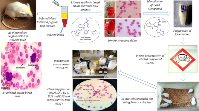
The difficulty of developing an efficient malaria vaccine along with increasing spread of multidrug resistant strain of Plasmodium falciparum to the available antimalarial drugs poses the need to discover safe and efficacious antimalarial drugs to control malaria. An alternative strategy is to synthesize compounds possessing structures similar to the active natural products or marketed drugs. Several biologically active natural products and drugs contain β-carboline moiety. In the present study, few selected β-carboline derivatives have been synthesized and tested for their in vitro and in vivo antiplasmodial activity against the rodent malaria parasite Plasmodium berghei (NK-65). The designed analogs exhibited considerable in vitro antimalarial activity. Two compounds (1R,3S)-methyl 1-(benzo[d][1,3]dioxol-5-yl)-2,3,4,9-tetrahydro-1H-pyrido[3,4-b]indole-3-carboxylate (9a) and (1R,3S)-methyl 1-(pyridin-3-yl)-2,3,4,9-tetrahydro-1H-pyrido[3,4-b]indole-3-carboxylate (9b) were further selected for in vivo studies. Both the lead compounds (9a and 9b) were observed to be safe for oral administration. The therapeutic effective dose (ED50) for 9a and 9b were determined and in the animal model, 9a (at 50 mg/kg dose) exhibited better activity in terms of parasite clearance and enhancement of host survival. Biochemical investigations also point toward the safety of the compound to the hepatic and renal functions of the rodent host. Further studies are underway to explore its activity alone as well as in combination therapy with artesunate against the human malaria parasite P. falciparum.
1. Introduction
Despite many efforts, malaria is one of the most significant infectious diseases, characterized by an intrinsic ability to acquire resistance against drugs. As per the World Malaria Report 2017, there were 216 million cases and 445 000 deaths from the disease globally in 2016 with Plasmodium falciparum, accounting for maximum (99%) cases of malaria in the African region.1 For malaria control, multifaceted approaches with a wide range of effective tools such as vector control, vaccines, and chemotherapy have been used worldwide. The antimalarial drug chloroquine (CQ) played a very significant role in controlling of malaria till the development of resistant strains of the parasite. The absence of an effective malaria vaccine, mutation in the vector, and resistance of the parasite to the available antimalarial drugs urge the need to look for newer approaches to combat the disease. An alternative strategy is to synthesize compounds possessing structures similar to the active natural products or marketed drugs.
1,2,3,4-Tetrahydro-β-carbolines (THβC’s) possess a wide range of biological activities and constantly studied for their use in novel pharmacological applications.2,3 THβC is a privileged structure present in several drugs currently available in the market as well as observed in many drug candidates under the development. Many pharmacologically interesting compounds comprise THβC scaffold4−9 and also represent an important moiety for the discovery of potent antimalarial drugs.10 Among the natural products, manzamine A, a β-carboline alkaloid isolated from marine sponge in 1986, demonstrated potent antiplasmodial activity both in vitro and in vivo.11 Whereas, moderate antimalarial activity in alkyl guanidine-substituted β-carboline derivatives was observed by Chan et al.6 The synthetically and biologically interesting subgroup among the THβC’s is the optically active C1–C3-substituted THβC’s.
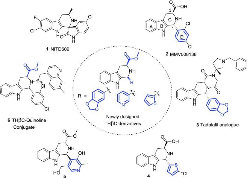
NITD609 1 (also named cipargamin), a C1–C3 substituted THβC analogue (Figure 1), was identified using high-throughput screening as a new class of potent and orally efficacious compound for the treatment of malaria and is currently undergoing phase II clinical trial.12,13 This compound is known to act against P. falciparum with a mechanism distinct from that of the existing antimalarial drugs.14
Figure 1.
C1–C3-substituted THβC’s as the antimalarial agent.
Similarly, MMV008138 2, a C1–C3 substituted THβC analogue, having potent antimalarial activity (showing >95% growth inhibition at 5 μM), was discovered during both in vivo and in vitro screening of 400 compounds within the malaria box and exhibited >60% rescue following the addition of 200 μM isopentenyl pyrophosphate.15 During this investigation, Yao et al. reported in vitro structure–activity relationship (SAR) studies of 2 and revealed that 1R,3S-stereochemistry at C1–C3 position is important for better potency and improved growth inhibition. Later, by more studies they found that the active compound specifically targets the enzyme 2-C-methyl-d-erythritol-4-phosphate cytidylyltransferase (PfIspD) in the MEP pathway.16 Another C1–C3-substituted THβC showing potent antimalarial activity is C1-piperonyl derivative 3 (discovered by SAR studies of tadalafil derivative on P. falciparum strain),17 C1-thiophene derivative 4,16 C1-pyridine derivative 5,18 and THβC–quinoline conjugate 6.8 It is noteworthy to mention that only the lead compounds 1 and 2 were investigated to show their in vivo efficacy in animal models, whereas none of the other active THβC antimalarials were further investigated in vivo.
On the basis of this systematic literature survey and the antimalarial activity of the known THβC’s, a small library of C1-pyridyl, thiophene, and piperonyl-substituted THβC’s were synthesized from methyl ester derivative of l-tryptophan (an essential amino acid) using classic Pictet–Spengler reaction. Both cis- and trans-isomers of C1, C3-substituted THβC’s as well as fully aromatic β-carboline analogue were synthesized to check the effect of stereochemistry at C1 position on the biological activity. In addition, both cis and trans, C1 n-hexyl derivatives were synthesized and evaluated for their antimalarial activity against murine malaria. In vitro antiplasmodial activity of these derivatives was assessed against Plasmodium berghei by WHO method based on schizont maturation inhibition. The combination of lead THβC with commercially available drug leucovorin (10 mg/kg) has also been tested in vivo. The limit test of Lorke was used to assess the acute toxicity of the compounds. In vivo suppressive activity against P. berghei was assessed and the ED50 (effective dose) and therapeutic index were also calculated. Furthermore, the biochemical assays were performed to monitor the hepatic and renal toxicity.
2. Results and Discussion
2.1. Design and Chemistry
The THβC scaffold is found in a wide range of natural products with interesting biological properties and attracted great attention of synthetic chemist for many years. In vitro SAR studies available on THβC’s shown in Figure 1 clearly demonstrated the importance of 1R,3S-stereochemistry in this scaffold for the antimalarial activity.15,16 It was also evident that the C1 aromatic substituents improve the activity, whereas incorporation of aliphatic substituents at this position abrogates the antimalarial property. The presence of carbonyl group at C3 position was also found to be important for the enhancement of activity.16
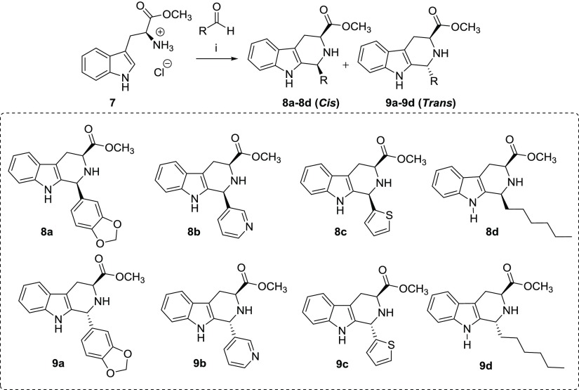
On the basis of the lack of in vivo data on this scaffold and to study the effect of stereochemistry as well as substituents at C1 and C3 position of THβC’s and to investigate this scaffold in combination therapy for malaria, we synthesized a set of eight THβC derivatives including both cis- and trans-isomers by reaction between methyl ester derivative of l-tryptophan and aldehydes containing medicinally important heterocyclic rings such as benzo[1,3]dioxole (8a and 9a), pyridine (8b and 9b) and thiophene (8c and 9c) rings. The selected heterocycles are privileged structures and found in several drugs such as tadalafil, niacin, clopidogrel, and many more.19,20 Additional aliphatic hydrocarbon chain-substituted THβC’s (8d and 9d) were also synthesized to confirm the exact requirement of C1 aromatic substitution for the desired antimalarial activity (Scheme 1).
Scheme 1. Synthesis of C1–C3-Substituted THβC Derivatives (i) Piperonal/Pyridine 3-Carboxaldehyde/Thiophene 2-Carboxaldehyde/Heptanal (1.2 equiv), Isopropanol, Reflux, 8 h.
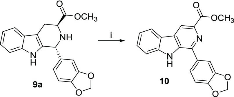
The most efficient and short route for the synthesis of this scaffold is stereoselective Pictet–Spengler reaction. Alternatively, the Bischler–Napieralski reaction to give 3,4-dihydro β-carboline followed by reduction to THβC is also possible. To synthesize C1–C3 substituted optically active THβC derivatives, l-tryptophan was treated with freshly distilled trimethyl silyl chloride in dry methanol at room temperature (RT), which furnished methyl ester derivative in 90% yield.21 Thionylchloride with methanol in refluxing condition also produced the desired product in 95% yield.22 Having synthesized a methyl ester derivative, the next target was to synthesize both cis- and trans-isomers of THβC derivatives in equal ratio and good yield for the ease of purification. In the Pictet–Spengler reaction, the cis product is predominantly formed under kinetically controlled conditions and selectivity switch toward trans-isomer under thermodynamically controlled conditions. The nature of reagents and temperature has a dramatic effect on kinetic and thermodynamic control of the reaction. When the reaction was executed in most commonly used reaction condition [trifluoroacetic acid (TFA) and dry dichloromethane (DCM) at 0 °C to RT], formation of both isomers of THβC (cis/trans, 80:20) was observed but the starting material was not completely consumed even after addition of excess amount of TFA and long reaction time. The overall yield of this reaction was also observed to be very poor. This investigation resulted in the isolation of desired product, and interestingly, it was found that the retention factor for the trans-isomer on thin layer chromatography (TLC) was less as compared to that of cis-isomers. The high mobility of cis-isomer was attributed to the low absorptivity of cis-isomer on the silica gel as a result of steric inaccessibility of polar groups.23 In 1H NMR, proton at C1 position of the trans-isomer was showing downfield signal as compared to that of cis-isomer.24 In an another attempt, the use of 10% TFA in water did not produce any product in this reaction,25 whereas Et3N in DCM resulted in the formation of only a trace amount of the product.26 The reaction carried out using nitromethane/toluene was good in yield (70%) but resulted in the formation of cis-isomer as a major product (cis/trans ratio was 95:5). Finally, the reaction performed in isopropanol solvent under refluxing condition27 resulted in the formation of desired product in about 85% yield with approximately equal amounts of both the isomers (cis/trans, 40:60). All the Pictet–Spengler reactions were carried out using this method.28 Fully aromatized β carboline derivative 10 was synthesized by reaction of compound 9a with a catalytic amount of iodine (20 mol %) at 150 °C for 6 h in dimethylformamide (DMF) (Scheme 2).29
Scheme 2. Synthesis of Fully Aromatized β-Carboline (10) (i) I2 (20 mol %), DMF, 150 °C, 8 h.
The column chromatographic purification of all the compounds was performed using 230–400 mesh silica gel to isolate the pure isomers, which were characterized by various spectroscopic techniques, and the purity was confirmed by high-performance liquid chromatography–mass spectrometry (HPLC–MS).
2.2. In Vitro Antiplasmodial Efficacy against P. berghei
The compounds were tested for their potential to inhibit P. berghei schizont maturation in vitro. The majority of compounds exhibited significant inhibitory activity against the parasite with IC50 < 5 μg/mL. The standard drug CQ (10 μM) exhibited 90.66 ± 0.48% inhibition, whereas leucovorin (L, 5 μg/mL) exhibited 70.91 ± 2.16% inhibition. The compounds 9a (5 μg/mL) and 9b (5 μg/mL) exhibited 71.79 ± 8.77 and 86.17 ± 13.9% inhibition of P. berghei schizont maturation, respectively (Figure 2). According to the WHO recommendations and other studies, synthetic chemicals/drugs are classified as highly active antimalarials having IC50 values of <5 μg/mL. The promising antimalarials demonstrate activity in the range of 5–15 μg/mL, whereas the compounds with IC50 of 15–50 μg/mL are described as moderately active antimalarials.30 Thus, all the compounds were categorized as highly active against the CQ-sensitive (NK-65) strain of rodent malaria parasite P. berghei. On the basis of these results, two compounds 9a and 9b were selected for assessment of antimalarial activity in vivo using rodent malaria model.
Figure 2.
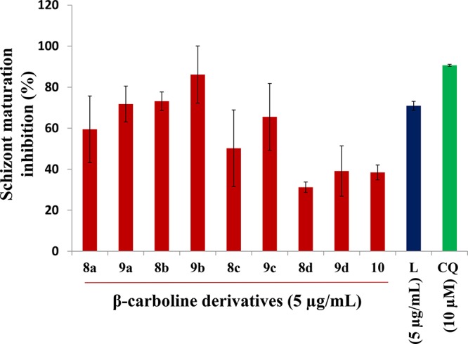
Graph showing the effect of β carboline derivatives (5 μg/mL) on schizont maturation of P. berghei in vitro. The standard drug CQ (10 μM) exhibited 90.66 ± 0.48% inhibition, whereas leucovorin (L, 5 μg/mL) exhibited 70.91 ± 2.16% inhibition.
Rodent-infecting Plasmodium species are recognized as valuable experimental models to study various aspects of mammalian malaria,31 such as their developmental biology, interactions between the parasite and its host, development of vaccines, and testing of novel drug candidates. This is because of their conserved genome organization, basic biology, house-keeping genes, and biochemical processes, as well as similar characteristic patterns of drug-sensitivity and resistance. Also, techniques are available for their genetic modification and their life-cycle stages can be easily manipulated.32 Rodent malaria parasites are very easy to maintain in vitro in cultures and in vivo in laboratory animals. P. berghei is also considered a useful model for studying Plasmodium vivax infection and alternative forms of CQ resistance.33,34
2.3. Acute Toxicity
The safety of any compound exhibiting good antiplasmodial activity is a major concern during antimalarial drug development. The compounds were evaluated for their in vivo acute toxicity using the limit test of Lorke, which is the standard test for evaluation of acute toxicity of any compound/drug.35 Mortality of mice was evident at a dose of 5 g/kg, which is the highest testing dose limit for any drug/compound in rodents. At 4 g/kg of 9a and 2.5 g/kg of 9b, all the mice survived longer than the specified 14 day test duration, which illustrated the safety of these compounds at these doses. No behavioral changes were observed within 2 h of 9b administration. However, abdominal contractions respiratory problems and swelling in the hind foot were observed in mice after administration of 9a. All these side effects disappeared within 2 weeks of dose administration. On the basis of these dose values and in vitro activity data, two different concentrations (50 and 100 mg/kg) were selected for in vivo suppressive activity of 9a and 9b against P. berghei.
2.4. In Vivo Suppressive Activity against P. berghei
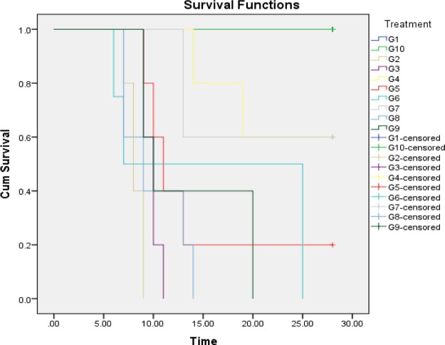
Compound 9a (50 mg/kg) in monotherapy (G4) and its combination with leucovorin (G7) exhibited considerable chemotherapeutic activity in vivo with 62.9 and 63.97% chemosuppression, respectively (Table 1). The ED50 of the compound was determined to be <50 mg/kg. The therapeutic index was determined to be >80. Combination of 9a with leucovorin also showed considerable activity (ED50 < 50 mg/kg) against the parasite in vivo. The % parasitaemia was observed to decline in all 9a-treated groups by day 28 post inoculation. Very low infection of 2.43 ± 0.51% (G4), 0.73 ± 0% (G5), and 3 ± 1% (G7) were recorded in the surviving mice on day 28. Mean survival time (MST) of 23.4 ± 6.54 days was recorded at 50 mg/kg of the compound 9a (Figure 3), which was extremely statistically significant as compared to the infected control (8.2 ± 0.83 days) (Figure 4). Leucovorin (10 mg/kg) and CQ (10 mg/kg) exhibited MST of 15.75 ± 10.68 and 28 ± 0 days, respectively. Behavioral changes such as lethargy and reduction in locomotion were not evident at 50 mg/kg of the compound and in the combination therapy-treated mice (Table 2). Some physical signs of illness were seen at higher dose (100 mg/kg).
Table 1. Suppressive Activity of Different Concentrations of 9a and 9b against P. berghei Infectiona.
| groups n = 5 | dose (D0–D3) (0.2 mL/mouse/OD/oral) | parasitaemia (%), day 5 | chemosuppression (%), day 5 | parasitaemia (%), day 28 |
|---|---|---|---|---|
| G1 | normal control | |||
| G2 | infected control | 29.76 ± 8.70 | ||
| G3 | vehicle control | 30.7 ± 5.22 | ||
| G4 | 9a (50 mg/kg) | 11.07 ± 3.85** | 62.9 | 2.43 ± 0.51 |
| G5 | 9a (100 mg/kg) | 16.36 ± 7.94* | 45.02 | 0.73 ± 0 |
| G6 | leucovorin (10 mg/kg) | 9.32 ± 6.14** | 68.68 | |
| G7 | 9a (50 mg/kg) + leucovorin (10 mg/kg) | 10.79 ± 4.80** | 63.97 | 3 ± 1 |
| G8 | 9b (50 mg/kg) | 15.17 ± 6.26* | 49.02 | |
| G9 | 9b (100 mg/kg) | 8.83 ± 3.07*** | 70.32 | |
| G10 | positive control, CQ (10 mg/kg) | 1.68 ± 1.06*** | 94.35 | 0 ± 0 |
Data are expressed as mean ± standard deviation (SD) for five mice per group as compared to infected control. p-Value in comparison to infected control is shown as ***p < 0.0005, extremely statistically significant; **p < 0.005, very statistically significant; and *p < 0.05, statistically significant.
Figure 3.
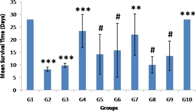
MST of various mice groups in the suppressive test. Data are expressed as mean ± SD for five mice per group as compared to infected control. p-Value in comparison to infected control is shown as ***p < 0.0005, extremely statistically significant; **p < 0.005, very statistically significant; #p > 0.05, not statistically significant.
Figure 4.
Kaplan Meier survival analysis of mice in various treatment groups.
Table 2. Physical Symptoms of Illness in 9a- and 9b-Treated and Malaria-Infected Micea.
| monotherapy |
|||||||
|---|---|---|---|---|---|---|---|
|
9a |
9b |
combination therapy | |||||
| clinical symptoms appeared in test population | DPI | NC | 50 mg/kg | 100 mg/kg | 50 mg/kg | 100 mg/kg | 9a + L |
| behavioural lethargy | 5 | + | – | + | + | + | – |
| 7 | +++ | – | + | +++ | ++ | – | |
| 14 | ××× | – | ××× | ××× | +++ | – | |
| 21 | ××× | – | ××× | ××× | ××× | – | |
| 28 | ××× | – | ××× | ××× | ××× | – | |
| reduction in movement | 5 | + | + | ++ | + | + | – |
| 7 | ++ | – | ++ | +++ | ++ | – | |
| 14 | ××× | – | ××× | ××× | +++ | – | |
| 21 | ××× | – | ××× | ××× | ××× | – | |
| 28 | ××× | – | ××× | ××× | ××× | – | |
| passage of dark urine | 5 | + | – | – | + | + | – |
| 7 | + | – | – | + | ++ | – | |
| 14 | ××× | – | ××× | ××× | ++ | – | |
| 21 | ××× | – | ××× | ××× | ××× | – | |
| 28 | ××× | – | ××× | ××× | ××× | – | |
| impaired consciousness | 5 | – | – | – | + | + | – |
| 7 | – | – | – | + | ++ | – | |
| 14 | ××× | – | ××× | ××× | ++ | – | |
| 21 | ××× | – | ××× | ××× | ××× | – | |
| 28 | ××× | – | ××× | ××× | ××× | – | |
Indicator: (−), absent; (+), mild; (++), moderate; (+++), severe; mortality; (×××), DPI: day post inoculation; and NC: negative control.
Compound 9b-treated groups, G8 (50 mg/kg) and G9 (100 mg/kg), recorded dose-dependent suppressive activity against P. berghei infection with 49.02 and 70.32% chemosuppression, respectively, on day 5. The ED50 of the compound was calculated to be =51.8 mg/kg. The therapeutic index was determined to be =48.26. However, all the mice in both groups died by day 14 post-inoculation recording a MST of 10 ± 3.31 and 13.6 ± 5.85 days, respectively (Figure 3). Mice treated with the standard drug CQ exhibited a MST of 28 ± 0 days (Figure 4). No behavioral changes were observed in 9b-treated groups at a dose of 100 mg/kg (Table 2).
The results of in vivo suppressive activity have been classified in accordance with the classification given by Muñoz et al., which categorizes antiplasmodial activity of a plant extract/synthetic compounds as very good, good, moderate, and inactive if it displays a percent inhibition equal to or more than 50% at concentration of 100, 250 mg/kg, between 500–1000, and above 1000 mg/kg, respectively.36 On the basis of this classification, both 9a and 9b can be categorized as very good antimalarials. However, the compound 9a exhibited better in vivo activity in terms of parasite clearance and enhancement of the MST of the rodent host at 50 mg/kg. The mortality of rodent host at higher concentration (100 mg/kg) can be due to the high drug pressure in the infected condition. The mice were observed to be anemic with decreased body weight. Infection was seen primarily in reticulocytes. The changes in the behavioral patterns were not much evident in the 9a-treated mice as compared to the negative control indicative of suppression of parasite development. In the negative control, these symptoms were evident due to anemia and reticulocytosis.
It is not necessary that compounds exhibiting good inhibition of parasite in vitro would always be active in rodent models.37 The compound 9b showing better inhibition of parasite in vitro did not show promising results in vivo suggesting a specific role of C1 pyridine-functionalized THβC in the observed toxicity. The drug leucovorin is a reduced form of folic acid. It is readily converted to other reduced folic acid derivatives and is known to increase the bioavailability of compound/drug. The combination of 9a with leucovorin also exhibited very good antiplasmodial efficacy with very low parasitaemia levels in surviving mice on day 28. Such findings illustrate the potential of this combination as an effective antimalarial.
2.5. Biochemical Assays
Many enzymes act as markers of disease states and their levels in the intracellular fluids are used in the diagnosis of diseases. Serum levels of alkaline phosphatase (ALP), serum glutamate oxaloacetate transaminase (SGOT), serum glutamate pyruvate transaminase (SGPT), and bilirubin levels are used as biomarkers of hepatic function, whereas urea and creatinine are used for evaluation of renal function.
2.5.1. Liver Function Tests
Impairment of normal hepatic function is most commonly observed in malaria infection. Significant elevations in the serum level of liver enzymes such as SGOT, SGPT, and ALP are observed in malaria infection.38 One of the factors responsible for impairment of liver function in acute malaria infection is centrilobular liver damage, which results in hyper bilirubinaemia.39 The results of the present study are consistent with these reports as significantly (p < 0.0005) elevated ALP, SGOT, SGPT, and bilirubin levels were observed in the infected control (G2) as compared to normal mice. In 9a-treated mice (G4–G8), serum ALP activity and bilirubin level were significantly (p < 0.0005) lower than the infected control on day 10 (Table 3). Slight increase in the bilirubin level (p < 0.05) was evident among the mice treated with 9b (G8, G9) on day 10 (Table 2). In the surviving mice of 9a and 9a + L-treated groups, a decrease in ALP and bilirubin levels was observed on day 28 post-inoculation. However, a slight increase in bilirubin levels was seen in surviving mice of combination therapy group (G7) on day 28.
Table 3. Liver Function Tests of Mice Treated with 9a and 9b on Day 10 and Survivors of Experimental Groups on Day 28a.
| groups n = 5 | dose (D0–D3) (0.2 mL/mouse/OD/oral) | ALP (IU/L) | bilirubin (mg/dL) | SGOT (IU/L) | SGPT (IU/L) | |
|---|---|---|---|---|---|---|
| G1 | normal control | 44.41 ± 11.51 | 0.43 ± 0.14 | 38.40 ± 4.94 | 68.72 ± 8.05 | |
| G2 | infected control | 209.33 ± 9.44*** | 1.56 ± 0.28*** | 101.79 ± 5.31*** | 119.97 ± 8.87*** | |
| G3 | vehicle control (SSV) | 195.6 ± 7.87*** | 1.67 ± 0.20*** | 104.56 ± 9.28*** | 120.50 ± 4.75*** | |
| G4 | 9a (50 mg/kg) | day 10 | 110.55 ± 0.81*** | 0.97 ± 0.007*** | 90.5 ± 3.53** | 83.31 ± 2.65*** |
| day 28 | 100.19 ± 2.94 | 0.85 ± 0.07 | 79.59 ± 0.79 | 74.89 ± 3.77 | ||
| G5 | 9a (100 mg/kg) | day 10 | 109.54 ± 4.94*** | 1.03 ± 0.20** | 97.92 ± 1.98# | 97.13 ± 9.80** |
| day 28 | 99.31 ± 0 | 1.03 ± 0 | 100.1 ± 0 | 95.67 ± 0 | ||
| G6 | leucovorin (L) (10 mg/kg) | day 10 | 113.35 ± 1.10*** | 1.2 ± 0.14* | 116.54 ± 1.50*** | 105.55 ± 3.50** |
| G7 | 9a (50 mg/kg) + L (10 mg/kg) | day 10 | 105.71 ± 2.57*** | 1.05 ± 0.09** | 93.56 ± 4.90* | 84.81 ± 1.83*** |
| day 28 | 97.31 ± 2.58 | 1.13 ± 0.07 | 80.11 ± 2.84 | 73.55 ± 3.57 | ||
| G8 | 9b (50 mg/kg) | day 10 | 121.80 ± 1.98*** | 1.21 ± 0.10* | 96.91 ± 3.59# | 91.23 ± 2.99*** |
| G9 | 9b (100 mg/kg) | day 10 | 129.60 ± 2.22*** | 1.24 ± 0.08* | 97.3 ± 9.33# | 104.08 ± 5.68** |
| G10 | CQ (10 mg/kg) | day 10 | 92.13 ± 3.42*** | 1.06 ± 0.46* | 59.96 ± 4.54*** | 79.34 ± 10.34*** |
| day 28 | 92.61 ± 6.35 | 0.9 ± 0.39 | 55.8 ± 6.47 | 81.86 ± 9.65 | ||
Data are expressed as mean ± SD for five mice per group as compared to infected control. p-Value in comparison to infected control is shown as ***p < 0.0005, extremely statistically significant; **p < 0.005, very statistically significant; *p < 0.05, statistically significant; and #p > 0.05, not statistically significant.
Activity of serum SGOT was significantly lower than the infected control on day 10 in 9a (p < 0.005) and its combination therapy (p < 0.05)-treated groups. However, a slight increase in SGOT activity was evident at the higher dose (100 mg/kg). In 9b-treated mice, a rise in SGOT activity (p > 0.05) was evident. Serum SGPT activity was significantly (p < 0.005) lower than the infected control in all compound-treated groups on day 10. Levels of both the enzymes were observed to decline in the surviving mice of 9a (50 mg/kg) and its combination (G7) on day 28 (Table 3). In these groups, SGPT activity was observed to be lower than the CQ-treated mice (81.86 ± 9.65 IU/L) on day 28. The significantly lower levels of these hepatic function biomarkers in the 9a-treated groups as compared to the infected control point to the safety of this compound to the liver function of the rodent host.
2.5.2. Kidney Function Tests
Acute renal failure is evident in severe malaria infection caused by P. falciparum and P. vivax,40 characterized by a rise in serum levels of urea and creatinine. The levels of these biomarkers are used for the assessment of kidney function. In the present study, a significant increase (p < 0.0005) in serum urea and creatinine level was recorded in negative control indicating impairment of renal function. Among the treated mice, a rise in the serum creatinine level was observed at higher concentration (100 mg/kg) of both the compounds- and leucovorin-treated groups on day 10 (Figure 5A). Serum urea levels were significantly (p < 0.05) lower than the infected control (G2) on day 10 at low dose of 9a (G4)- and its combination-treated groups (G7) (Figure 5B). Elevation in urea levels was observed (p > 0.05) in 9b-treated groups on day 10. In the surviving mice (G4, G7), the serum creatinine and urea levels were observed to decrease by day 28. These findings highlight the safety of compound 9a to kidney function of the host at lower doses.
Figure 5.
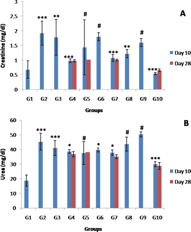
Serum creatinine [A] and urea [B] concentration in 9a- and 9b-treated mice on day 10 and survivors of experimental groups on day 28. Data are expressed as mean ± SD for five mice per group as compared to infected control. p-Value in comparison to infected control is shown as ***p < 0.0005, extremely statistically significant; **p < 0.005, very statistically significant; *p < 0.05, statistically significant; and #p > 0.05 not statistically significant.
3. Conclusions
The current study highlights the substantial potential of THβC’s as a novel antimalarial agent along with their safety on the hepatic and renal functions of the rodent host. The majority of the β carboline derivatives exhibited prominent in vitro activity against the parasite. The compound 9a and its combination with leucovorin exhibited best antimalarial activity in vivo in terms of parasite clearance as well as enhancement of survival of the rodent host. The compound is also safe to the hepatic and renal functions of the host as evident from biochemical studies. Further studies are underway to assess its potential against the human malaria parasite P. falciparum and its combination therapy with artesunate. The encouraging results of this preliminary antimalarial activity screening and in vivo efficacy highlights the need for further detailed analysis of THβC’s to provide greater insights into their antiplasmodial potential.
4. Experimental Methods
4.1. General
Commercially available reagents were used without further purification. TLC was carried out on Merck silica gel F254 aluminum sheets and visualized under UV light at 254 and/or 360 nm and/or by chemical staining with ninhydrin or iodine. Diastereomers were separated by flash column chromatography using 230–400 mesh size silica gel (fisher). 1H and 13C spectra were recorded on a Bruker Avance II 400 MHz spectrophotometer in CDCl3 and DMSO-d6 or using mixture of these solvents. IR spectra were recorded on a Thermo Scientific Nicolet 50 FT-IR system. Mass spectrometry was carried out using a Waters Q-TOF micromass system. Chemical shifts are reported in parts per million (ppm) relative to tetramethylsilane (TMS) as the internal standard. Following abbreviations were used in reporting spectra: s (singlet), d (doublet), t (triplet), m (multiple).
4.2. General Procedure for the Synthesis of 8a and 9a
l-Tryptophan (1 g, 4.89 mmol) was taken in a round bottom flask, freshly distilled TMSCl (1.23 mL, 9.88 mmol) was added followed by the addition of dry methanol (20 mL), and the resulting solution was stirred at RT. After completion of the reaction (monitored by TLC), the mixture was concentrated on a rotary evaporator and coevaporated 3–4 times with methanol to give l-tryptophan methylester hydrochloride (7) in 96% yield (1.2 g, 4.71 mmol). To a solution of pipernol (707 mg, 4.711 mmol, 1.2 equiv) in 10 mL of isopropanol, l-tryptophan methylester hydrochloride 7 (1 g, 3.926 mmol) was added, and the resulting suspension was heated at reflux. After the completion of reaction (monitored by TLC, approximate 8 h), the mixture was concentrated on a rotary evaporator. The reaction mixture was further basified with saturated solution of aq K2CO3 (40 mL) and extracted with ethylacetate. The organic layer was washed with brine, dried over anhydrous Na2SO4, and evaporated to give mixture of 8a and 9a, which were separated using flash column chromatography (silica gel. 230–400).
4.2.1. (1S,3S)-Methyl-1-(benzo[d][1,3]dioxol-5-yl)-2,3,4,9-tetrahydro-1H-pyrido[3,4-b]indole-3-carboxylate (8a)
Yield 30%, off-white solid, Rf = 0.40 (ethyl acetate/hexanes, 55%), mp 100–105 °C, IR (neat) ν: 3363, 3011, 2953, 2909, 1727 cm–1. 1H NMR (400 MHz, CDCl3): δ 7.52 (d, J = 7.2 Hz, 1H), 7.47 (s, 1H), 7.22–7.20 (m, 1H), 7.15–7.09 (m, 2H), 6.87 (dd, J = 8.0, 1.6 Hz, 1H), 6.80 (m, 2H), 5.94 (s, 2H), 5.16 (s, 1H), 3.94 (dd, J = 12.4, 4.4 Hz, 1H), 3.80 (s, 3H), 3.20 (ddd, J = 15.2, 4, 1.6 Hz, 1H), 3.02–2.94 (m, 1H). ESI-MS m/z: 351.13 [M + H]+ (1H NMR data and melting point were consistent with that reported in the literature.41 Purity analysis is reported in the Supporting Information file).
4.2.2. (1R,3S)-Methyl-1-(benzo[d][1,3]dioxol-5-yl)-2,3,4,9-tetrahydro-1H-pyrido[3,4-b]indole-3-carboxylate (9a)
Yield 25%, off-white solid, Rf = 0.25 (ethyl acetate/hexane, 50%), mp 160–170 °C, IR (neat) ν: 3397, 3313, 2958, 2933, 1724 cm–1. 1H NMR (400 MHz, CDCl3): δ 7.60 (s, 1H), 7.54 (d, J = 8 Hz, 1H), 7.24–7.22 (m, 1H), 7.14 (dtd, J = 16, 6.8, 1.2 Hz, 2H), 6.75 (s, 3H), 5.93 (s, 2H), 5.33 (s, 1H), 3.98 (t, J = 6.4 Hz, 1H), 3.71 (s, 3H), 3.25 (ddd, J = 15.4, 5.4, 1.1 Hz, 1H), 3.12 (ddd, J = 15.4, 6.6, 1.4 Hz, 1H). ESI-MS m/z: 351.13 [M + H]+. (1H NMR data and melting point were consistent with that reported in the literature.41 Purity analysis is reported in the Supporting Information file).
4.2.3. (1S,3S)-Methyl-1-(pyridin-3-yl)-2,3,4,9-tetrahydro-1H-pyrido[3,4-b]indole-3-carboxylate (8b)
Yield 32%, off-white solid, Rf = 0.47 (MeOH/DCM, 5%), IR (neat): ν 3328, 3146, 3056, 2923, 2851, 1731, 1583 cm–1. mp 235–240 °C, 1H NMR (400 MHz, CDCl3): δ 10.26 (s, 1H), 8.64 (d, J = 1.8 Hz, 1H), 8.55 (dd, J = 4.8, 1.7 Hz, 1H), 7.98 (s, 1H), 7.71 (dt, J = 7.9, 1.9 Hz, 1H), 7.44 (d, J = 7.1 Hz, 11H), 7.33 (dd, J = 7.7, 4.9 Hz, 1H), 7.26–7.20 (m, 1H), 7.04–6.96 (m, 2H), 5.31 (s, 1H), 3.93 (dd, J = 11.1, 4.1 Hz, 1H), 3.77 (s, 3H), 3.15 (ddd, J = 14.8, 4.0, 1.7 Hz, 1H), 2.96–2.89 (m, 1H). ESI-MS m/z: 308.13 [M + H]+. (1H NMR data and melting point were consistent with that reported in the literature.24 Purity of analysis is reported in the Supporting Information file).
4.2.4. (1R,3S)-Methyl 1-(pyridin-3-yl)-2,3,4,9-tetrahydro-1H-pyrido[3,4-b]indole-3-carboxylate (9b)
Yield 38%, off-white solid, Rf = 0.38 (MeOH/DCM, 5%), IR (neat): ν 3288, 3178, 2921, 2851, 1739, 1588 cm–1. mp 218–224 °C, 1H NMR (400 MHz, CDCl3): δ 8.55 (d, J = 2.0 Hz, 1H), 8.47 (dd, J = 4.8, 1.6 Hz, 1H), 8.18 (s, 1H), 7.59–7.54 (m, 2H), 7.26–7.24 (m, 1H), 7.22–7.11 (m, 3H), 5.46 (s, 1H), 3.97–3.94 (m, 1H), 3.72 (s, 3H), 3.28 (ddd, J = 15.2, 5.6, 1.2 Hz, 1H), 3.17 (ddd, J = 15.6, 6.4, 0.4 Hz, 1H). ESI-MS m/z: 308.14 [M + H]+. (1H NMR data and melting point were consistent with that reported in the literature.24 Purity analysis is reported in the Supporting Information file).
4.2.5. (1R,3S)-Methyl-1-(thiphen-2-yl)-2,3,4,9-tetrahydro-1H-pyrido[3,4-b]indole-3-carboxylate (8c)
Yield 25%, off-white solid, Rf = 0.40 (ethyl acetate/hexanes, 30%), IR (neat): ν 3379, 3328, 2929, 2782, 1735 cm–1. 1H NMR (400 MHz, CDCl3): δ 7.60 (s, 1H), 7.53 (d, J = 7.2 Hz, 1H), 7.32 (dd, J = 5.2, 1.2 Hz, 1H), 7.24 (d, J = 1.2 Hz, 1H), 7.18–7.10 (m, 3H), 7.03 (dd, J = 5, 3.6 Hz, 1H), 5.61 (s, 1H), 3.97 (dd, J = 11.2, 4.4 Hz, 1H), 3.82 (s, 3H), 3.21 (ddd, J = 15.2, 4, 1.6 Hz, 1H), 3.00 (ddd, J = 15.1, 11.2, 2.5 Hz, 1H). ESI-MS m/z: 313.10 [M + H]+. (1H NMR analysis was consistent with that reported in the literature.26 Purity analysis is reported in the Supporting Information file).
4.2.6. (1S,3S)-Methyl-1-(thiophen-2-yl)-2,3,4,9-tetrahydro-1H-pyrido[3,4-b]indole-3-carboxylate (9c)
Yield 30%, off-white solid, Rf = 0.30 (ethyl acetate/hexanes, 30%), IR (neat): ν 3398, 3317, 2954, 2847, 1723 cm–1. 1H NMR (400 MHz, CDCl3): δ 7.72 (s, 1H), 7.54 (d, J = 8 Hz, 1H), 7.28–7.26 (m, 6H), 7.17–7.12 (m, 2H), 6.98–6.95 (m, 2H), 5.75 (s, 1H), 4.07 (dd, J = 7.6, 5.6 Hz, 1H), 3.74 (s, 3H), 3.25 (dd, J = 15.5, 5.3 Hz, 1H), 3.08 (ddd, J = 15.4, 7.5, 1.4 Hz, 1H). ESI-MS m/z: 313.10 [M + H]+. (1H NMR analysis was consistent with that reported in the literature.26 Purity analysis is reported in the Supporting Information file).
4.2.7. (1S,3S)-Methyl-1-hexyl-2,3,4,9-tetrahydro-1H-pyrido[3,4-b]indole-3-carboxylate Hydrochloride Salt (8d)
Yield 25%, white solid, IR (neat) ν: 3255, 2956, 2922, 2863, 2714, 2603, 2473, 1760, 1454 cm–1. 1H NMR (400 MHz, DMSO-d6 + CDCl3) δ: 11.21 (s, 1H), 9.99 (br, 2H), 7.43 (d, J = 7.8 Hz, 1H), 7.38 (d, J = 8.1 Hz, 1H), 7.11 (t, J = 7.5 Hz, 1H), 7.02 (t, J = 7.4 Hz, 1H), 4.71 (d, J = 6.7 Hz, 1H), 4.46 (dd, J = 11.6, 4.4 Hz, 1H), 3.89 (s, 3H), 3.29 (dd, J = 15.8, 4.6 Hz, 1H), 3.14–3.07 (m, 1H), 2.35–2.29 (m, 1H), 2.06–2.01 (m, 1H), 1.61–1.35 (m, 8H), 0.91 (t, J = 6.7 Hz, 3H). 13C NMR (101 MHz, DMSO-d6 + CDCl3): δ 168.90, 136.49, 129.78, 125.41, 121.71, 119.07, 117.68, 111.39, 104.42, 55.08, 53.79, 52.80, 31.01, 30.93, 28.65, 24.47, 22.26, 22.07, 13.86. ESI-MS m/z: 315.5 [M + H]+.
4.2.8. (1R,3S)-Methyl-1-hexyl-2,3,4,9-tetrahydro-1H-pyrido[3,4-b]indole-3-carboxylate Hydrochloride Salt (9d)
Yield 30%, white solid, IR (neat) ν: 3385, 3067, 2922, 2863, 2725, 2644, 2579, 2434, 1738, 1455 cm–1. 1H NMR (400 MHz, DMSO-d6 + CDCl3) δ: 11.25 (s, 1H), 10.22 (s, 2H), 7.49 (d, J = 7.8 Hz, 1H), 7.37 (d, J = 8.1 Hz, 1H), 7.14–7.10 (m, 1H), 7.04–7.00 (m, 1H), 4.72 (q, J = 6.6 Hz, 2H), 3.76 (s, 3H), 3.42–3.35 (m, 1H), 3.15 (dd, J = 16.0, 6.5 Hz, 1H), 2.12–2.04 (m, 2H), 1.59–1.53 (m, 2H), 1.35–1.30 (m, 6H), 0.89 (t, J = 6.6 Hz, 3H). 13C NMR (101 MHz, DMSO-d6 + CDCl3) δ: 169.04, 136.29, 129.97, 125.53, 121.86, 119.03, 118.04, 111.42, 103.66, 52.99, 51.75, 51.30, 32.04, 30.93, 28.51, 24.87, 22.00, 21.62, 13.92. ESI-MS m/z: 315.5 [M + H]+.
4.3. Procedure for the Synthesis of (10)
To a solution of compound 9a (50 mg, 0.142 mmol) in DMF (5 mL), 30% H2O2 (200 μL) and I2 (8 mg, 20 mol %) were added with continuous stirring at RT and the reaction mixture was further heated at 150 °C for 6 h. After the completion of reaction (monitored by TLC), the reaction mixture was treated with 5% hypo solution (Na2S2O3) and the product was extracted in EtOAc. The organic layer was dried over anhydrous Na2SO4 and concentrated under reduced pressure. The crude product was purified over column chromatography to afford compound 10.
4.3.1. Methyl 1-(benzo[d][1,3]dioxol-5-yl)-9H-pyrido[3,4-b]indole-3-carboxylate (10)
Yield 50%, off-white solid, Rf = 0.50 (ethyl acetate/hexane, 60%), IR (neat) ν: 3338, 2949, 2817, 1710, 1494 cm–1. 1H NMR (400 MHz, DMSO-d6): δ 11.91 (s, 1H), 8.89 (s, 1H), 8.41 (d, J = 7.9 Hz, 1H), 7.70 (d, J = 8.2 Hz, 1H), 7.62–7.54 (m, 3H), 7.32 (t, J = 7.4 Hz, 1H), 7.17 (d, J = 8.5 Hz, 1H), 6.17 (s, 2H), 3.93 (s, 3H). The 1H NMR analysis was consistent with that reported in the literature.42
4.4. Experimental Animals
White Swiss albino mice, Mus musculus of Laca strain 4–6 weeks old, weighing 25–30 g were used as an experimental model in the present study. Mice were obtained from and kept in the Central Animal House, Panjab University, Chandigarh, India. The animals were kept under controlled temperature and humidity conditions and were fed upon standard pellet diet and water ad libitum. The guidelines of the committee for the purpose of control and supervision on experiments on animals (45/GO/ReBi/S/99/CPCSEA) were followed during the experimental procedures.
4.5. Parasite Strain
Asexual blood stages of CQ-sensitive strain of P. berghei (NK-65) were maintained in vivo in Laca mice. Experimental infections were initiated by intraperitoneal inoculation of 1 × 10⁶P. berghei-infected erythrocytes/reticulocytes in citrate saline from infected to naive mice.
4.6. In Vitro Antiplasmodial Efficacy against P. berghei
In vitro antiplasmodial activity of the compounds was assessed against P. berghei by schizont maturation inhibition assay.43 The stock solution of compounds (10 mg/mL) was prepared by dissolving a known quantity of compound in DMSO (1%). The stock solution was further diluted with culture medium, to make the required concentration (5 μg/mL) of the compounds. CQ (10 μM) was used as the positive control. Complete medium (1 mL; RPMI-1640) supplemented with 10% fetal calf serum and mixture of normal and infected mouse erythrocytes at 2% parasitaemia contained either 10 μL of compounds (different concentrations in duplicate) or standard drug in each well. The titer plate was shaken gently to mix contents. Smears were prepared at 0 h, and the culture plate was incubated at 37 °C in a candle jar (5% CO2, 3% O2, 78% N2) using the Trager and Jensen method.44 The percent schizont maturation inhibition was calculated by the following formula:
4.7. Acute Toxicity
The limit test of Lorke was used to assess the acute toxicity of the compounds.35 The compounds were dissolved in the standard suspension vehicle (SSV) as done in our earlier study.45 Normal female mice were divided into two groups consisting of three mice each. A highest testing dose of 5 g/kg was orally administered to the mice following 4 h fasting. In case of mortality, lower doses of compounds were administered, till LD50 was determined. The mice were kept under observation for 2 weeks after the administration of compounds to assess the various side effects including mortality. Guidelines of the Canadian Council on Animal Care (CCAC) were followed for the acceptable oral dose volumes in rodents (ACC-2012-Tech09).
4.8. In Vivo Suppressive Activity against P. berghei
The suppressive activity of the compounds was evaluated by the Knight and Peters method.46 Mice were divided into 10 experimental groups (G1 to G10) consisting of five mice each (Table 2). The sample size of 5 animals per group was determined based on the calculations done by Charan and Kantharia.47 On day 1 (D0), all the mice were inoculated with 1 × 10⁶P. berghei-infected erythrocytes (except G1) as done in our earlier study.45 Drug treatment was initiated 1 h post-parasite-inoculation and was continued once daily for 4 days (D0–D3). In the combination therapy group, the second drug was administered at an interval of 20 min. Twenty four hours after the completion of 4-day dose regimen, thin blood smears were made from the tail of each mouse on day 5 (D4). The slides were fixed in methanol followed by Giemsa staining. The percent chemosuppression was calculated by using the following formula:
where A is the average parasitaemia in infected control and B is the average parasitaemia in the treated group.
The ED50 representing 50% suppression of parasite due to the compounds was also calculated. The therapeutic index (ratio of LD50 and ED50) for the compounds was also determined.
4.9. Measurements of Basic Parameters
All treated and control group mice were observed visually throughout the experiment for behavioral changes and signs of illness which include lethargy, reduction of movement, passage of dark urine, and impaired consciousness.48 These signs of illness were recorded as either absent (−), mild (+), moderate (++), or severe (+++). Mortality was recorded throughout the experiment.
4.10. Biochemical Assays
The biochemical assays were performed by taking blood from mice by tail vein drainage on day 10 and survivors of experimental groups on day 28. All biochemical assays were carried out using commercially available diagnostic kits (Reckon Diagnostic P. Ltd. Gorwa, Baroda, Gujarat, India and Transasia Bio Medicals Ltd., Baddi, Distt. Solan, Himachal Pradesh, India).
4.10.1. Liver Function Tests
ALP,49 SGOT,50 and SGPT51 activities and bilirubin51 levels were checked in serum of mice for assessment of hepatic function.
4.10.2. Kidney Function Tests
Serum levels of urea52 and creatinine53 were measured for assessment for renal function.
Acknowledgments
D.B.S. is thankful to UGC New Delhi for start-up research grant and DBT New Delhi for the award of Ramalingaswami Fellowship. The support from UGC-CAS, DST-PURSE-II, DST-SAIF, and Panjab University Development fund is gratefully acknowledged. R.S. is thankful to CSIR, New Delhi, for the award of Research Fellowship. N.S.W. is thankful to CSIR for the award of Research Associateship.
Supporting Information Available
The Supporting Information is available free of charge on the ACS Publications website at DOI: 10.1021/acsomega.8b01833.
Complete spectroscopic characterization details of compounds 8a–8d, 9a–9d, and 10 including copies of 1H NMR, IR, and LC–MS; reaction mechanism for the Pictet–Spengler reaction as well as reaction optimization details (PDF)
Author Contributions
§ V.G. and R.S. contributed equally to this work.
The authors declare no competing financial interest.
Supplementary Material
References
- WHO . World Malaria Report 2017; WHO Press: Geneva, 2017; ISBN 978-92-4-156552-3.
- Rao R. N.; Maiti B.; Chanda K. Application of Pictet-Spengler Reaction to Indole-Based Alkaloids Containing Tetrahydro-β-carboline Scaffold in Combinatorial Chemistry. ACS Comb. Sci. 2017, 19, 199–228. 10.1021/acscombsci.6b00184. [DOI] [PubMed] [Google Scholar]
- Laine A.; Lood C.; Koskinen A. Pharmacological Importance of Optically Active Tetrahydro-β-carbolines and Synthetic Approaches to Create the C1 Stereocenter. Molecules 2014, 19, 1544–1567. 10.3390/molecules19021544. [DOI] [PMC free article] [PubMed] [Google Scholar]
- Daugan A.; Grondin P.; Ruault C.; Le Monnier de Gouville A.-C.; Coste H.; Kirilovsky J.; Hyafil F.; Labaudinière R. The Discovery of Tadalafil: A Novel and Highly Selective PDE5 Inhibitor. 1: 5,6,11,11a-Tetrahydro-1H-imidazo[1’,5’:1,6]pyrido[3,4-b]indole-1,3(2H)-dione Analogues. J. Med. Chem. 2003, 46, 4525–4532. 10.1021/jm030056e. [DOI] [PubMed] [Google Scholar]
- Davis R. A.; Duffy S.; Avery V. M.; Camp D.; Hooper J. N. A.; Quinn R. J. (+)-7-Bromotrypargine: an antimalarial β-carboline from the Australian marine sponge Ancorina sp. Tetrahedron Lett. 2010, 51, 583–585. 10.1016/j.tetlet.2009.11.055. [DOI] [Google Scholar]
- Chan S. T. S.; Pearce A. N.; Page M. J.; Kaiser M.; Copp B. R. Antimalarial β-Carbolines from the New Zealand Ascidian Pseudodistoma opacum. J. Nat. Prod. 2011, 74, 1972–1979. 10.1021/np200509g. [DOI] [PubMed] [Google Scholar]
- Gellis A.; Dumètre A.; Lanzada G.; Hutter S.; Ollivier E.; Vanelle P.; Azas N. Preparation and antiprotozoal evaluation of promising β-carboline alkaloids. Biomed. Pharmacother. 2012, 66, 339–347. 10.1016/j.biopha.2011.12.006. [DOI] [PubMed] [Google Scholar]
- Gupta L.; Srivastava K.; Singh S.; Puri S. K.; Chauhan P. M. S. Synthesis of 2-[3-(7-Chloro-quinolin-4-ylamino)-alkyl]-1-(substituted phenyl)-2,3,4,9-tetrahydro-1H-β-carbolines as a new class of antimalarial agents. Bioorg. Med. Chem. Lett. 2008, 18, 3306–3309. 10.1016/j.bmcl.2008.04.030. [DOI] [PubMed] [Google Scholar]
- Rinehart K. L.; Kobayashi J.; Harbour G. C.; Hughes R. G.; Mizsak S. A.; Scahill T. A. Eudistomins C, E, K, and L, potent antiviral compounds containing a novel oxathiazepine ring from the Caribbean tunicate Eudistoma olivaceum. J. Am. Chem. Soc. 1984, 106, 1524–1526. 10.1021/ja00317a079. [DOI] [Google Scholar]
- Yeung B. K. S.; Zou B.; Rottmann M.; Lakshminarayana S. B.; Ang S. H.; Leong S. Y.; Tan J.; Wong J.; Keller-Maerki S.; Fischli C.; Goh A.; Schmitt E. K.; Krastel P.; Francotte E.; Kuhen K.; Plouffe D.; Henson K.; Wagner T.; Winzeler E. A.; Petersen F.; Brun R.; Dartois V.; Diagana T. T.; Keller T. H. Spirotetrahydro β-Carbolines (Spiroindolones): A New Class of Potent and Orally Efficacious Compounds for the Treatment of Malaria. J. Med. Chem. 2010, 53, 5155–5164. 10.1021/jm100410f. [DOI] [PMC free article] [PubMed] [Google Scholar]
- Ashok P.; Ganguly S.; Murugesan S. Manzamine alkaloids: isolation, cytotoxicity, antimalarial activity and SAR studies. Drug Discovery Today 2014, 19, 1781–1791. 10.1016/j.drudis.2014.06.010. [DOI] [PubMed] [Google Scholar]
- IFPMA developing world health partnerships: Novartis R&D for Malaria. Available online: http://partnerships.ifpma.org/partnership/novartis-r-d-for-malaria (accessed on July 23, 2013).
- Rottmann M.; McNamara C.; Yeung B. K. S.; Lee M. C. S.; Zou B.; Russell B.; Seitz P.; Plouffe D. M.; Dharia N. V.; Tan J.; Cohen S. B.; Spencer K. R.; Gonzalez-Paez G. E.; Lakshminarayana S. B.; Goh A.; Suwanarusk R.; Jegla T.; Schmitt E. K.; Beck H.-P.; Brun R.; Nosten F.; Renia L.; Dartois V.; Keller T. H.; Fidock D. A.; Winzeler E. A.; Diagana T. T. Spiroindolones, a potent compound class for the treatment of malaria. Science 2010, 329, 1175–1180. 10.1126/science.1193225. [DOI] [PMC free article] [PubMed] [Google Scholar]
- Bowman J. D.; Merino E. F.; Brooks C. F.; Striepen B.; Carlier P. R.; Cassera M. B. Antiapicoplast and gametocytocidal screening to identify the mechanisms of action of compounds within the malaria box. Antimicrob. Agents Chemother. 2014, 58, 811–819. 10.1128/aac.01500-13. [DOI] [PMC free article] [PubMed] [Google Scholar]
- Yao Z.-K.; Krai P. M.; Merino E. F.; Simpson M. E.; Slebodnick C.; Cassera M. B.; Carlier P. R. Determination of the active stereoisomer of the MEP pathway-targeting antimalarial agent MMV008138, and initial structure-activity studies. Bioorg. Med. Chem. Lett. 2015, 25, 1515–1519. 10.1016/j.bmcl.2015.02.020. [DOI] [PMC free article] [PubMed] [Google Scholar]
- Ghavami M.; Merino E. F.; Yao Z.-K.; Elahi R.; Simpson M. E.; Fernández-Murga M. L.; Butler J. H.; Casasanta M. A.; Krai P. M.; Totrov M. M.; Slade D. J.; Carlier P. R.; Cassera M. B. Biological Studies and Target Engagement of the 2-C-Methyl-d-Erythritol 4-Phosphate Cytidylyltransferase (IspD)-Targeting Antimalarial Agent (1R,3S)-MMV008138 and Analogs. ACS Infect. Dis. 2018, 4, 549–559. 10.1021/acsinfecdis.7b00159. [DOI] [PMC free article] [PubMed] [Google Scholar]
- Beghyn T. B.; Charton J.; Leroux F.; Laconde G.; Bourin A.; Cos P.; Maes L.; Deprez B. Drug to Genome to Drug: Discovery of New Antiplasmodial Compounds. J. Med. Chem. 2011, 54, 3222–3240. 10.1021/jm1014617. [DOI] [PubMed] [Google Scholar]
- Brokamp R.; Bergmann B.; Müller I. B.; Bienz S. Stereoselective preparation of pyridoxal 1,2,3,4-tetrahydro-β-carboline derivatives and the influence of their absolute and relative configuration on the proliferation of the malaria parasite Plasmodium falciparum. Bioorg. Med. Chem. 2014, 22, 1832–1837. 10.1016/j.bmc.2014.01.057. [DOI] [PubMed] [Google Scholar]
- Kumar S. A. Review on the anticonvulsant activity of 1,3-Benzodixolo ring system based compounds. Int. J. Pharma Sci. Res. 2013, 4, 3296–3303. 10.13040/IJPSR.0975-8232.4(9).3296-03. [DOI] [Google Scholar]
- Baumann M.; Baxendale I. R. An overview of the synthetic routes to the best selling drugs containing 6-membered heterocycles. Beilstein J. Org. Chem. 2013, 9, 2265–2319. 10.3762/bjoc.9.265. [DOI] [PMC free article] [PubMed] [Google Scholar]
- Li J.; Sha Y. A Convenient Synthesis of Amino Acid Methyl Esters. Molecules 2008, 13, 1111–1119. 10.3390/molecules13051111. [DOI] [PMC free article] [PubMed] [Google Scholar]
- Alqahtani N.; Porwal S. K.; James E. D.; Bis D. M.; Karty J. A.; Lane A. L.; Viswanathan R. Synergism between genome sequencing, tandem mass spectrometry and bio-inspired synthesis reveals insights into nocardioazine B biogenesis. Org. Biomol. Chem. 2015, 13, 7177–7192. 10.1039/c5ob00537j. [DOI] [PubMed] [Google Scholar]
- Preobrazhenskaya M. N.; Mikhailova L. N.; Chemerisskaya A. A. Thin-layer chromatographic mobility of diastereoisomers. J. Chromatogr. A 1971, 61, 269–278. 10.1016/s0021-9673(00)92419-1. [DOI] [Google Scholar]
- Ungemach F.; Soerens D.; Weber R.; DiPierro M.; Campos O.; Mokry P.; Cook J. M.; Silverton J. V. General method for the assignment of stereochemistry of 1,3-disubstituted 1,2,3,4-tetrahydro-.beta.-carbolines by carbon-13 spectroscopy. J. Am. Chem. Soc. 1980, 102, 6976–6984. 10.1021/ja00543a012. [DOI] [Google Scholar]
- Saha B.; Sharma S.; Sawant D.; Kundu B. Water as an efficient medium for the synthesis of tetrahydro-β-carbolines via Pictet-Spengler reactions. Tetrahedron Lett. 2007, 48, 1379–1383. 10.1016/j.tetlet.2006.12.112. [DOI] [Google Scholar]
- Sharma S. D.; Bhaduri S.; Kaur G. Diastereoselective synthesis of 1,3-disubstituted Tetrahydro-β-carbolines using pictet Spengler reaction in non-acidic aprotic media. Indian J. Chem. 2006, 45B, 2710–2715. [Google Scholar]
- Xiao S.; Lu X.; Shi X.-X.; Sun Y.; Liang L.-L.; Yu X.-H.; Dong J. Syntheses of chiral 1,3-disubstituted tetrahydro-β-carbolines via CIAT process: highly stereoselective Pictet-Spengler reaction of d-tryptophan ester hydrochlorides with various aldehydes. Tetrahedron: Asymmetry 2009, 20, 430–439. 10.1016/j.tetasy.2009.01.026. [DOI] [Google Scholar]
- The mechanism for the Pictet–Spengler reaction as well as the details of the reaction optimization is included in the Supporting Information.
- Meesala R.; Arshad A. S. M.; Mordi M. N.; Mansor S. M. Iodine-catalyzed one-pot decarboxylative aromatization of tetrahydro-β-carbolines. Tetrahedron 2016, 72, 8537–8541. 10.1016/j.tet.2016.10.069. [DOI] [Google Scholar]
- Lekana-Douki J. B.; Bongui J. B.; Oyegue Liabagui S. L.; Zang Edou S. E.; Zatra R.; Bisvigou U.; Druilhe P.; Lebibi J.; Toure Ndouo F. S.; Kombila M. In vitro antiplasmodial activity and cytotoxicity of nine plants traditionally used in Gabon. J. Ethnopharmacol. 2011, 133, 1103–1108. 10.1016/j.jep.2010.11.056. [DOI] [PubMed] [Google Scholar]
- Thurston J. P. The action of antimalarial drugs in mice infected with Plasmodium berghei. Br. J. Pharmacol. 1950, 5, 409–416. 10.1111/j.1476-5381.1950.tb00590.x. [DOI] [PMC free article] [PubMed] [Google Scholar]
- Janse C.General introduction rodent malaria parasite. Leids universitair medisch centrum. Available online: https://www.lumc.nl/org/parasitologie/research/malaria/berghei-model/general-introduction (accessed on March 21, 2018).
- Espinosa D. A.; Yadava A.; Angov E.; Maurizio P. L.; Ockenhouse C. F.; Zavala F. Development of a chimeric Plasmodium berghei strain expressing the repeat region of the P. vivax circumsporozoite protein for in vivo evaluation of vaccine efficacy. Infect. Immun. 2013, 81, 2882–2887. 10.1128/iai.00461-13. [DOI] [PMC free article] [PubMed] [Google Scholar]
- Pisciotta J. M.; Scholl P. F.; Shuman J. L.; Shualev V.; Sullivan D. J. Quantitative characterization of hemozoin in Plasmodium berghei and vivax. Int. J. Parasitol.: Drugs Drug Resist. 2017, 7, 110–119. 10.1016/j.ijpddr.2017.02.001. [DOI] [PMC free article] [PubMed] [Google Scholar]
- Lorke D. A new approach to practical acute toxicity testing. Arch. Toxicol. 1983, 54, 275–287. 10.1007/bf01234480. [DOI] [PubMed] [Google Scholar]
- Muñoz V.; Sauvain M.; Bourdy G.; Callapa J.; Rojas I.; Vargas L.; Tae A.; Deharo E. The search for natural bioactive compounds through a multidisciplinary approach in Bolivia. Part II. Antimalarial activity of some plants used by Mosetene indians. J. Ethnopharmacol. 2000, 69, 139–155. 10.1016/s0378-8741(99)00096-3. [DOI] [PubMed] [Google Scholar]
- Soh P. N.; Benoit-Vical F. Are West African plants a source of future antimalarial drugs?. J. Ethnopharmacol. 2007, 114, 130–140. 10.1016/j.jep.2007.08.012. [DOI] [PubMed] [Google Scholar]
- Onyesom I.; Onyemakonor N. Levels of parasitaemia and changes in some liver enzymes among malarial infected patients in Edo-Delta Region of Nigeria. Curr. Res. J. Biol. Sci. 2011, 3, 78–81. [Google Scholar]
- Adekunle A. S.; Adekunle O. C.; Egbewale B. E. Serum status of selected biochemical parameters in malaria: An animal model. Biomed. Res. 2007, 18, 109–113. [Google Scholar]
- Kochar D. K.; Saxena V.; Singh N.; Kochar S. K.; Kumar S. V.; Das A. Plasmodium vivax Malaria. Emerg. Infect. Dis. 2005, 11, 132–134. 10.3201/eid1101.040519. [DOI] [PMC free article] [PubMed] [Google Scholar]
- Daugan A.; Grondin P.; Ruault C.; Le Monnier de Gouville A.-C.; Coste H.; Linget J. M.; Kirilovsky J.; Hyafil F.; Labaudinière R. The discovery of tadalafil: a novel and highly selective PDE5 inhibitor. 2: 2,3,6,7,12,12a-hexahydropyrazino[1’,2’:1,6]pyrido[3,4-b]indole-1,4-dione analogues. J. Med. Chem. 2003, 46, 4533–4542. 10.1021/jm0300577. [DOI] [PubMed] [Google Scholar]
- Sathish M.; Kavitha B.; Nayak V. L.; Tangella Y.; Ajitha A.; Nekkanti S.; Alarifi A.; Shankaraiah N.; Nagesh N.; Kamal A. Synthesis of podophyllotoxin linked β-carboline congeners as potential anticancer agents and DNA topoisomerase II inhibitors. Eur. J. Med. Chem. 2018, 144, 557–571. 10.1016/j.ejmech.2017.12.055. [DOI] [PubMed] [Google Scholar]
- WHO . In Vitro Micro Test (Mark III) for the Assessment of Response of Plasmodium falciparum to Chloroquine, Mefloquine, Quinine, Amodiaquine, Sulfadoxine, Pyrimethamine and Artemisinin; CTD/MAL/97.20; World Health Organization: Geneva, 2001. [Google Scholar]
- Trager W.; Jensen J. Human malaria parasites in continuous culture. Science 1976, 193, 673–675. 10.1126/science.781840. [DOI] [PubMed] [Google Scholar]
- Walter N. S.; Bagai U.; Kalia S. Antimalarial activity of Bergenia ciliata (Haw.) Sternb. against Plasmodium berghei. Parasitol. Res. 2013, 112, 3123–3128. 10.1007/s00436-013-3487-z. [DOI] [PubMed] [Google Scholar]
- Knight D. J.; Peters W. The antimalarial activity of N-benzyloxydihydrotriazines. Ann. Trop. Med. Parasitol. 1980, 74, 393–404. 10.1080/00034983.1980.11687360. [DOI] [PubMed] [Google Scholar]
- Charan J.; Kantharia N. D. How to calculate sample size in animal studies?. J. Pharmacol. Pharmacother. 2013, 4, 303–306. 10.4103/0976-500x.119726. [DOI] [PMC free article] [PubMed] [Google Scholar]
- Basir R.; Fazalul Rahiman S. S.; Hasballah K.; Chong W. C.; Talib H.; Yam M. F.; Jabbarzare M.; Tie T. H.; Othman F.; Moklas M. A. M.; Abdullah W. O.; Ahmad Z. Plasmodium berghei ANKA infection in ICR mice as a model of cerebral malaria. Iran. J. Parasitol. 2012, 7, 62–74. [PMC free article] [PubMed] [Google Scholar]
- German Society for Clinical Chemistry (GSCC). Recommendations of the German Society for Clinical Chemistry standardization of methods for the estimation of the enzyme activity in biological fluids. J. Clin. Chem. Clin. Biochem. 1972, 8, 182–192. [Google Scholar]
- Bergmeyer H. U. IFCC methods for the measurement of catalytic concentrations of enzymes. Clin. Chim. Acta 1980, 105, 147–154. 10.1016/0009-8981(80)90105-9. [DOI] [PubMed] [Google Scholar]
- Jendrassik L.; Grof P. Quantitative estimation of some biochemicals and biomolecules in serum. Biochem. Z. 1938, 297, 81–89. [Google Scholar]
- Chaney A. L.; Marbach E. P. Modified reagents for determination of urea and ammonia. Clin. Chem. 1962, 8, 130–132. [PubMed] [Google Scholar]
- Kaplan A.; Szabo L. L.. Clinical Chemistry: Interpretation and Techniques, 2nd ed.; Lea and Febiger: Philadelphia, 1983; p 157. [Google Scholar]
Associated Data
This section collects any data citations, data availability statements, or supplementary materials included in this article.


