Abstract
The Mediterranean diet is considered to prevent several diseases. In the present study, the antioxidant properties of six extracts from Mediterranean plant foods were assessed. The extracts' chemical composition analysis showed that the total polyphenolic content ranged from 56 to 408 GAE mg/g dw of extract. The major polyphenols identified in the extracts were quercetin, luteolin, caftaric acid, caffeoylquinic acid isomers, and cichoric acid. The extracts showed in vitro high scavenging potency against ABTS•+ and O2 •− radicals and reducing power activity. Also, the extracts inhibited peroxyl radical-induced cleavage of DNA plasmids. The three most potent extracts, Cichorium intybus, Carthamus lanatus, and Cichorium spinosum, inhibited OH•-induced mutations in Salmonella typhimurium TA102 cells. Moreover, C. intybus, C. lanatus, and C. spinosum extracts increased the antioxidant molecule glutathione (GSH) by 33.4, 21.5, and 10.5% at 50 μg/ml, respectively, in human endothelial EA.hy926 cells. C. intybus extract was also shown to induce in endothelial cells the transcriptional expression of Nrf2 (the major transcription factor of antioxidant genes), as well as of antioxidant genes GCLC, GSR, NQO1, and HMOX1. In conclusion, the results suggested that extracts from edible plants may prevent diseases associated especially with endothelium damage.
1. Introduction
Reactive oxygen species (ROS) are generated within living organisms by different physiological processes such as metabolism and inflammation [1, 2]. Although basic levels of ROS are needed for cellular homeostasis, they can be harmful when they are overproduced, a condition called oxidative stress [1, 2]. An excessive production of ROS intracellularly may induce oxidative damage to important biological macromolecules [3]. Thus, oxidative stress may be the aetiological factor for a number of pathological conditions, such as cancer, neurodegenerative diseases, diabetes, and cardiovascular diseases [4, 5].
Especially, oxidative stress-induced damage of the vascular endothelium is considered a major cause of cardiovascular ailments [6–8]. For instance, oxidative stress may induce acute and chronic phases of leukocyte adhesion to the endothelium [6, 9]. Moreover, the interplay between ROS and nitric oxide induces a vicious circle that may cause further endothelial activation and inflammation [6, 7]. Furthermore, ROS like hydrogen peroxide (H2O2) may enter into endothelial cells and interact with cysteine groups in proteins to alter their function [6, 10]. Consequently, oxidative stress may induce different abnormalities to endothelial cells such as progress to senescence loss of integrity and detach into the circulation [11].
Living organisms produce antioxidant molecules, enzymatic and nonenzymatic, for protection against oxidative stress [1, 3]. Moreover, an organism may also obtain antioxidant compounds through diet, especially from plant foods [12, 13]. The antioxidant properties of plant foods are mainly attributed to polyphenols, a large group of secondary metabolites acting as free radical scavengers and metal chelators and affecting the activity of antioxidant enzymes [14]. Consumption of plant products is of great importance in the Mediterranean diet known for its benefits on human health [15]. For example, wild and semidomesticated edible plants containing high polyphenolic content and exhibiting strong antioxidant activity form a major part of the Mediterranean diet [16–20]. Specifically, in Greece, the term “chórta” means wild or semidomesticated edible herbaceous plants, which are cooked or consumed as raw salads as part of the Mediterranean-style Greek diet [16, 17, 21–23]. Currently, there have been few studies on the antioxidant activity of wild edible greens of Greece and especially on the molecular mechanisms accounting for this activity. In a recent preliminary study, we have found that extracts from “chórta” species possessed anticarcinogenic and antioxidant potential [24].
Therefore, the aim of the present study was a further investigation of the antioxidant properties of extracts derived from six wild edible greens (i.e., Carthamus lanatus, Crepis sancta, Cichorium intybus, Cichorium spinosum, Amaranthus blitum, and Sonchus asper) from Greece. Thus, the extracts were examined for their free radical scavenging activity against the ABTS•+ radical and superoxide anion radical (O2•−), for their reducing power activity and for their antimutagenic activity against ROS-induced mutagenicity. Moreover, the extracts' possible enhancement of antioxidant defense in endothelial cells and the molecular mechanisms accounting for these effects was investigated.
2. Materials and Methods
2.1. Plant Material and Isolation of Extracts
Six plant species, C. lanatus (gkourounáki), C. intybus (kavouráki), C. sancta (ladáki), S. asper (zochós), C. spinosum (stamnagkáthi), and A. blitum (vlíto), were obtained from local markets in Athens (Greece; spring of 2015). Five of them were from the family of Asteraceae and one from the family of Amaranthaceae (i.e., A. blitum). The samples were botanically characterized at the Laboratory of Pharmacognosy and Natural Products Chemistry. As described previously [24], the leaves and stems were boiled with water (500 g of plant material/1 l of water), for 20 minutes. After cooling at room temperature, the decoctions were filtered through paper and evaporated to dryness. Moreover, three of the extracts and more specifically those of C. lanatus, C. sancta, and C. intybus were enriched by using XAD7 HP Amberlite® adsorption resin. All of the dry extracts were submitted to HPLC-PDA chemical analysis.
2.2. HPLC-PDA Analysis
For the HPLC analysis of the extracts, a Thermo Finnigan® HPLC-PDA System (P4000 Pump, AS3000 Autosampler, PDA Detector UV8000, ChromQuest™ 4.2 Software) and a Supelco® RP18 Discovery HS-C18 (250 mm, 4.6 mm, and 5 μm) column were employed. 20 μl of water extracts at 1.5 mg/ml was injected. The mobile phase was 0.1% formic acid in water (A) and MeOH (B). Elution started with 2% (B), reaching 100% (B) in 60 minutes. These conditions were kept for 4 minutes before getting back to initial conditions in 2 min for a 4 min reequilibration. The flow rate was maintained at 1 ml/min and the column temperature at 25°C. Relative quantification of the main secondary metabolites was performed at 280 nm absorbance.
2.3. Evaluation of the Total Polyphenolic Content (TPC) of the Extracts
The evaluation of the TPC of the plant extracts was assessed spectrophotometrically at 765 nm by using the Folin-Ciocalteu reagent as described previously [25]. TPC was determined by a standard curve of absorbance values in correlation with standard concentrations (50–1500 μg/ml) of gallic acid. The TPC was expressed as mg of gallic acid equivalents (GAE) per g of dry weight (dw) of extract.
2.4. ABTS•+ Radical Scavenging Assay
ABTS•+ radical scavenging capacity of the extracts was performed as described previously [26]. Briefly, ABTS•+ radical was generated by mixing 2 mM ABTS with 30 μM H2O2 and 6 μM horseradish peroxidase (HRP) enzyme in 1 ml of distilled water. The mixture was vortexed vigorously and left at room temperature in the dark for 45 min. Subsequently, 10 μl of extract at different concentrations was added in the reaction solution and the absorbance at 730 nm was read. In each experiment, a blank was used consisting of the tested sample in distilled water, ABTS•+, and H2O2. The ABTS•+ radical solution with 10 μl H2O was used as control. After measuring the absorbance, the percentage of radical scavenging capacity (RSC) of the tested extracts was calculated. In addition, for comparison of the extracts' radical scavenging efficiency, IC50 value indicating the concentration that caused 50% scavenging of ABTS•+ radical was calculated from the graph-plotted RSC percentage against extract concentration. At least two independent experiments in triplicate were performed for each tested compound.
2.5. Superoxide Radical Scavenging Assay
The superoxide anion radical (O2 •−) scavenging activity of the extracts was evaluated as described previously [27]. In brief, O2 • is produced by the PMS-NADH system through oxidation of NADH and is measured spectrophotometrically at 560 nm by the reduction of nitroblue tetrazolium (NBT). Antioxidants may scavenge O2 •− and consequently reduce absorbance. The RSC and the IC50 values for O2 •− were evaluated as mentioned above for ABTS•+ radical. At least two independent experiments in triplicate were performed for each tested compound.
2.6. Reducing Power Assay
Reducing power was determined spectrophotometrically as described previously [28]. RP0.5AU value showing the extract concentration-caused absorbance of 0.5 at 700 nm was calculated from the graph-plotted absorbance against extract concentration. At least two independent experiments in triplicate were performed for each tested compound.
2.7. Peroxyl Radical-Induced DNA Plasmid Strand Cleavage
The assay was performed as described previously [29]. In brief, peroxyl radicals (ROO•) were produced from thermal decomposition of 2,2′-azobis (2-amidinopropane hydrochloride) (AAPH). The reaction mixture (10 μl) containing 1 μg pBluescript-SK+ plasmid DNA, 2.5 mM AAPH in phosphate-buffered saline (PBS), and the tested extract at different concentrations was incubated in the dark for 45 min at 37°C. Then the reaction was stopped by the addition of 3 μl loading buffer (0.25% bromophenol blue and 30% glycerol). After analyzing the DNA samples by agarose gel electrophoresis, they were photographed and analyzed using the Alpha Innotech Multi Image (ProteinSimple, California, USA). In addition, plasmid DNA was treated with each extract alone at the highest concentration used in the assay in order to test their effects on plasmid DNA conformation. The percentage of the protective activity of the tested extracts from ROO•-induced DNA strand breakage was calculated using the following formula:
| (1) |
where S control is the percentage of supercoiled DNA in the negative control sample (plasmid DNA alone), S 0 is the percentage of supercoiled plasmid DNA in the positive control sample (without tested extracts but in the presence of the radical initiating factor), and S is the percentage of supercoiled plasmid DNA in the sample with the tested extracts and the radical initiating factor. Moreover, IC50 values showing the concentration that inhibited the AAPH-induced relaxation by 50% were calculated from the graph-plotted percentage inhibition against extract concentration. At least two independent experiments in triplicate were performed for each tested compound.
2.8. Bacterial Strain
Seven hundred microliters of the stock culture of Salmonella typhimurium TA102 strain (MOLTOX, Boone, NC) was used to inoculate 30 ml of Oxoid nutrient broth no. 2. The inoculated cultures were placed on a shaker (100 rpm) and incubated in the dark at 37°C until the cells reached a density of 1-2 × 10⁹ colony forming units (CFU/ml, OD540 between 0.1 and 0.2).
2.9. The Antimutagenicity Test
Two of the extracts (i.e., C. lanatus and C. intybus) enriched with polyphenols that exhibited the highest protective activity against ROO•-induced DNA plasmid damage were also examined for their inhibitory activity against ROS-induced mutagenicity in S. typhimurium TA102 bacterial cells. Similarly, C. spinosum extract, the most potent among nonenriched extracts, was also examined for its antimutagenic activity in S. typhimurium TA102 bacterial cells.
For the antimutagenicity examination, the standard plate incorporation procedure was used as described previously [27, 30, 31]. Tert-butyl hydroperoxide (t-BOOH) was used as mutagenic agent. Specifically, the following substances were added in screwed sterile tubes maintained at 45°C ± 2°C: 2 ml top agar, 100 μl bacterial culture of S. typhimurium TA102 strain, 50 μl t-BOOH solution (0.4 mM final concentration), and 50 μl extract at various concentrations. The contents of the tubes were mixed and poured onto the surface of glucose minimal agar plates. Then the plates were inverted and placed in an incubator, at 37°C ± 2°C for 48 h. Afterwards, the histidine revertant colonies (His+) were counted. Before counting, the agar plates were microscopically checked for toxicity [31]. Each assay included both positive (oxidizing agent alone) and negative (plates without oxidizing agent or tested extract) controls. Also, each antioxidant was checked at the two highest concentrations used in the antimutagenicity assay, for possible induction of mutations.
For evaluation of the percent inhibition of mutagenicity, the number of induced revertants was obtained by subtracting the number of spontaneous revertants from the number of revertants on the plates containing the mutagen and/or antioxidant. The percentage inhibition of mutagenicity was calculated as follows:
| (2) |
At least two independent experiments in triplicate were performed for each tested compound.
2.10. Cell Culture Conditions
As described previously [32], human endothelial EA.hy926 cells were cultured in normal Dulbecco's modified Eagle's medium (DMEM) in plastic disposable tissue culture flasks at 37°C in 5% carbon dioxide.
2.11. XTT Assay
To examine the extracts' antioxidant activity in endothelial cells, noncytotoxic concentrations were used. For selection of these concentrations, extracts' cytotoxicity in endothelial cells was checked using the cell viability XTT assay kit (Roche, Switzerland) as described previously [28]. Briefly, EA.hy926 cells were seeded into a 96-well plate with 1 × 10⁴ cells per well in DMEM medium. After 24 h incubation, the cells were treated with different concentrations of the extracts in serum-free DMEM medium for 24 h. Then 50 μl of XTT test solution was added to each well. After 4 h of incubation, absorbance was measured at 450 nm and also at 630 nm as a reference wavelength in a Biotek ELx800 microplate reader (Winooski, Vermont, USA). Negative control was DMEM serum-free medium. The absorbance values of the control and samples were used for calculation of the percentage inhibition of cell growth caused by the extract treatment. All experiments were carried out in triplicate and on two separate occasions.
2.12. Treatment of EA.hy926 Cells with the Extracts
C. lanatus and C. intybus extracts which exhibited the highest free radical scavenging potency among extracts enriched with polyphenols were examined for their antioxidant capacity in endothelial EA.hy926 cells. C. spinosum extract, the most potent of nonenriched with polyphenols extracts, was also examined in endothelial cells. The cells were cultured in flasks for 24 h. Afterwards, the medium was replaced with serum-free medium containing the tested extracts at noncytotoxic concentrations. The cells were treated with the extracts for 24 h, and then they were trypsinized, collected, and centrifuged twice at 300 × g for 10 min at 5°C. At the end of the first centrifugation, the supernatant fluid was discarded and the cellular pellet was resuspended in PBS. After the second centrifugation, the cell pellet was collected and used to measure the glutathione (GSH) and ROS levels and the mRNA levels of antioxidant genes.
2.13. Assessment of GSH and ROS Levels by Flow Cytometry Analysis in Endothelial Cells
The GSH and ROS levels in EA.hy926 cells were assessed using mercury orange and DCF-DA, respectively, as described previously [33, 34]. In brief, for assessment of the GSH and ROS levels, the cells were resuspended in PBS at 1 × 10⁶ cells/ml and incubated in the presence of mercury orange (10 μΜ) and DCF-DA (40 μΜ), respectively, in the dark at 37°C for 30 min. Then the cells were washed, resuspended in PBS, and submitted to flow cytometric analysis using a FACSCalibur flow cytometer (Becton Dickinson, New Jersey, USA) with excitation and emission wavelengths at 488 and 530 nm for ROS and at 488 and 580 nm for GSH. Data were analyzed using “BD CellQuest” software (Becton Dickinson). Each experiment was repeated at least three times.
2.14. Quantitative Real-Time PCR (qRT-PCR) of Antioxidant Genes
The extract that exhibited the highest antioxidant potency in endothelial cells (i.e., C. intybus) was examined for its effects on the transcriptional expression of major antioxidant genes, as described previously [35]. Specifically, EA.hy926 cells were treated with C. intybus extract at 50 μg/ml for 3, 12, and 24 h. Then RNA was extracted from cell pellet (see Section 2.11) using an RNA isolation kit (PureLink™ RNA kit, Invitrogen, USA). The extracted RNA (~10 μg) was treated with DNase (RQ1 RNase-Free DNase, 1 U/μl, Promega, USA). DNA-free RNA was then reverse transcribed to obtain cDNA (SuperScript II Reverse Transcriptase, Invitrogen, USA) using oligo (dT) 12-18 primers (Invitrogen, USA). Amplification of cDNAs for the NFE2L2, GCLC, GSR, GPX1, HMOX1, CAT, SOD1, NQO1, TXN, and GAPDH genes was carried out in 10 μl reactions containing SYBR® Select Master Mix 2X (Applied Biosystems, CA, USA), 0.25 μΜ of each primer, 50 nM ROX Low, and 25 ng cDNA for the amplification of all genes. The utilized primers are shown in Table 1. The thermocycling conditions used for the amplification of the aforementioned genes were the following: 3 min at 95°C, 45 cycles of 15 sec at 95°C, and 30 sec at 53°C followed by 30 sec at 72°C. Finally, a melting curve was carried out from 53°C to 95°C to check the specificity of the products. All qRT-PCR were performed on a μx3005P system (Stratagene, UK). Amplification efficiencies were >86% with r 2 values > 0.981 for all genes.
Table 1.
The sequence of primers used for the assessment of mRNA levels of NFE2L2, GCLC, GSR, GPX1, HMOX1, CAT, NQO1, SOD1, and TXN genes in EA.hy926 cells by qRT-PCR.
| Gene | Access no. | Primer (5′-3′) |
|---|---|---|
| CAT | 847 | Forward: CCAGAAGAAAGCGGTCAAGAA |
| Reverse: TGGATGTGGCTCCCGTAGTC | ||
| SOD1 | 6647 | Forward: AGGGCATCA TCAATTTCGAG |
| Reverse: GGGCCTCAGACTACATCCAA | ||
| TXN | 7295 | Forward: TTTCCATCGGTCCTTACAGC |
| Reverse: TTGGCTCCAGAAAATTCACC | ||
| HMOX1 | 3162 | Forward: GGCCTGGCCTTCTTCACCTT |
| Reverse: GAGGGGCTCTGGTCCTTGGT | ||
| NFE2L2 | 4780 | Forward: ATTGCCTGTAAGTCCTGGTCA |
| Reverse: ACTGCTCTTTGGACATCATTTCG | ||
| NQO1 | 1728 | Forward: GGGCAAGTCCATCCCAACTG |
| Reverse: GCAAGTCAGGGAAGCCTGGA | ||
| GCLC | 2729 | Forward: GAAGAAGATATTTTTCCTGTCATTGAT |
| Reverse: CCATTCATGTATTGAAGAGTGAATTT | ||
| GSR | 2936 | Forward: CCAGCTTAGGAATAACCAGCGATGG |
| Reverse: GTCTTTTTAACCTCCTTGACCTGGGAGAAC | ||
| GPX1 | 2876 | Forward: CGCTTCCAGACCATTGACATC |
| Reverse: CGAGGTGGTATTTTCTGTAAGATCA | ||
| GAPDH | 2597 | Forward: TGCACCACCAACTGCTTAG |
| Reverse: GATGCAGGGATGATGTTC |
2.15. Statistical Analysis
All results were expressed as mean ± SD. Differences were considered significant at p < 0.05. One-way ANOVA was performed followed by Tukey's test for multiple pair-wise comparisons using the SPSS 20.0 software.
3. Results and Discussion
3.1. Polyphenolic Composition of Extracts
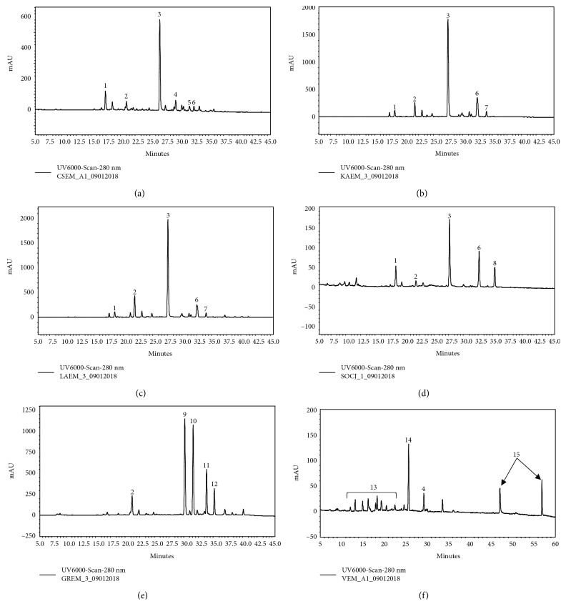
The range of the TPC in the tested extracts was from 56 to 408 mg GAE/gr dw (Table 2). As expected, C. lanatus, C. intybus, and C. sancta exhibited the higher TPC values (408, 320, and 288 mg GAE/gr dw of extract, respectively), since they were enriched with polyphenols by using absorption resin (Table 2). All the extracts from the Asteraceae family were rich in phenolic compounds and particularly hydroxycinnamic acids and flavonoid derivatives. All the members of the Cichoriae tribe (i.e., C. spinosum, C. intybus, C. sancta, and S. asper) presented a similar chemical profile (Figure 1) with variations to the relative percentages of specific secondary metabolites in each extract (supplementary material (available here)). Based on the literature [36] and previous LC-MS analyses (data not shown) of the extracts, we estimated that two caffeoyl tartaric acid derivatives, namely, caftaric acid and cichoric acid, constituted the predominant compounds of these extracts and could be responsible for the decoctions' biological activities. C. lanatus-enriched decoction appeared to possess extremely high amounts of phenolic substances, most probably glycosides of the flavonoids quercetin and luteolin, while its profile was complimented by the presence of caffeoylquinic acid isomers and dimers. On the other hand, the A. blitum extract was quite poor in phenolic compounds with only a few, minor peaks observed in the medium polarity area of its chromatogram. However, as expected from a member of the Amaranthaceae family, the presence of the nonpolar triterpene saponins was evident.
Table 2.
Total phenolic content, free radical scavenging activity against ABTS•+ and O2 •− radicals, protective activity against peroxyl (ROO•) radical-induced DNA damage, and reducing power of the extracts.
| Plant variety | TPCa (mg GAE/gr dw) | IC50 (μg/ml)e | |||
|---|---|---|---|---|---|
| ABTS•+ | O2 •− | ROO• | Reducing powere (RP0.5AU)d | ||
| Carthamus lanatus | 408 | 7.9 ± 0.9b | 6.3 ± 0.4b | 110.0 ± 8.2c | 5.0 ± 0.3b |
| Cichorium intybus | 320 | 9.1 ± 0.6 | 8.2 ± 0.7 | 105.0 ± 7.6 | 6.0 ± 0.2 |
| Cichorium spinosum | 117 | 28.0 ± 3.1 | 21.0 ± 1.4 | 300.0 ± 17.3 | 8.0 ± 0.6 |
| Crepis sancta | 288 | 12.0 ± 1.5 | 7.5 ± 0.6 | 132.0 ± 9.8 | 10.5 ± 0.9 |
| Sonchus asper | 56 | 66.0 ± 7.2 | 56.0 ± 3.8 | 970.0 ± 53.4 | 47.0 ± 3.3 |
| Amaranthus blitum | 63 | 72.0 ± 9.6 | 21.0 ± 1.2 | 443.0 ± 19.5 | 65.0 ± 4.6 |
aTPC: total polyphenolic content. bValues are the mean ± SD of at least two separate triplicate experiments. cValues are the mean ± SD from three independent experiments. dRP0.5AU: extract concentration (μg/ml) caused absorbance of 0.5 at 700 nm. eValues are statistically significant, p < 0.05.
Figure 1.
HPLC-PDA profiles of plant decoctions: (a) Cichorium spinosum, (b) Cichorium intybus, (c) Crepis sancta, (d) Sonchus asper, (e) Carthamus lanatus, (f) Amaranthus blitum, and their main metabolites: 1: caftaric acid; 2: caffeoylquinic acid isomer; 3: cichoric acid; 4: luteolin diglycoside; 5: quercetin glucuronide; 6: luteolin glucuronide; 7: dicaffeoylquinic acid isomer; 8: apigenin glucuronide; 9: quercetin glucoside; 10: luteolin glucoside; 11: quercetin acetyl glucoside; 12: luteolin acetyl glucoside; 13: hydroxycinnamates; 14: rutin; 15: triterpene saponins.
3.2. Free Radical Scavenging Activity and Reducing Power of Extracts
The IC50 values against ABTS•+ and O2 •− radicals are shown in Table 2. Low IC50 values mean strong antioxidant potency. In the ABTS•+ method, the IC50 extended from 7.9 (C. lanatus) to 72.0 μg/ml (A. blitum), and in O2 •− radical assay, it was from 6.3 (C. lanatus) to 56.0 μg/ml (S. asper) (Table 2). It was remarkable that C. lanatus extract had the highest potency in ABTS•+ and O2 •− radical assays and exhibited activity at too low concentrations. As mentioned, C. lanatus had also the highest TPC, and so, its rich polyphenolic content may explain the high antioxidant activity. Moreover, the polyphenols (i.e., quercetin, luteolin, and caffeoylquinic acid derivatives) found in C. lanatus have been known as strong free radical scavengers [37, 38]. In general, the three extracts (i.e., C. lanatus, C. intybus, and C. sancta) enriched with polyphenols due to use of absorption resin exhibited IC50 values which were at least 2-fold lower than those of the other extracts (Table 2). Although ABTS•+ radical is one of the most used for examining compounds' antioxidant activity, it is a synthetic radical. However, O2 •− is one of the most common and reactive radicals found in living organisms [3]. Superoxide radical may be produced in vivo by the reactions of the electron transport chains in mitochondria (1–3% of electrons forming O2 •−), activated phagocytic cells, enzymatic activity (e.g., P450 enzymes and xanthine oxidase), and autooxidation reactions of biomolecules (e.g., adrenalin and FADH2) [2]. Superoxide radical may cause damage to DNA, proteins, and lipids, and these effects seem to increase with aging [3]. Thus, it is important for prevention of oxidative stress-induced diseases to find out compounds which effectively scavenge O2 •−.
In the reducing power assay, RP0.5AU values ranged from 5.0 (C. lanatus) to 65.0 (A. blitum) (Table 2). Like IC50 values, the lower the RP0.5AU value is, the higher is the reducing power activity. The reducing power of a compound is an indication of its antioxidant activity, because it shows its ability to act as an electron donor and consequently to neutralize free radicals [39]. Again, C. lanatus extract was the most potent in this assay, while the other two extracts (i.e., C. intybus and C. sancta) that were enriched with polyphenols had also high reducing activity (Table 2). However, in this assay, C. spinosum extract also exhibited high reducing potency (RP0.5AU: 8.0), although it was not processed with absorption resins (Table 2). This C. spinosum extract's high activity may be explained by the type of its polyphenols or by the presence of other chemical compounds which may be very effective as hydrogen donors.
3.3. Antimutagenic Activity of Extracts against ROS-Induced DNA Damage
All the tested extracts inhibited ROO•-induced DNA plasmid breakage, with IC50 values ranging from 105 to 970 μg/ml (Table 2 and Figure 2). The potency order was C. lanatus = C. intybus > C. sancta > C. spinosum > A. blitum > S. asper (Table 2). Similar to antioxidant assays, the three extracts enriched with polyphenols by passage through resin column exhibited at least 2-fold higher protective activity compared to other extracts. The ROO• radicals that caused the DNA damage in this assay are produced within cells after the addition of O2 to carbon-centered radicals [40]. Subsequently, ROO• can oxidize DNA bases to their hydroxyl derivatives resulting in mutations and manifestation of diseases [41]. These detrimental effects may be prevented by the use of the tested extracts through diet as the present findings suggested. Interestingly, quercetin, luteolin, and caffeoylquinic acid found in C. lanatus extract have been demonstrated to scavenge ROO• [42, 43].
Figure 2.
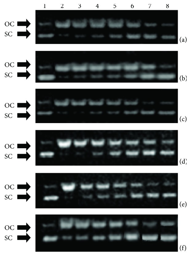
Protective activity of extracts from (a) Crepis sancta, (b) Cichorium intybus, (c) Carthamus lanatus, (d) Amaranthus blitum, (e) Cichorium spinosum, and (f) Sonchus asper plants against ROO• radicals: 1: pBluescript-SK+ plasmid DNA without any treatment; 2: plasmid DNA exposed to ROO• radicals alone; 3, 4, 5, 6, and 7: plasmid DNA exposed to ROO• radicals in the presence of extract concentrations: 0.015, 0.030, 0.060, 0.120, and 0.240 mg/mL, respectively (a–c) and 0.030, 0.060, 0.120, 0.240, and 0.480 mg/ml, respectively (e) and 0.120, 0.240, 0.480, 0.960, and 1.920 mg/ml, respectively (d, f); 8: plasmid DNA exposed to the maximum tested concentration of each extract alone. OC: open circular; SC: supercoiled.
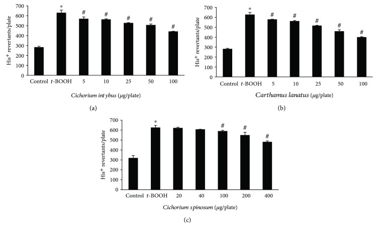
The two most potent extracts enriched with polyphenols due to passage through resin (i.e., C. lanatus and C. intybus) and the most potent (i.e., C. spinosum) among nonenriched extracts were also tested for their inhibitory potency against ROS-induced mutagenicity in S. typhimurium TA102 bacterial cells. The results from this assay supported those from DNA plasmid cleavage assay, since all three extracts inhibited dose-dependent t-BOOH-induced mutagenicity (Figure 3). Specifically, C. lanatus extract inhibited significantly t-BOOH-induced mutations by 12.3, 17.9, 31.7, 48.7, and 66.9% at 5, 10, 25, 50, and 100 μg/plate, respectively, C. intybus extract by 17.0, 19.6, 30.0, 38.0, and 56.9% at 5, 10, 25, 50, and 100 μg/plate, respectively, and C. spinosum extract by 12.2, 24.7, and 48.1% at 100, 200, and 400 μg/plate, respectively (Figure 3). The two extracts enriched with polyphenols had higher inhibitory activity than the nonenriched extract, indicating that the polyphenols accounted mainly for the observed protection from mutagenicity. The t-BOOH reacts with Fe2+ in cells and generates HO• causing DNA damage [3]. The OH• radicals are the major ROS to react with either DNA bases or deoxyribose resulting in DNA damage and mutations [3]. Thus, it was important that the tested extracts protected from OH•-induced DNA mutations. C. lanatus extract was again the most potent due, at least in part, to its identified polyphenols, since previous studies have shown that quercetin and luteolin inhibited t-BOOH-induced mutagenicity in TA102 cells [44]. Moreover, Jho et al. [45] have reported that a caffeoylquinic derivative inhibited t-BOOH-induced DNA damage in human liver HepG2 cells.
Figure 3.
Antimutagenic effects of C. intybus, C. lanatus, and C. spinosum extracts on t-BOOH-induced mutagenicity in S. typhimurium TA102 cells. Values are the mean ± SD number of histidine revertants of three independent experiments carried out in triplicate. The concentration of t-BOOH was 0.4 mM/plate. ∗ p < 0.05 when compared with control. # p < 0.05 when compared with the t-BOOH alone sample.
3.4. Effects of Extracts on the Antioxidant Status of Endothelial Cells
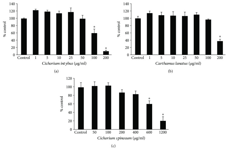
C. lanatus and C. intybus extracts, the two most potent extracts enriched with polyphenols, and C. spinosum extract, the most potent among nonenriched extracts, were examined for their antioxidant activity in human endothelial EA.hy926 cells. Firstly, the extracts' cytotoxicity was evaluated, so as noncytotoxic concentrations to be used for the assessment of the antioxidant activity. The results from XTT assay showed that C. intybus, C. lanatus, and C. spinosum extracts exhibited significant cytotoxicity above 100, 200, and 600 μg/ml, respectively (Figure 4). Thus, the selected concentrations of the C. intybus, C. lanatus, and C. spinosum extracts in the following assays were up to 50, 100, and 400 μg/ml, respectively.
Figure 4.
Cell viability following treatment with C. intybus, C. lanatus, and C. spinosum extracts in EA.hy926 cells. The results are presented as the means ± SEM of three independent experiments carried out in triplicate. ∗ p < 0.05 indicates significant difference from the control value.
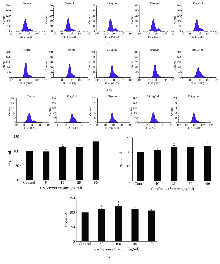
The assessment of the extracts' effects on the antioxidant capacity of endothelial cells was based on the measurement of GSH and ROS levels by flow cytometry. The results showed that C. intybus increased significantly GSH levels by 10.8, 15.2, and 33.4% at 10, 25, and 50 μg/ml, respectively, compared to control, C. lanatus by 15.9, 21.5, and 24.7% at 25, 50, and 100 μg/ml, respectively, and C. spinosum by 10.5 and 21.6% at 50 and 100 μg/ml, respectively (Figure 5). The increase in GSH after extract treatment is important, since GSH is considered as a significant endogenous antioxidant molecule in cells [46]. GSH may scavenge directly free radicals by donating one hydrogen atom from its sulfhydryl group or is used as substrate by antioxidant enzymes such as glutathione transferase (GST) and glutathione peroxidase (GPx) [46]. Especially for endothelial cells, GSH is important not only as antioxidant but also as a crucial regulator of cell signaling [47, 48]. Although the effects of C. intybus and C. lanatus on GSH were dose dependent, the C. spinosum extract did not affect GSH at higher concentrations than 100 μg/ml (Figure 5). This intriguing observation may be explained by the fact that C. spinosum at 200 and 400 μg/ml exhibited a tension to decrease cell viability (Figure 4). That is, for C. spinosum, 100 μg/ml seemed to be a crucial concentration, above which cytotoxicity was caused. In turn, this cytotoxicity was encountered by GSH consumption. It is known that polyphenols sometimes have a biphasic effect, namely, at low concentrations, they act as antioxidants and at high concentrations, they act as prooxidants resulting in cytotoxicity [49]. It is also worth mentioning that polyphenols identified in the tested extracts have been reported to increase GSH levels. For example, quercetin has been demonstrated recently to enhance GSH levels in human aortic endothelial cells (HAEC) through increased expression of glutamate-cysteine ligase (GCL), one of the major enzymes involved in GSH synthesis [50]. Moreover, luteolin has been demonstrated to decrease oxidative stress in the mouse lung through, among other mechanisms, increase of GSH [51]. Additionally, administration of caftaric acid to rats inhibited lead-induced decrease in GSH in the kidney [52].
Figure 5.
Effects of treatment with C. intybus, C. lanatus, and C. spinosum extracts at different concentrations for 24 h on GSH levels in EA.hy926 cells. The histograms of cell counts versus fluorescence of 10,000 cells analyzed by flow cytometry for the detection of GSH levels after treatment with (a) C. intybus, (b) C. lanatus, and (c) C. spinosum. FL2 represents the detection of fluorescence using 488 and 580 nm as the excitation and emission wavelength, respectively. Bar charts indicate the GSH levels as % of control as estimated by the histograms in EA.hy926 cells after treatment with C. intybus, C. lanatus, and C. spinosum extracts. All values of bar charts are presented as the mean ± SEM of 3 independent experiments. ∗ p < 0.05 indicates significant difference from the control.
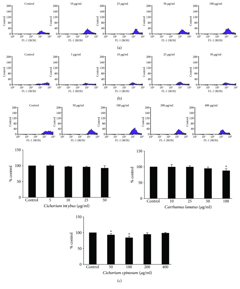
Unlike GSH, extract treatment did not affect ROS levels too intensively (Figure 6). Only C. lanatus extract reduced significantly ROS by 12.1% and C. spinosum extract by 6.8 and 15.6% at 50 and 100 μg/ml, respectively, compared to control (Figure 6). The weak extracts' effect on ROS levels may be attributed to the fact that their impact was examined on the baseline ROS levels, that is, there was not an oxidant stimulus to cells. Nevertheless, the observed decrease in ROS, even only at the higher extract concentrations, was in accordance with and might be attributed to the corresponding increase in the antioxidant molecule of GSH by the extracts (Figure 5). Interestingly, C. spinosum at concentrations higher than 100 μg/ml did not decrease further ROS levels (Figure 6). This finding supported our hypothesis mentioned above, that is, C. spinosum at concentrations above 100 μg/ml may exhibit prooxidant effect and cytotoxicity.
Figure 6.
Effects of treatment with C. intybus, C. lanatus, and C. spinosum extracts at different concentrations for 24 h on ROS levels in EA.hy926 cells. The histograms of cell counts versus fluorescence of 10,000 cells analyzed by flow cytometry for the detection of ROS levels after treatment with (a) C. intybus, (b) C. lanatus, and (c) C. spinosum. FL2 represents the detection of fluorescence using 488 and 530 nm as the excitation and emission wavelength, respectively. Bar charts indicate the ROS levels as % of control as estimated by the histograms in EA.hy926 cells after treatment with C. intybus, C. lanatus, and C. spinosum extracts. All values of bar charts are presented as the mean ± SEM of 3 independent experiments. ∗ p < 0.05 indicates significant difference from the control.
Since C. intybus extract induced the highest increase in antioxidant mechanism (i.e., GSH) in endothelial cells, its effects on the expression at a transcriptional level of antioxidant genes were assessed (Figure 7). Specifically, it was examined if the extract affected the expression of the nuclear factor (erythroid-derived 2)-like 2 (Nrf2), the most important transcription factor regulating antioxidant genes [53]. The results from the qRT-PCR showed that C. intybus treatment upregulated significantly the expression of NFE2L2 gene encoding for Nrf2 by 7.3-fold and 8.5-fold at 12 and 24 h, respectively, compared to control (Figure 7). This finding was significant, because it indicated that the extract's compounds exerted antioxidant activity not only as direct free radical scavengers but also as modulators of molecular mechanisms. Interestingly, chicoric acid found in C. intybus extract has been reported to increase Nrf2 expression in mouse muscle [54]. The increase in Nrf2 expression was supported by the extract treatment-induced increase in expression of genes regulated by Nrf2. Namely, extract treatment upregulated significantly the expression of GCLC by 6.2-fold, 6.3-fold, and 3.4-fold at 3, 12, and 24 h, respectively, GSR by 9.8-fold, 8.0-fold, and 5.0-fold at 3, 12, and 24 h, respectively, NQO1 by 8.4-fold, 12.1-fold, and 6.7-fold at 3, 12, and 24 h, respectively, and HMOX1 by 4.0-fold, 4.5-fold, and 2.7-fold at 3, 12, and 24 h, respectively, compared to control (Figure 7). Especially, the increase in GCLC and GSR expression was important, since it accounted for the C. intybus extract-induced increase in GSH levels (Figure 5). GCLC gene encodes the catalytic subunit of the GCL protein, the main enzyme involved in GSH synthesis [40]. GSR encodes for glutathione reductase (GR) enzyme regenerating GSH from the oxidized glutathione (GSSG) [40]. HMOX1 and NQO1, the other two upregulating genes, encode for heme oxygenase 1 (HO-1) and NAD (P)H:quinone oxidoreductase 1 (NQO1), respectively. The increase in the expression of these enzymes supported further the ability of the C. intybus extract to enhance endothelial cells' antioxidant capacity, since HO-1 and NQO1 are important antioxidant enzymes participating in iron sequestration and quinone detoxification, respectively [55, 56]. C. intybus treatment did not affect the expression of CAT, SOD1, and GPX1 genes, while it downregulated significantly TXN gene expression by 35.7-fold, 10.3-fold, and 10.0-fold at 3, 12, and 24 h, respectively (Figure 7). The lack of effect on CAT, SOD1, and GPX1 and the downregulation of TXN gene were intriguing, since Nrf2 activates their expression [53]. This contradiction may be explained by the high complexity of the regulation of the antioxidant mechanisms and the interaction between them, namely, when some antioxidant mechanisms are enhanced, some others remain inactive as a compensation adaptive response of the cell [57].
Figure 7.
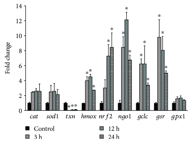
Gene expression profiles of NFE2L2, GCLC, GSR, GPX1, HMOX1, NQO1, CAT, SOD1, and TXN genes in EA.hy926 cells after treatment with C. intybus extract at 50 μg/ml for 3, 12, and 24 h. mRNA levels were determined by qRT-PCR, and relative levels were expressed as fold of control (untreated cells) after normalization to GAPDH. The results are expressed as mean ± SD of three independent experiments. ∗ p < 0.05 indicates significant difference from the control.
4. Conclusions
In conclusion, the present findings demonstrated for the first time that extracts from the edible plants C. lanatus, C. intybus, and C. spinosum enhanced antioxidant defense mechanism such as GSH in endothelial cells. Especially, C. spinosum extract was shown to mediate this antioxidant effect through increased expression of Nrf2, the most crucial transcription factor of antioxidant genes, and subsequent upregulation of important antioxidant genes including those involved in GSH synthesis. Since, these plants constitute a part of the Mediterranean diet, their observed bioactivities may explain, at least in part, the prevention of this type of diet against diseases associated with endothelial function such as the cardiovascular disease [58]. Moreover, the results suggested that these extracts may be used for the development of food supplements or biofunctional foods that would protect from diseases caused by oxidative stress-induced endothelium damage.
Acknowledgments
The work was funded by the “Toxicology” MSc program in the Department of Biochemistry and Biotechnology at the University of Thessaly. The current work was also supported by the EU and project MediHealth-H2020-MSCA-RISE-2015—“Novel natural products for healthy ageing from Mediterranean diet and food plants of other global sources” (Proposal number: 691158) under the Horizon 2020 framework.
Data Availability
The data used to support the findings of this study are available from the corresponding author upon request.
Conflicts of Interest
The authors have no conflicts of interest to declare.
Authors' Contributions
Dimitrios Balabanos, Salomi Savva, and Zoi Skaperda contributed equally to the present study.
Supplementary Materials
Values of retention time and area (%) of the relative quantification of main secondary metabolites detected in the extracts by using HPLC-PDA analysis.
References
- 1.Ray P. D., Huang B. W., Tsuji Y. Reactive oxygen species (ROS) homeostasis and redox regulation in cellular signaling. Cellular Signalling. 2012;24(5):981–990. doi: 10.1016/j.cellsig.2012.01.008. [DOI] [PMC free article] [PubMed] [Google Scholar]
- 2.Priftis A., Stagos D., Konstantinopoulos K., et al. Comparison of antioxidant activity between green and roasted coffee beans using molecular methods. Molecular Medicine Reports. 2015;12(5):7293–7302. doi: 10.3892/mmr.2015.4377. [DOI] [PMC free article] [PubMed] [Google Scholar]
- 3.Halliwell B. Free Radicals and Other Reactive Species in Disease. John Wiley & Sons, Ltd, eLS; 2005. [DOI] [Google Scholar]
- 4.Sosa V., Moliné T., Somoza R., Paciucci R., Kondoh H., LLeonart M. E. Oxidative stress and cancer: an overview. Ageing Research Reviews. 2013;12(1):376–390. doi: 10.1016/j.arr.2012.10.004. [DOI] [PubMed] [Google Scholar]
- 5.Rochette L., Zeller M., Cottin Y., Vergely C. Diabetes, oxidative stress and therapeutic strategies. Biochimica et Biophysica Acta (BBA) - General Subjects. 2014;1840(9):2709–2729. doi: 10.1016/j.bbagen.2014.05.017. [DOI] [PubMed] [Google Scholar]
- 6.Kouka P., Priftis A., Stagos D., et al. Assessment of the antioxidant activity of an olive oil total polyphenolic fraction and hydroxytyrosol from a Greek Olea europea variety in endothelial cells and myoblasts. International Journal of Molecular Medicine. 2017;40(3):703–712. doi: 10.3892/ijmm.2017.3078. [DOI] [PMC free article] [PubMed] [Google Scholar]
- 7.Deanfield J. E., Halcox J. P., Rabelink T. J. Endothelial function and dysfunction: testing and clinical relevance. Circulation. 2007;115(10):1285–1295. doi: 10.1161/CIRCULATIONAHA.106.652859. [DOI] [PubMed] [Google Scholar]
- 8.Victor V. M., Rocha M., Solá E., Bañuls C., Garcia-Malpartida K., Hernández-Mijares A. Oxidative stress, endothelial dysfunction and atherosclerosis. Current Pharmaceutical Design. 2009;15(26):2988–3002. doi: 10.2174/138161209789058093. [DOI] [PubMed] [Google Scholar]
- 9.Kokura S., Wolf R. E., Yoshikawa T., Granger D. N., Aw T. Y. Molecular mechanisms of neutrophil-endothelial cell adhesion induced by redox imbalance. Circulation Research. 1999;84(5):516–524. doi: 10.1161/01.RES.84.5.516. [DOI] [PubMed] [Google Scholar]
- 10.Rhee S. G. Cell signaling. H2O2, a necessary evil for cell signaling. Science. 2006;312(5782):1882–1883. doi: 10.1126/science.1130481. [DOI] [PubMed] [Google Scholar]
- 11.Woywodt A., Bahlmann F. H., De Groot K., Haller H., Haubitz M. Circulating endothelial cells: life, death, detachment and repair of the endothelial cell layer. Nephrology, Dialysis, Transplantation. 2002;17(10):1728–1730. doi: 10.1093/ndt/17.10.1728. [DOI] [PubMed] [Google Scholar]
- 12.Landete J. M. Dietary intake of natural antioxidants: vitamins and polyphenols. Critical Reviews in Food Science and Nutrition. 2013;53(7):706–721. doi: 10.1080/10408398.2011.555018. [DOI] [PubMed] [Google Scholar]
- 13.Fang Y. Z., Yang S., Wu G. Free radicals, antioxidants, and nutrition. Nutrition. 2002;18(10):872–879. doi: 10.1016/S0899-9007(02)00916-4. [DOI] [PubMed] [Google Scholar]
- 14.Landete J. M. Updated knowledge about polyphenols: functions, bioavailability, metabolism, and health. Critical Reviews in Food Science and Nutrition. 2012;52(10):936–948. doi: 10.1080/10408398.2010.513779. [DOI] [PubMed] [Google Scholar]
- 15.Battino M., Mezzetti B. Update on fruit antioxidant capacity: a key tool for Mediterranean diet. Public Health Nutrition. 2006;9(8A):1099–1103. doi: 10.1017/S1368980007668554. [DOI] [PubMed] [Google Scholar]
- 16.Leonti M., Nebel S., Rivera D., Heinrich M., Leonti M. Wild gathered food plants in the European Mediterranean: a comparative analysis. Economic Botany. 2006;60(2):130–142. doi: 10.1663/0013-0001(2006)60[130:WGFPIT]2.0.CO;2. [DOI] [Google Scholar]
- 17.Hadjichambis A. C. H., Paraskeva-Hadjichambi D., Della A., et al. Wild and semi-domesticated food plant consumption in seven circum-Mediterranean areas. International Journal of Food Sciences and Nutrition. 2008;59(5):383–414. doi: 10.1080/09637480701566495. [DOI] [PubMed] [Google Scholar]
- 18.Vasilopoulou E., Trichopoulou A. Green pies: the flavonoid rich Greek snack. Food Chemistry. 2011;126(3):855–858. doi: 10.1016/j.foodchem.2010.11.051. [DOI] [Google Scholar]
- 19.Vasilopoulou E., Georga K., Joergensen M. B., Naska A., Trichopoulou A. The antioxidant properties of Greek foods and the flavonoid content of the Mediterranean menu. Current Medicinal Chemistry - Immunology, Endocrine & Metabolic Agents. 2005;5(1):33–45. doi: 10.2174/1568013053005508. [DOI] [Google Scholar]
- 20.Conforti F., Sosa S., Marrelli M., et al. The protective ability of Mediterranean dietary plants against the oxidative damage: the role of radical oxygen species in inflammation and the polyphenol, flavonoid and sterol contents. Food Chemistry. 2009;112(3):587–594. doi: 10.1016/j.foodchem.2008.06.013. [DOI] [Google Scholar]
- 21.Zeghichi S., Kallithraka S., Simopoulos A. P., Kypriotakis Z. Nutritional composition of selected wild plants in the diet of Crete. World Review of Nutrition and Dietetics. 2003;91:22–40. doi: 10.1159/000069928. [DOI] [PubMed] [Google Scholar]
- 22.Vardavas C. I., Majchrzak D., Wagner K. H., Elmadfa I., Kafatos A. The antioxidant and phylloquinone content of wildly grown greens in Crete. Food Chemistry. 2006;99(4):813–821. doi: 10.1016/j.foodchem.2005.08.057. [DOI] [Google Scholar]
- 23.Nebel S., Pieroni A., Heinrich M. Ta chòrta: wild edible greens used in the Graecanic area in Calabria, Southern Italy. Appetite. 2006;47(3):333–342. doi: 10.1016/j.appet.2006.05.010. [DOI] [PubMed] [Google Scholar]
- 24.Mikropoulou E. V., Vougogiannopoulou K., Kalpoutzakis E., et al. Phytochemical composition of the decoctions of Greek edible greens (chórta) and evaluation of antioxidant and cytotoxic properties. Molecules. 2018;23(7, article 1541) doi: 10.3390/molecules23071541. [DOI] [PMC free article] [PubMed] [Google Scholar]
- 25.Makri S., Kafantaris I., Savva S., et al. Novel feed including olive oil mill wastewater bioactive compounds enhanced the redox status of lambs. In Vivo. 2018;32(2):291–302. doi: 10.21873/invivo.11237. [DOI] [PMC free article] [PubMed] [Google Scholar]
- 26.Stagos D., Soulitsiotis N., Tsadila C., et al. Antibacterial and antioxidant activity of different types of honey derived from Mount Olympus in Greece. International Journal of Molecular Medicine. 2018;42(2):726–734. doi: 10.3892/ijmm.2018.3656. [DOI] [PMC free article] [PubMed] [Google Scholar]
- 27.Priftis A., Mitsiou D., Halabalaki M., et al. Roasting has a distinct effect on the antimutagenic activity of coffee varieties. Mutation Research. 2018;829-830:33–42. doi: 10.1016/j.mrgentox.2018.03.003. [DOI] [PubMed] [Google Scholar]
- 28.Kerasioti E., Stagos D., Priftis A., et al. Antioxidant effects of whey protein on muscle C2C12 cells. Food Chemistry. 2014;155:271–278. doi: 10.1016/j.foodchem.2014.01.066. [DOI] [PubMed] [Google Scholar]
- 29.Spanou C., Stagos D., Tousias L., et al. Assessment of antioxidant activity of extracts from unique Greek varieties of Leguminosae plants using in vitro assays. Anticancer Research. 2007;27(5A):3403–3410. [PubMed] [Google Scholar]
- 30.Maron D. M., Ames B. N. Revised methods for the Salmonella mutagenicity test. Mutation Research. 1983;113(3-4):173–215. doi: 10.1016/0165-1161(83)90010-9. [DOI] [PubMed] [Google Scholar]
- 31.Mortelmans K., Zeiger E. The Ames Salmonella/microsome mutagenicity assay. Mutation Research. 2000;455(1-2):29–60. doi: 10.1016/S0027-5107(00)00064-6. [DOI] [PubMed] [Google Scholar]
- 32.Priftis A., Panagiotou E. M., Lakis K., et al. Roasted and green coffee extracts show antioxidant and cytotoxic activity in myoblast and endothelial cell lines in a cell specific manner. Food and Chemical Toxicology. 2018;114:119–127. doi: 10.1016/j.fct.2018.02.029. [DOI] [PubMed] [Google Scholar]
- 33.Goutzourelas N., Stagos D., Demertzis N., et al. Effects of polyphenolic grape extract on the oxidative status of muscle and endothelial cells. Human & Experimental Toxicology. 2014;33(11):1099–1112. doi: 10.1177/0960327114533575. [DOI] [PubMed] [Google Scholar]
- 34.Kerasioti E., Stagos D., Georgatzi V., et al. Antioxidant effects of sheep whey protein on endothelial cells. Oxidative Medicine and Cellular Longevity. 2016;2016:10. doi: 10.1155/2016/6585737.6585737 [DOI] [PMC free article] [PubMed] [Google Scholar]
- 35.Priftis A., Goutzourelas N., Halabalaki M., et al. Effect of polyphenols from coffee and grape on gene expression inmyoblasts. Mechanisms of Ageing and Development. 2018;172:115–122. doi: 10.1016/j.mad.2017.11.015. [DOI] [PubMed] [Google Scholar]
- 36.Brieudes V., Angelis A., Vougogiannopoulou K., et al. Phytochemical analysis and antioxidant potential of the phytonutrient-rich decoction of Cichorium spinosum and C. intybus. Planta Medica. 2016;82(11-12):1070–1078. doi: 10.1055/s-0042-107472. [DOI] [PubMed] [Google Scholar]
- 37.Boots A. W., Haenen G. R., Bast A. Health effects of quercetin: from antioxidant to nutraceutical. European Journal of Pharmacology. 2008;585(2-3):325–337. doi: 10.1016/j.ejphar.2008.03.008. [DOI] [PubMed] [Google Scholar]
- 38.Zhang Y. C., Gan F. F., Shelar S. B., Ng K. Y., Chew E. H. Antioxidant and Nrf 2 inducing activities of luteolin, a flavonoid constituent in Ixeris sonchifolia Hance, provide neuroprotective effects against ischemia-induced cellular injury. Food and Chemical Toxicology. 2013;59:272–280. doi: 10.1016/j.fct.2013.05.058. [DOI] [PubMed] [Google Scholar]
- 39.Chanda S., Dave R. In vitro models for antioxidant activity evaluation and some medicinal plants possessing antioxidant properties: an overview. African Journal of Microbiology Research. 2009;3:981–996. [Google Scholar]
- 40.Dix T. A., Aikens J. Mechanisms and biological relevance of lipid peroxidation initiation. Chemical Research in Toxicology. 1993;6(1):2–18. doi: 10.1021/tx00031a001. [DOI] [PubMed] [Google Scholar]
- 41.Simandan T., Sun J., Dix T. A. Oxidation of DNA bases, deoxyribonucleosides and homopolymers by peroxyl radicals. The Biochemical Journal. 1998;335(2):233–240. doi: 10.1042/bj3350233. [DOI] [PMC free article] [PubMed] [Google Scholar]
- 42.Rodrigues E., Mariutti L. R., Mercadante A. Z. Carotenoids and phenolic compounds from Solanum sessiliflorum, an unexploited Amazonian fruit, and their scavenging capacities against reactive oxygen and nitrogen species. Journal of Agricultural and Food Chemistry. 2013;61(12):3022–3029. doi: 10.1021/jf3054214. [DOI] [PubMed] [Google Scholar]
- 43.Terashima M., Kakuno Y., Kitano N., et al. Antioxidant activity of flavonoids evaluated with myoglobin method. Plant Cell Reports. 2012;31(2):291–298. doi: 10.1007/s00299-011-1163-2. [DOI] [PubMed] [Google Scholar]
- 44.Edenharder R., Grünhage D. Free radical scavenging abilities of flavonoids as mechanism of protection against mutagenicity induced by tert-butyl hydroperoxide or cumene hydroperoxide in Salmonella typhimurium TA102. Mutation Research. 2003;540(1):1–18. doi: 10.1016/S1383-5718(03)00114-1. [DOI] [PubMed] [Google Scholar]
- 45.Jho E. H., Kang K., Oidovsambuu S., et al. Gymnaster koraiensis and its major components, 3,5-di-O-caffeoylquinic acid and gymnasterkoreayne B, reduce oxidative damage induced by tert-butyl hydroperoxide or acetaminophen in HepG2 cells. BMB Reports. 2013;46(10):513–518. doi: 10.5483/BMBRep.2013.46.10.037. [DOI] [PMC free article] [PubMed] [Google Scholar]
- 46.Aquilano K., Baldelli S., Ciriolo M. R. Glutathione: new roles in redox signaling for an old antioxidant. Frontiers in Pharmacology. 2014;5:p. 196. doi: 10.3389/fphar.2014.00196. [DOI] [PMC free article] [PubMed] [Google Scholar]
- 47.Elliott S. J., Koliwad S. K. Redox control of ion channel activity in vascular endothelial cells by glutathione. Microcirculation. 1997;4(3):341–347. doi: 10.3109/10739689709146798. [DOI] [PubMed] [Google Scholar]
- 48.Espinosa-Díez C., Miguel V., Vallejo S., et al. Role of glutathione biosynthesis in endothelial dysfunction and fibrosis. Redox Biology. 2018;14:88–99. doi: 10.1016/j.redox.2017.08.019. [DOI] [PMC free article] [PubMed] [Google Scholar]
- 49.Mileo A. M., Miccadei S. Polyphenols as modulator of oxidative stress in cancer disease: new therapeutic strategies. Oxidative Medicine and Cellular Longevity. 2016;2016:17. doi: 10.1155/2016/6475624.6475624 [DOI] [PMC free article] [PubMed] [Google Scholar]
- 50.Li C., Zhang W. J., Choi J., Frei B. Quercetin affects glutathione levels and redox ratio in human aortic endothelial cells not through oxidation but formation and cellular export of quercetin-glutathione conjugates and upregulation of glutamate-cysteine ligase. Redox Biology. 2016;9:220–228. doi: 10.1016/j.redox.2016.08.012. [DOI] [PMC free article] [PubMed] [Google Scholar]
- 51.Liu B., Yu H., Baiyun R., et al. Protective effects of dietary luteolin against mercuric chloride-induced lung injury in mice: involvement of AKT/Nrf 2 and NF-κB pathways. Food and Chemical Toxicology. 2018;113:296–302. doi: 10.1016/j.fct.2018.02.003. [DOI] [PubMed] [Google Scholar]
- 52.Koriem K. M. M., Arbid M. S. Role of caftaric acid in lead-associated nephrotoxicity in rats via antidiuretic, antioxidant and anti-apoptotic activities. Journal of Complementary and Integrative Medicine. 2017;15(2, article 20170024) doi: 10.1515/jcim-2017-0024. [DOI] [PubMed] [Google Scholar]
- 53.Bellezza I., Giambanco I., Minelli A., Donato R. Nrf2-Keap1 signaling in oxidative and reductive stress. Biochimica et Biophysica Acta (BBA) - Molecular Cell Research. 2018;1865(5):721–733. doi: 10.1016/j.bbamcr.2018.02.010. [DOI] [PubMed] [Google Scholar]
- 54.Zhu D., Zhang X., Niu Y., et al. Cichoric acid improved hyperglycaemia and restored muscle injury via activating antioxidant response in MLD-STZ-induced diabetic mice. Food and Chemical Toxicology. 2017;107, Part A:138–149. doi: 10.1016/j.fct.2017.06.041. [DOI] [PubMed] [Google Scholar]
- 55.Ndisang J. F. Synergistic interaction between heme oxygenase (HO) and nuclear-factor E2- related factor-2 (Nrf 2) against oxidative stress in cardiovascular related diseases. Current Pharmaceutical Design. 2017;23(10):1465–1470. doi: 10.2174/1381612823666170113153818. [DOI] [PubMed] [Google Scholar]
- 56.Dinkova-Kostova A. T., Talalay P. NAD (P)H:quinone acceptor oxidoreductase 1 (NQO1), a multifunctional antioxidant enzyme and exceptionally versatile cytoprotector. Archives of Biochemistry and Biophysics. 2010;501(1):116–123. doi: 10.1016/j.abb.2010.03.019. [DOI] [PMC free article] [PubMed] [Google Scholar]
- 57.Alvarez S., Boveris A. Antioxidant adaptive response of human mononuclear cells to UV-B: effect of lipoic acid. Journal of Photochemistry and Photobiology. B. 2000;55(2-3):113–119. doi: 10.1016/S1011-1344(00)00030-0. [DOI] [PubMed] [Google Scholar]
- 58.Erwin C. M., McEvoy C. T., Moore S. E., et al. A qualitative analysis exploring preferred methods of peer support to encourage adherence to a Mediterranean diet in a Northern European population at high risk of cardiovascular disease. BMC Public Health. 2018;18(1):p. 213. doi: 10.1186/s12889-018-5078-5. [DOI] [PMC free article] [PubMed] [Google Scholar]
Associated Data
This section collects any data citations, data availability statements, or supplementary materials included in this article.
Supplementary Materials
Values of retention time and area (%) of the relative quantification of main secondary metabolites detected in the extracts by using HPLC-PDA analysis.
Data Availability Statement
The data used to support the findings of this study are available from the corresponding author upon request.


