Abstract
Essential metabolic reactions are shaping constituents of metabolic networks, enabling viable and distinct phenotypes across diverse life forms. Here we analyse and compare modelling predictions of essential metabolic functions with experimental data and thereby identify core metabolic pathways in prokaryotes. Simulations of 15 manually curated genome-scale metabolic models were integrated with 36 large-scale gene essentiality datasets encompassing a wide variety of species of bacteria and archaea. Conservation of metabolic genes was estimated by analysing 79 representative genomes from all the branches of the prokaryotic tree of life. We find that essentiality patterns reflect phylogenetic relations both for modelling and experimental data, which correlate highly at the pathway level. Genes that are essential for several species tend to be highly conserved as opposed to non-essential genes which may be conserved or not. The tRNA-charging module is highlighted as ancestral and with high centrality in the networks, followed closely by cofactor metabolism, pointing to an early information processing system supplied by organic cofactors. The results, which point to model improvements and also indicate faults in the experimental data, should be relevant to the study of centrality in metabolic networks and ancient metabolism but also to metabolic engineering with prokaryotes.
Author summary
If we tried to list every known chemical reaction within an organism–human, plant or even bacteria–we would get quite a long and confusing read. But when this information is represented in so-called genome-scale metabolic networks, we have the means to access computationally each of those reactions and their interconnections. Some parts of the network have alternatives, while others are unique and therefore can be essential for growth. Here, we simulate growth and compare essential reactions and genes for the simplest type of unicellular species–prokaryotes–to understand which parts of their metabolism are universally essential and potentially ancestral. We show that similar patterns of essential reactions echo phylogenetic relationships (this makes sense, as the genome provides the building plan for the enzymes that perform those reactions). Our computational predictions correlate strongly with experimental essentiality data. Finally, we show that a crucial step of protein synthesis (tRNA charging) and the synthesis and transformation of small molecules that enzymes require (cofactors) are the most essential and conserved parts of metabolism in prokaryotes. Our results are a step further in understanding the biology and evolution of prokaryotes but can also be relevant in applied studies including metabolic engineering and antibiotic design.
Introduction
Prokaryotes are the simplest contemporary life forms known, and nevertheless are characterized by an immense complexity. The debate on the features of such complexity and its breadth in the primordial life forms has been around for years [1–3], and was furthermore expanded and detailed since the advent of systems biology [4–6]. The study of essential genes has been a crucial contribution for detangling this complexity, relating some proteins with cell viability in specific conditions [7] and others with cell viability in apparently all conditions [8,9]. Genome-wide essentiality studies based on collections of targeted mutants or generated by random mutagenesis have been conducted for a number of species, aiming mainly at antibiotic design or identifying industrially relevant targets [10–15]. These data have been made available in databases such as the Online Gene Essentiality Database (OGEE) [16] and the database of essential genes (DEG) [17] but their comparative and integrative analysis, although having already provided relevant insights, is still emerging. Early work comparing genome-scale essentiality data of Mycoplasma genitalium, Haemophilus influenza, Bacillus subtilis and Escherichia coli found that it is essentiality, not expressiveness, that drives gene strand bias [18]. Later, a critical review of genome-scale essentiality datasets conducted a preliminary analysis integrating six assays corresponding to four different species [19]. Functional differences were highlighted, as the smaller number of essential genes in flavin synthesis in B. subtilis, a species known to have an active riboflavin salvage capability. The authors of the DEG database have also conducted a pair of integrative analyses on large-scale essentiality data. The first concluded that there are less essential genes inside than outside genomic islands, and some of those are related with virulence [20]. The second study [21] added to a previous finding based only on E. coli essentiality data where it was proposed that essential genes are more evolutionarily conserved than non-essential genes [22]. Luo and others used the same type of analysis, based on synonymous and non-synonymous substitution rates for 23 bacterial species, to corroborate this finding [21]. The study indicates that the most evolutionarily conserved COG categories of essential genes are carbohydrate transport and metabolism; coenzyme transport and metabolism; transcription; translation, ribosomal structure and biogenesis; lipid transport and metabolism, and replication, recombination and repair.
Genome-scale metabolic models (GSMs) are large curated repositories of metabolic data for individual species that expand possibilities of analysis of cellular physiology [23]. Apart from improving or suggesting new functional annotations by reconstructing whole pathways [24], GSMs can be used for calculating metabolic fluxes that permit the prediction of, among others, lethal phenotypes [25]. Multi-species analysis of this type of phenotype predictions with different manually curated models has been scarce [26], in part impaired by the poor knowledge basis for other species than the usual model organisms, but also by the deficient use of standards in building such models [27].
Comparative genomics is commonly used to find core essential genes for several species, and being based on the key evolutionary notion of orthology, to infer genes present in common ancestors [28]. Evolutionary parsimony indicates that genes present in a set of species have been vertically inherited from a common ancestor. Horizontal Gene Transfer (HGT) might have played a role even before the divergence of the three main domains [29], but when a gene is present in all or most species of a phylogenetic branch, the most parsimonious scenario is vertical inheritance.
In parallel with genetic comparisons, genome-scale metabolic models allow for functional comparisons at the level of metabolic capacities (reactions). Building up on this methodological advantage, in this study, 36 experimental genome-scale essentiality assays were integrated with simulation results from 15 genome-scale metabolic models to reveal common patterns of essentiality. To this analysis the screening of full genome sequences of 79 prokaryotic species was added in order to find core conserved functions in prokaryotic biology. It is expected that this knowledge on the minimal metabolic functions of prokaryotic cells can not only help untangling the fundamental complexity of cellular systems but also, by building up on the concept of orthogonalization of metabolic modules [30], here analysed in the form of metabolic subsystems (pathways), improve future engineering approaches that use this type of organisms.
Methods
Genome-scale metabolic models used in essentiality predictions
For all essentiality predictions performed in this study, 15 genome-scale metabolic models were chosen based on curation, validation, and comparability of the nomenclature of metabolites and reactions. These comprise 7 prokaryotic phyla, including one archaea. Ten of these models include more than 20% of the total of ORFs of the corresponding species. Table 1 summarizes the details on the models used, including species name, model ID, content and references.
Table 1. Details on models and corresponding species used in in silico essentiality simulations.
| Phylum | Species | Model ID | # Reactions |
# Metabolites |
% ORFs | Reference |
|---|---|---|---|---|---|---|
| Firmicutes | Bacillus subtilis | iYO844 | 1020 | 988 | 21% | [31] |
| Clostridium beijerinckii NCIMB 8052 | iCB925 | 938 | 881 | 18% | [32] | |
| Staphylococcus aureus N315 | iSB619 | 641 | 571 | 24% | [33] | |
| Proteobacteria | Escherichia coli K12 | iAF1260 | 2077 | 1039 | 29% | [34] |
| Escherichia coli W (ATCC9637) | iCA1273 | 2477 | 1111 | 27% | [35] | |
| Helicobacter pylori 16695 | iIT341 | 476 | 485 | 21% | [36] | |
| Klebsiella pneumoniae MGH 78578 | iYL1228 | 1970 | 1658 | 24% | [37] | |
| Pseudomonas putida KT2440 | iJN746 | 950 | 911 | 14% | [38] | |
| Salmonella typhimurium LT2 | STM_v1.0 | 2201 | 1119 | 28% | [39] | |
| Shewanella oneidensis MR-1 | iSO783 | 774 | 634 | 15% | [40] | |
| Actinobacteria | Mycobacterium tuberculosis H37Rv | iNJ661 | 939 | 828 | 15% | [41] |
| Chloroflexi | Dehalococcoides ethenogenes | iAI549 | 518 | 549 | 27% | [42] |
| Cyanobacteria | Synechocystis sp. PCC6803 | iJN678 | 863 | 795 | 21% | [43] |
| Thermotogales | Thermotoga maritima MSB8 | (None) | 562 | 503 | 25% | [44] |
| Euryarchaeota | Methanosarcina barkeri str. Fusaro | iAF692 | 476 | 485 | 14% | [45] |
Environmental conditions for simulations
All GSMs were collected in SBML format and then parsed to model an environmental condition corresponding to rich media: all original exchange reactions in the model were set to a maximum uptake limit of -20 mmol gDW-1 h-1 to allow for the import of all transported metabolites (including oxygen, whenever possible).
In silico, single deletion of metabolic reactions
Flux Balance Analysis (FBA) [46,47] was used to predict the essentiality of each metabolic reaction in all models. A threshold of 10% of the flux through the biomass reaction compared to the wild type was set as the limit to define an essential metabolic reaction. All modelling procedures were implemented in C++ and solved using IBM ILOG CPLEX solver. The Optflux platform [48] was used occasionally to benchmark results.
Standardizing the nomenclature of essential metabolic reactions
For comparison of the essential reactions calculated for the 15 GSMs, some nomenclature inconsistencies were resolved: standardization of suffixes used in reaction IDs, removal of unnecessary or redundant indications of reversibility and species names allocated to reactions and other redundant tags. Irrelevant and irregular characters such as dashes were filtered out of the nomenclature (the final list of standardized reaction ids for reactions essential at least once is provided in the S1 File).
Experimental data and subsystem mapping
Large-scale experimental data on gene essentiality were collected from two databases, OGEE [16] and DEG [17]. The content of the databases was compared and DEG was chosen for it was considerably larger, including wider and clearer annotation metadata for 36 prokaryotic datasets (Table 2). Genes were mapped to the subsystems present in the latest Escherichia coli genome-scale metabolic model [49] using the DEG integrated nomenclature system of gene identifiers. All essential reactions obtained after GSMs analysis were also mapped according to this updated list of subsystems, using their standardized nomenclature (see above; S2 File).
Table 2. Large-scale essentiality assays used in this study, number of essential genes in each and respective original reference of publication.
| Species name | Number of essential genes | Reference a | |
|---|---|---|---|
| Acinetobacter baylyi ADP1 | 499 | [12] | |
| Bacillus subtilis 168 | 271 | [50] | |
| Bacteroides fragilis 638R | 547 | [51] | |
| Bacteroides thetaiotaomicron VPI-5482 | 325 | [52] | |
| Burkholderia pseudomallei K96243 | 505 | [53] | |
| Burkholderia thailandensis E264 | 406 | [54] | |
| Campylobacter jejuni subsp. jejuni NCTC 11168 = ATCC 700819 | 228 | [55] | |
| Caulobacter crescentus | 480 | [56] | |
| Escherichia coli MG1655 I | 609 | [10] | |
| Escherichia coli MG1655 II | 296 | [57] | |
| Francisella novicida U112 | 392 | [11] | |
| Haemophilus influenzae Rd KW20 | 642 | [58] | |
| Helicobacter pylori 26695 | 323 | [59] | |
| Methanococcus maripaludis S2 | 519 | [60] | |
| Mycobacterium tuberculosis H37Rv | 614 | [61] | |
| Mycobacterium tuberculosis H37Rv II | 771 | [62] | |
| Mycobacterium tuberculosis H37Rv III | 687 | [63] | |
| Mycoplasma genitalium G37 | 381 | [64] | |
| Mycoplasma pulmonis UAB CTIP | 310 | [65] | |
| Porphyromonas gingivalis ATCC 33277 | 463 | [66] | |
| Pseudomonas aeruginosa PAO1 | 117 | [67] | |
| Pseudomonas aeruginosa UCBPP-PA14 | 335 | [68] | |
| Salmonella enterica serovar Typhi | 353 | [14] | |
| Salmonella enterica serovar Typhi Ty2 | 358 | [69] | |
| Salmonella enterica serovar Typhimurium SL1344 | 353 | [69] | |
| Salmonella enterica subsp. enterica serovar Typhimurium str. 14028S | 105 | [70] | |
| Salmonella typhimurium LT2 | 230 | [71] | |
| Shewanella oneidensis MR-1 | 403 | [72] | |
| Sphingomonas wittichii RW1 | 535 | [73] | |
| Staphylococcus aureus N315 | 302 | [74] | |
| Staphylococcus aureus NCTC 8325 | 351 | [15] | |
| Streptococcus pneumoniae | 244 | [75] | |
| Streptococcus pyogenes MGAS5448 | 227 | [76] | |
| Streptococcus pyogenes NZ131 | 241 | [76] | |
| Streptococcus sanguinis | 218 | [77] | |
| Vibrio cholerae N16961 | 779 | [13] | |
a The corresponding annotated data was obtained from the DEG database [17].
Analysis of genetic conservation
To analyse the conservation and infer ancestry of all the genes annotated in metabolic subsystems of GSMs, a local protein blast was performed against representative genomes of all the 35 prokaryotic phyla with at least one fully sequenced quality genome in the NCBI genome database (version June 2015). For this task, translated genomes were selected and downloaded for 53 unique species of prokaryotes for which an available GSM could be found; to these, 26 representative genomes for phyla not modelled with GSMs were added. This totalled in 79 translated genomes representing the 35 fully sequenced phyla in the current tree of life of prokaryotes (see S2 Fig, built with iTOL v. 4.2.1 [78] which can be reproduced in iTOL with the corresponding NCBI taxonomy). All of the protein-encoding genes of E. coli K12 (RefSeq genome NC_000913.3) were used as queries and annotated to the subsystems of the latest E. coli model [49]. The threshold e-value considered was 1e-10. All the procedures were implemented using the Biopython package [79].
Numerical and statistical analysis of essentiality and conservation
For assessing the conservation of essential reactions and essential genes in each metabolic subsystem, the weighted sum of essentiality (W) was calculated for each subsystem m, as:
| (1) |
ni being the number of reactions or genes of that subsystem essential in i models or datasets, where t is the total of models or datasets, 15 and 36 respectively.
The score sum of experimental essentiality for each individual gene Sg was calculated as the number of times a gene was found essential (E) minus the number of times it was found not essential (N) in all experimental assays datasets:
| (2) |
All statistical analyses were performed using R (statistical software, version 3.1). Hierarchical clustering was performed using the ‘pvclust’ R package [80] with binary distance as the dissimilarity metric and Ward 1 method as the linkage criterion. Pvclust was also used for assessing uncertainty by calculating approximately unbiased p-values via multiscale bootstrap resampling. Both the Fisher and Kolmogorov Smirnov tests were performed in R as well with the corresponding default parameters.
Results
Single-reaction and single-gene essentiality data reflect phylogenetic patterns
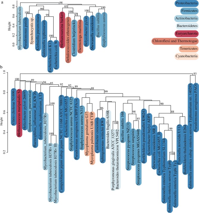
To analyse the validity of the essentiality results on a large scale, the different models were clustered based on single-reaction essentiality predictions and the different datasets available on DEG [17] were clustered based on the content of essential genes (Fig 1).
Fig 1.
Clustering of (A) simulated and (B) experimental genome-scale essentialities of prokaryotes. Clusters show approximately unbiased p-values greater than 80% calculated by multiscale bootstrap re-sampling with 1000 replicas (see Methods for details). Phyla are coloured according to the taxonomical representation of the prokaryotic tree of life built with iTOL [78] (tree in S2 Fig).
In the case of the simulated essentiality (Fig 1A), strongly supported clusters (more than 80% of 1000 bootstrap replicas) are phylogenetically consistent at the level of the phylum–they cluster in a statistically significant manner with at least one sister species–with the exception of the models of C. beijerinckii and P. putida. H. pylori and S. oneidensis show up in the same cluster, but not together with the rest of the Proteobacteria, but the two high-level clusters (that exclude M. tuberculosis with a p-value of 98%) are not statistically supported (p-values of 58 and 62% for the left and right cluster respectively). The lower number of available exchange reactions in the models of H. pylori, S. oneidensis and P. putida (74, 95 and 89 respectively) compared with the models for other Proteobacteria (K. pneumoniae, E. coli K12, S. typhimurium and E. coli W with 289, 299, 305 and 310 respectively) points to a justification for these results, as less exchanges cause more reactions in the network to be essential. C. beijerinckii’s model is also very restricted with regards to exchange reactions, with only 19 metabolic drains available.
Regarding the experimental data (Fig 1B), there is also a pattern of clustering taxonomically related species. One well-supported phylogenetic cluster is that of several gamma and beta-proteobacteria, including Acinetobacter baylyi, dataset II of E. coli K12, three Salmonellas, one Shewanella and one Francisella. Others are the cluster of Bacteroidetes, the cluster with all three datasets of M. tuberculosis and the cluster of the alpha-proteobacteria, Sphingomonas and Caulobacter. The datasets of Pseudomonas aeruginosa PAO1 and Salmonella enterica subsp. Enterica serovar Typhimurium str. 14028S cluster together likely due to not being saturated genome-wide gene-essentiality screens (these datasets are considerably smaller, with 117 and 105 genes respectively–see Table 2 for context). Surprisingly, Firmicutes are spread all across the tree. Also, both E. coli sets are very distant from each other. Although they were performed under rich media conditions, one yielded 609 essential genes and the other only 296 (Table 2). This difference is likely due to the use of different technologies to perform the large-scale assays, the first being random mutagenesis and the screening of mixed populations, and the second the screening of libraries of targeted mutants, as reviewed in [19].
Cofactor metabolism is the most essential subsystem both in simulated and experimental data
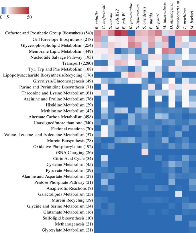
Next, all the essential reactions calculated for the 15 GSMs were mapped to the corresponding metabolic subsystem (see Methods; S1 and S2 Files). Different models show different proportions of essential reactions for each subsystem (Fig 2).
Fig 2. Essentiality for biomass production of each metabolic subsystem in 15 genome-scale manually curated metabolic models.
The colour bar represents the ratio of essential reactions (in that subsystem) to the total of essential reactions in each model (0%—blue; 5%—white; 50%—red). In parenthesis next to the subsystem name is the total of reactions in that subsystem (for all models).
For the majority of the models, the most essential subsystem is that of cofactor and prosthetic group biosynthesis. Nearly 48% of the essential reactions in the simulations with the GSM of E. coli K12–54 reactions–were related with this subsystem (Fig 2). Several of the predicted essential reactions were confirmed to be essential steps in the biosynthesis of the active forms of cofactors that cannot be directly uptaken, e.g. dihydrofolate synthase and dihydrofolate reductase for the biosynthesis of tetrahydrofolate and derivatives [81], NAD kinase for obtaining NADP [82] and riboflavin synthase in some species [83]. For M. tuberculosis, D. ethenogenes, S. typhimurium and K. pneumoniae the most represented subsystems were glycerophospholipid or membrane lipids metabolism (30.5, 25.1, 21.8 and 29.2% of all essential reactions, respectively). Discrepancies regarding results for each individual model are not only related with differences in the metabolic network but are also dependent on the formulations of the biomass equation (e.g. the biomass equation in Klebsiella pneumoniae’s model lacks cofactors) [84].
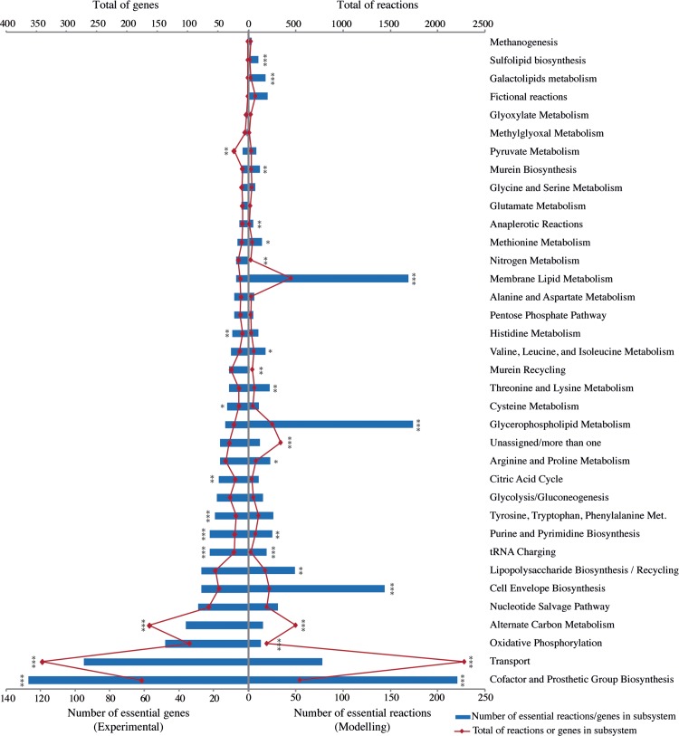
To validate the predictions of essentiality of metabolic subsystems obtained with GSMs, each experimentally essential gene in DEG was annotated according to its function, using the same system used in GSM’s. This system (see Methods) covered more annotations when compared with COG annotations (1363 essential metabolic genes annotated in total, when compared with a total of 906 unique metabolic COGs–S1 Fig). Moreover, this curated dataset could be directly compared to the modelling results and included some genes annotated in the “General function prediction only” COG category. After annotation, all unique essential genes were identified and the same was done for essential reactions in the models. Both the total number of unique essential reactions (modelling) or genes (experimental) varies significantly between subsystems (Fig 3). The total of reactions and genes in each subsystem (for both modelling and experimental data, respectively) also varies significantly. To test for independence of the totals of essentials from the sizes of the subsystems, we performed a Fisher’s exact test, and for the majority of subsystems the null hypothesis of dependence was rejected (p-value less than 0.05).
Fig 3. Number of essential reactions predicted by genome-scale metabolic models and essential genes in genome-scale experimental assays for different metabolic subsystems.
Each reaction and gene were counted only once, even if present (or present and essential) in more than one model or experimental dataset. Single, double and triple asterisks indicate p-values less than 0.05, 0.01 and 0.0001, respectively, after a Fisher's exact test for count data testing independence of the number of essential vs the number of non-essential mapped genes and reactions.
In Fig 3 the subsystem of Cofactor and Prosthetic Groups biosynthesis appears isolated with the maximum number of unique essential enzymes both in experimental and modelling data. Three subsystems show a striking difference in the ranking between experimental and modelling data, being more represented in the latter, all related with membrane and cell wall metabolism. Several justifications can be raised for this difference. First and foremost, often in those subsystems the number of different reactions that can be encoded by the same gene is high [85], (thus there will be several essential reactions for each essential gene). For instance, in the model of Synechocystis there are twelve essential reactions related with fatty acid biosynthesis all encoded by the same gene, fabZ (sll1605 in the model), a gene that is essential in rich medium experimentally, but in that case, counted only once. Similarly, all of the twenty essential fatty acid synthase reactions in the model of M. tuberculosis are encoded by only 3 different genes: Rv1663 and Rv1662 or Rv2940c. There might also be some lack of integration in nomenclature of the reactions in these subsystems in the different GSMs that should not contribute significantly as all models follow the same nomenclature scheme, which was still manually curated for the essential reactions.
Conservation of essentiality validates predictions by GSMs with experimental data and pinpoints specific model gaps
To further explore essentiality at the level of genes and reactions, the conservation of essentiality across models and experimental datasets was analysed for each reaction and gene in each metabolic subsystem. Strikingly, no reaction was essential in all the models analysed. Three reactions annotated within aromatic amino acids metabolism (shikimate kinase, 3-phosphoshikimate 1-carboxyvinyltransferase and chorismate synthase) were essential in all models except for K. pneumoniae. Although there are differences in the models regarding the capacity to uptake aromatic amino acids, this does not justify the observed differences in terms of essentiality. The notable difference between the model of K. pneumoniae and the others is that it lacks cofactors and prosthetic groups in its biomass equation. Those three reactions correspond to the three last steps in the synthesis of chorismate, which is part of the shikimate pathway, which connects central metabolism with aromatic amino acids metabolism. However, this pathway is also the route taken to synthesize several other compounds in the cell, including quinones and folates [86], which are not present in the biomass equation of Klebsiella, which is likely the cause for the difference in the results as discussed in [84] and above.
Reactions essential in several models annotated within the cofactor and prosthetic group biosynthesis subsystem are dihydrofolate synthase, essential in 13 out of the 15 models, and dihydrofolate reductase and NAD kinase, both essential in 12 models. These are related with the biosynthesis of folates and the phosphorylation of NAD to produce NADP. Two reactions involved in the salvage pathways of nucleotides–the biosynthesis of GDP and dTTP—were also essential for 14 models. Three reactions related with the biosynthesis of cell wall components are essential in all models except for B. subtilis and D. ethenogenes—glucosamine-1-phosphate N-acetyltransferase, phosphoglucosamine mutase and UDP-N-acetylglucosamine 1-carboxyvinyltransferase. Acetyl-CoA carboxylase, related with membrane lipid metabolism, is essential in 12 of the 15 models. One reaction not assigned to any subsystem, the HCO3- equilibration reaction, was essential in 11 of all 15 models.
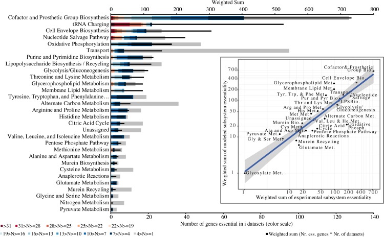
To overview the relationship between modelling and experimental results, the conservation of essentiality for each set of results was compared. The inset plot in Fig 4 shows the high correlation obtained between the weighted essentiality for each subsystem between simulated and experimental data. However, there are some differences to be noted. Firstly, regarding the tRNA charging subsystem, it is modelled in only one GSM (S. oneidensis, Fig 2). This causes this category to appear much more evidently as the second most conserved essential subsystem in experimental data, in contrast with the low result in the simulations of GSMs. It is expected that future GSMs will include this subsystem, but for the sake of comparison of these results, it was excluded from the correlation.
Fig 4. Conservation of essentiality of metabolic subsystems in 36 large-scale gene essentiality datasets and correlation with modelling predictions (inset).
Red indicates highest conservation (genes essential in more than 31 datasets) and grey the least (essential in less than 4 datasets); black bar: weighted sum of essential genes given the number of datasets in which they are essential (see Methods). Inset plot: Correlation between modelling and experimental genome-scale essentiality data at subsystem level (adjusted R2 of 0.821, Pearson correlation coefficient of 0.909 with p-value 9.65e-14); axes are represented in log scale for visualization purposes only.
Interestingly, regarding the three highly essential reactions in modelling results related with chorismate biosynthesis, these were not found as significantly essential in experimental data. It is known that in minimal media the knock-out of chorismate synthase (aroC) in E. coli impairs growth [87]. The non-essentiality of this enzyme in the rich media analysed here indicates that there must be a compound in the media compensating for its absence. It has been shown that, when provided with p-aminobenzoic acid (PABA), para-hydroxybenzoic acid (PHBA) or a combination of a precursor from PABA with a non-biological catalyst, the growth of E. coli aroC mutant in M9 minimal medium can be rescued [88]. PABA and its derivatives cannot be uptaken in the genome-scale model of Escherichia coli or any other of the working set. Transporters for these compounds or others that might compensate in rich media for lethal phenotypes in minimal media remain to be integrated in the genome-scale metabolic models and further explored.
Again, the subsystem of cofactor and prosthetic groups metabolism has the highest number of reactions appearing as essential in more datasets in experimental data, in accordance with modelling data. Dihydrofolate synthase and reductase (highest ranking in modelling) are essential in 11 and 25 experimental datasets, respectively, indicating that either several organisms can overcome the lack of both enzymes by intermediate pathways not yet modelled, or that the experimental assays have produced false negatives (see the differences between the sizes of the two experimental assays for E. coli in Table 2 discussed above; a special case, dihydrofolate synthase, is further explored in the Discussion). NAD kinase appears as highly essential, also in accordance with simulations, in 24 datasets. Several other genes encoding for enzymes essential for the biosynthesis of cofactors are highly essential for cell viability experimentally, even in rich media (eg. nadE for NAD; coaD and coaE for coenzyme A; hemC for tetrapyrroles; dxr for isoprenoids). Cell envelope biosynthesis genes follow as the third most conserved essential functional module, in accordance with the modelling results as well.
Core conserved metabolic subsystems
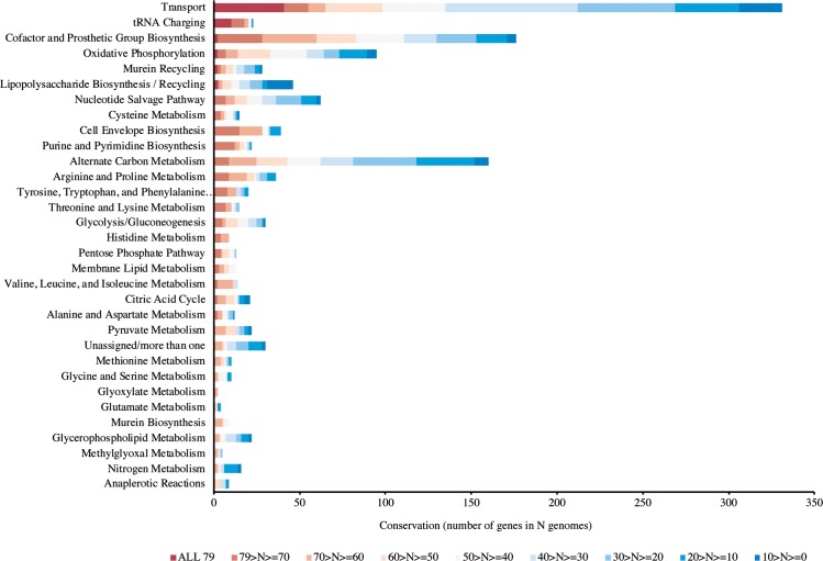
Based on the premises of evolutionary parsimony and orthology [28], we proceeded to the analysis at a large scale of the conservation of metabolic genes in the prokaryotic tree of life to infer potential ancestral metabolic functions. Seventy-nine genomes were assayed, representing all the known prokaryotic phyla with a fully sequenced genome (see Methods for details). A phylogenetic tree with these 79 species is available in S2 Fig. All annotated metabolic genes of E. coli K12 were used as queries to search the set of genomes for conserved metabolic genes and respective functions. The results on conservation of metabolic genes are summarized in Fig 5.
Fig 5. Conservation of metabolic subsystems in genomes of all prokaryotic phyla with at least one fully sequenced genome.
Dark red indicates the highest conservation (genes that are present in all 79 genomes accessed) and dark blue the least (present in less than 10 genomes).
The metabolic subsystem with more prevalent genes is Transport, followed by the tRNA charging subsystem with 18 nearly universal aminoacyl-tRNA synthetases. It should be noted though that nearly all of the 41 transport genes conserved in all 79 genomes correspond to ABC transporters (S1 Table) with a ubiquitous ATP-binding domain. Two genes involved in oxidative phosphorylation were also conserved in all genomes analysed: atpA and atpD (ATP synthase subunits alpha and beta, respectively). In the subsystem of cofactors and prosthetic group biosynthesis, glutX and sufC were also conserved in all genomes analysed. It should be noted that glutX corresponds to a tRNA charging protein, a glutamyl-tRNA synthetase involved in the biosynthesis of heme, that should have a double annotation; sufC is an atypical cytoplasmic ABC/ATPase, required for the assembly of iron-sulphur clusters [89]. Although the vast majority of genes found conserved in all the genomes analysed correspond to ABC ubiquitous domains, the high conservation (between 70 and 79 genomes) of 214 other genes is still prominent. Twenty-eight of those are classified in the subsystem of cofactor and prosthetic group biosynthesis genes (Fig 5, S2 Table).
Common essential genes are rarer and prone to be highly conserved, contrarily to common non-essential genes
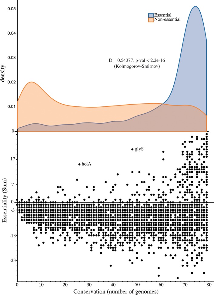
On a first look, there is no correlation between essentiality (Fig 4) and conservation (Fig 5) at the individual gene level. The same is even more evident in the case of the highly conserved ABC domains in transporters (S1 Table), with the majority (20/33) not being essential in any dataset in DEG. This substantiates the fact that highly conserved genes are not necessarily highly essential, likely due to genetic redundancy (see Discussion). The only subsystem for which the correlation is positive and significant (Pearson coefficient 0.95, p-value 1.07e-06) is Membrane Lipid Metabolism. To assess the unbiased relationship between essentiality and conservation at the individual gene level, we compared data on conservation with the experimental data on essentiality. For this purpose, we calculated a sum score for each gene’s experimental essentiality and compared it with its conservation (Methods, Fig 6). This allows for measuring the level of evidence of essentiality for each gene individually with precision.
Fig 6. Conservation and essentiality of protein-encoding genes.
Conservation is calculated as the number of genomes where a gene is present and essentiality as the number of datasets where a gene is essential minus the datasets where it is non-essential. The full list of genes is given in S3 File. In the bottom panel, all data is plotted, with conservation on the x axis and essentiality on the y axis. The top panel depicts the density distribution of the conservation of genes (on the shared x axis) with a sum of essentiality larger than 0 –blue–and that of the genes with a sum of essentiality equal or smaller than 0 –orange. The results of a Kolmogorov–Smirnov test for the independence of both distributions are shown, D for the value of the test statistic and the p-value of the test.
In the bottom panel of Fig 6, the vast majority of genes lie on the bottom area of the plot (sum of essentiality below zero), meaning that they show up in more datasets as non-essential than as essential. However, on the top side of the same plot, where the genes with a positive sum of essentiality lie, the clear majority are shifted to the right–meaning that they are highly conserved. There are some interesting outliers, such as glyS–glycine-tRNA ligase–highlighted in Fig 6. This corresponds to one instance in which the monophyly rule is violated: E. coli’s type is common for most bacteria, but another type is common to some other bacteria, archaea and eukarya [90,91]. Another interesting case is holA, also highlighted, a DNA polymerase delta subunit that is also divergent in its evolutionary history [50,92] but which has been poorly studied. Upon splitting the genes into two sets–those with a positive sum of essentiality and those with a null or negative sum, two very different distributions are obtained, shown in the top panel of Fig 6. A Kolmogorov-Smirnov test states on the independence of both distributions–essential genes are much more likely to be conserved, whereas non-essential genes can or not be highly conserved. The full data on conservation and essentiality sums can be found in S3 File.
Discussion
The integration done here was, to our knowledge, the first of the kind for a wide variety of phyla of the bacteria and archaea domains, encompassing experimental phenotypic data, results of large-scale computational simulations and sequence data. The experimental genome-scale essentiality data reveal that approximately 25% of prokaryotic essential genes encode for unknown or general functions (categories S and R in S1 Fig), which is a strong warning on the need for experimental studies on the phenotype of these essential proteins for prokaryotic physiology. Moreover, the organisms for which genome-wide essentiality data are available are relatively scarce. While more experimental data are not available, computational models can be valuable tools aiding in the task of decoding prokaryotic metabolism. It is well known that GSMs are limited by the quality of the genome annotations, the formulation of the biomass equation and the pre-defined environmental conditions and other modelling artefacts [27,84], of which the impact in our results we expand below. Here we tried to reduce the impact of these limitations by basing the choice of the models on a large survey of high-quality manually curated models [84] from which 15 balanced, validated, comparable models were chosen, that at the same time represented a wide phylogenetic diversity (Table 1). The analysis of common patterns of essentiality filtered out the unique essential reactions that might represent specific errors related with individual models. In this manner, we can find core and common features in the overlap of all models. Using GSMs is particularly interesting in this sense, as they allow us to step one level above that of genomes and all their redundancy in the form of isozymes, duplications and confusing phylogenetic events as lateral gene transfers and gene losses. Manually curated GSMs are not mere reflections of genomes–they include thorough revisions of the network and addition of necessary reactions that are encoded by unknown genes or spontaneous chemical transformations. These are also assigned subsystems and counted in our results. Last, as recognized by other authors, the predictive power of comparative analysis can be significantly enhanced by using it within the functional context of pathways and subsystems [19].
The GSMs prediction of which metabolic subsystem has more genes that are commonly essential in multiple species–Cofactor and Prosthetic Group Biosynthesis–was accurate (Fig 4). The exception of the experimentally highly essential tRNA-charging functionality that was not reflected in the simulations is due to the hindrance of just one model including this subsystem [40] but it should be fixed if all the models represent appropriately this subsystem in the future. However, the analysis done here, by integrating experimental data with several different models still allowed us to identify this subsystem as highly essential and conserved (Fig 4 and Fig 5). The problem of the unstandardized biomass composition, evidenced by the GSM of K. pneumoniae not predicting any essential reaction involved in cofactor and prosthetic group biosynthesis due to the fact that none of those compounds is present in the biomass equation (Fig 2) is relevant to the results, a subject that we have already addressed in a recent publication [84]. Due to the incompleteness of the networks, it was not possible to complete the biomass equations with the missing cofactors without an impractical manual editing and curation of most models. However, considering the results obtained here, this incompleteness could readily be identified (Fig 2) and did not impair the prediction of an overwhelming majority of essential reactions related with the subsystem of cofactor and prosthetic group biosynthesis (Fig 4)–the overlap of all other models and the experimental data reveals the conserved essentiality of this subsystem.
The comparison of the modelling and experimental results can help raise specific hypotheses and directions for more detailed investigation, as discussed above for the essentiality of chorismate synthase in GSMs. In the analysed experimental datasets obtained in rich media, chorismate synthase is not essential, but is has been shown that in minimal media in E. coli the knock-out of its gene impairs growth [87] that can be restored with p-aminobenzoic acid (PABA) or derivatives [88]. The transporters for these compounds should be added to the models, and we expect that a curated analysis of experimental studies of auxotrophies in the literature can point several more additions to GSMs that will improve predictions at the gene-level. Moreover, these results point also to the necessity of performing more often essentiality experiments in defined media. On the other direction, genome-scale models can also indicate improvements for experimental assays. At the moment of writing of this manuscript, a new genome-wide screen for Bacillus subtilis was published [93], where folC (dihydrofolate synthase) was found to be essential, confirming our modelling results and contradicting the previous experimental results for B. subtilis that were used here [50].
The analysis of conservation of metabolic genes here was the first performed using a manually curated annotation system for metabolic pathways and subsystems, with the most complete genome-scale metabolic model of a prokaryote to date [49]. Regarding inferences on ancestry, there are some limitations to our approach. We chose to use a single e-value threshold in a local BLAST–this might be a lax threshold, but at the same time it allows us to recover potential very ancient homologs, tracing back all the way to the Last Universal Common Ancestor (LUCA), and not to incur in debates about in and out-paralogs. Moreover, we used only a sample of prokaryotic genomes– 79 –although we made sure to include representatives of all sequenced phyla to date (S2 Fig), and we took a stringent threshold to indicate and not affirm ancestry (at least 70 out of 79 genomes). Looking at the conservation of subsystems (Fig 5) also allows us to overcome the phylogenetic distribution bias. On another note, looking only for universal genes as markers of ancestry can be a limited approach, due to the phenomena of gene loss and lateral gene transfer–ideally, phylogenetic trees should be built for all genes. A recent study used an innovative and large-scale approach to infer on the genome of LUCA, building all trees for protein families based on 1981 prokaryotic genomes [94]. Interestingly, although using a completely alternative approach, the study also concluded on tRNA charging and cofactor metabolism as being ancient subsystems. These findings corroborate that the genes identified here as present in all genomes of all representative phyla are most likely genes present in the last common ancestor [28]. Overall, our results of high conservation of the tRNA charging system, Transport and Oxidative Phosphorylation point to a last common ancestor metabolic network of prokaryotes where most of the nutrients were uptaked with nonspecific transporters at the expense of ATP and in which tRNA charging was already present. The results also suggest that the catalytic role of cofactors and prosthetic groups was a coin highly sought for in early prebiotic systems still maintained today, as this is the most conserved metabolic subsystem after transport and tRNA charging. It is highly likely that genes encoding for enzymes aiding in cofactor biosynthesis were selected for early in primordial evolution, as was suggested elsewhere for the origin of anabolic pathways in prebiotic systems [95].
This work expanded considerably on previous related studies regarding the relationship between gene conservation and essentiality in width and depth. The demonstration that essential genes are more evolutionary conserved that non-essential [22], corroborated later with more datasets [21], used the ratio of non-synonymous substitutions to synonymous substitutions in the genomes to estimate conservation (Ka/Ks). Here, 36 experimentally essential datasets were used, that included one Archaea (Table 2). The conservation was analysed by looking at the presence of each gene in 79 genomes that were manually selected to represent all the phyla with one fully-sequenced genome in the prokaryotic tree of life. Because each gene might be essential in some datasets in DEG, non-essential in others and not assayed in yet others, instead of analysing essential genes separately from non-essential as in the two aforementioned studies, we used a measure of essentiality for each gene (sum of essentiality) that takes into account the datasets where it shows up as essential and those where it is non-essential (Fig 6). The results show that genes with a positive sum of essentiality (more datasets showing essential than non-essential) are much scarcer than those with a negative sum; however, it is much more likely that they are highly conserved. For genes with a negative sum of essentiality, there is no tendency for high or low conservation, with a uniform distribution of these genes for all the values of conservation (corroborating results by Fang et al. [92]). We also expanded on previous studies [21,22] by integrating in silico simulations and functional assessment of the data, with the conclusion that with the exception of the tRNA charging subsystem, the majority of highly conserved genes related with transport and cofactor biosynthesis are not highly essential (Fig 6, S1 and S2 Tables). These two subsystems show low single-gene essentiality most likely due to metabolic redundancy caused by known alternative metabolic routes (for which multiple knock-outs ought to be performed to test for subsystem-level essentiality) complemented with enzymatic activities not yet known (supported by the percentage of genes with general function prediction only S1 Fig) that might also include promiscuous enzymes [96]. The remarkable redundancy of metabolic networks is reflected in the resilience and robustness of prokaryotic life for the billions of years that it has inhabited Earth.
Supporting information
COG metabolic functional categories are less detailed than those used in the annotation of metabolic models: both the “Energy production and Conversion” and “Amino acid transport and metabolism” functional categories encompass several of those that are detailed within GSMs, except for the “Transport” category lumped in GSMs, and distributed for each of the major biomolecules in the COG system. Misleading COG annotations included thiO, an essential gene for the biosynthesis of thiamine diphosphate annotated in category E (Amino acid transport and metabolism) and csd, a cysteine desulfurase, essential in 7 datasets and involved in the formation of Fe-S clusters, also annotated in category E.
(PDF)
(PDF)
The list includes genes conserved in all 79 prokaryotic genomes analysed. Essentiality is given as the number of datasets in DEG (out of 36) in which each gene is essential. The description is that of the corresponding annotated ORF in the genome of E. coli K12.
(PDF)
Essentiality is given as the number of datasets in DEG (out of 36) in which each gene is essential. Conservation is given as the percentage of the 79 genomes where a significant homolog for this gene was found. The description is that of the corresponding annotated ORF in the genome of E. coli K12. Biosynthesized cofactors were manually retrieved from the detailed information available in the Metacyc database.
(PDF)
Presence-absence matrix of essential reactions in the 15 GSMs used.
(XLSX)
Lists of original subsystem classifications and corresponding translation for all models and all reactions.
(XLSX)
List of genes with corresponding scores of essentiality and conservation.
(XLSX)
Acknowledgments
We thank Fábio Ramalho and Sergej Andrejev for crucial assistance with bioinformatics tools, in particular with the Biopython package and FBA simulations, respectively.
Data Availability
The experimental essentiality datasets used in this study are listed within the paper with the original references and are also available in accessible formats in the OGEE (http://ogee.medgenius.info/downloads/) and DEG databases (http://tubic.org/deg/). The genome-scale metabolic models used are listed in the paper with the corresponding reference from where they can be obtained in SBML format. Supplementary data including the results of simulations, translations for metabolic subsystems and the analysis of genetic conservation are provided in supplementary files.
Funding Statement
This work was supported by grants from: the Fundação para a Ciência e a Tecnologia (http://www.fct.pt) with award number UID/BIO/04469/2013, the European Regional Development Fund (http://www.norte2020.pt) with award number NORTE-01-0145-FEDER-000004 (https://www.ceb.uminho.pt/Projects/Details/6040), Horizon 2020 (https://ec.europa.eu/programmes/horizon2020) with award number 686070 (http://dd-decaf.eu/) and COMPETE 2020 with award number POCI-01-0145-FEDER-006684 to JCX and IR and the Fundação para a Ciência e a Tecnologia (http://www.fct.pt) with award number SFRH/BD/81626/2011 to JCX. The funders had no role in study design, data collection and analysis, decision to publish, or preparation of the manuscript.
References
- 1.Kauffman S. At Home in the Universe: The Search for the Laws of Self-Organization and Complexity. Oxford University Press; 1995. [Google Scholar]
- 2.Rasmussen S, Bedau MA, Chen L, Deamer D, Krakauer DC, Packard NH, et al. , editors. Protocells. 1st ed London: The MIT Press; 2008. [Google Scholar]
- 3.Schuster P. How does complexity arise in evolution:Nature’s recipe for mastering scarcity, abundance, and unpredictability. Complexity. 1996;2: 22–30. [Google Scholar]
- 4.Kim KM, Caetano-Anollés G. The proteomic complexity and rise of the primordial ancestor of diversified life. BMC Evol Biol. 2011;11: 140 10.1186/1471-2148-11-140 [DOI] [PMC free article] [PubMed] [Google Scholar]
- 5.Oltvai ZN, Barabási A-L. Systems biology. Life’s complexity pyramid. Science. 2002;298: 763–764. 10.1126/science.1078563 [DOI] [PubMed] [Google Scholar]
- 6.Peretó J. Out of fuzzy chemistry: from prebiotic chemistry to metabolic networks. Chem Soc Rev. 2012;41: 5394 10.1039/c2cs35054h [DOI] [PubMed] [Google Scholar]
- 7.Skouloubris S, Thiberge JM, Labigne A, De Reuse H. The Helicobacter pylori UreI protein is not involved in urease activity but is essential for bacterial survival in vivo. Infect Immun. 1998;66: 4517–21. [DOI] [PMC free article] [PubMed] [Google Scholar]
- 8.Fayet O, Ziegelhoffer T, Georgopoulos C. The groES and groEL heat shock gene products of Escherichia coli are essential for bacterial growth at all temperatures. J Bacteriol. 1989;171: 1379–85. [DOI] [PMC free article] [PubMed] [Google Scholar]
- 9.Wu J, Ohta N, Zhao J-L, Newton A. A novel bacterial tyrosine kinase essential for cell division and differentiation. Proc Natl Acad Sci. 1999;96: 13068–13073. 10.1073/pnas.96.23.13068 [DOI] [PMC free article] [PubMed] [Google Scholar]
- 10.Gerdes SY, Scholle MD, Campbell JW, Balazsi G, Ravasz E, Daugherty MD, et al. Experimental Determination and System Level Analysis of Essential Genes in Escherichia coli MG1655. J Bacteriol. 2003;185: 5673–5684. 10.1128/JB.185.19.5673-5684.2003 [DOI] [PMC free article] [PubMed] [Google Scholar]
- 11.Gallagher LA, Ramage E, Jacobs MA, Kaul R, Brittnacher M, Manoil C. A comprehensive transposon mutant library of Francisella novicida, a bioweapon surrogate. Proc Natl Acad Sci U S A. 2007;104: 1009–14. 10.1073/pnas.0606713104 [DOI] [PMC free article] [PubMed] [Google Scholar]
- 12.de Berardinis V, Vallenet D, Castelli V, Besnard M, Pinet A, Cruaud C, et al. A complete collection of single-gene deletion mutants of Acinetobacter baylyi ADP1. Mol Syst Biol. 2008;4: 174 10.1038/msb.2008.10 [DOI] [PMC free article] [PubMed] [Google Scholar]
- 13.Cameron DE, Urbach JM, Mekalanos JJ. A defined transposon mutant library and its use in identifying motility genes in Vibrio cholerae. Proc Natl Acad Sci U S A. 2008;105: 8736–41. 10.1073/pnas.0803281105 [DOI] [PMC free article] [PubMed] [Google Scholar]
- 14.Langridge GC, Phan M-D, Turner DJ, Perkins TT, Parts L, Haase J, et al. Simultaneous assay of every Salmonella Typhi gene using one million transposon mutants. Genome Res. 2009;19: 2308–16. 10.1101/gr.097097.109 [DOI] [PMC free article] [PubMed] [Google Scholar]
- 15.Chaudhuri RR, Allen AG, Owen PJ, Shalom G, Stone K, Harrison M, et al. Comprehensive identification of essential Staphylococcus aureus genes using Transposon-Mediated Differential Hybridisation (TMDH). BMC Genomics. 2009;10: 291 10.1186/1471-2164-10-291 [DOI] [PMC free article] [PubMed] [Google Scholar]
- 16.Chen W-H, Minguez P, Lercher MJ, Bork P. OGEE: an online gene essentiality database. Nucleic Acids Res. 2012;40: D901–6. 10.1093/nar/gkr986 [DOI] [PMC free article] [PubMed] [Google Scholar]
- 17.Luo H, Lin Y, Gao F, Zhang C-TT, Zhang R. DEG 10, an update of the database of essential genes that includes both protein-coding genes and noncoding genomic elements. Nucleic Acids Res. 2014;42: 574–580. 10.1093/nar/gkt1131 [DOI] [PMC free article] [PubMed] [Google Scholar]
- 18.Rocha EPC, Danchin A. Gene essentiality determines chromosome organisation in bacteria. Nucleic Acids Res. 2003;31: 6570–6577. 10.1093/nar/gkg859 [DOI] [PMC free article] [PubMed] [Google Scholar]
- 19.Gerdes S, Edwards R, Kubal M, Fonstein M, Stevens R, Osterman A. Essential genes on metabolic maps. Curr Opin Biotechnol. 2006;17: 448–56. 10.1016/j.copbio.2006.08.006 [DOI] [PubMed] [Google Scholar]
- 20.Zhang X, Peng C, Zhang G, Gao F. Comparative analysis of essential genes in prokaryotic genomic islands. Sci Rep. Nature Publishing Group; 2015;5: 12561 10.1038/srep12561 [DOI] [PMC free article] [PubMed] [Google Scholar]
- 21.Luo H, Gao F, Lin Y. Evolutionary conservation analysis between the essential and nonessential genes in bacterial genomes. Sci Rep. Nature Publishing Group; 2015;5: 13210 10.1038/srep13210 [DOI] [PMC free article] [PubMed] [Google Scholar]
- 22.Jordan IK, Rogozin IB, Wolf YI, Koonin E V. Essential genes are more evolutionarily conserved than are nonessential genes in bacteria. Genome Res. 2002;12: 962–8. 10.1101/gr.87702 [DOI] [PMC free article] [PubMed] [Google Scholar]
- 23.Bordbar A, Monk JM, King ZA, Palsson BO. Constraint-based models predict metabolic and associated cellular functions. Nat Rev Genet. Nature Research; 2014;15: 107–20. 10.1038/nrg3643 [DOI] [PubMed] [Google Scholar]
- 24.Overbeek R, Olson R, Pusch GD, Olsen GJ, Davis JJ, Disz T, et al. The SEED and the Rapid Annotation of microbial genomes using Subsystems Technology (RAST). Nucleic Acids Res. 2014;42: D206–14. 10.1093/nar/gkt1226 [DOI] [PMC free article] [PubMed] [Google Scholar]
- 25.Edwards JS, Palsson BO. The Escherichia coli MG1655 in silico metabolic genotype: Its definition, characteristics, and capabilities. Proc Natl Acad Sci. 2000;97: 5528–5533. 10.1073/pnas.97.10.5528 [DOI] [PMC free article] [PubMed] [Google Scholar]
- 26.Oberhardt MA, Palsson BØ, Papin JA. Applications of genome-scale metabolic reconstructions. Mol Syst Biol. Department of Biomedical Engineering, University of Virginia, Health System, Charlottesville, VA, USA.; 2009;5 10.1038/msb.2009.77 [DOI] [PMC free article] [PubMed] [Google Scholar]
- 27.Ebrahim A, Almaas E, Bauer E, Bordbar A, Burgard AP, Chang RL, et al. Do genome-scale models need exact solvers or clearer standards? Mol Syst Biol. EMBO Press; 2015;11: 831–831. doi: 10.15252/msb.20156157 [DOI] [PMC free article] [PubMed] [Google Scholar]
- 28.Koonin E V. Comparative genomics, minimal gene-sets and the last universal common ancestor. Nat Rev Microbiol. 2003;1: 127–36. 10.1038/nrmicro751 [DOI] [PubMed] [Google Scholar]
- 29.Fournier GP, Andam CP, Gogarten JP. Ancient horizontal gene transfer and the last common ancestors. BMC Evol Biol. 2015;15: 70 10.1186/s12862-015-0350-0 [DOI] [PMC free article] [PubMed] [Google Scholar]
- 30.Mampel J, Buescher JM, Meurer G, Eck J. Coping with complexity in metabolic engineering. Trends Biotechnol. 2013;31: 52–60. 10.1016/j.tibtech.2012.10.010 [DOI] [PubMed] [Google Scholar]
- 31.Oh Y-K, Palsson BO, Park SM, Schilling CH, Mahadevan R. Genome-scale Reconstruction of Metabolic Network in Bacillus subtilis Based on High-throughput Phenotyping and Gene Essentiality Data. J Biol Chem. 2007;282: 28791–28799. 10.1074/jbc.M703759200 [DOI] [PubMed] [Google Scholar]
- 32.Milne CB, Eddy JA, Raju R, Ardekani S, Kim P-J, Senger RS, et al. Metabolic network reconstruction and genome-scale model of butanol-producing strain Clostridium beijerinckii NCIMB 8052. BMC Syst Biol. 2011;5: 130 10.1186/1752-0509-5-130 [DOI] [PMC free article] [PubMed] [Google Scholar]
- 33.Becker S a, Palsson BØ. Genome-scale reconstruction of the metabolic network in Staphylococcus aureus N315: an initial draft to the two-dimensional annotation. BMC Microbiol. 2005;5: 8 10.1186/1471-2180-5-8 [DOI] [PMC free article] [PubMed] [Google Scholar]
- 34.Feist AM, Henry CS, Reed JL, Krummenacker M, Joyce AR, Karp PD, et al. A genome-scale metabolic reconstruction for Escherichia coli K-12 MG1655 that accounts for 1260 ORFs and thermodynamic information. Mol Syst Biol. 2007;3: 121 10.1038/msb4100155 [DOI] [PMC free article] [PubMed] [Google Scholar]
- 35.Archer CT, Kim JF, Jeong H, Park JH, Vickers CE, Lee SY, et al. The genome sequence of E. coli W (ATCC 9637): comparative genome analysis and an improved genome-scale reconstruction of E. coli. BMC Genomics. BioMed Central Ltd; 2011;12: 9 10.1186/1471-2164-12-9 [DOI] [PMC free article] [PubMed] [Google Scholar]
- 36.Thiele I, Vo TD, Price ND, Palsson BØ. Expanded Metabolic Reconstruction of Helicobacter pylori (i IT341 GSM / GPR): an In Silico Genome-Scale Characterization of Single- and Double-Deletion Mutants. J Bacteriol. 2005;187: 5818–5830. 10.1128/JB.187.16.5818-5830.2005 [DOI] [PMC free article] [PubMed] [Google Scholar]
- 37.Liao YC, Huang TW, Chen FC, Charusanti P, Hong JSJ, Chang HY, et al. An experimentally validated genome-scale metabolic reconstruction of Klebsiella pneumoniae MGH 78578, iYL1228. J Bacteriol. 2011;193: 1710–1717. 10.1128/JB.01218-10 [DOI] [PMC free article] [PubMed] [Google Scholar]
- 38.Nogales J, Palsson BØ, Thiele I. A genome-scale metabolic reconstruction of Pseudomonas putida KT2440: iJN746 as a cell factory. BMC Syst Biol. 2008;2: 79 10.1186/1752-0509-2-79 [DOI] [PMC free article] [PubMed] [Google Scholar]
- 39.Thiele I, Hyduke DR, Steeb B, Fankam G, Allen DK, Bazzani S, et al. A community effort towards a knowledge-base and mathematical model of the human pathogen Salmonella Typhimurium LT2. BMC Syst Biol. BioMed Central Ltd; 2011;5: 8 10.1186/1752-0509-5-8 [DOI] [PMC free article] [PubMed] [Google Scholar]
- 40.Pinchuk GE, Hill EA, Geydebrekht O V, De Ingeniis J, Zhang X, Osterman A, et al. Constraint-based model of Shewanella oneidensis MR-1 metabolism: a tool for data analysis and hypothesis generation. PLoS Comput Biol. 2010;6: e1000822 10.1371/journal.pcbi.1000822 [DOI] [PMC free article] [PubMed] [Google Scholar]
- 41.Jamshidi N, Palsson BØ. Investigating the metabolic capabilities of Mycobacterium tuberculosis H37Rv using the in silico strain iNJ661 and proposing alternative drug targets. BMC Syst Biol. 2007;1: 26 10.1186/1752-0509-1-26 [DOI] [PMC free article] [PubMed] [Google Scholar]
- 42.Ahsanul Islam M, Edwards E a., Mahadevan R. Characterizing the metabolism of Dehalococcoides with a constraint-based model. PLoS Comput Biol. 2010;6 10.1371/journal.pcbi.1000887 [DOI] [PMC free article] [PubMed] [Google Scholar]
- 43.Nogales J, Gudmundsson S, Knight EM, Palsson BØ, Thiele I. Detailing the optimality of photosynthesis in cyanobacteria through systems biology analysis. Proc Natl Acad Sci U S A. 2012;109: 2678–83. 10.1073/pnas.1117907109 [DOI] [PMC free article] [PubMed] [Google Scholar]
- 44.Zhang Y, Thiele I, Weekes D, Li Z, Jaroszewski L, Ginalski K, et al. Three-dimensional structural view of the central metabolic network of Thermotoga maritima. Science. 2009;325: 1544–1549. 10.1126/science.1174671 [DOI] [PMC free article] [PubMed] [Google Scholar]
- 45.Feist AM, Scholten JCM, Palsson BØ, Brockman FJ, Ideker T. Modeling methanogenesis with a genome-scale metabolic reconstruction of Methanosarcina barkeri. Mol Syst Biol. 2006;2 10.1038/msb4100046 [DOI] [PMC free article] [PubMed] [Google Scholar]
- 46.Savinell JM, Palsson BØ. Network analysis of intermediary metabolism using linear optimization. I. Development of mathematical formalism. J Theor Biol. 1992;154: 421–54. 10.1016/S0022-5193(05)80161-4 [DOI] [PubMed] [Google Scholar]
- 47.Varma A, Palsson BØ. Metabolic Capabilities of Escherichia coli: I. Synthesis of Biosynthetic Precursors and Cofactors. J Theor Biol. 1993; 10.1006/jtbi.1993.1202 [DOI] [PubMed] [Google Scholar]
- 48.Rocha I, Maia P, Evangelista P, Vilaça P, Soares S, Pinto JP, et al. OptFlux: an open-source software platform for in silico metabolic engineering. BMC Syst Biol. 2010;4: 45 10.1186/1752-0509-4-45 [DOI] [PMC free article] [PubMed] [Google Scholar]
- 49.Orth JD, Conrad TM, Na J, Lerman J a, Nam H, Feist AM, et al. A comprehensive genome-scale reconstruction of Escherichia coli metabolism—2011. Mol Syst Biol. 2011;7: 1–9. 10.1038/msb.2011.65 [DOI] [PMC free article] [PubMed] [Google Scholar]
- 50.Kobayashi K, Ehrlich SD, Albertini a, Amati G, Andersen KK, Arnaud M, et al. Essential Bacillus subtilis genes. Proc Natl Acad Sci U S A. 2003;100: 4678–83. 10.1073/pnas.0730515100 [DOI] [PMC free article] [PubMed] [Google Scholar]
- 51.Veeranagouda Y, Husain F, Tenorio EL, Wexler HM. Identification of genes required for the survival of B. fragilis using massive parallel sequencing of a saturated transposon mutant library. BMC Genomics. 2014;15: 429 10.1186/1471-2164-15-429 [DOI] [PMC free article] [PubMed] [Google Scholar]
- 52.Goodman AL, McNulty NP, Zhao Y, Leip D, Mitra RD, Lozupone C a., et al. Identifying Genetic Determinants Needed to Establish a Human Gut Symbiont in Its Habitat. Cell Host Microbe. 2009;6: 279–289. 10.1016/j.chom.2009.08.003 [DOI] [PMC free article] [PubMed] [Google Scholar]
- 53.Moule MG, Hemsley CM, Seet Q, Guerra-Assuncao JA, Lim J, Sarkar-Tyson M, et al. Genome-wide saturation mutagenesis of Burkholderia pseudomallei K96243 predicts essential genes and novel targets for antimicrobial development. MBio. 2014;5: e00926–13. 10.1128/mBio.00926-13 [DOI] [PMC free article] [PubMed] [Google Scholar]
- 54.Baugh L, Gallagher LA, Patrapuvich R, Clifton MC, Gardberg AS, Edwards TE, et al. Combining functional and structural genomics to sample the essential Burkholderia structome. PLoS One. 2013;8: e53851 10.1371/journal.pone.0053851 [DOI] [PMC free article] [PubMed] [Google Scholar]
- 55.Metris A, Reuter M, Gaskin DJH, Baranyi J, van Vliet AHM. In vivo and in silico determination of essential genes of Campylobacter jejuni. BMC Genomics. 2011;12: 535 10.1186/1471-2164-12-535 [DOI] [PMC free article] [PubMed] [Google Scholar]
- 56.Christen B, Abeliuk E, Collier JM, Kalogeraki VS, Passarelli B, Coller JA, et al. The essential genome of a bacterium. Mol Syst Biol. 2011;7: 528 10.1038/msb.2011.58 [DOI] [PMC free article] [PubMed] [Google Scholar]
- 57.Baba T, Ara T, Hasegawa M. Construction of Escherichia coli K-12 in-frame, single-gene knockout mutants: the Keio collection. Mol Syst Biol. 2006;2 10.1038/msb4100050 [DOI] [PMC free article] [PubMed] [Google Scholar]
- 58.Akerley BJ, Rubin EJ, Novick VL, Amaya K, Judson N, Mekalanos JJ. A genome-scale analysis for identification of genes required for growth or survival of Haemophilus influenzae. Proc Natl Acad Sci U S A. 2002;99: 966–71. 10.1073/pnas.012602299 [DOI] [PMC free article] [PubMed] [Google Scholar]
- 59.Salama NR, Shepherd B, Falkow S. Global transposon mutagenesis and essential gene analysis of Helicobacter pylori. J Bacteriol. 2004;186: 7926–35. 10.1128/JB.186.23.7926-7935.2004 [DOI] [PMC free article] [PubMed] [Google Scholar]
- 60.Sarmiento F, Mrazek J, Whitman WB. Genome-scale analysis of gene function in the hydrogenotrophic methanogenic archaeon Methanococcus maripaludis. Proc Natl Acad Sci U S A. 2013; 10.1073/pnas.1220225110 [DOI] [PMC free article] [PubMed] [Google Scholar]
- 61.Sassetti CM, Boyd DH, Rubin EJ. Genes required for mycobacterial growth defined by high density mutagenesis. Mol Microbiol. 2003;48: 77–84. 10.1046/j.1365-2958.2003.03425.x [DOI] [PubMed] [Google Scholar]
- 62.Griffin JE, Gawronski JD, Dejesus MA, Ioerger TR, Akerley BJ, Sassetti CM. High-resolution phenotypic profiling defines genes essential for mycobacterial growth and cholesterol catabolism. PLoS Pathog. 2011;7: e1002251 10.1371/journal.ppat.1002251 [DOI] [PMC free article] [PubMed] [Google Scholar]
- 63.Zhang YJ, Ioerger TR, Huttenhower C, Long JE, Sassetti CM, Sacchettini JC, et al. Global assessment of genomic regions required for growth in Mycobacterium tuberculosis. PLoS Pathog. 2012;8: e1002946 10.1371/journal.ppat.1002946 [DOI] [PMC free article] [PubMed] [Google Scholar]
- 64.Glass JI, Assad-Garcia N, Alperovich N, Yooseph S, Lewis MR, Maruf M, et al. Essential genes of a minimal bacterium. Proc Natl Acad Sci U S A. 2006;103: 425–430. 10.1073/pnas.0510013103 [DOI] [PMC free article] [PubMed] [Google Scholar]
- 65.French CT, Lao P, Loraine AE, Matthews BT, Yu H, Dybvig K. Large-scale transposon mutagenesis of Mycoplasma pulmonis. Mol Microbiol. 2008;69: 67–76. 10.1111/j.1365-2958.2008.06262.x [DOI] [PMC free article] [PubMed] [Google Scholar]
- 66.Klein B a, Tenorio EL, Lazinski DW, Camilli A, Duncan MJ, Hu LT. Identification of essential genes of the periodontal pathogen Porphyromonas gingivalis. BMC Genomics. 2012. 10.1186/1471-2164-13-578 [DOI] [PMC free article] [PubMed] [Google Scholar]
- 67.Gallagher LA, Shendure J, Manoil C. Genome-scale identification of resistance functions in Pseudomonas aeruginosa using Tn-seq. MBio. 2011;2: e00315–10. 10.1128/mBio.00315-10 [DOI] [PMC free article] [PubMed] [Google Scholar]
- 68.Liberati NT, Urbach JM, Miyata S, Lee DG, Drenkard E, Wu G, et al. An ordered, nonredundant library of Pseudomonas aeruginosa strain PA14 transposon insertion mutants. Proc Natl Acad Sci U S A. 2006;103: 2833–8. 10.1073/pnas.0511100103 [DOI] [PMC free article] [PubMed] [Google Scholar]
- 69.Barquist L, Langridge GC, Turner DJ, Phan M-D, Turner AK, Bateman A, et al. A comparison of dense transposon insertion libraries in the Salmonella serovars Typhi and Typhimurium. Nucleic Acids Res. 2013;41: 4549–64. 10.1093/nar/gkt148 [DOI] [PMC free article] [PubMed] [Google Scholar]
- 70.Khatiwara A, Jiang T, Sung S-S, Dawoud T, Kim JN, Bhattacharya D, et al. Genome scanning for conditionally essential genes in Salmonella enterica Serotype Typhimurium. Appl Environ Microbiol. 2012;78: 3098–107. 10.1128/AEM.06865-11 [DOI] [PMC free article] [PubMed] [Google Scholar]
- 71.Knuth K, Niesalla H, Hueck CJ, Fuchs TM. Large-scale identification of essential Salmonella genes by trapping lethal insertions. Mol Microbiol. 2004;51: 1729–44. [DOI] [PubMed] [Google Scholar]
- 72.Deutschbauer A, Price MN, Wetmore KM, Shao W, Baumohl JK, Xu Z, et al. Evidence-based annotation of gene function in Shewanella oneidensis MR-1 using genome-wide fitness profiling across 121 conditions. PLoS Genet. 2011;7: e1002385 10.1371/journal.pgen.1002385 [DOI] [PMC free article] [PubMed] [Google Scholar]
- 73.Roggo C, Coronado E, Moreno-Forero SK, Harshman K, Weber J, Van der Meer JR. Genome-wide transposon insertion scanning of environmental survival functions in the polycyclic aromatic hydrocarbon degrading bacterium Sphingomonas wittichiiRW1. Environ Microbiol. 2013;15: 2681–2695. 10.1111/1462-2920.12125 [DOI] [PubMed] [Google Scholar]
- 74.Ji Y, Zhang B, Van SF, Horn Warren P, Woodnutt G, et al. Identification of critical staphylococcal genes using conditional phenotypes generated by antisense RNA. Science. 2001;293: 2266–9. 10.1126/science.1063566 [DOI] [PubMed] [Google Scholar]
- 75.Thanassi JA, Hartman-Neumann SL, Dougherty TJ, Dougherty BA, Pucci MJ. Identification of 113 conserved essential genes using a high-throughput gene disruption system in Streptococcus pneumoniae. Nucleic Acids Res. 2002;30: 3152–3162. 10.1093/nar/gkf418 [DOI] [PMC free article] [PubMed] [Google Scholar]
- 76.Le Breton Y, Belew AT, Valdes KM, Islam E, Curry P, Tettelin H, et al. Essential genes in the core genome of the human pathogen Streptococcus pyogenes. Sci Rep. 2015;5: 9838 10.1038/srep09838 [DOI] [PMC free article] [PubMed] [Google Scholar]
- 77.Xu P, Ge X, Chen L, Wang X, Dou Y, Xu JZ, et al. Genome-wide essential gene identification in Streptococcus sanguinis. Sci Rep. 2011;1: 1–9. 10.1038/srep00001 [DOI] [PMC free article] [PubMed] [Google Scholar]
- 78.Letunic I, Bork P. Interactive tree of life (iTOL) v3: an online tool for the display and annotation of phylogenetic and other trees. Nucleic Acids Res. Oxford University Press; 2016;44: W242–W245. 10.1093/nar/gkw290 [DOI] [PMC free article] [PubMed] [Google Scholar]
- 79.Cock PJA, Antao T, Chang JT, Chapman BA, Cox CJ, Dalke A, et al. Biopython: freely available Python tools for computational molecular biology and bioinformatics. Bioinformatics. 2009;25: 1422–3. 10.1093/bioinformatics/btp163 [DOI] [PMC free article] [PubMed] [Google Scholar]
- 80.Suzuki R, Shimodaira H. Pvclust: An R package for assessing the uncertainty in hierarchical clustering. Bioinformatics. 2006;22: 1540–1542. 10.1093/bioinformatics/btl117 [DOI] [PubMed] [Google Scholar]
- 81.Bermingham A, Derrick JP. The folic acid biosynthesis pathway in bacteria: evaluation of potential for antibacterial drug discovery. BioEssays. 2002;24: 637–648. 10.1002/bies.10114 [DOI] [PubMed] [Google Scholar]
- 82.Magni G, Orsomando G, Raffaelli N. Structural and functional properties of NAD kinase, a key enzyme in NADP biosynthesis. Mini Rev Med Chem. 2006;6: 739–746. 10.2174/138955706777698688 [DOI] [PubMed] [Google Scholar]
- 83.Saeed-Kothe A, Yang W, Mills SD. Use of the riboflavin synthase gene (ribC) as a model for development of an essential gene disruption and complementation system for Haemophilus influenzae. Appl Environ Microbiol. American Society for Microbiology; 2004;70: 4136–43. 10.1128/AEM.70.7.4136-4143.2004 [DOI] [PMC free article] [PubMed] [Google Scholar]
- 84.Xavier JCJC, Patil KR, Rocha I. Integration of Biomass Formulations of Genome-Scale Metabolic Models with Experimental Data Reveals Universally Essential Cofactors in Prokaryotes. Metab Eng. 2017;39: 200–208. 10.1016/j.ymben.2016.12.002 [DOI] [PMC free article] [PubMed] [Google Scholar]
- 85.Rock CO, Jackowski S. Forty years of bacterial fatty acid synthesis. Biochem Biophys Res Commun. 2002;292: 1155–1166. 10.1006/bbrc.2001.2022 [DOI] [PubMed] [Google Scholar]
- 86.Coggins JR, Abell C, Evans LB, Frederickson M, Robinson D a, Roszak a W, et al. Experiences with the shikimate-pathway enzymes as targets for rational drug design. Biochem Soc Trans. 2003;31: 548–552. 10.1042/ [DOI] [PubMed] [Google Scholar]
- 87.Joyce AR, Reed JL, White A, Edwards R, Osterman A, Baba T, et al. Experimental and computational assessment of conditionally essential genes in Escherichia coli. J Bacteriol. 2006;188: 8259–71. 10.1128/JB.00740-06 [DOI] [PMC free article] [PubMed] [Google Scholar]
- 88.Lee Y, Umeano A, Balskus EP. Rescuing auxotrophic microorganisms with nonenzymatic chemistry. Angew Chem Int Ed Engl. 2013;52: 11800–3. 10.1002/anie.201307033 [DOI] [PMC free article] [PubMed] [Google Scholar]
- 89.Nachin L. SufC: an unorthodox cytoplasmic ABC/ATPase required for [Fe-S] biogenesis under oxidative stress. EMBO J. 2003;22: 427–437. 10.1093/emboj/cdg061 [DOI] [PMC free article] [PubMed] [Google Scholar]
- 90.Mazauric M-H, Reinbolt J, Lorber B, Ebel C, Keith G, Giege R, et al. An Example of Non-Conservation of Oligomeric Structure in Prokaryotic Aminoacyl-tRNA Synthetases. Biochemical and Structural Properties of Glycyl-tRNA Synthetase from Thermus thermophilus. Eur J Biochem. 1996;241: 814–826. 10.1111/j.1432-1033.1996.00814.x [DOI] [PubMed] [Google Scholar]
- 91.Woese CR, Olsen GJ, Ibba M, Soll D. Aminoacyl-tRNA Synthetases, the Genetic Code, and the Evolutionary Process. Microbiol Mol Biol Rev. 2000;64: 202–236. 10.1128/MMBR.64.1.202–236.2000 [DOI] [PMC free article] [PubMed] [Google Scholar]
- 92.Fang G, Rocha E, Danchin A. How Essential Are Nonessential Genes? Mol Biol Evol. Oxford University Press; 2005;22: 2147–2156. 10.1093/molbev/msi211 [DOI] [PubMed] [Google Scholar]
- 93.Koo B-M, Kritikos G, Farelli JD, Todor H, Tong K, Kimsey H, et al. Construction and Analysis of Two Genome-Scale Deletion Libraries for Bacillus subtilis. Cell Syst. ASM Press; 2017;4: 291–305.e7. 10.1016/j.cels.2016.12.013 [DOI] [PMC free article] [PubMed] [Google Scholar]
- 94.Weiss MC, Sousa FL, Mrnjavac N, Neukirchen S, Roettger M, Nelson-Sathi S, et al. The physiology and habitat of the last universal common ancestor. Nat Microbiol. Nature Publishing Group; 2016;1: 16116 10.1038/nmicrobiol.2016.116 [DOI] [PubMed] [Google Scholar]
- 95.Fani R, Fondi M. Origin and evolution of metabolic pathways. Phys Life Rev. Elsevier B.V.; 2009;6: 23–52. 10.1016/j.plrev.2008.12.003 [DOI] [PubMed] [Google Scholar]
- 96.Patrick WM, Quandt EM, Swartzlander DB, Matsumura I. Multicopy suppression underpins metabolic evolvability. Mol Biol Evol. 2007;24: 2716–22. 10.1093/molbev/msm204 [DOI] [PMC free article] [PubMed] [Google Scholar]
Associated Data
This section collects any data citations, data availability statements, or supplementary materials included in this article.
Supplementary Materials
COG metabolic functional categories are less detailed than those used in the annotation of metabolic models: both the “Energy production and Conversion” and “Amino acid transport and metabolism” functional categories encompass several of those that are detailed within GSMs, except for the “Transport” category lumped in GSMs, and distributed for each of the major biomolecules in the COG system. Misleading COG annotations included thiO, an essential gene for the biosynthesis of thiamine diphosphate annotated in category E (Amino acid transport and metabolism) and csd, a cysteine desulfurase, essential in 7 datasets and involved in the formation of Fe-S clusters, also annotated in category E.
(PDF)
(PDF)
The list includes genes conserved in all 79 prokaryotic genomes analysed. Essentiality is given as the number of datasets in DEG (out of 36) in which each gene is essential. The description is that of the corresponding annotated ORF in the genome of E. coli K12.
(PDF)
Essentiality is given as the number of datasets in DEG (out of 36) in which each gene is essential. Conservation is given as the percentage of the 79 genomes where a significant homolog for this gene was found. The description is that of the corresponding annotated ORF in the genome of E. coli K12. Biosynthesized cofactors were manually retrieved from the detailed information available in the Metacyc database.
(PDF)
Presence-absence matrix of essential reactions in the 15 GSMs used.
(XLSX)
Lists of original subsystem classifications and corresponding translation for all models and all reactions.
(XLSX)
List of genes with corresponding scores of essentiality and conservation.
(XLSX)
Data Availability Statement
The experimental essentiality datasets used in this study are listed within the paper with the original references and are also available in accessible formats in the OGEE (http://ogee.medgenius.info/downloads/) and DEG databases (http://tubic.org/deg/). The genome-scale metabolic models used are listed in the paper with the corresponding reference from where they can be obtained in SBML format. Supplementary data including the results of simulations, translations for metabolic subsystems and the analysis of genetic conservation are provided in supplementary files.


