Abstract
The understanding of and control over light emission from quantum tunneling has challenged researchers for more than four decades due to the intricate interplay of electrical and optical properties in atomic scale volumes. Here we introduce a device architecture that allows for the disentanglement of electronic and photonic pathways—van der Waals quantum tunneling devices. The electronic properties are defined by a stack of two-dimensional atomic crystals whereas the optical properties are controlled via an external photonic architecture. In van der Waals heterostructures made of gold, hexagonal boron nitride and graphene we find that inelastic tunneling results in the emission of photons and surface plasmon polaritons. By coupling these heterostructures to optical nanocube antennas we achieve resonant enhancement of the photon emission rate in narrow frequency bands by four orders of magnitude. Our results lead the way towards a new generation of nanophotonic devices that are driven by quantum tunneling.
Electrical and optical properties of light emitting devices driven by quantum tunneling are closely intertwined. Parzefall et al. show that these properties can be individually controlled by cointegrating van der Waals heterostructures with optical antennas.
Introduction
The desire to develop an optical implementation of the classical antenna in its role as a transducer between an electronic source and electromagnetic radiation has revived interest in light emission from inelastic electron tunneling (IET). This process, originally discovered in the 1970s1, allows for the conversion of electrical energy into optical excitations on length scales considerably smaller than what is achievable with other optoelectronic devices. Recently, IET-driven optical antennas were demonstrated2,3 and continued development has lead to the achievement of greatly improved device efficiencies4 and directionality5, the development of scalable fabrication processes6,7 as well as the exploration of new applications in sensing8 and catalysis9.
The vast majority of IET studies, both on integrated devices1,10–13 as well as in scanning tunneling microscopy (STM)14–19, are centered around metal-insulator-metal (MIM) tunnel junctions. In these devices the tunnel junction defines both the electrical circuit and the optical mode confinement, which severely limits the degrees of freedom for device design and optimization. Furthermore, as light emission from MIM devices is the result of a two-step process in which the tunneling electrons first excite a propagating or localized surface plasmon polariton (SPP) mode that subsequently decays via radiation, it is difficult to disentangle the influence of optical and electrical device properties on the light emission process.
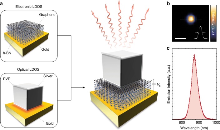
A device structure that allows the optical mode to be defined independently of the tunnel junction would provide additional degrees of freedom for device design, adaptability and optimization of performance. Here we introduce and present a first realization of van der Waals quantum tunneling (vdWQT) devices, hybrid van der Waals heterostructures20,21 comprised of a vertical stack of gold (plasmonic metal), hexagonal boron nitride (h-BN, two-dimensional insulator) and graphene (Gr, two-dimensional semimetal), as illustrated in Fig. 1a. We show that the optical mode confinement in these vdWQT devices can be defined independently of the electrical tunnel junction—setting a new paradigm in interfacing optics and electronics at the nanoscale. As graphene is almost transparent to light; only 2.3% of photons propagating through it are being absorbed22, we observe—in addition to the two-step process—the direct emission of photons from tunnel junctions that are not coupled to MIM cavities. A comparison to a theoretical model allows us to determine IET to be the source of the observed emission.
Fig. 1.
Visualization of the vdWQT device concept. a Illustration (not to scale) of a gold–few-layer h-BN–graphene vdWQT device, integrated with a (silver, PVP-coated) nanocube antenna. In this device configuration, the electronic LDOS is controlled by the hybrid vdW heterostructure whereas the optical LDOS is governed by the nanocube antenna. Applying a voltage Vb across the insulating few-layer h-BN crystal results in antenna-mediated photon emission (wavy arrows) due to quantum tunneling. b, c Measured spatial (b) and spectral (c) photon distribution from a nanocube antenna coupled to a vdWQT device, demonstrating a diffraction-limited spot and a narrow emission spectrum. The inset in b shows a line-cut, featuring a line-width (FWHM) of ~460 nm, close to the expected value of λ/(2NA) ~ 480 nm. Scale bar: 1 μm
We further demonstrate that photon emission from IET can be locally enhanced by coupling to an optical antenna, cf. Fig. 1a. vdWQT device-coupled nanocube antennas emit light from a nanoscopic volume, resulting in the diffraction-limited emission pattern shown in Fig. 1b. Nanocube antennas provide a high local density of optical states (LDOS) and give rise to a narrow emission spectrum, cf. Fig. 1c. On resonance, we achieve an increase of the spectral photon emission rate by more than four orders of magnitude.
To fully understand these results and their implications for future research we first analyze the photon emission from vdWQT devices in the absence of optical antennas before studying the effect of antenna-coupling.
Results
Light from uncoupled vdWQT devices
Figure 2a shows an optical microscope image of a set of devices. They are composed of a vertical stack of a gold electrode, a few-layer h-BN crystal and monolayer graphene (cf. Fig. 1a), assembled on top of a glass (SiO2) substrate. Details on the fabrication process are described in the Methods section. The tunneling devices are formed in the regions where all of the three constituting materials overlap, i.e., at the end of the three left electrodes in Fig. 2a. The electrodes on the right-hand side serve as electrical contacts to the graphene sheet.
Fig. 2.
Light emission from vdWQT devices. a Optical microscope image of a set of devices. The three areas on the left, where gold electrodes (50 nm), h-BN crystal (N = 7) and graphene overlap, form the tunneling devices. Electrodes on the right serve as electrical contacts to the graphene sheet. Scale bar: 10 μm. b Spatial distribution of photons emitted into the air/upper half-space above the graphene sheet from the devices at an applied voltage of 2.0 V. Light is emitted from the entire area of the devices. Scale bar: 5 μm. c Corresponding distribution of photons emitted into to substrate/lower half-space. Light is observed primarily at the edges of the devices. Scale bar: 5 μm. Straight lines in b, c mark the outlines of gold electrodes (gold) and graphene (gray). Intensity units are as measured and not corrected for system efficiency. d Sketch of the vertical cross-section through the device, illustrating the mechanism of light generation. The inelastic component of the tunneling current j couples to the optical modes of the vdWQT device, i.e., the free-space continuum of optical states as well as surface-bound SPP modes (not to scale). Photons emitted into the air half-space give rise to the image in b, SPP scattering at the edges of the gold electrodes generate the image in c
When a voltage between the graphene and gold electrodes is applied, we observe photon emission into both half-spaces, i.e., the air above the device and the glass substrate. Our analysis regarding the origin of this emission is segmented into two parts. First, we analyze our results in terms of the optical modes that are excited. Second, we will determine which physical mechanism causes the mode excitation.
Direct and indirect photon emission
Photons that are emitted into the air half-space give rise to the image shown in Fig. 2b. Evidently, photons are emitted from the entire area of the devices. On the contrary, photons emitted into the glass half-space give rise to the image shown in Fig. 2c, where the emission is localized near the edges of the device. The origin of these images is found by identifying the optical modes of the planar geometry. As we show in Supplementary Note 4, these modes are the radiation continuum of free space and SPP modes bound to the gold surfaces, as illustrated in Fig. 2d (additionally, our geometry supports graphene plasmons which are not considered here, cf. Supplementary Note 4).
On the one hand, tunneling electrons may couple to the radiation continuum, emitting photons without an intermediate excitation. This direct emission process, enabled by the transparency of the top graphene electrode, explains the emission into the top half-space from the device area seen in Fig. 2b. It is important to note that this observation cannot be explained by the random scattering of SPPs by surface roughness as the emission area is restricted to the area of the tunnel junction whereas SPPs are free to propagate across the left edge of the junction area due to the negligible mode mismatch (cf. Supplementary Fig. 3f). On the other hand, photons may also be emitted indirectly as the result of a two-step process. First, a SPP is excited by a tunneling event. Second, since the SPP is bound to the surface it may only emit a photon when it is scattered, that is, at the edges of the gold electrodes. Since photons are preferentially scattered into the medium with the higher refractive index, i.e., glass, we observe photon emission from the edges of the device in Fig. 2c. The relative strength of the coupling to the radiation continuum and the SPP modes is governed by the LDOS associated with each mode. Due to the low mode confinement of the SPP mode the associated LDOS is of the same order of magnitude as the LDOS associated with direct photon emission (cf. Supplementary Note 4), resulting in similar intensities of the two emission pathways, cf. Fig. 2b, c.
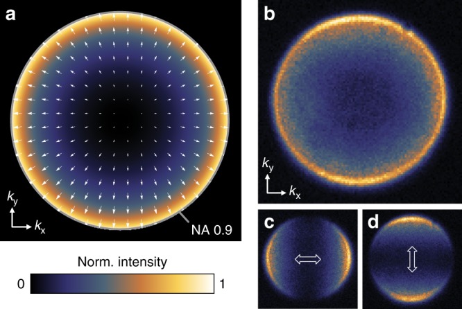
To provide further evidence for the direct photon emission process we analyze the angular distribution of photons emitted into the air half-space, shown in Fig. 3. If photons are indeed directly emitted by tunneling electrons their radiation pattern should resemble the radiation pattern of a vertical dipole above a metal surface (cf. Fig. 2d), as calculated in Fig. 3a23. The calculation shows that photons are primarily emitted into high angles. Furthermore, as indicated by the white arrows, the emission is radially polarized. Experimentally, this distribution is obtained by imaging the back focal plane of the microscope objective that collects the photons. The measured radiation pattern, as well as the decomposition into two orthogonal polarization directions, is shown in Fig. 3b–d. The experimental results stand in excellent agreement with the direct emission mechanism.
Fig. 3.

Radiation pattern of vdWQT device emission. a Calculated emission pattern of an electric point dipole located at a distance of 1 nm above a gold half-space, radiating a free space wavelength of 800 nm. Arrows indicate the position-dependent orientation of the electric field vector. The maximum value of the in-plane wave vector component is given by k||,max = NA × k0 where NA = 0.9 is the numerical aperture of the objective and k0= 2π/λ. b Measured angular distribution of photons emitted into the upper half-space. c, d As b when measured through a polarizer whose transmitting axis is oriented horizontally/vertically, respectively
Mode excitation by quantum tunneling
The remaining question relates to the nature of the emission: What is the physical process that leads to the observed light emission? While it is generally accepted that inelastic electron tunneling—where one1,15 or multiple24,25 electrons generate an optical excitation while traversing the barrier—is the dominating mechanism in light emission from MIM tunnel junctions13,15, it is not a given that the same is true for the devices presented here due to the two-dimensional nature of the graphene electrode and its unique band structure26. The different mechanisms that are discussed in the context of light emission from tunnel junctions are
Inelastic electron tunneling.
Hot electron photoemission, where the electron first traverses the barrier and subsequently transitions to an energetically lower-lying unoccupied state through the generation of an optical excitation27, and
Thermal emission, where electron tunneling gives rise to a ‘hot’ electron distribution, which in turn generates black-body-like radiation28,29.
Irrespective of the mechanism, the spectrum of the light that is emitted is ultimately not only determined by the mechanism itself but also by the frequency-dependence of the LDOS (ρopt) and radiation efficiency (ηrad) of the associated modes in a given sample geometry. In particular, for the case of light emission from MIM tunnel junctions, this interplay of a series of frequency-dependent factors has made it historically difficult to distinguish between the different physical mechanisms that might be at work13,27. The direct photon emission from our devices is governed by an optical mode density that is nearly constant in frequency (cf. Supplementary Note 4), allowing us to get an unobstructed view on the underlying mechanisms.
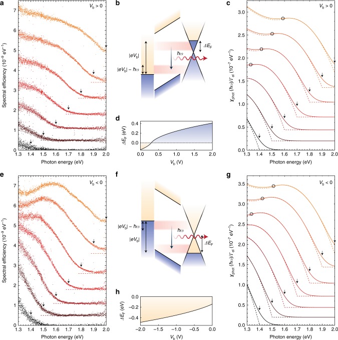
To identify the dominating emission mechanism, we now turn to the spectral distribution of these directly emitted photons, shown for applied voltages in the range from 1.4 to 2.0 V in Fig. 4a, e. First, we find that the spectral efficiency of light emission is very similar for both bias polarities. In MIM devices this finding is in agreement with both inelastic electron tunneling and hot electron photoemission because of the symmetry of the device. The efficiency of hot electron photoemission, that relies on the spontaneous generation of an optical excitation after reaching the counter electrode, depends on the efficacy of competing decay channels. With hot carrier lifetimes in graphene and gold being of the order of 100 fs30 and 10 fs31, respectively, one would expect a pronounced bias asymmetry in the emission efficiency, which is not observed. Second, we observe a clear correlation between the spectral cutoff ħωmax and the applied voltage Vb given by ħωmax ~ |eVb|. This indicates that multiple-electron IET processes do not play a dominant role24,25. Furthermore, it is not compliant with blackbody-like thermal emission, where the emitted spectrum is determined by the effective temperature of the electron gas rather than the applied voltage28,29. Hence, we identify single-electron IET to be the most likely mechanism underlying the observed emission.
Fig. 4.
Spectral distribution of directly emitted photons from vdWQT devices. a, e Measured efficiency spectra (spectral distribution of emitted photons per tunneling electron, cf. Supplementary Note 1) for positive/negative voltages Vb from a Au–7L h-BN–Gr device. Subsequent spectra are acquired with increasing voltage from |Vb| = 1.4 V (black) to |Vb| = 2.0 V (orange) in steps of 0.1 V. Spectra are offset with respect to each other, dashed lines indicate the respective zero line. Arrows indicate the spectral position where ħω = eVb. b, f Schematic of inelastic electron tunneling at positive/negative voltages. Optical mode excitations (here: photons) are generated by spontaneous transitions from occupied states in one electrode to unoccupied states in the other electrode. The energy window available for spontaneous excitation of a mode of energy ħω is given by |eVb| − ħω. c, g Theoretical photon emission rate spectra (ρrad = 0.04ρ0) for positive/negative voltages Vb, normalized to the elastic tunneling rate Γel. Spectra are calculated for voltages from |Vb| = 1.4 V to |Vb| = 2.0 V in steps of 0.1 V. Straight and dashed lines show calculations for T = 300 K and T = 0 K, respectively. Black circles mark the positions on each curve for which the condition |eVb| − ħω = |ΔEF| is fulfilled. Arrows indicate the spectral position where ħω = eVb. d, h Electrostatically induced graphene Fermi level shift ΔEF (cf. Methods) as a function of (positive/negative) applied voltage
Theoretical device modeling
To validate this hypothesis we compare our experimental results with theoretical calculations, describing the IET process—as depicted in Fig. 4b, f—as a spontaneous transition between occupied electronic states of one electrode to unoccupied states of the other electrode13,32–35. Based on Fermi’s golden rule and within the validity range of the dipole approximation, we may express the spectral rate of inelastic electron tunneling as35
| 1 |
where ρopt is the partial LDOS along the direction of electron tunneling, is the momentum matrix element (cf. Supplementary Note 2), ω is the angular frequency, ħω the energy of the optical excitation and ρAu/Gr is the electronic density of states of gold/graphene. The equation as stated applies to the positive voltage range. For negative voltages, the roles of the two electronic state densities are interchanged. The process is schematically depicted in Fig. 4b, f for positive/negative bias. At positive voltages electrons excite optical modes by transitioning from occupied states in the graphene electrode to unoccupied states in the gold electrode. Correspondingly, at negative voltages, optical modes are excited by transitions from occupied states in the gold electrode to unoccupied states in the graphene electrode. At any given mode energy ħω the energetic range over which inelastic electron tunneling may occur is given by the applied voltage as |eVb| − ħω. While and ρAu are approximately constant (cf. Supplementary Note 3), ρGr depends linearly on ΔEF, cf. Eq. (4). It is important to realize that the gold layer does not only act as one of the electrodes of the tunnel junction but also as a gate electrode36,37. This causes the charge carrier density of the graphene sheet and hence the shift of the Fermi level with respect to the Dirac point (ΔEF) to depend on the tunnel voltage, as depicted in Fig. 4d, h, cf. Methods. Differences in the band alignment and dipole layers at the gold/h-BN and graphene/h-BN interfaces result in a finite value of ΔEF even at Vb = 0, cf. Supplementary Note 1.
From Eq. (1) we calculate the spectral photon emission rate as γphot = ηrad × γinel = ρrad × γinel/ρopt, where ηrad is the radiation efficiency and ρrad = ρopt × ηrad is the radiative LDOS. For the planar heterostructure we find that ρrad = 0.04ρ0, where ρ0 = ω2π−2c−3 is the vacuum LDOS and c is the speed of light in vacuum (cf. Supplementary Note 4). The resulting γphot spectra, normalized by the calculated elastic tunneling rate Γel (cf. Supplementary Note 1), are displayed in Fig. 4c, where dashed and straight lines correspond to calculations for T = 0 K and T = 300 K, respectively. At T = 0 K, the quantum cut-off condition ħωmax = |eVb| is perfectly fulfilled such that the emission rate is zero for energies larger than the applied voltage. At finite temperatures this condition is relaxed by the thermal broadening due to the Fermi-Dirac distribution function, in agreement with experimental results. Most importantly, we are able to reproduce the experimentally observed spectral shape, confirming IET to be the underlying physical mechanism. It is determined by the electronic density of states of graphene, in particular by the voltage-dependent energetic position of the Dirac point (cf. Fig. 4d, h and Methods). Black circles on each curve mark the points for which the condition |eVb| − ħω = |ΔEF| is fulfilled. This condition determines the spectral location of the observed inflection point. The effect of the density of states minimum is more strongly pronounced for negative voltages. In our modeling we assume a symmetric and energy-independent electronic wave function decay (cf. Supplementary Note 2). Deviations from this assumption, as well as potential effects related to the gate-dependence of the barrier height at the graphene–h-BN interface38, are possible causes for the observed asymmetry. We emphasize that no fitting parameters are employed in the theoretical model with the exception of the extraction of the residual Fermi level shift at zero applied bias (cf. Supplementary Note 1).
Comparing the absolute values of the experimental and theoretical spectra of Fig. 4a, c, respectively, we find that the experimental efficiencies are lower than the theoretically calculated ones by a factor of ~40. This deviation is caused by the presence of an additional phonon-enhanced tunneling channel in metal–graphene tunnel junctions39–42, see Supplementary Note 1 for details.
vdWQT devices coupled to nanocube antennas
Until now we have analyzed the coupling of tunneling electrons to photons and SPPs of a planar, layered heterostructure—the optical properties of which are characteristic of a single gold film with a nearly energy-independent LDOS. Our findings demonstrate that we can view tunneling electrons as quantum emitters with emission properties that depend on the electronic density of states as well as the optical LDOS, cf. Eq. (1), and can thus be controlled by coupling to optical antennas43,44 or cavities45 to increase emission rates and shape emission spectra and emission pattern. To illustrate this concept we couple the vdWQT devices to nanocube antennas46,47.
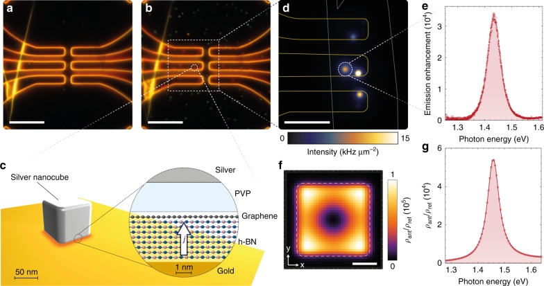
Silver nanocubes are deposited on top of the vdWQT devices from solution (cf. Methods) and can be directly observed with a darkfield microscope. Before nanocube deposition, as seen in Fig. 5a, scattering is dominated by the edges of the gold film. After nanocube deposition, as seen in Fig. 5b, we additionally observe randomly distributed spots, which are associated with optical resonances formed in the junctions between nanocubes and gold electrodes, as schematically illustrated in Fig. 5c. It is interesting to note that nanocubes sitting on top of the gold electrodes and the heterostructure produce a red ring-shaped pattern whereas (single) nanocubes sitting on the glass surface appear green. The ring-like pattern is caused by the symmetry of a higher-order MIM resonator mode that dominates the scattering response in the visible spectral range, cf. ref. 48. The green color of nanocubes on glass originates from the nanocubes dipolar resonance. The distance between the two metallic domains is determined by the number of h-BN layers and the thickness of the PVP coating (~3 nm) that stabilizes the nanocubes in solution.
Fig. 5.
Nanocube antennas driven by a vdWQT device. a Darkfield microscope image of the same devices as shown in Fig. 2a. Scale bar: 10 μm. b Darkfield image after the deposition of silver nanocubes onto the sample. The red ring-like patterns on top of the electrodes indicate the presence of nanocube antennas. Scale bar: 10 μm. c Schematic illustration of the nanocube antenna configuration. The inset shows a vertical cross-section of the modified heterostucture configuration which determines the optical modes “seen” by tunneling electrons. d Spatial intensity distribution of light emission from nanocube antennas. Outlines indicate the spatial location of the edges of graphene and gold electrodes. Scale bar: 5 μm. e Emission spectrum of the antenna marked by a dashed circle in d at Vb = 2 V, normalized by the corresponding emission spectrum of the planar heterostructure (Fig. 4a). f Spatial (on resonance) and g spectral dependence of the simulated, radiative fraction of the optical mode density of the nanocube antenna ρant for a vertically oriented dipole located in the center of the h-BN crystal, normalized by the radiative LDOS of the planar geometry ρref, cf. Supplementary Note 4. The white dashed line in f marks the outline of the silver nanocube (75 nm with a 7.5 nm edge radius)
While the formation of nanocube antennas leaves the electronic properties, i.e., the elastic and phonon-enhanced tunneling channels (c.f. Supplementary Note 1), largely unaffected—it strongly alters the light emission properties of the device. Without nanocubes we observe emission from the entire device area, cf. Fig. 2b. After antenna-coupling, as shown in Fig. 5d, the spatial intensity distribution is dominated by point-like sources, the size of which is given by the point-spread function of our imaging system. In the following we will focus on and selectively analyze (cf. Methods) the emission spot marked by the dashed circle in Fig. 5d. Further cube antennas are discussed in Supplementary Note 5. Figure 5e shows the photon emission spectrum of this particular nanocube antenna, normalized by the corresponding emission spectrum of the planar heterostructure (Fig. 4a) from an area equivalent to the nanocube footprint (75 × 75 nm2, cf. Supplementary Note 5). Previously, before the deposition of nanocubes, the emission spectrum was broad (cf. Fig. 4a) and primarily determined by the electronic density of states of the electrodes, whereas now photons are emitted into a narrow spectral window centered at ~1.43 eV with a full width at half maximum (FWHM) of ~57 meV. Compared to the emission spectrum of the bare vdWQT device, we find that—on resonance—the probability for an electron tunneling event, occurring within the active area of the nanocube antenna, to result in the generation of a photon is enhanced by more than four orders of magnitude. This enhancement is independent of applied voltage and polarity (cf. Supplementary Note 5).
The origin of these alterations is found by analyzing the modified optical properties of the system. According to Eq. (1), the spectral rate of photon emission by inelastic electron tunneling is determined by the optical mode density ρopt(ħω). The formation of nanocube antennas modifies ρopt(ħω) in two ways. Firstly, SPPs are confined to the nanoscale gap between the silver nanocube and the gold surface. The optical mode density of these MIM-SPPs is strongly enhanced as a result of the spatial field confinement and the increased propagation constant. Additionally, the termination of the MIM gap by the facets of the cube act as reflectors—forming a resonator—which leads to an additional increase in the mode density by the Purcell effect49–51. While ρopt determines the rate of inelastic electron tunneling γinel, the fraction associated with photon emission γphot is determined by the radiative LDOS ρrad.
Classically, the LDOS enhancement ρopt/ρ0 corresponds to an increase of the power dissipated by a point dipole P/P0 due to its interaction with the environment23. This correspondence allows us to determine the LDOS by means of classical electrodynamics (cf. Methods and Supplementary Note 4). The spectral variation of the radiative LDOS of the nanocube antenna ρant, normalized by the radiative LDOS of the planar geometry ρref (cf. Supplementary Note 4), is shown in Fig. 5g. The radiative LDOS is resonantly enhanced at a photon energy of ħωres ~1.46 eV and over a spectral range given by the FWHM of ~57 meV. We further analyze the nature of the enhancement by varying the position of the dipole, oscillating at ωres, as shown in Fig. 5f. The lateral variation of ρrad maps out the fundamental mode of the MIM resonator. The average LDOS enhancement is found to be ~5 × 10⁴, in excellent agreement with the experimentally observed photon emission rate enhancement of ~3 × 10⁴. These findings validate the dependence of the strength of the inelastic electron tunneling channel on the LDOS experienced by tunneling electrons, as suggested by Eq. (1).
Discussion
In conclusion, we have introduced and experimentally demonstrated a device platform that allows for the excitation of optical modes at the atomic scale by inelastic electron tunneling—van der Waals quantum tunneling devices. In particular, we have demonstrated the independent control over the electronic and optical properties of the system, both of which determine the spectral rate of inelastic electron tunneling in the form of the respective densities of states, cf. Eq. (1).
The electronic properties and the optical source spectrum are determined by a hybrid van der Waals heterostructure, here: gold–hexagonal boron nitride–graphene. The optical modes of this structure are the photon modes of free space and SPP modes. Based on a careful analysis of the spatial, angular and spectral distribution of the emitted photons, we unambiguously determine the direct emission of light from tunneling electrons to be the source of the observed emission. The emission is well described by a spontaneous emission model, confirming inelastic electron tunneling as its source. The spectral flatness of the optical mode density of the planar heterostucture allows us to observe the influence of the electronic density of states on the emission spectrum.
The optical properties are further modified by an external photonic architecture, here: nanocube antennas. The integration with these optical antennas leads to a spectral shaping and enhancement of the photon emission rate. Nanocube antennas constitute nanoscopic resonators for MIM-SPPs, accompanied by a local enhancement of the LDOS. Additionally, the geometry allows for the efficient coupling of MIM-SPPs to photons, resulting in the observed enhancement of the photon emission rate by more than four orders of magnitude.
This concept is easily translated to more complex systems. To optimize the source efficiency or introduce other device functionalities one may choose materials constituting the van der Waals heterostructure20,21 from an entire library of two-dimensional atomic crystals52. For example, utilizing semiconducting atomic crystals enables the shaping of the energy-dependence of the electronic density of states and may allow for the suppression of the elastic tunneling channel in favor of the inelastic process. In the devices presented here the source efficiency is reduced compared to traditional MIM tunneling devices due to the dominance of the phonon-assisted tunneling channel (cf. Supplementary Note 1). However, this may be overcome in future device configurations since tunneling between identical atomic crystals may allow for significantly higher efficiencies due to favorable momentum selection rules that have not yet been harnessed in any other material system53–55.
The individual two-dimensional crystals constituting vdWQT devices very weakly interact with optical modes that are polarized perpendicular to the sample plane as shown here for the case of the long-range SPPs of the planar geometry and the MIM-SPP mode inside the nanocube antenna. The same modes however couple most efficiently to tunneling electrons. This feature of vdWQT devices may be further explored by their integration with optical systems that support corresponding modes such as certain types of photonic cavities45, hybrid plasmonic waveguide resonators56,57, or SPP resonators58–60.
Methods
Device fabrication
Devices are fabricated on commercially available glass coverslips (22 × 22 × 0.13 mm3). Electrodes are defined by photolithography and electron beam evaporation of 1 nm titanium (Ti, 0.05 nm/s) and 50 nm gold (Au, 0.1 nm/s) at a pressure of <10−7 mbar. Lift-off is performed sequentially in acetone, isopropyl alcohol and deionized water. The RMS roughness of the gold electrodes is 0.4–0.5 nm.
Graphene and few-layer h-BN are mechanically exfoliated from bulk crystals onto oxidized silicon wafers (100 nm SiO2). h-BN crystals were grown as described in ref. 61. Suitable crystals are identified optically. Their thickness, i.e., the number of layers, is determined by a combination of optical contrast and atomic force microscopy. h-BN crystals with thicknesses of 6–7 layers (corresponding to ~2–2.3 nm) are chosen as a compromise between the voltage that may be applied without reaching the electrostatic breakdown field of ~1 V/nm and the tunneling current which decreases exponentially with crystal thickness62,63. To assemble the tunneling devices, graphene and few-layer h-BN are picked up successively with a polydimethylsiloxane (PDMS)/polycarbonate (PC) stack. The vdW stack is subsequently transfered on top of the gold electrodes. For pickup and transfer we follow the procedure developed by Zomer et al.64. The PC film employed as a carrier is dissolved in chloroform after successful transfer. Finally, the samples are annealed at 200 °C in a tube furnace while flowing 380 sccm argon and 20 sccm hydrogen to reduce residues on top of the heterostructure.
Nanocube antennas are assembled by drop-casting 50 µL of silver nanocubes (size: 78 ± 5 nm) in ethanol (concentration approximately 0.1 mg/mL, purchased from Nanocomposix) on top of the gold–h-BN–graphene assembly. After a short incubation time of several seconds the sample is spun dry at ~3000 rpm.
Following this approach we have assembled ten gold–h-BN–graphene heterostructures with varying numbers of individual devices and nanocube antennas, the properties of which are in agreement with the representative set of experimental data and their evaluation shown in this work.
Optical and electrical device characterization
The devices are characterized on a customized, inverted Nikon TE300 microscope under ambient conditions. Electrical measurements are carried out using a Keithley 2602B source meter unit. Light emitted from the devices into the air half-space is collected by an air objective (Nikon, 100×, NA 0.9) whereas light emitted into the glass half-space is collected by an oil-immersion objective (Nikon, 100×, NA 1.4). For real space and Fourier space (backfocal plane) imaging, the respective plane is imaged onto a electron-multiplying charge-coupled device (EMCCD, Andor iXon Ultra). The emission from individual nanocube antennas is selectively analyzed by spatial filtering in an intermediate image plane using a pinhole with a diameter of 100 μm, corresponding to a diameter of 1 μm in the sample plane. Spectra are acquired using a Princeton Instruments Acton SpectraPro 300i spectrometer. Spatial intensity distribution maps as shown in Figs. 2b, c and 5d are not corrected for system transmission, detection efficiency and the point spread function of the imaging system that determines the size of the emission spots in Fig. 5d. Spectral efficiency and photon flux spectra as shown in Figs. 4a, e and 5e and Supplementary Figs. 4a, 5a are corrected for the transmission function of the optical system and detection efficiency. They are further normalized to the area from which the photons are emitted, i.e., the entire device area in Fig. 4a and the footprint of the nanocube antenna in Fig. 5e as well as Supplementary Figs. 4a, 5a.
Device electrostatics
The dependence of the charge carrier density of graphene (nGr) on the applied voltage can be described as
| 2 |
where cg = ε0εhBN/dhBN is the geometric capacitance per unit area, ε0 is the vacuum permittivity and εhBN and dhBN = N × dil are the DC permittivity and thickness of the h-BN crystal, respectively, with N being the number of h-BN layers and dil = 0.33 nm being the h-BN interlayer distance65. The second term, ΔEF, accounts for the quantum capacitance of the two-dimensional graphene sheet66,67. The charge carrier density is related to the electronic density of states (ρGr) through
| 3 |
Near the Dirac point energy ED, ρGr depends linearly on energy as26
| 4 |
where vF is the Fermi velocity of graphene.
Combining Eqs. 2–4 results in a quadratic equation in ΔEF that allows us to explicitly calculate ΔEF as a function of voltage. For calculations, we assume vF = 1.1 × 10⁶ m/s68 and εhBN = 3.569.
Local density of optical states
The inelastic electron tunneling rate, described in Eq. (1), depends on the LDOS ρopt. For uncoupled vdWQT devices we calculate the LDOS semi-analytically (Supplementary Note 4). The LDOS of antenna-coupled vdWQT devices is calculated numerically (Supplementary Note 4). In both approaches we make use of the fact that the normalized LDOS ρopt/ρ0 is equivalent to the normalized power P/P0 dissipated by a classical point dipole with moment p23.
Supplementary information
Acknowledgements
The authors thank A. Jain, N. Flöry, S. Heeg, M. Frimmer and D. Svintsov for valuable discussions. This work was supported by the Swiss National Science Foundation (grant no. 200021_165841) and the ETH Zürich (ETH-32 15-1). The authors further acknowledge the use of facilities at the FIRST Center for Micro- and Nanotechnology at ETH Zürich. K.W. and T.T. acknowledge support from the Elemental Strategy Initiative conducted by the MEXT, Japan and the CREST (JPMJCR15F3), JST.
Author contributions
M.P. and L.N. conceived the device concept. M.P. fabricated and characterized the devices. M.P. performed all analytical calculations and numerical simulations. Á.Sz. and M.L. performed ab initio device simulations. K.W. and T.T. synthesized the h-BN crystals. M.P. and L.N. discussed results and co-wrote the paper with contributions from Á.Sz. and M.L.
Data availability
The data that support the findings of this study are available from the corresponding author upon reasonable request.
Competing interests
The authors declare no competing interests.
Footnotes
Journal peer review information: Nature Communications thanks Reuven Gordon, Jean-Jacques Greffet and the other anonymous reviewer(s) for their contribution to the peer review of this work. Peer reviewer reports are available.
Publisher’s note: Springer Nature remains neutral with regard to jurisdictional claims in published maps and institutional affiliations.
Contributor Information
Markus Parzefall, Email: mparzefall@ethz.ch.
Lukas Novotny, Email: lnovotny@ethz.ch.
Electronic supplementary information
Supplementary Information accompanies this paper at 10.1038/s41467-018-08266-8.
References
- 1.Lambe J, McCarthy SL. Light emission from inelastic electron tunneling. Phys. Rev. Lett. 1976;37:923–925. doi: 10.1103/PhysRevLett.37.923. [DOI] [Google Scholar]
- 2.Parzefall M, et al. Antenna-coupled photon emission from hexagonal boron nitride tunnel junctions. Nat. Nanotechnol. 2015;10:1058–1063. doi: 10.1038/nnano.2015.203. [DOI] [PubMed] [Google Scholar]
- 3.Kern J, et al. Electrically driven optical antennas. Nat. Photon. 2015;9:582–586. doi: 10.1038/nphoton.2015.141. [DOI] [Google Scholar]
- 4.Qian H, et al. Efficient light generation from enhanced inelastic electron tunnelling. Nat. Photon. 2018;12:485–488. doi: 10.1038/s41566-018-0216-2. [DOI] [Google Scholar]
- 5.Gurunarayanan SP, et al. Electrically driven unidirectional optical nanoantennas. Nano Lett. 2017;17:7433–7439. doi: 10.1021/acs.nanolett.7b03312. [DOI] [PubMed] [Google Scholar]
- 6.Du W, et al. On-chip molecular electronic plasmon sources based on self-assembled monolayer tunnel junctions. Nat. Photon. 2016;10:274–280. doi: 10.1038/nphoton.2016.43. [DOI] [Google Scholar]
- 7.Namgung S, et al. Ultrasmall plasmonic single nanoparticle light source driven by a graphene tunnel junction. ACS Nano. 2018;12:2780–2788. doi: 10.1021/acsnano.7b09163. [DOI] [PubMed] [Google Scholar]
- 8.Dathe A, Ziegler M, Hübner U, Fritzsche W, Stranik O. Electrically excited plasmonic nanoruler for biomolecule detection. Nano Lett. 2016;16:5728–5736. doi: 10.1021/acs.nanolett.6b02414. [DOI] [PubMed] [Google Scholar]
- 9.Wang P, Krasavin AV, Nasir ME, Dickson W, Zayats AV. Reactive tunnel junctions in electrically driven plasmonic nanorod metamaterials. Nat. Nanotechnol. 2018;13:159. doi: 10.1038/s41565-017-0017-7. [DOI] [PMC free article] [PubMed] [Google Scholar]
- 10.Davis LC. Theory of surface-plasmon excitation in metal-insulator-metal tunnel junctions. Phys. Rev. B. 1977;16:2482–2490. doi: 10.1103/PhysRevB.16.2482. [DOI] [Google Scholar]
- 11.Hansma PK, Broida HP. Light emission from gold particles excited by electron tunneling. Appl. Phys. Lett. 1978;32:545–547. doi: 10.1063/1.90123. [DOI] [Google Scholar]
- 12.Hone D, Mühlschlegel B, Scalapino DJ. Theory of light emission from small particle tunnel junctions. Appl. Phys. Lett. 1978;33:203–204. doi: 10.1063/1.90275. [DOI] [Google Scholar]
- 13.Persson BNJ, Baratoff A. Theory of photon emission in electron tunneling to metallic particles. Phys. Rev. Lett. 1992;68:3224–3227. doi: 10.1103/PhysRevLett.68.3224. [DOI] [PubMed] [Google Scholar]
- 14.Johansson P, Monreal R, Apell P. Theory for light emission from a scanning tunneling microscope. Phys. Rev. B. 1990;42:9210–9213. doi: 10.1103/PhysRevB.42.9210. [DOI] [PubMed] [Google Scholar]
- 15.Berndt R, Gimzewski JK, Johansson P. Inelastic tunneling excitation of tip-induced plasmon modes on noble-metal surfaces. Phys. Rev. Lett. 1991;67:3796–3799. doi: 10.1103/PhysRevLett.67.3796. [DOI] [PubMed] [Google Scholar]
- 16.Berndt R, Gimzewski JK. Photon emission in scanning tunneling microscopy: Interpretation of photon maps of metallic systems. Phys. Rev. B. 1993;48:4746–4754. doi: 10.1103/PhysRevB.48.4746. [DOI] [PubMed] [Google Scholar]
- 17.Sakurai M, Thirstrup C, Aono M. New aspects of light emission from STM. Appl. Phys. A. 2005;80:1153–1160. doi: 10.1007/s00339-004-3180-2. [DOI] [Google Scholar]
- 18.Rossel F, Pivetta M, Schneider WD. Luminescence experiments on supported molecules with the scanning tunneling microscope. Surf. Sci. Rep. 2010;65:129–144. doi: 10.1016/j.surfrep.2010.06.001. [DOI] [Google Scholar]
- 19.Bharadwaj P, Bouhelier A, Novotny L. Electrical excitation of surface plasmons. Phys. Rev. Lett. 2011;106:226802. doi: 10.1103/PhysRevLett.106.226802. [DOI] [PubMed] [Google Scholar]
- 20.Geim A, Grigorieva I. Van der waals heterostructures. Nature. 2013;499:419–425. doi: 10.1038/nature12385. [DOI] [PubMed] [Google Scholar]
- 21.Novoselov K, Mishchenko A, Carvalho A, Neto AC. 2d materials and van der waals heterostructures. Science. 2016;353:aac9439. doi: 10.1126/science.aac9439. [DOI] [PubMed] [Google Scholar]
- 22.Nair RR, et al. Fine structure constant defines visual transparency of graphene. Science. 2008;320:1308–1308. doi: 10.1126/science.1156965. [DOI] [PubMed] [Google Scholar]
- 23.Novotny L, Hecht B. Principles of Nano-Optics. 2nd edn. Cambridge: Cambridge University Press; 2012. [Google Scholar]
- 24.Schull G, Néel N, Johansson P, Berndt R. Electron-plasmon and electron-electron interactions at a single atom contact. Phys. Rev. Lett. 2009;102:057401. doi: 10.1103/PhysRevLett.102.057401. [DOI] [PubMed] [Google Scholar]
- 25.Peters PJ, et al. Quantum coherent multielectron processes in an atomic scale contact. Phys. Rev. Lett. 2017;119:066803. doi: 10.1103/PhysRevLett.119.066803. [DOI] [PubMed] [Google Scholar]
- 26.Castro Neto AH, Guinea F, Peres NMR, Novoselov KS, Geim AK. The electronic properties of graphene. Rev. Mod. Phys. 2009;81:109–162. doi: 10.1103/RevModPhys.81.109. [DOI] [Google Scholar]
- 27.Kirtley J, Theis T, Tsang J, DiMaria D. Hot-electron picture of light emission from tunnel junctions. Phys. Rev. B. 1983;27:4601–4611. doi: 10.1103/PhysRevB.27.4601. [DOI] [Google Scholar]
- 28.Downes A, Dumas P, Welland ME. Measurement of high electron temperatures in single atom metal point contacts by light emission. Appl. Phys. Lett. 2002;81:1252–1254. doi: 10.1063/1.1497188. [DOI] [Google Scholar]
- 29.Buret M, et al. Spontaneous hot-electron light emission from electron-fed optical antennas. Nano Lett. 2015;15:5811–5818. doi: 10.1021/acs.nanolett.5b01861. [DOI] [PubMed] [Google Scholar]
- 30.Breusing M, et al. Ultrafast nonequilibrium carrier dynamics in a single graphene layer. Phys. Rev. B. 2011;83:153410. doi: 10.1103/PhysRevB.83.153410. [DOI] [Google Scholar]
- 31.Brown AM, Sundararaman R, Narang P, Goddard WA, III, Atwater HA. Nonradiative plasmon decay and hot carrier dynamics: effects of phonons, surfaces, and geometry. ACS Nano. 2015;10:957–966. doi: 10.1021/acsnano.5b06199. [DOI] [PubMed] [Google Scholar]
- 32.Schimizu T, Kobayashi K, Tsukada M. Theoretical approach to the microscopic mechanism of light emission from a scanning tunneling microscope. Appl. Surf. Sci. 1992;60-61:454–459. doi: 10.1016/0169-4332(92)90459-B. [DOI] [Google Scholar]
- 33.Downes A, Taylor ME, Welland ME. Two-sphere model of photon emission from the scanning tunneling microscope. Phys. Rev. B. 1998;57:6706–6714. doi: 10.1103/PhysRevB.57.6706. [DOI] [Google Scholar]
- 34.Uskov AV, Khurgin JB, Protsenko IE, Smetanin IV, Bouhelier A. Excitation of plasmonic nanoantennas with nonresonant and resonant electron tunnelling. Nanoscale. 2016;8:14573. doi: 10.1039/C6NR01931E. [DOI] [PubMed] [Google Scholar]
- 35.Parzefall, M., Bharadwaj, P. & Novotny, L. in Quantum Plasmonics (eds Bozhevolnyi, S. I., Martin-Moreno, L. & Garcia-Vidal, F.) 211–236 (Springer, Cham, 2017).
- 36.Bokdam M, Khomyakov PA, Brocks G, Zhong Z, Kelly PJ. Electrostatic doping of graphene through ultrathin hexagonal boron nitride films. Nano Lett. 2011;11:4631–4635. doi: 10.1021/nl202131q. [DOI] [PubMed] [Google Scholar]
- 37.Britnell L, et al. Field-effect tunneling transistor based on vertical graphene heterostructures. Science. 2012;335:947–950. doi: 10.1126/science.1218461. [DOI] [PubMed] [Google Scholar]
- 38.Ponomarenko L, et al. Field-effect control of tunneling barrier height by exploiting graphene’s low density of states. J. Appl. Phys. 2013;113:136502. doi: 10.1063/1.4795542. [DOI] [Google Scholar]
- 39.Brar VW, et al. Scanning tunneling spectroscopy of inhomogeneous electronic structure in monolayer and bilayer graphene on sic. Appl. Phys. Lett. 2007;91:122102. doi: 10.1063/1.2771084. [DOI] [Google Scholar]
- 40.Zhang Y, et al. Giant phonon-induced conductance and scanning tunneling spectroscopy of gate-tunable graphene. Nat. Phys. 2008;4:627–630. doi: 10.1038/nphys1022. [DOI] [Google Scholar]
- 41.Wehling TO, Grigorenko I, Lichtenstein AI, Balatsky AV. Phonon-mediated tunneling into graphene. Phys. Rev. Lett. 2008;101:216803. doi: 10.1103/PhysRevLett.101.216803. [DOI] [PubMed] [Google Scholar]
- 42.Decker R, et al. Local electronic properties of graphene on a bn substrate via scanning tunneling microscopy. Nano Lett. 2011;11:2291–2295. doi: 10.1021/nl2005115. [DOI] [PubMed] [Google Scholar]
- 43.Bharadwaj P, Deutsch B, Novotny L. Optical antennas. Adv. Opt. Phot. 2009;1:438–483. doi: 10.1364/AOP.1.000438. [DOI] [Google Scholar]
- 44.Biagioni P, Huang JS, Hecht B. Nanoantennas for visible and infrared radiation. Rep. Prog. Phys. 2012;75:024402. doi: 10.1088/0034-4885/75/2/024402. [DOI] [PubMed] [Google Scholar]
- 45.Vahala KJ. Optical microcavities. Nature. 2003;424:839. doi: 10.1038/nature01939. [DOI] [PubMed] [Google Scholar]
- 46.Moreau A, et al. Controlled-reflectance surfaces with film-coupled colloidal nanoantennas. Nature. 2012;492:86. doi: 10.1038/nature11615. [DOI] [PMC free article] [PubMed] [Google Scholar]
- 47.Akselrod GM, et al. Probing the mechanisms of large purcell enhancement in plasmonic nanoantennas. Nat. Photon. 2014;8:835–840. doi: 10.1038/nphoton.2014.228. [DOI] [Google Scholar]
- 48.Chikkaraddy R, et al. How ultranarrow gap symmetries control plasmonic nanocavity modes: from cubes to spheres in the nanoparticle-on-mirror. ACS Photonics. 2017;4:469–475. doi: 10.1021/acsphotonics.6b00908. [DOI] [Google Scholar]
- 49.Bozhevolnyi SI, Søndergaard T. General properties of slow-plasmon resonant nanostructures: nano-antennas and resonators. Opt. Expr. 2007;15:10869–10877. doi: 10.1364/OE.15.010869. [DOI] [PubMed] [Google Scholar]
- 50.Kuttge M, Garcia de Abajo FJ, Polman A. Ultrasmall mode volume plasmonic nanodisk resonators. Nano Lett. 2009;10:1537–1541. doi: 10.1021/nl902546r. [DOI] [PubMed] [Google Scholar]
- 51.Faggiani R, Yang J, Lalanne P. Quenching, plasmonic, and radiative decays in nanogap emitting devices. ACS Photonics. 2015;2:1739–1744. doi: 10.1021/acsphotonics.5b00424. [DOI] [Google Scholar]
- 52.Mounet N, et al. Two-dimensional materials from high-throughput computational exfoliation of experimentally known compounds. Nat. Nanotechnol. 2018;13:246. doi: 10.1038/s41565-017-0035-5. [DOI] [PubMed] [Google Scholar]
- 53.Svintsov D, Vyurkov V, Ryzhii V, Otsuji T. Voltage-controlled surface plasmon-polaritons in double graphene layer structures. J. Appl. Phys. 2013;113:053701. doi: 10.1063/1.4789818. [DOI] [Google Scholar]
- 54.Svintsov D, Devizorova Z, Otsuji T, Ryzhii V. Plasmons in tunnel-coupled graphene layers: Backward waves with quantum cascade gain. Phys. Rev. B. 2016;94:115301. doi: 10.1103/PhysRevB.94.115301. [DOI] [Google Scholar]
- 55.de Vega S, Garcia de Abajo FJ. Plasmon generation through electron tunneling in graphene. ACS Photonics. 2017;4:2367–2375. doi: 10.1021/acsphotonics.7b00695. [DOI] [Google Scholar]
- 56.Oulton RF, et al. Plasmon lasers at deep subwavelength scale. Nature. 2009;461:629–632. doi: 10.1038/nature08364. [DOI] [PubMed] [Google Scholar]
- 57.Lu YJ, et al. Plasmonic nanolaser using epitaxially grown silver film. Science. 2012;337:450–453. doi: 10.1126/science.1223504. [DOI] [PubMed] [Google Scholar]
- 58.Kress SJ, et al. Wedge waveguides and resonators for quantum plasmonics. Nano Lett. 2015;15:6267–6275. doi: 10.1021/acs.nanolett.5b03051. [DOI] [PMC free article] [PubMed] [Google Scholar]
- 59.Kress SJ, et al. A customizable class of colloidal-quantum-dot spasers and plasmonic amplifiers. Sci. Adv. 2017;3:e1700688. doi: 10.1126/sciadv.1700688. [DOI] [PMC free article] [PubMed] [Google Scholar]
- 60.Zhu W, et al. Surface plasmon polariton laser based on a metallic trench fabry-perot resonator. Sci. Adv. 2017;3:e1700909. doi: 10.1126/sciadv.1700909. [DOI] [PMC free article] [PubMed] [Google Scholar]
- 61.Taniguchi T, Watanabe K. Synthesis of high-purity boron nitride single crystals under high pressure by using Ba–BN solvent. J. Cryst. Growth. 2007;303:525–529. doi: 10.1016/j.jcrysgro.2006.12.061. [DOI] [Google Scholar]
- 62.Lee GH, et al. Electron tunneling through atomically flat and ultrathin hexagonal boron nitride. Appl. Phys. Lett. 2011;99:243114. doi: 10.1063/1.3662043. [DOI] [Google Scholar]
- 63.Britnell L, et al. Electron tunneling through ultrathin boron nitride crystalline barriers. Nano Lett. 2012;12:1707–1710. doi: 10.1021/nl3002205. [DOI] [PubMed] [Google Scholar]
- 64.Zomer P, Guimarães M, Brant J, Tombros N, Van Wees B. Fast pick up technique for high quality heterostructures of bilayer graphene and hexagonal boron nitride. Appl. Phys. Lett. 2014;105:013101. doi: 10.1063/1.4886096. [DOI] [Google Scholar]
- 65.Levinshtein, M. E., Rumyantsev, S. L. & Shur, M. S. Properties of Advanced Semiconductor Materials: GaN, AIN, InN, BN, SiC, SiGe (Wiley, Hoboken, 2001).
- 66.Luryi S. Quantum capacitance devices. Appl. Phys. Lett. 1988;52:501–503. doi: 10.1063/1.99649. [DOI] [Google Scholar]
- 67.Dröscher S, et al. Quantum capacitance and density of states of graphene. Appl. Phys. Lett. 2010;96:152104. doi: 10.1063/1.3391670. [DOI] [Google Scholar]
- 68.Ponomarenko LA, et al. Density of states and zero landau level probed through capacitance of graphene. Phys. Rev. Lett. 2010;105:136801. doi: 10.1103/PhysRevLett.105.136801. [DOI] [PubMed] [Google Scholar]
- 69.Jang SK, Youn J, Song YJ, Lee S. Synthesis and characterization of hexagonal boron nitride as a gate dielectric. Sci. Rep. 2016;6:30449. doi: 10.1038/srep30449. [DOI] [PMC free article] [PubMed] [Google Scholar]
Associated Data
This section collects any data citations, data availability statements, or supplementary materials included in this article.
Supplementary Materials
Data Availability Statement
The data that support the findings of this study are available from the corresponding author upon reasonable request.


