Abstract
Colloidal-based solution syntheses offer a scalable and cost-efficient means of producing 2D nanomaterials in high yield. While much progress has been made towards the controlled and tailorable synthesis of semiconductor nanocrystals in solution, it remains a substantial challenge to fully characterize the products’ inherent electronic transport properties. This is often due to their irregular morphology or small dimensions, which demand the formation of colloidal assemblies or films as a prerequisite to performing electrical measurements. Here, we report the synthesis of nearly monodisperse 2D colloidal nanocrystals of semiconductor SnS and a thorough investigation of the intrinsic electronic transport properties of single crystals. We utilize a combination of multi-point contact probe measurements and ultrafast terahertz spectroscopy to determine the carrier concentration, carrier mobility, conductivity/resistivity, and majority carrier type of individual colloidal semiconductor nanocrystals. Employing this metrological approach, we compare the electronic properties extracted for distinct morphologies of 2D SnS and relate them to literature values. Our results indicate that the electronic transport of colloidal semiconductors may be tuned through prudent selection of the synthetic conditions. We find that these properties compare favorably to SnS grown using vapor deposition techniques, illustrating that colloidal solution synthesis is a promising route to scalable production of nanoscale 2D materials.
Keywords: colloidal nanocrystals, 2D materials, solution synthesis, electronic properties, electrical measurements, time-resolved terahertz spectroscopy, tin sulfide
Graphical Abstract
Two-dimensional nanomaterials have garnered immense research interest in the last decade due to their potential as building blocks for the next generation of optoelectronics. Unlike graphene, many metal chalcogenides have a band gap which, as a semiconductor, allows them to serve as the principle constituent of solid-state devices.1 Metal chalcogenides with a layered crystal structure tend to naturally form 2D crystals, a consequence of stronger in-plane atomic bonding relative to their weaker out-of-plane van der Waals interactions between layers. When nanoscale-thin, this 2D morphology lends itself well for their incorporation into optoelectronics, both due to their ability to stack efficiently with other device components and their substantial mechanical elasticity, which engenders the fabrication of flexible or wearable electronics.2,3 Additionally, 2D semiconductor nanocrystals display attractive electrical properties, such as higher intrinsic mobilities than are found in organic electronics or assemblies of 0D nanoparticles, as well as a multitude of emerging, novel physics.4,5 Two-dimensional materials initially were prepared using micromechanical exfoliation;6 however, more recently substantial progress has been made in developing in vacuo techniques such as chemical vapor deposition (CVD) and molecular beam epitaxy (MBE) that utilize gas-phase reactants.7 Although large, high-quality single crystals are accessible, scalability and reproducibility remain ongoing issues for these techniques, which currently limit their efficacy for commercial applications.8,9
A promising alternative to gas-phase deposition of 2D materials is solution-based synthetic strategies. These low-temperature methods yield liquid “inks” of dispersed freestanding 2D semiconductors that allow for the straightforward fabrication of flexible devices, assemblies, and thin films through means such as inkjet printing, spray coating, or roll-to-roll processing. Employing these fabrication techniques has allowed for solution-processed 2D semiconductors to be effectively incorporated into field-effect transistors, photodetectors, solar cells, light-emitting diodes (LEDs), photocatalysts, memory devices, and more.10-12 Most of the dispersed 2Ds used in devices have been produced by top-down liquid processing techniques, which involve the exfoliation of layered materials in solution followed by extensive post-processing centrifugal separation.13,14 However, the scale-up of these methods is hampered by poor yields, low throughput, and difficulty in achieving morphological uniformity. Conversely, bottom-up solution-phase syntheses of 2D materials lend themselves better to commercialization, as they utilize the pre-existing chemical manufacturing infrastructure and paradigms.15 Colloidal chemistry has proven effective in the mass production of 0D semiconductor nanomaterials with fine control of nanocrystal size and morphology. Consequently, the past two decades has seen colloidal semiconductors successfully complete the evolution from small-scale curiosities to consumer products. Wet chemical methods have successfully accessed a variety of 2D semiconductor nanocrystals,16 including Sn(S,Se),17,18 PbS,19 Ge(S,Se,Te), 20-22 GaSe,23 InSe,24 Cd(S,Se,Te),25 Mo(S,Se,Te)2,26-28 and various lead halide perovskites.29 However, achieving high yields of morphologically uniform, individual, unagglomerated crystals via solution routes requires extensive reaction and processing optimization. Relative to bottom-up gas-phase deposition techniques, to date it remains a significant challenge to produce monodisperse 2D materials when employing colloidal chemistries.
Among the more intriguing emerging 2D materials is tin(II) sulfide (SnS). The tin sulfides are a family of earth-abundant semiconductors being actively investigated for use in a variety of technological application spaces. Although several stable and metastable phases of 1:1 tin sulfide are accessible, the most thermodynamically favored is the GeS-type orthorhombic α-phase,30 found naturally as the mineral herzenbergite, and which we will generally refer to hereafter in this report simply as “SnS”. The structure of SnS is a compound analogue of black phosphorus, with two-atom-thick layers of Sn and S orthogonal to the [100] direction. Atoms are bonded strongly to three nearest neighbors within the layer, forming a zigzag arrangement in the b-direction and an arm chair arrangement in the c-direction (see Figure S1).31 The overall atomic geometry of the crystal can be thought of as a distorted rock salt structure, where the 5s2 lone pair electrons of Sn2+ lead to the formation of gaps between the two-atom-thick planes due to electrostatic repulsion. Weaker van der Waals interactions between atoms across the gap result in the formation of a pseudo-octahedral coordination environment.32 The layered atomic structure resulting from these bonding arrangements affords the driving force for SnS to crystallize primarily in 2D morphologies, and imparts electrical anisotropy within the {100} facets of individual 2D crystals. SnS is intrinsically p-type due to the presence of Sn vacancies, which act as shallow acceptors,33 resulting in relatively high conductivities ranging from 10−4to 0.1 S / cm.34 Undoped carrier concentrations of holes are reported to be on the order of 1015 cm−3 to 1017 cm−3 with Hall mobilities of up to 90 cm2 / V·s in single crystals.35,36 SnS also displays excellent optical properties, including an indirect band gap of 1.07 eV and absorption coefficient values > 104 cm−1 across the visible and near-infrared spectrum,37 which is higher than Si and most other absorber materials. Finally, unlike many other semiconductors, SnS is a non-toxic, low-cost compound, making it a promising candidate for widespread commercial application.
Nanocrystals of SnS have been reported using solution-based syntheses in a variety of sizes and morphologies, although there are limited examples of nearly uniform products. Zero-dimensional nanoparticles have been accessed with a high degree of monodispersity as spheres,17,38-41 cubes,17,40,42 and tetrahedra.39,40 Owing to SnS having an exciton Bohr radius of ≈ 7 nm,43 many of these 0D nanocrystals exhibit an increased band gap due to quantum confinement, a phenomenon which also occurs for thin 2D SnS.44 Several colloidal methods to prepare 2D nanocrystals of SnS have been developed;17,38,45,46 however, to date, high-yield syntheses of 2D SnS have been limited to small or intermediate-sized crystals with dimensions of ~ 250 nm or less. The synthesis of larger (>1 μm), high-quality, single-crystalline 2D materials is of nanoelectronic and optoelectronic interest due to the potential for larger mobilities, fewer interface recombination sites in assemblies, and the ability to lithographically fabricate devices using individual crystals. Although colloidal synthesis routes to larger 2D SnS nanosheets41,42,47,48 and nanoribbons49-52 have been reported previously, their morphological and dimensional uniformity is limited. To our knowledge, there has been no report of a high-yield solution-based route to μm-scale, monodisperse, single-crystalline 2D SnS nanocrystals.
A pressing technological obstacle for the application of colloidally produced materials in optoelectronics is the lack of a straightforward means to measure the inherent electronic transport properties of the individual synthesized crystals. While structural and optical characterization of semiconductor nanocrystals has become routine, electronic features are substantially more challenging to interrogate. Contact probe and spectroscopic measurements typically are performed on films or superlattices of assembled colloidal nanocrystals;12,53,54 however, such a strategy does not allow for the disentanglement of the innate carrier transport within individual crystals from that resulting due to charge transfer between separate colloidal domains. Carrier transport is strongly influenced by capping agents,55 inter-crystalline charge transfer ligands,56 and packing order.57 As such, the properties of assemblies are not representative of the individual nanocrystals contained therein, and the full dissemination of inherent electronic behavior for discrete colloidal semiconductors has proven exceptionally challenging. Further, relative to single-crystalline 2D nanomaterials, assemblies of 0D nanocrystals have often been plagued by poor mobility and conductivity resulting from imperfect assembly and the prevalence of surface recombination sites. Although it is desirable to produce devices using individual, high-quality, 2D single crystals of colloidal materials, electrical characterization is rare and generally limited to two-contact measurements,21,23,52,58-61 while the four-point probe method, which measures the intrinsic conductivity of a material, is rarely achieved. In light of the tremendous interest towards employing colloidal nanocrystals as components of optoelectronic devices, the development of techniques that determine the inherent electronic transport properties of solution-synthesized semiconductors, such as time-resolved terahertz spectroscopy (TRTS),10,41,62-64 is desperately needed.
Here, we report a comprehensive analysis of the electronic transport properties in individual colloidal semiconductor nanocrystals. First, we present the development of a solution chemistry-based synthetic approach to produce nearly monodisperse μm-scale 2D tin(II) sulfide nanoribbons and square nanosheets using a one-pot, one-step, easily scalable synthetic route. These syntheses represent a rare example in this size regime of essentially uniform, single-crystalline, 2D nanocrystals produced using colloidal chemistry. Next, we detail the structural characterization of these SnS materials, and describe how they are processed from solution to fabricate back-gated, top-contact solid-state devices from individual colloidal crystals. Finally, we interrogate their electronic transport properties using a combination of multi-point contact probe electrical conductivity measurements and time-resolved terahertz spectroscopy. These combined studies allow for the direct determination of carrier concentration, carrier mobility, conductivity, and the majority carrier type within an individual 2D nanocrystal. We report findings illustrating that careful manipulation of solution chemistries can selectively afford products with substantively disparate charge carrier behavior. These measurands are challenging to extract using common experimental practices, and underpins that this metrological strategy represents a significant and valuable advancement in the characterization of colloidally synthesized semiconductors.
RESULTS AND DISCUSSION
Solution Synthesis and Structural Characterization of μm-scale Colloidal 2D SnS.
We developed high-yield, solution-based routes to μm-scale 2D crystals of colloidal SnS in two distinct morphologies. Their 2D structure and relatively large in-plane size were essential for the subsequent fabrication of solid-state devices using individual colloidal nanocrystals as the semiconductor component. Concurrently, a high degree of shape and size monodispersity was desired as, in addition to being an atom-efficient and cost-effective synthetic process, a substantial degree of crystalline homogeneity allowed for bulk structural characterization of the product. To these ends, we pursued a heat-up strategy of nanocrystal synthesis in solution (see materials and methods section for details), as such approaches have proven consistently effective at producing large quantities of monodisperse shape-controlled nanocrystals.65,66
SnS 2D Nanoribbons
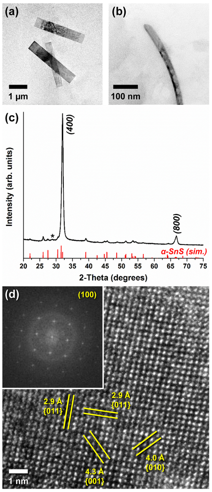
One of our devised syntheses produced a high yield of nearly morphologically-uniform, high aspect ratio, 2D SnS colloidal crystals that hereafter we refer to as nanoribbons. Briefly, SnCl2 and elemental sulfur were dissolved in oleylamine (OLAM) solvent and heated to 180 °C (453 K) under an inert atmosphere. The product was collected, separated from solution by centrifugation, and redispersed in toluene. Figure 1a shows a representative transmission electron microscope (TEM) image of the resulting 2D nanoribbons lying flat, and additional images can be found in Figure S2. Statistical analysis of the crystals revealed that our product had a morphological yield of 95 % nanoribbons, with an average length of 2.81 γm ± 0.28 μm (C.V. = 10 %) and width of 0.48 μm ±0.19 μm among the nanoribbons. Notably, such high yield and monodispersity is rarely reported for bottom-up synthesized colloidal semiconductors in the >1 μm size regime. We found that variations in regent concentration, Sn:S precursor ratio, and temperature all adversely affect the homogeneity of our nanocrystals; however, the reaction is robust enough to tolerate small perturbations to the synthetic conditions. Additionally, the selection of SnChl2 was essential to produce monodisperse nanoribbons as both SnBr2 and Sn acetate yielded mixtures of nanoribbons, likely due to differences in thermal decomposition kinetics afforded by the anion (Figure S3). Figure 1b displays the view of an individual nanoribbon from the side, indicating that the μm-scale crystals possess smooth top and bottom facets with a thickness of approximately 17 nm to 20 nm. Their morphological smoothness was also observed using high-angle annular dark-field scanning TEM (HAADF-STEM), and the few-layer thickness of the nanoribbons was confirmed by atomic force microscopy (AFM) (Figures S4, S5).
Figure 1.
TEM images of μm-scale colloidal SnS nanoribbons, viewed from the (a) top and (b) side, displaying their 2D morphology. (c) Powder XRD pattern of the nanoribbons, which can be indexed to phase-pure orthorhombic α-SnS (* = surface oxide present in air) and displays strong preferred orientation in the [100] direction. (d) HRTEM image of a single SnS nanoribbon and (inset) the resulting FFT, both of which reveal that they are single-crystalline with a surface that can be indexed to α-SnS(100).
The structural composition of the 2D nanoribbons was investigated using powder X-ray diffraction (XRD). The resulting pattern (Figure 1c) was indexed to phase-pure orthorhombic α-SnS (JCPDS No. 73-1859, space group Pnma, lattice constants a = 1.118 nm, b = 0.398 nm, c = 0.433 nm). The strong relative intensity of the (400) and (800) reflections indicated substantial preferred orientation towards [100], which is expected as the layered planes of covalently-bound Sn-S extend in the b and c crystallographic directions (Figure S1). Aside from the reduced relative sharpness of the reflections, due to smaller crystalline domain sizes, the XRD pattern matches well with that collected from commercially-purchased bulk herzenbergite SnS, including a small amount of native surface oxide detectable at approximately 29 °2θ (Figure S6). High-resolution TEM (HRTEM) analysis of the μm-scale nanoribbon surface revealed that they were singlecrystalline, free of linear and planar defects such as dislocations and twinning (Figure 1d). Lattice fringes with spacings of 2.9 Å (0.29 nm) were visible between a given atom and its four nearest neighbors, which agrees well with the expected interatomic distance for planes of the form {011}. The (010) and (001) planes were also visible, with interatomic lattice fringe distances of 4.0 Å (0.40 nm) and 4.3 Å (0.43 nm), respectively. A fast Fourier transform (FFT) of the collected HRTEM image (inset) matched well with the [100] zone axis of SnS, in good agreement with our powder XRD results. Selected area electron diffraction (SAED) of an individual nanoribbon produced a pattern which confirmed its single-crystalline nature and can be indexed to α-SnS(100) (Figure S7). Additionally, when compared to the orientation of the nanoribbon being characterized, reciprocal space distances between diffraction spots along the length of the crystal correspond to the SnS<010> directions, while the diffraction spots along the width match that of SnS<001>. This indicates that the nanocrystals grow more favorably along the zigzag b-axis relative to the armchair c-axis, the latter of which is identified as the lateral direction of the ribbons. We verified these results by performing HRTEM analysis on the corner of a nanoribbon (Figure S8), and found that the atomic fringes along the length of the crystal matched well to the lattice spacings of SnS(010), and those of the width to SnS(001). These crystalline orientations concur with those previously observed in the case of belt-like SnS nanowires that similarly grew primarily in the <010> directions, which was ascribed to the higher surface energy of the SnS {010} facets relative to those of {100} or {001}.67
SnS 2D Square Nanosheets
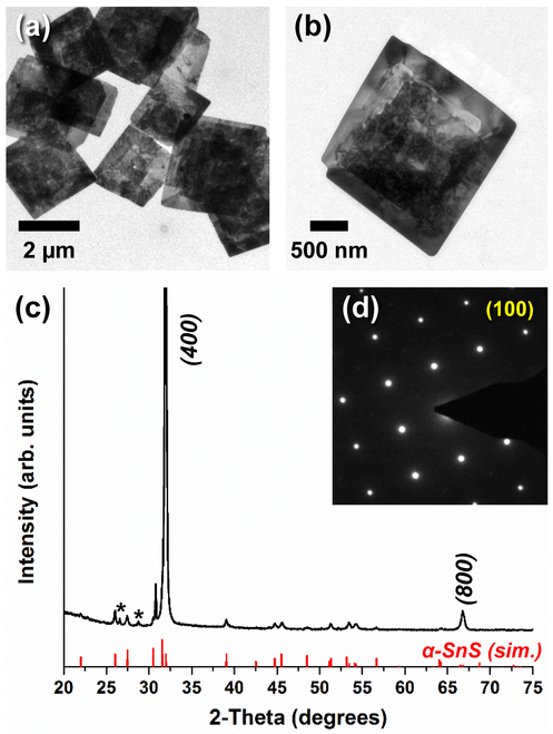
A second synthetic procedure we developed yields SnS square nanosheets with high morphological uniformity. Briefly, an excess of Sn(IV) chloride was heated in OLAM under an inert atmosphere while in the presence of hexamethyldisilazane (HMDS) and elemental sulfur to form μm-scale colloidal 2D SnS nanocrystals. The silvery-dark product was collected, purified by centrifugation, and redispersed in toluene. Figures 2a,b show demonstrative TEM characterization of the resulting square nanosheets (Figure S9 for additional images). Particle counting confirmed that the nanosheets had nearly uniform lateral dimensions of 2.58 ± 0.37 μm per side (C.V. = 14 %), and our synthesis produced a yield of 89 % square nanosheets. Unlike the μm-scale nanoribbons, the SnS nanosheets have an irregular, uneven surface. HAADF-STEM imaging shows the broad face of the nanosheets resemble a series of cliffs and plateaus (Figure S10). The coarse morphology of the nanosheets was further confirmed by AFM, showing a series of steps that rise from the edges of the nanostructure (Figure Sll). The nanosheets are also revealed to be substantially thicker than the nanoribbons, with vertical dimensions ranging from 75 nm around the periphery of the nanostructure to 150 nm along the thickest interior regions. The impetus for this unusual morphology is uncertain; although it resembles the terraced morphology that can result from screw dislocation-driven growth, it lacks the distinctive spiral pattern that results from such a mechanism. More likely, it is the result of a nonclassical, kinetic-driven crystallization mechanism such as oriented attachment68 or the “wedding cake” growth model, which consists of successive adatom-driven nucleation events.69 The reaction kinetics may be a result of the presence of HMDS during synthesis, which is known to increase the reactivity of Sn complexes it coordinates to and often influences product crystallinity when employed in solution-based syntheses 45,52 Our use of Sn4+, instead of the Sn2+ utilized for nanoribbons, also requires an additional reduction process to take place during the formation of SnS that could alter nucleation and growth kinetics. We also found that the reaction temperature and an excess of Sn are important synthetic parameters in this synthesis (Figure S12).
Figure 2.
(a and b) TEM images of μm-scale 2D colloidal SnS square nanosheets. (c) Powder XRD pattern of the square nanosheets, which can be indexed ssto phase-pure orthorhombic α-SnS (* = surface oxide present in air) and displays strong preferred orientation in the [100] direction. (d) SAED pattern collected from an individual square nanosheet, confirming that it is single crystalline SnS oriented towards the [100] zone axis.
Figure 2c shows the powder X-ray diffraction pattern of a drop-cast ensemble of square nanosheets, confirming that they too could be indexed to phase-pure GeS-structural type α-SnS with a small amount of native surface oxide. As with the nanoribbons, the as-prepared nanocrystals were oriented preferably along [100], which is consistent with their layered, anisotropic, 2D structure. The increased sharpness of the reflections, relative to that of the nanoribbons, indicated a larger single-crystalline domain size, which we attribute to both the greater thickness and higher cross-sectional area of the nanosheets. Importantly, the sharpness of the reflections in the XRD pattern suggests that, despite their coarse morphology, these nanocrystals are not polycrystalline. This observation was confirmed by electron diffraction (Figure 2d). The spot pattern produced from SAED revealed that the square nanosheets, or at least very large domain therein, were single-crystalline and that, like the nanoribbons, the surface normal of the nanosheets corresponded to the [100] zone axis. The in-plane orientation of the electron diffraction pattern indicated that the four edges of the nanosheets extended in the zigzag (<010>) and armchair (<001 >) directions, while <011> aligned with the corners of the square (Figure SI3).
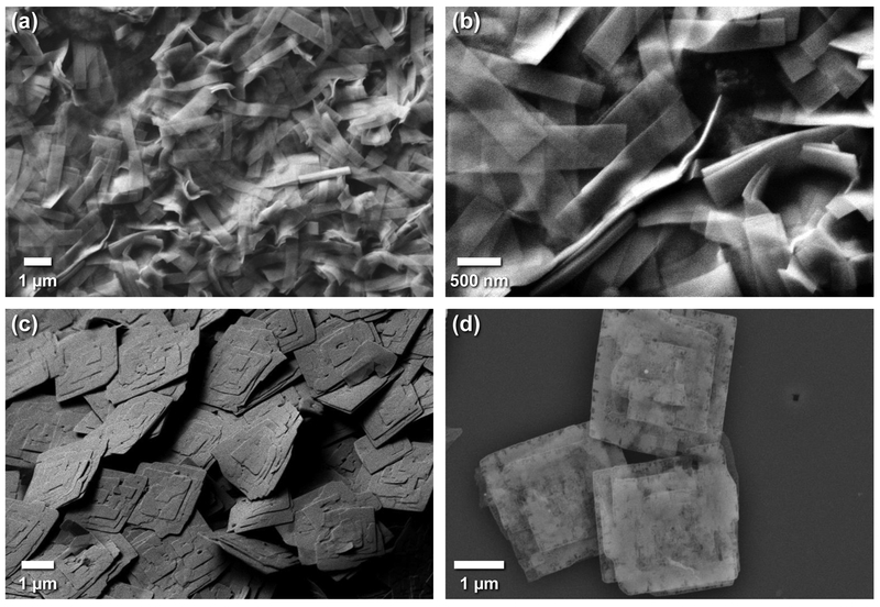
The 2D morphology and large-scale monodispersity of centrifugally washed, drop-cast SnS nanocrystals was verified by scanning electron microscopy (SEM) analysis (Figures 3, S14, S15). The high morphological uniformity of our two products was evident through examination of large- area films of deposited SnS, which show 100 or more nearly identical nanocrystals at a time. The nanoscale thinness of the μm-scale 2D materials (≈ 17 nm – 20 nm for the nanoribbons, ≈ 75 nm –150 nm for the nanosheets) is also observable by side view inspection of individual crystals. The difference in flexibility between the morphologies, likely as a function of their thickness, is apparent. Whereas the nanoribbons curve and bend with ease, the square nanosheets stack in a rigid, inelastic manner. The elasticity of drop-cast nanoribbons makes them potentially of interest for flexible devices and straintronics (Figure SI 6). The distinct tiered structure of the nanosheets was also easily observable; however, each individual crystal appeared to have a distinct pattern of steps and plateaus, despite approximately uniform lateral dimensions. Additionally, we generated elemental maps of SnS nanoribbons and square nanosheets using energy-dispersive X-ray spectroscopy coupled with SEM (SEM-EDX), which confirmed a homogeneous distribution of Sn and S throughout the nanostructures (Figures S17, S18). Notably, SEM-EDX also detected carbon and nitrogen on the 2D nanocrystals, suggesting that molecular surfactants are still adsorbed to the surface of the nanocrystals even after washing.
Figure 3.
SEM images of (a,b) SnS nanoribbons and (c,d) SnS square nanosheets dropcast on a substrate, indicating the high morphological uniformity of individual crystals within the colloidal solution. The greater rigidity and coarseness of the square nanosheets, relative to the nanoribbons, can also be clearly seen.
Fabrication of Back-Gated Devices from Individual Crystals of Colloidal 2D SnS.
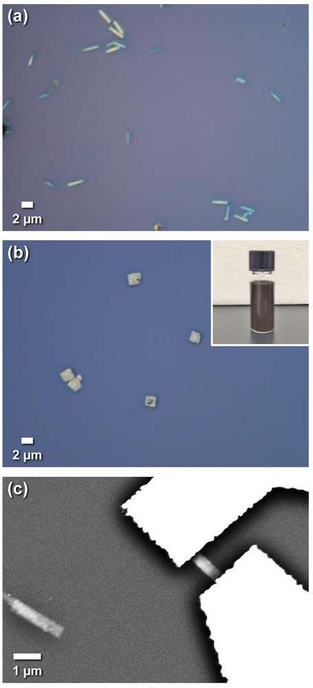
To explore the electronic transport properties of our synthesized 2D SnS nanocrystals, we fabricated back-gated field-effect transistor (FET) devices from individual SnS nanoribbons and square nanosheets taken from solution using standard lithographic techniques. Although long-chain surfactants adsorbed to the surface of the 2D SnS afford colloidal stability and prevent agglomeration of nanocrystals, these organic species are highly insulating and must be removed prior to SEM analysis or device fabrication. Accordingly, first we diluted and centrifugally washed solutions of our samples, which consists of separating the precipitate from the supernatant and then redispersing the former in neat toluene. The effect of this washing step is manifest when the SEM images in Figure 3 are compared to those of unwashed 2D nanocrystals, Figure S19. Without washing, an amorphous matrix of dried organic residue can be seen encapsulating the crystals, preventing both effective charge transfer between domains and the isolation of individual 2D SnS semiconductors for subsequent device fabrication. Drop-casting dilute solutions afforded deposited 2D SnS with a free proximity of 10 μm or more surrounding them, making these regions suitable for the deposition of metal contacts to a series of individual semiconductor nanocrystals (Figure 4). Unlike most exfoliation-based 2D material techniques, using solution-synthesized nanocrystals allowed us to fabricate on the order of 6 – 10 separate devices in each lithographic write, and fabricating up to hundreds at a time is achievable due to the preponderance of nearly uniform 2D materials available on each substrate (supporting information and Figures S20-S23 for more details).
Figure 4.
Optical microscopy images of (a) SnS nanoribbons and (b) SnS square nanosheets dropcast from (inset) colloidal solution on p+Si/SiO2 substrates prior to device fabrication. (c) SEM image of a back-gated, 2-contact device fabricated from an individual SnS nanoribbon that was cast onto the substrate.
Our initial electronic device measurements revealed that our device did not display ohmic behavior (Figure S24). The nonlinearity of the initial IDS-VDS curves was attributed to a Schottky barrier arising from poor contact between the deposited Cr/Au and the SnS channel. To resolve this problem, a second annealing step was necessary, this time with the entire device array being treated at 350 °C (623 K) in 5 % forming gas. The subsequent linearity of the IDS-VDS curve is indicative of an ohmic-like interface between the semiconductor and the contact. Previous studies have also shown a marked decrease in contact resistance between SnS and metal contacts following annealing, and this improvement is usually attributed to a solid-state reaction at the interface which results in the formation of a CrSx or other metal-sulfide compound70,71 To ensure that the fidelity of the SnS crystal had been preserved during this reaction and throughout the fabrication process, we performed Raman spectroscopy as a means of structural analysis. As shown in Figure S25, the Raman spectra of 2D SnS as-synthesized, following centrifugal washing, and after the device fabrication process changed minimally, indicating the SnS structure remained intact. A slight hardening of most phonon modes can be attributed to the removal of surface ligands by washing and, especially, by thermal annealing. The peaks matched well with the previously-reported Raman spectra of α-SnS,37,72 and all are induced from first-order scattering from optical phonons.30 Further confirmation was provided by powder XRD (Figure S26). Although alloys may have formed at the device interface, SEM-EDX elemental mapping of the devices confirmed that there was no detectable metal diffusion from the contacts into the semiconductor channel during fabrication (Figure S27).73
Electrical Characterization of Individual Solution-Synthesized 2D Crystals of SnS.
We explored the electrical properties of solution-synthesized, μm-scale 2D SnS nanocrystals using the test structures fabricated from single nanoribbons and square nanosheets. A schematic of the completed top-contact, back-gated FET devices is displayed in Figure S28. First, output characteristics of individual 2D SnS nanocrystals were collected using a two-terminal configuration at room temperature in the dark. Evaluation of the devices was performed without ambient light to avoid the generation of photocurrent, as the high absorptivity of SnS in the visible spectrum, which lends itself towards application in photodetectors and photovoltaics, is well-documented.37,74 Figure 5 shows typical IDS-VDS curves generated from output measurements of individual solution-synthesized SnS nanoribbons while varying the back gate bias, VGS, between – 40 V and 40 V. The linearity of the output curves indicated that the interface between the contacts and the semiconductor was ohmic-like. Additionally, gate action was observed in that the drain-source current showed a consistent response to applied VGS; as the back gate bias became more negative, the output current increased. This is indicative of an injection of holes, and implies that the Fermi level within the band gap lies towards to the top of the valence band of SnS.
Figure 5.
Back-gated two-contact devices fabricated from individual colloidal SnS nanoribbons, (a) Representative optical microscopy image and the resulting room temperature (b) output (IDS-VDS) and (c) transfer (IDS-VGS) characteristics. The output characteristics confirm ohmic contact between the metal and semiconductor. The p-type behavior observed in the transfer characteristics confirm that holes are the majority carrier in the SnS nanoribbons.
Transfer characteristics of a nanoribbon device, generated by sweeping the back gate voltage VGS with an applied bias VDS ranging from 0.3 V to 0.6 V, are displayed in Figure 5c. The measured drain-source current decreased with a more positive VGS, reinforcing their p-type semiconductor character. This hole-dominated carrier transport is expected, as the relatively low formation energy of Sn vacancies generally results in an excess of holes for bulk, thin film, and nanoscale SnS,33,75 thereby resulting in intrinsic p-type conductivity.34,35 The consistent influence of VGS on IGS indicates that these devices function as FETs; however, the relatively low subthreshold slope and the inability to fully turn the devices off limit their effectiveness in this application. Notably, the room temperature FET performance of these colloidally synthesized single-crystalline SnS devices, while modest, is comparable to those using exfoliated and vapor-deposited 2D SnS.52,76-78 Previous reports of single-crystalline 2D SnS (and SnSe) FETs displayed poor on/off ratio, no higher than 10 and as low as 1.5, with a fully “off’ current in the tens of nA. This appreciable “off’ current has been attributed to strong carrier screening that prevents the induction of effective switching behavior by the field effect of the gate. SnS inherently has a relatively short Debye screening length,77 which results in a depletion of carriers towards the bottom of the channel but a shunting effect as carriers continue to pass beyond screening length near the top surface. The poor gate tuning behavior of SnS FETs was somewhat ameliorated by thinning the channel down to several 10s of nm.77 Although this could explain a low gate response in our thicker 2D square nanosheets, the SnS nanoribbons are only 20 nm or less in height. We therefore ascribe the inability to turn our nanoribbon devices off to the relatively thick (300 nm) SiO2 dielectric layer, and posit that improved device performance could be achieved by employing either a dielectric layer on the order of 30 nm or a top-gated configuration.
The output current of the SnS square nanosheet devices displays an ohmic response that is consistently lower than that of nanoribbons with similar channel dimensions. As seen by the two-contact IDS-VDS behavior illustrated in Figure 6, IDS is consistently half or less than that of the nanoribbons for a given applied bias and back gate voltage. These devices display a consistent response to applied VGS; but surprisingly, the current increased with higher applied back gate potential, suggesting that electrons, not holes, were the majority carrier in these solution-synthesized 2D materials. This was corroborated by the collected transfer curves of the devices (Figure 6c), which confirm the n-type character of the square nanosheets. The field effect-induced gate action is even lower than that of the nanoribbons, with a nearly linear subthreshold slope, which was not unexpected considering their larger channel height. As mentioned above, SnS generally exhibits p-type transport behavior due to the thermodynamic favorability of forming Sn2+ vacancies, and these defects’ ability to act as shallow acceptors.75 The n-type conductivity measured in our SnS square nanosheets represents one of the few reported cases of electrons acting as the majority carrier. Previously, this has been achieved in SnS through external doping using elements such as Pb or Cl,79 and, notably, isoelectronic black phosphorus was recently n-doped by surface deposition of silicon nitride.80 Although we did not deliberately dope our square nanosheets, we hypothesize that HMDS or SnCl4, neither of which were present during the synthesis of the nanoribbons, may have acted as a source of extrinsic Cl, Si, or N dopants. To test this theory, we performed detailed X-ray photoelectron spectroscopy (XPS) analysis on the square nanosheets. As seen in Figure S29, Cl 2p and Si 2p photoelectrons were not detected at the surface of the nanocrystals. Nitrogen is observed in the centrifugally washed samples; however, this is due to OLAM surfactant adsorbed to the nanosheet surface. Following removal of the ligands by annealing the signal due to the N 1s photoelectrons is no longer present, in agreement with FTIR data (Figure S21), indicating that there does not appear to be extrinsic doping.
Figure 6.
Back-gated, two-contact devices fabricated from individual colloidal SnS square nanosheets, (a) Representative SEM image and the resulting room temperature (b) output (IDS-VDS) and (c) transfer (IDS-VGS) characteristics. Although limited current and gate action is observed, n-type ohmic behavior is displayed indicating, unlike nanoribbons, electrons are the majority carrier.
An alternative explanation of the n-type behavior exhibited by the square nanosheets is intrinsic doping, specifically a disproportionally Sn-rich stoichiometry. Quantitative EDX elemental analysis of a wide-area, large ensemble of nanocrystals found that the μm-scale SnS nanoribbons had a slight excess of sulfur (Figure S30), in conjunction with most previous reports of nanoscale and bulk SnS.38 Conversely, the elemental stoichiometry of the SnS square nanosheets, determined by ensemble EDX measurements (Figure S31), indicated a ≈ 7.5 % majority of tin, which is a significant margin of difference even when accounting for the limitations of quantitative EDX. Non-stoichiometry is known to be an effective means of switching the majority carrier from holes to electrons in metal chalcogenides, both through the formation of anion vacancies81 and the presence of excess cations.82 Indeed, an excess of Sn or deficiency of S has previously resulted in n-type SnS.76,83,84 The Sn-heavy elemental composition of the square nanosheets, and its resulting influence on carrier transport and band structure, can result from either a deviation in stoichiometry within the crystal lattice, including the formation of S vacancies, or from cationic Sn Lewis acid complexes adsorbed to the surface.56,85 We believe this is a reasonable explanation for the n-type behavior of these nanocrystals, especially in light of the excess Sn4+ present during their synthesis, which was not the case for our 2D nanoribbons. However, we note that extrinsic doping at concentrations below the limits of XPS detection cannot be discounted as a possible source of this unexpected charge transport.
We hypothesized that the relatively low currents displayed by our nanosheets may be due to oxide formation. XPS analysis confirmed the presence of a thin native surface oxide, but of insufficient thickness to strongly affect device performance (Figure S32). A more likely impetus to the modest currents measured in our two-terminal SnS devices are oxides resulting from our choice of contact metal. Although Cr is commonly used as a semiconductor-contact adhesion layer due to its low work function, which is especially important for facilitating charge transfer to n-type semiconductors, it has been reported to form insulating oxides (CrOx) at the interface during deposition, even under high vacuum conditions.71 These oxides, which also can form with other common low work function contact metals such as Ti, will act as a Schottky barrier to the channel, assuming no Fermi level pinning. Indeed, we recently have found that higher work function metals significantly improve the performance of our two-terminal SnS devices.86 This uncertainty about the band alignment of our contacts underpins the importance of performing four-point measurements, instead of two-point measurements, when extracting the fundamental electronic properties of our materials.
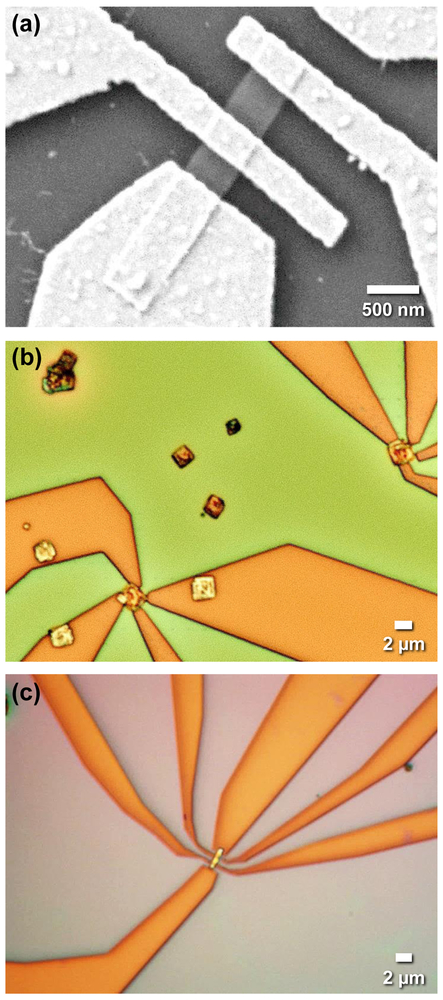
To determine the inherent conductivity of the individual solution-synthesized SnS nanocrystals, devices with additional contacts were patterned (Figure 7). These are among the smallest many-terminal devices ever fabricated from individual 2D semiconductor crystals, and approach the scaling limit for our lithographic process. Using four-point probe techniques, with a linear arrangement of terminals along the length of the nanoribbons (Figure S33) and a van der Pauw arrangement on the square nanosheets (Figure S34), allowed for deconvolution of contact resistance from the inherent resistance of the material. Our SnS nanoribbons had an average room temperature resistivity of 10.3 Ω·cm, which corresponds to a conductivity (σ) of 0.097 S / cm and compares favorably to the conductivity of single-crystalline SnS reported elsewhere.33 As determined using SAED and HRTEM, the length of the ribbon corresponds to the b-axis zigzag structural orientation of the anisotropic SnS crystal. Previous reports74,76,78 conductivity is higher in this direction, relative to that of the armchair direction, and so, although the ribbon width is too narrow for direct four-probe measurement, we can reasonably postulate that the conductivity along this direction would be lower. By comparing the device resistance obtained from two-point measurements with the channel resistance extracted from four-point results on the same nanoribbon, the magnitude of contact resistance may be quantified by
| (1) |
for a linear arrangement of 4 contacts, and assuming contacts 2 and 3 have an equal resistance, Rcontact. Our average measured two-point resistance between contacts 2 and 3 was 5.21 × 10⁶ Ω, which indicates that the contact resistance of these devices was in the range of 8.5 × 10⁴ Ω. This indicates that, although our contacts function reasonably well, further engineering of the semiconductor-metal band alignment could improve device performance.
Figure 7.
(a) SEM and (b and c) optical microscopy images of more complex back-gated device architectures fabricated from individual colloidal SnS nanoribbons and square nanosheets with 3 or more separate metal contacts.
Four-point probe analysis of the square nanosheets utilized a van der Pauw configuration, which affords measurements along two directions set off 90° from each other, in this case corresponding to the edges of the square nanocrystals. We found the measured resistances to be highly anisotropic, with an average value of 5.35 × 10⁵ Ω in one direction and 8.55 × 10⁵ Ω for the orthogonal in-plane direction, yielding an overall room temperature value ρ = 23.1 Ω·cm. This corresponds to a conductivity of 0.043 S / cm and confirms that the solution-synthesized SnS square nanosheets are less conductive than the SnS nanoribbons. However, it is important to note that the resistivity value determined for the square nanosheets assumes isotropic in-plane carrier transport, which is not the case for 2D nanocrystals of SnS. Indeed, our measured resistance along one edge of the square nanosheet was 1.6 times higher than along the other. Although we cannot verify it structurally, considering previous experimental and computational transport studies of SnS single crystals74-76,87 we can reasonably assume that the direction of lower resistance corresponds to the zigzag b-axis, and the higher resistance is along the orthogonal armchair c-axis. These measurements confirm the impact of structural orientation on device performance and underpin the importance of probing electronic transport anisotropy in solution-synthesized colloidal nanocrystals.
Extracting the Electronic Transport Properties through a Combination of Four-Point Probe and Time-Resolved Terahertz Spectroscopy.
Accurate measurement of the intrinsic charge carrier mobility, and subsequently the carrier concentration, is often a major challenge in the characterization of low-dimensional materials. Field-effect mobility (μFET), extracted from the transfer curve of a FET device, is frequently reported; however, unaccounted for contact resistance or device-substrate interactions produce error that often leads this value to be underestimated and inconsistent.88 Our attempt to extract μFET from the linear region of device transfer measurements at several low VDS values yielded discrepant results (Scheme S1). Magnetotransport Hall measurements of fabricated test structures, yielding the Hall mobility (μH) of the material, is regarded as a more effective approach of elucidating the carrier mobility of a semiconductor.89 Unfortunately, reliable magnetotransport measurements on the nm-scale are complicated by the small spacing between electrical terminals and associated lithographic limitations. As an alternative to Hall measurements, we turned to time-resolved terahertz spectroscopy to determine the charge carrier mobility. TRTS has emerged as a useful technique for the investigation of electronic transport properties in colloidal semiconductor nanocrystals,41,62,63 and recent reports have indicated that the carrier mobility values generated from TRTS (μT) are comparable to μH.10,64
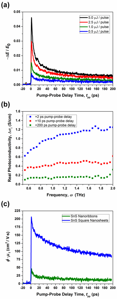
TRTS is an ultrafast spectroscopic technique that utilizes a visible pump and far-infrared probe to generate and interrogate the dynamics of free charge carriers. The materials’ photoconductivity can be approximated by measuring the change in transmission of a THz frequency probe wave packet following photoexcitation with visible light above the band gap energy. The probe waveform (covering frequencies 0.3 THz to 2.0 THz) was collected from samples of drop-cast centrifugally washed 2D SnS nanocrystals both following 800 nm excitation and again with the pump beam blocked, the latter scenario hereafter referred to as terahertz time-domain spectroscopy (THz-TDS). For TRTS, the delay between the pump and gate pulses, tpg was held constant at the point of maximum attenuation in the electric field transmission waveform relative to the unpumped THz-TDS waveform (Figure S35), while the pump-probe delay time, tpp, was varied between 0 ps and 200 ps to monitor the magnitude of change in THz probe transmission due to photogenerated free carriers (Figure 8a).
Figure 8.
Terahertz mobility, μT, of solution-synthesized 2D SnS nanocrystals determined by TRTS at room temperature. (a) Fluence-dependence of ΔE / E0 for SnS nanoribbons at pump-probe delay times, tpp, 0 to 200 ps using 800 nm pump excitation. (b) Frequency-dependence of the real part of the generated change in photoconductivity in SnS nanoribbons with a pump fluence of 0.5 μJ / pulse, indicating that at low frequencies and longer pump delay times it nearly matches the measured electrical conductivity of ~ 0.1 S/cm. (c) The product of the carrier photogeneration yield and terahertz mobility at tpp 0 – 200 ps when pumped with a fluence of 0.5 μJ / pulse, as determined using Equation 3, for both SnS nanoribbons and SnS square nanosheets.
The time-dependent photoconductivity was calculated from the optically induced decrease in THz transmission following photoexcitation using the thin film equation,90 which is valid because our deposited nanocrystal films are substantially thinner (≈ 3-4 μm) than the wavelength of the probe radiation (hundreds of μm):
| (2) |
where ΔE(tpp) is the differential change in transmission, relative to the TDS measurement, at the peak of the THz probe waveform resulting from a photoexcited sample at pump-probe delay tpp, E0 is the sample transmission at the same point in the THz waveform without photoexcitation, nTHz is the index of refraction of the substrate at THz frequencies, Z0 is the impedance of free space, and d is the thin film thickness (additional details, Figures S36, S37).
Measurement of ΔE / E0 (Figure 8a), and therefore the transient photoconductivity, is a function of both the pump-probe delay time, tpp, and the pump fluence. At very early pump-probe delay times, hot carriers with excess energy quickly relax to their respective band edges through phonon emission before reaching a long-lived state (ns timescale) where ΔE / E0 decay occurs gradually through recombination mediated by typical Auger or Shockley-Read-Hall processes. This temporal stage several picoseconds after excitation is more representative of the inherent mobility of charge carriers within a given material, and so we and others focus our analysis on, in this case, pump-probe delay times of tpp = 10 ps or greater.10,62,63,91,92 By employing Equation 2 and performing a Fourier transform on the THz transmission differential waveforms for a deposited film of known thickness (Figure S38), we obtain the frequency-dependent real and imaginary conductivity. At low frequencies and longer pump-probe delay times, we found that Δσ1(ω) was close to the DC conductivity determined from our four-point probe measurements when pumped with an excitation of 0.5 μJ / pulse (Figure 8b). Therefore, we selected this low fluence for our carrier transport measurements, as photogenerating too many electron-hole pairs can influence the mobility due to carrier crowding and increased Auger recombination. Importantly, at later tpp the real photoconductivity Δσ1(ω) was largely frequency-independent and the imaginary portion of the photoconductivity μσ2(ω) was positive across all measured frequencies (Figure S39). These findings indicate Drude-like charge transport with isotropic scattering and minimal back-scattering at defects or interfaces.41,92
To determine the photogenerated carrier mobility in our samples, we utilize the following equation (see Scheme S2):
| (3) |
where φ is the charge carrier photogeneration efficiency, F is the excitation fluence, A is the absorbance, and μT is the terahertz mobility, which is equal to the sum of the carrier mobilities, (μe + μh). The resulting time-dependent μT values for our 2D SnS nanocrystals is presented in Figure 8c. In our analysis, φ is assumed to be at or nearly unity, which is reasonable considering the strong absorptivity of SnS. However, we note that the values presented represent a lower bound to our mobility calculation. As with the ΔE / E0 values, the mobility quickly decreases within the first several ps following excitation, likely from carrier-to-phonon cooling, and then gradually diminishes over time due to carrier recombination, which is not incorporated into Equation 3. We extract our room temperature μT value from the spectra at tpp = 10 ps, where we can safely assume that all hot carrier energy transfers have ceased and very limited carrier recombination has taken place, yielding values of μT = 26.5 cm2 / V·s for the SnS nanoribbons and 158.0 cm2 / V·s in the square nanosheets. Although TRTS is an ensemble measurement, the high monodispersity of the samples allows us to assume that the mobility values extracted are confined within an individual crystalline domain if the diffusion length, L, is substantially shorter than the lateral size of our 2D nanocrystals.10,41 Using the Einstein relation, L = (τμTkBT/e)1/2, at τ = 10 ps we find L is only on the order of 26 nm in our SnS nanoribbons and 64 nm for our square nanosheets, confirming that backscattering at the edges should not be strongly limiting our mobility measurement.
A major limitation of the TRTS technique is that the extracted mobility corresponds to a combination of both electron and holes, while disentanglement of the individual free carrier mobilities remains challenging. Obtaining the majority carrier mobility of semiconductor is desirable because it is a more accurate representation of the material performance in electronic devices, and potentially allows for the calculation of the room temperature carrier concentration if the materials’ conductivity is known. In an effort to ameliorate this issue, we devised an approximation to deconvolute the individual carrier mobilities from the combined values captured by TRTS (see Scheme S3). By assuming that carrier scattering at room temperature is primarily due to acoustic phonon scattering and that transport in these layered 2D materials is overwhelmingly in-plane,4,87,92 we derived the following relationships for TRTS-measured in-plane carrier mobility of holes (μh,xy) and electrons (μe,xy):
| (4) |
where m* is the carrier effective mass and Ed is the deformation potential for that carrier. Using values of m* and Ed collected from reported density functional theory (DFT) calculations (Scheme S4), we find that the in-plane mobility of the majority carriers for our 2D SnS nanocrystals is μh,xy = 4.8 cm2 / V·s in the nanoribbons and μe,xy, = 158 cm2 / V·s for our square nanosheets.
The anisotropic conductivity we observed in our Van der Pauw measurements, which has also been predicted and observed by others in single-crystalline SnS,74,76,77 is primarily due to disparate mobilities in the zigzag and armchair directions, and thus likely manifest by differences in reduced mass and deformation potential experienced by a charge carrier in these directions. We estimate the zigzag and armchair contribution to the composite in-plane carrier mobility of 2D SnS using the following expressions (Schemes S5,S6),:
| (5) |
where x and y denote two orthogonal in-plane directions within a 2D or layered material, using reported values of effective mass and deformation potential for a given carrier type.87 A summary of the results can be found in Table 1. In the p-type nanoribbons we estimate hole mobilities of 5.8 cm2 / V·s along the zigzag direction and 3.9 cm2 / V·s in the armchair direction, while for the n-type square nanosheets the electron mobilities are 170 cm2 / V·s and 97 cm2 / V·s along zigzag and armchair, respectively. This indicates a ~ 1.5 times higher mobility for holes in the zigzag direction, relative to armchair, in our SnS nanocrystals and likewise a ~ 1.75 times higher mobility of electron carriers along the zigzag direction, relative to armchair. These values are comparable to what has been previously reported for 2D SnS and other anisotropic 2D materials. 74,76,77 The ratio of to μe to μh of ~ 4.5 that we report is somewhat higher than expected,93 but this may be due to our mobility relation’s dependence of the square of the effective mass, rather than the linear approximation between μ and m that is often reported.
Table 1.
Summary of the electronic transport properties obtained from our combined four-point probe and TRTS measurements on solution-synthesized colloidal nanocrystals of 2D SnS. For comparison, a summary of previously reported transport properties obtained from Hall measurements on both single-crystalline and thin films of SnS is provided. All SnS samples are undoped and results are at room temperature. See also supporting information, Table S1.
| SnS Material | Conductivity (S / cm) |
Mobility (cm2 / V·s) [direction undefined] |
Majority Carrier Concentration (cm−3) |
Mobility (cm2 / V·s) [approximated zigzag] |
Mobility (cm2 / V·s) [approximated armchair] |
|---|---|---|---|---|---|
| Nanoribbons, this work | 0.097 (zigzag) | holes: 4.8 electrons: 22 |
1.04 ×10¹⁷, holes [p-type] |
holes: 5.8 electrons: 29 |
holes: 3.9 electrons: 16 |
| Square nanosheets, this work | 0.043 (in-plane) | electrons: 130 holes: 29 |
2.07 ×10¹⁵, electrons [n-type] |
electrons: 170 holes: 35 |
electrons: 97 holes: 23 |
| Single crystals, consensus from literature |
~0.01 - ~ 0.2 | 10 - 90 | 1013 - 1017, holes | (1.15 - 2.5 times higher μ in zigzag direction relative to armchair) |
|
| Thin films, consensus from literature |
~0.001 - ~ 0.07 | 0.8 - 37 | 1014 - 1017, holes 1015, electrons |
-- | -- |
Finally, the carrier concentrations of our 2D colloidal SnS nanocrystals were calculated using the four-point probe-measured conductivity and our direction-dependent estimated majority carrier mobility values. The results (Table 1) indicated that the SnS nanoribbons have several orders of magnitude higher carrier concentration of holes (1.04 × 1017 cm−3) relative to the concentration of free electrons found in our square nanosheets (2.07 × 1015 cm−3), which accounts for the former’s higher conductivity despite substantially lower carrier mobility. According to Kroger-Vink defect chemistry theory,94 the higher carrier concentration and lower mobility of the nanoribbons, relative to the square nanosheets, collectively suggest that they possess a larger population of vacancies. While surface passivation of the nanoribbons with Sn complexes or other metal cations likely would improve the carrier mobility by eliminating surface trap states resulting from Sn vacancies, it is probable that doing so would result in a concomitant decreased density of free carriers. We hypothesize that a relatively small concentration of sulfur vacancies would engender the high μe, but also markedly lower ne, exhibited by the square nanosheets. Indeed, previous calculations have indicated that small concentrations of sulfur defects in SnS lead to a markedly lower carrier concentration relative to typical tin-deficient bulk SnS.95
When compared to previous measurements of SnS electronic transport properties (Table S1 and summarized in Table 1), we find that our values correspond well with bulk SnS, which is expected as the thinness of our μm-scale 2D nanocrystals is not sufficient for quantum confinement. This outcome underscores the comparable crystal quality and electrical properties that are achievable when employing easily, scalable solution-based, colloidal syntheses relative to vapor-based techniques. While the electrical properties of solution-synthesized semiconductors are typically measured across large assemblies of nanocrystals, resulting in extraneously high resistivities due to losses from inefficient charge transfer between domains, the consensus between our measurements and preceding reports substantiates the veracity of our proposed metrological strategy. Although our measured carrier mobilities are comparable to exfoliated ultrathin transition metal dichalcogenides and higher than most solution-synthesized organic semiconductor or nanocrystal assembly devices, we stress that TRTS has the potential to slightly underreport carrier mobilities, as a photocarrier generation rate less than unity and a nontrivial population of photoinduced carriers lost to recombination at early pump-probe delay times will diminish μ values.
These results conclusively demonstrate that colloidal nanocrystals can exhibit a substantial, yet inconspicuous, disparity in electronic transport properties. Despite being grown using similar precursors and reaction conditions, producing 2D materials of equivalent crystalline structure, our SnS nanoribbons and square nanosheets possessed dissimilar transport properties. This highlights the crucial role of judicious and precise synthetic parameter selection, and emphasizes how indispensable these measurements are towards the development of colloidal nanostructures appropriate for electronic and optoelectronic applications. As thorough investigations of inherent electronic transport in discrete nanocrystals are rare to date, this study may serve as a roadmap to the characterization of future solution-synthesized 2D electronic material systems.
CONCLUSIONS
In summary, colloidal solution-based routes were applied to achieve nearly monodisperse, single-crystalline, 2D SnS semiconductor nanocrystals in high yield using low-temperature, one-pot chemistries. Two distinct morphologies of 2D SnS were accessed, and utilized to demonstrate an innovative metrological strategy to extract the intrinsic electronic transport properties of these colloidal 2D nanocrystals by employing a combination of four-terminal and TRTS measurements. The conductivity/resistivity and majority carrier were determined using four-point probe techniques on solid-state devices fabricated from individual SnS nanoribbons and square nanosheets. We proposed an interpretation of TRTS data which deconvolutes the intrinsic electron and hole mobility in each of our nanocrystal morphologies, as well as providing an estimation of the relative carrier transport along disparate crystallographic directions within an anisotropic crystal structure. These results were summarily combined to afford a quantification of their carrier concentration. We found that our SnS nanoribbons and square nanosheets had unexpected and substantially dissimilar electronic transport properties, likely due to differences in their synthetic procedure, which illustrates the importance of pursuing these less routine measurands when conducting nanocrystal characterization. Colloidal SnS is an attractive model system to illustrate the potential for bottom-up synthesized 2D materials to be manufactured cost-effectively and subsequently integrated into functional composites or utilized as building blocks for the construction of solution-processed nanoelectronic devices. The routine quantification of the full electronic transport properties of colloidal 2D materials will prove invaluable toward their eventual incorporation into commercial electronic or optoelectronic applications, and we believe that the metrological strategy laid out herein will prove applicable and of paramount importance to the characterization of other solution-synthesized semiconductor nanocrystal systems.
MATERIALS AND METHODS
Materials.
Tin(IV) chloride (99.995 %), tin(IV) bromide (99 %), tin(II) acetate, tin(IV) acetate, sulfur powder (99.98 %), and oleylamine (70 %, technical grade) were purchased from Sigma- Aldrich. Tin(II) chloride (99 %), tin(II) bromide (99.2 %) hexamethyldisilazane (>99 %), tri-n-octylphosphine (90 %, technical grade), and tin(II) sulfide powder (99.5 %) were purchased from Alfa Aesar. Poly(methyl methacrylate) 495 A4 and 950 A4 were purchased from Microchem. Hydrofluoric acid (49 %) was purchased from J.T. Baker. Solvents, including toluene, ethanol, and acetone, were of analytical grade. All chemicals were used as received.
Synthesis.
All reactions were performed under an argon atmosphere using standard Schlenk techniques. Micrometer-scale colloidal 2D SnS nanoribbons were synthesized using the following developed heat-up route. First, 0.032 g (1 mmol) of sulfur was dissolved in 10 mL of oleylamine (OLAM), forming an orange solution, and then added to a 25 mL three-neck flask containing a magnetic Teflon-coated stir bar and equipped with a condenser fitted to a Schlenk line. 0.0948 g (0.5 mmol) of SnCl2 was then added directly into the reaction vessel. The temperature was controlled using a digital controller with glass-coated thermocouple (Gemini, J-KEM Scientific) and a 25 mL heating mantle (Glas-Col). After sealing the flask with a glass stopper, it was evacuated and the solution was heated to 120 °C (393 K) with vigorous stirring. The temperature was maintained for 20 min to remove residual water. After putting the reaction solution under an argon blanket, the temperature was raised to 180 °C (453 K) at ~ 5 ° / min, causing the solution to turn from orange to dark brown, and maintained at that temperature for 1 h before quenching the reaction by immersing the flask in cold water. SnS nanoribbons were separated from the reaction solution by adding 30 mL of ethanol antisolvent and centrifuging at 5000 rpm (524 rad / s) for 1 min. After decanting the yellow supernatant, the precipitate was redispersed in toluene and centrifuged at 3000 rpm (314 rad / s) for 1 min. The clear supernatant was discarded, and the precipitate was redispersed in toluene before being centrifuged a final time at 2000 rpm 209 (rad / s) for 1 min. Following decanting of the light brown supernatant, the precipitate was redispersed in 4 mL of toluene. 500 μL of OLAM and 250 μL of tri-n-octylphosphine (TOP) were added to afford improved colloidal stability to the nanoribbons during storage in solution.
A similar route was applied to synthesize μm-scale colloidal 2D SnS square nanosheets. 0.0038 g (0.118 mmol) of sulfur was dissolved in 10 mL of OLAM then added to a 25 mL reaction flask that was equivalently outfitted. To this, 20.2 μL (0.175 mmol) of SnCl4 was added all at once using a micropipette and the flask was sealed with a septum. While stirring vigorously at room temperature, the reaction was evacuated. After waiting several min for the resulting bubbling to subside, the solution was heated to 120 °C (393 K) and held there for 20 minutes under vacuum. Next, the reaction was switched to an argon blanket and 0.7 mL (3.34 mmol) of hexamethyldisilazane was injected through the septum with a syringe. The temperature was raised to 195 °C (468 K) at ~ 10 ° / min and then to 210 °C (483 K) at ~ 3 ° / min, resulting in a silvery-black solution, and the temperature was maintained for 5 min before quenching the reaction with cold water. To separate the nanosheets, 30 mL of ethanol antisolvent was added to the solution before centrifuging at 5000 rpm (524 rad / s) for 1 min and decanting the resulting light-yellow supernatant. The precipitate was redispersed in toluene and washed by centrifugation twice more under the same conditions. Finally, the SnS square nanosheets were dispersed in 2 mL of toluene before 500 μL of OLAM and 250 μL of TOP surface stabilizers were added to assist with dispersability during solution storage.
Device fabrication.
First, an aliquot of the nanoribbon and square nanosheet stock solutions were each washed of excess OLAM and TOP stabilizer by centrifugation at 13000 rpm (1361 rad / s) for 1 min and then redispersed in neat toluene. A drop of dilute, centrifugally washed colloidal 2D SnS was then cast on a heavily-doped p-type Si substrate with ≈ 300 nm of thermally grown SiO2 and prefabricated gold alignment marks. The p++Si/SiO2 served as the global back gate and gate dielectric, respectively, of the device. To remove the remaining surface organics, the sample was annealed in a tube furnace (Lindburg/Blue M, Thermo Scientific) under flowing 5 % H2 forming gas at 400 °C for 1 h. Next, isolated nanoribbons and square nanosheets were selected for contact fabrication using optical microscopy, and their positions relative to the alignment marks noted. After spin coating a resist of poly(methyl methacrylate) (PMMA) 495 A4 followed by PMMA 950 A4, contacts were patterned onto individual crystals using an electron beam lithography system (Raith Elphy Quantum). After writing the patterns, the substrate was dipped in 1 % hydrofluoric acid for 30 s to remove native oxide and then Cr/Au (5 nm/75 nm) metal contacts were deposited using an e-beam evaporator (Denton Vacuum Infinity 22). Resist liftoff was achieved through immersion in acetone overnight. Channel dimensions were recorded using optical microscopy. Finally, the sample was again annealed under forming gas at 350 °C (623 K) for 15 min to achieve ohmic contact between the semiconductor and metal.
Characterization.
Transmission electron microscope (TEM) images were collected using a Phillips EM-400 operating at an accelerating voltage of 120 kV. Samples were prepared by casting one drop of dilute dispersed sample in toluene onto a 300-mesh Formvar and carbon-coated copper grid (Ted Pella, Inc.). High-resolution TEM (HRTEM) images, selected area electron diffraction (SAED) patterns, high-angle annular dark field scanning TEM (HAADF-STEM) images, and energy-dispersive X-ray (EDX) spectra were obtained using a FEI Titan 80-300 TEM operating at an accelerating voltage of 300 kV and equipped with an EDAX r-TEMEDX spectrometer. TIA software (FEI) was used for EDX data processing, with the Sn L-shell and S K-shell transitions, which do not appreciably overlap, chosen for quantitative analysis. Lattice spacings were determined from the fast Fourier transform (FFT) of HRTEM images using Gatan Digital Micrograph software. Particle counting analysis used a minimum of 200 individual particles and size was determined using the ImageJ program (http://rsbweb.nih.gov/ij/). Scanning electron microscope (SEM) images and SEM-EDX elemental maps of washed samples drop-cast on Si/SiO2 were obtained using a Zeiss Ultra 60 field emission SEM operating at 2 kV – 10 kV with a secondary electron detector and Oxford X-Max EDX detector. Optical microscopy images were collected using a Nikon L200 compound optical microscope. Atomic force microscope (AFM) analysis was conducted using an Asylum Research Cypher AFM in tapping mode.
Powder X-ray diffraction (XRD) patterns of washed samples drop-cast on a zero-background plate were collected with a Rigaku SmartLab X-ray diffractometer in the Bragg-Brentano geometry using Cu Kα radiation. Simulated powder XRD and electron diffraction patterns were made using the CrystalMaker software suite. X-ray photoelectron spectroscopy (XPS) measurements were performed in a Kratos Axis Ultra X-ray photoelectron spectrometer with a monochromatic A1 Ka excitation source operating at 15 kV and 10 mA. The pass energy used was 160 eV for survey scans and 20 eV for detailed scans. XPS samples were prepared by drop-casting concentrated sample onto a gold-coated Si wafer and CasaXPS software was used for data analysis. Fourier transform infrared (FTIR) spectra were obtained from 100 co-added transmission scans at Brewster’s angle using a Thermo Scientific Nicolet 8700 FTIR spectrometer equipped with a deuterated triglycine sulfate potassium bromide (DTGS KBr) detector at 4 cm−1 resolution. Samples were prepared by drop-casting onto one side of double-sided, moderately-doped, polished Si(111) wafer. FTIR absorbance spectra were generated by referencing to measurements conducted on the bare Si substrate. Raman spectra were collected using a 514 nm excitation line from an Ar+ laser (Coherent Innova Sabre) and collecting in a 180° backscattering geometry through a triple-grating Raman spectrometer coupled to a CCD detector (Horiba T64000). The typical integration time was 300 s and samples were prepared by drop-casting on gold-coated Si.
Electrical measurements of individual fabricated devices were conducted in the dark at room temperature using a probe station (Cascade Microtech 11000) connected to a semiconductor parameter analyzer (Agilent 4156C). Output and transfer characteristics were obtained between two contacts while varying the applied back gate potential. Four-point measurements were taken for the nanoribbons by measuring the voltage between the inner two probes of the linear four-terminal structure and for the square nanosheets using the van der Pauw method.89 Time-resolved terahertz spectroscopy (TRTS) measurements were carried out on washed colloidal 2D SnS samples prepared by casting a single drop of dispersed material onto a fused quartz substrate (GM Associates) without annealing. The thickness of each deposited sample was measured with a Bruker Dektak XT contact profilometer. The TRTS apparatus is based on an amplified femtosecond Ti: sapphire laser system (Coherent Mira seed and Legend regenerative amplifier) with 1 kHz repetition rate that has been described previously in detail.41 Briefly, the 800 nm amplified pulse train is split into 3 arms which allow for photoexcitation, THz probe generation (0.3 THz to 2.0 THz) in a 1 mm thick ZnTe crystal, and gated, electro-optic detection of the THz probe in a 0.5 mm thick ZnTe crystal. The resulting gate pulse polarization is analyzed by a λ/4 wave plate and Wollaston prism, and then collected by a pair of balanced silicon photodetectors using lock-in amplification (Stanford Research Systems, SR830). Measurements were made in transmission configuration within a chamber purged with dry air to avoid absorption of the terahertz probe by atmospheric water. Terahertz time-domain spectroscopy (THz-TDS) was also carried out using the same apparatus with the excitation beam blocked. All measurements are the average of 10 sweeps of the pump or probe delay. A Perkin Elmer Lambda 950 UV-vis-NIR spectrometer equipped with an integrating sphere was used to determine the transmission, reflection, and absorption of the TRTS samples.
Supplementary Material
ACKNOWLEDGMENT
We would like to acknowledge the NIST Director’s Fellow Postdoctoral Research Associateship program (A.J.B.), the NIST/National Research Council Postdoctoral Research Associateship program (B.G.A.), and NIST-STRS for funding. Fabrication and some characterization was conducted at the NIST Center for Nanoscale Science and Technology. We are especially grateful to Steven Hudson for the kind use of his TEM. We also thank Kerry Siebein, Alline Myers, Albert Davydov, Jeffery Fagan, Christina Hacker, June Lau, and Emily Bittle for their assistance with characterization and helpful discussions. A.J.B. gratefully acknowledges emeritus NIST fellow Herbert Bennett.
Footnotes
Supporting Information. Further characterization of SnS 2D colloidal nanocrystals and the devices fabricated thereof, including additional TEM and SEM images, HAADF-STEM images, optical microscopy images, EDX spectra, AFM images, SEM-EDX elemental maps, FTIR spectra, XPS spectra, Raman spectra, and TRTS waveform spectra. This material is available free of charge at http://pubs.acs.org.
Disclaimer. The authors declare no competing financial interest. Certain commercial equipment or materials are identified in this paper to adequately specify the experimental procedures. In no case does the identification imply recommendation or endorsement by NIST, nor does it imply that the materials or equipment identified are necessarily the best available for the purpose.
REFERENCES
- 1.Jariwala D; Sangwan VK; Lauhon LJ; Marks TJ; Hersam MC Emerging Device Applications for Semiconducting Two-Dimensional Transition Metal Dichalcogenides. ACS Nano 2014, 8, 1102–1120. [DOI] [PubMed] [Google Scholar]
- 2.Novoselov KS; Mishchenko A; Carvalho A; Castro Neto AH 2D Materials and van der Waals Heterostructures. Science 2016, 353. [DOI] [PubMed] [Google Scholar]
- 3.Gao M; Li L; Song Y Inkjet Printing Wearable Electronic Devices. J. Mater. Chem. C 2017, 5, 2971–2993. [Google Scholar]
- 4.Li S-L; Tsukagoshi K; Orgiu E; Samori P Charge Transport and Mobility Engineering in Two-Dimensional Transition Metal Chalcogenide Semiconductors. Chem. Soc. Rev 2016, 45, 118–151. [DOI] [PubMed] [Google Scholar]
- 5.Chhowalla M; Jena D; Zhang H Two-Dimensional Semiconductors for Transistors. Nat. Rev. Mater 2016, 1, 16052. [Google Scholar]
- 6.Novoselov KS; Jiang D; Schedin F; Booth TJ; Khotkevich VV; Morozov SV; Geim AK Two-Dimensional Atomic Crystals. Proc. Natl. Acad. Sci. U.S.A 2005, 102, 10451–10453. [DOI] [PMC free article] [PubMed] [Google Scholar]
- 7.Bhimanapati GR; Lin Z; Meunier V; Jung Y; Cha J; Das S; Xiao D; Son Y; Strano MS; Cooper VR; Liang L; Louie SG; Ringe E; Zhou W; Kim SS; Naik RR; Sumpter BG; Terrones H; Xia F; Wang Y; et al. Recent Advances in Two-Dimensional Materials beyond Graphene. ACS Nano 2015, 9, 11509–11539. [DOI] [PubMed] [Google Scholar]
- 8.Ryder CR; Wood JD; Wells SA; Hersam MC Chemically Tailoring Semiconducting Two-Dimensional Transition Metal Dichalcogenides and Black Phosphorus. ACS Nano 2016, 10, 3900–3917. [DOI] [PubMed] [Google Scholar]
- 9.Schwierz F; Pezoldt J; Granzner R Two-Dimensional Materials and their Prospects in Transistor Electronics. Nanoscale 2015, 7, 8261–8283. [DOI] [PubMed] [Google Scholar]
- 10.Kelly AG; Hallam T; Backes C; Harvey A; Esmaeily AS; Godwin I; Coelho J; Nicolosi V; Lauth J; Kulkarni A; Kinge S; Siebbeles LDA; Duesberg GS; Coleman JN All-Printed Thin-Film Transistors from Networks of Liquid-Exfoliated Nanosheets. Science 2017, 356, 69–73. [DOI] [PubMed] [Google Scholar]
- 11.McManus D; Vranic S; Withers F; Sanchez-Romaguera V; Macucci M; Yang H; Sorrentino R; Parvez K; Son S-K; Iannaccone G; Kostarelos K; Fiori G; Casiraghi C Water-Based and Biocompatible 2D Crystal Inks for All-Inkjet-Printed Heterostructures. Nat. Nanotech 2017, 72, 343–350. [DOI] [PubMed] [Google Scholar]
- 12.Zhu J; Hersam MC Assembly and Electronic Applications of Colloidal Nanomaterials. Adv. Mater 2017, 29, 1603895. [DOI] [PubMed] [Google Scholar]
- 13.Kang J; Sangwan VK; Wood JD; Hersam MC Solution-Based Processing of Monodisperse Two-Dimensional Nanomaterials. Acc. Chem. Res 2017, 50, 943–951. [DOI] [PubMed] [Google Scholar]
- 14.Bonaccorso F; Bartolotta A; Coleman JN; Backes C 2D-Crystal-Based Functional Inks. Adv. Mater 2016, 28, 6136–6166. [DOI] [PubMed] [Google Scholar]
- 15.Lv R; Robinson JA; Schaak RE; Sun D; Sun Y; Mallouk TE; Terrones M Transition Metal Dichalcogenides and Beyond: Synthesis, Properties, and Applications of Single- and Few-Layer Nanosheets. Acc. Chem. Res 2015, 48, 56–64. [DOI] [PubMed] [Google Scholar]
- 16.Nasilowski M; Mahler B; Lhuillier E; Ithurria S; Dubertret B Two-Dimensional Colloidal Nanocrystals. Chem. Rev 2016, 116, 10934–10982. [DOI] [PubMed] [Google Scholar]
- 17.Biacchi AJ; Vaughn DD; Schaak RE Synthesis and Crystallographic Analysis of Shape-Controlled SnS Nanocrystal Photocatalysts: Evidence for a Pseudotetragonal Structural Modification. J. Am. Chem. Soc 2013, 135, 11634–11644. [DOI] [PubMed] [Google Scholar]
- 18.Vaughn DD II; In S-I; Schaak RE A Precursor-Limited Nanoparticle Coalescence Pathway for Tuning the Thickness of Laterally-Uniform Colloidal Nanosheets: The Case of SnSe. ACS Nano 2011, 5, 8852–8860. [DOI] [PubMed] [Google Scholar]
- 19.Schliehe C; Juarez BH; Pelletier M; Jander S; Greshnykh D; Nagel M; Meyer A; Foerster S; Kornowski A; Klinke C; Weller H Ultrathin PbS Sheets by Two-Dimensional Oriented Attachment. Science 2010, 329, 550–553. [DOI] [PubMed] [Google Scholar]
- 20.Vaughn DD II; Patel RJ; Hickner MA; Schaak RE Single-Crystal Colloidal Nanosheets of GeS and GeSe. J. Am. Chem. Soc 2010, 132, 15170–15172. [DOI] [PubMed] [Google Scholar]
- 21.Xue D-J; Tan J; Hu J-S; Hu W; Guo Y-G; Wan L-J Anisotropic Photoresponse Properties of Single Micrometer-Sized GeSe Nanosheet. Adv. Mater 2012, 24, 4528–4533. [DOI] [PubMed] [Google Scholar]
- 22.Buck MR; Biacchi AJ; Popczun EJ; Schaak RE Polymer-Assisted Synthesis of Colloidal Germanium Telluride Nano-Octahedra, Nanospheres, and Nanosheets. Chem. Mater 2013, 25, 2163–2171. [Google Scholar]
- 23.Xiong X; Zhang Q; Zhou X; Jin B; Li H; Zhai T One-Step Synthesis of p-Type GaSe Nanoribbons and their Excellent Performance in Photodetectors and Phototransistors. J. Mater. Chem. C 2016, 4, 7817–7823. [Google Scholar]
- 24.Lauth J; Gorris FES; Samadi Khoshkhoo M; Chasse T; Friedrich W; Lebedeva V; Meyer A; Klinke C; Kornowski A; Scheele M; Weller H Solution-Processed Two-Dimensional Ultrathin InSe Nanosheets. Chem. Mater. 2016, 28, 1728–1736. [Google Scholar]
- 25.Ithurria S; Tessier MD; Mahler B; Lobo RPSM; Dubertret B; Efros AL Colloidal Nanoplatelets with Two-Dimensional Electronic Structure. Nat. Mater 2011, 10, 936–941. [DOI] [PubMed] [Google Scholar]
- 26.Xie J; Zhang J; Li S; Grote F; Zhang X; Zhang H; Wang R; Lei Y; Pan B; Xie Y Controllable Disorder Engineering in Oxygen-Incorporated MoS2 Ultrathin Nanosheets for Efficient Hydrogen Evolution. J. Am. Chem. Soc 2013, 135, 17881–17888. [DOI] [PubMed] [Google Scholar]
- 27.Sun D; Feng S; Terrones M; Schaak RE Formation and Interlayer Decoupling of Colloidal MoSe2 Nanoflowers. Chem. Mater 2015, 27, 3167–3175. [Google Scholar]
- 28.Sun Y; Wang Y; Sun D; Carvalho BR; Read CG; Lee C.-h.; Lin Z; Fujisawa K; Robinson JA; Crespi VH; Terrones M; Schaak RE Low-Temperature Solution Synthesis of Few-Layer IT ′-MoTe2 Nanostructures Exhibiting Lattice Compression. Angew. Chem. Int. Ed 2016, 55, 2830–2834. [DOI] [PubMed] [Google Scholar]
- 29.Tan Z; Wu Y; Hong H; Yin J; Zhang J; Lin L; Wang M; Sun X; Sun L; Huang Y; Liu K; Liu Z; Peng H Two-Dimensional (C4H9NH3)2PbBr4 Perovskite Crystals for High- Performance Photodetector. J. Am. Chem. Soc 2016, 138, 16612–16615. [DOI] [PubMed] [Google Scholar]
- 30.Skelton JM; Burton LA; Oba F; Walsh A Chemical and Lattice Stability of the Tin Sulfides. J. Phys. Chem. C 2017, 121, 6446–6454. [DOI] [PMC free article] [PubMed] [Google Scholar]
- 31.Xin C; Zheng J; Su Y; Li S; Zhang B; Feng Y; Pan F Few-Layer Tin Sulfide: A New Black-Phosphorus-Analogue 2D Material with a Sizeable Band Gap, Odd–Even Quantum Confinement Effect, and High Carrier Mobility. J. Phys. Chem. C 2016, 120, 22663–22669. [Google Scholar]
- 32.Antunez PD; Buckley JJ; Brutchey RL Tin and Germanium Monochalcogenide IV-VI Semiconductor Nanocrystals for Use in Solar Cells. Nanoscale 2011, 3, 2399–2411. [DOI] [PubMed] [Google Scholar]
- 33.Burton LA; Colombara D; Abellon RD; Grozema FC; Peter LM; Savenije TJ; Dennler G; Walsh A Synthesis, Characterization, and Electronic Structure of Single-Crystal SnS, Sn2S3, and SnS2. Chem. Mater 2013, 25, 4908–4916. [Google Scholar]
- 34.Koteeswara Reddy N; Devika M; Gopal ESR Review on Tin(II) Sulfide (SnS) Material: Synthesis, Properties, and Applications. Crit. Rev. Solid State Mater. Sci 2015, 40, 359–398. [Google Scholar]
- 35.Banai RE; Horn MW; Brownson JRS A Review of Tin(II) Monosulfide and its Potential as a Photovoltaic Absorber. Sol. Energy Mater. Sol. Cells 2016, 150, 112–129. [Google Scholar]
- 36.Albers W; Haas C; van der Maesen F The Preparation and the Electrical and Optical Properties of SnS Crystals. J. Phys. Chem. Solids 1960, 15, 306–310. [Google Scholar]
- 37.Sinsermsuksakul P; Heo J; Noh W; Hock AS; Gordon RG Atomic Layer Deposition of Tin Monosulfide Thin Films. Adv. Energy Mater 2011, 1, 1116–1125. [Google Scholar]
- 38.de Kergommeaux A; Lopez-Haro M; Pouget S; Zuo J-M; Lebrun C; Chandezon F; Aldakov D; Reiss P Synthesis, Internal Structure, and Formation Mechanism of Monodisperse Tin Sulfide Nanoplatelets. J. Am. Chem. Soc 2015, 137, 9943–9952. [DOI] [PubMed] [Google Scholar]
- 39.Deng Z; Han D; Liu Y Colloidal Synthesis of Metastable Zincblende IV-VI SnS Nanocrystals with Tunable Sizes. Nanoscale 2011, 3, 4346–4351. [DOI] [PubMed] [Google Scholar]
- 40.Patra BK; Sarkar S; Guria AK; Pradhan N Monodisperse SnS Nanocrystals: In Just 5 Seconds. J. Phys. Chem. Lett 2013, 4, 3929–3934. [Google Scholar]
- 41.Alberding BG; Biacchi AJ; Hight Walker AR; Heilweil EJ Charge Carrier Dynamics and Mobility Determined by Time-Resolved Terahertz Spectroscopy on Films of Nano-to-Micrometer-Sized Colloidal Tin(II) Monosulfide. J. Phys. Chem. C 2016, 120, 15395–15406. [DOI] [PMC free article] [PubMed] [Google Scholar]
- 42.Yang H; Kim C-E; Giri A; Soon A; Jeong U Synthesis of Surfactant-Free SnS Nanoplates in an Aqueous Solution. RSC Adv 2015, 5, 94796–94801. [Google Scholar]
- 43.Koktysh DS; McBride JR; Rosenthal SJ Synthesis of SnS Nanocrystals by the Solvothermal Decomposition of a Single Source Precursor. Nanoscale Res. Lett 2007, 2, 144. [Google Scholar]
- 44.Brent JR; Lewis DJ; Lorenz T; Lewis EA; Savjani N; Haigh SJ; Seifert G; Derby B; O’Brien P Tin(II) Sulfide (SnS) Nanosheets by Liquid-Phase Exfoliation of Herzenbergite: IV-VI Main Group Two-Dimensional Atomic Crystals. J. Am. Chem. Soc 2015, 137, 12689–12696. [DOI] [PubMed] [Google Scholar]
- 45.Liu X; Li Y; Zhou B; Wang X; Cartwright AN; Swihart ΜT Shape-Controlled Synthesis of SnE (E = S, Se) Semiconductor Nanocrystals for Optoelectronics. Chem. Mater 2014, 26, 3515–3521. [Google Scholar]
- 46.Greyson EC; Barton JE; Odom TW Tetrahedral Zinc Blende Tin Sulfide Nano- and Microcrystals. Small 2006, 2, 368–371. [DOI] [PubMed] [Google Scholar]
- 47.Gao W; Wu C; Cao M; Huang J; Wang L; Shen Y Thickness Tunable SnS Nanosheets for Photoelectrochemical Water Splitting. J. Alloys Compd 2016, 688, 668–674. [Google Scholar]
- 48.Zhu H; Yang D; Ji Y; Zhang H; Shen X Two-Dimensional SnS Nanosheets Fabricated by a Novel Hydrothermal Method. J. Mater. Sci 2005, 40, 591–595. [Google Scholar]
- 49.Herron SM; Tanskanen JT; Roelofs KE; Bent SF Highly Textured Tin(II) Sulfide Thin Films Formed from Sheetlike Nanocrystal Inks. Chem. Mater 2014, 26, 7106–7113. [Google Scholar]
- 50.Chao J; Wang Z; Xu X; Xiang Q; Song W; Chen G; Hu J; Chen D Tin Sulfide Nanoribbons as High Performance Photoelectrochemical Cells, Flexible Photodetectors and Visible-Light-Driven Photocatalysts. RSC Adv 2013, 3, 2746–2753. [Google Scholar]
- 51.Zhang Y; Lu J; Shen S; Xu H; Wang Q Ultralarge Single Crystal SnS Rectangular Nanosheets. Chem. Commun 2011, 47, 5226–5228. [DOI] [PubMed] [Google Scholar]
- 52.Deng Z; Cao D; He J; Lin S; Lindsay SM; Liu Y Solution Synthesis of Ultrathin Single-Crystalline SnS Nanoribbons for Photodetectors via Phase Transition and Surface Processing. ACS Nano 2012, 6, 6197–6207. [DOI] [PubMed] [Google Scholar]
- 53.Kagan CR; Murray CB Charge Transport in Strongly Coupled Quantum Dot Solids. Nat. Nanotech 2015, 10, 1013–1026. [DOI] [PubMed] [Google Scholar]
- 54.Kholmicheva N; Moroz P; Eckard H; Jensen G; Zamkov M Energy Transfer in Quantum Dot Solids. ACS Energy Lett 2017, 2, 154–160. [Google Scholar]
- 55.Oh SJ; Berry NE; Choi J-H; Gaulding EA; Lin H; Paik T; Diroll BT; Muramoto S; Murray CB; Kagan CR Designing High-Performance PbS and PbSe Nanocrystal Electronic Devices through Stepwise, Post-Synthesis, Colloidal Atomic Layer Deposition. Nano Lett 2014, 14, 1559–1566. [DOI] [PubMed] [Google Scholar]
- 56.Kagan CR; Lifshitz E; Sargent EH; Talapin DV Building Devices from Colloidal Quantum Dots. Science 2016, 353, 885. [DOI] [PubMed] [Google Scholar]
- 57.Whitham K; Yang J; Savitzky BH; Kourkoutis LF; Wise F; Hanrath T Charge Transport and Localization in Atomically Coherent Quantum Dot Solids. Nat. Mater 2016, 15, 557–563. [DOI] [PubMed] [Google Scholar]
- 58.Dogan S; Bielewicz T; Cai Y; Klinke C Field–Effect Transistors Made of Individual Colloidal PbS Nanosheets. Appl. Phys. Lett 2012, 101, 073102. [Google Scholar]
- 59.Oh SJ; Kim DK; Kagan CR Remote Doping and Schottky Barrier Formation in Strongly Quantum Confined Single PbSe Nanowire Field-Effect Transistors. ACS Nano 2012, 6, 4328–4334. [DOI] [PubMed] [Google Scholar]
- 60.Ramasamy P; Kwak D; Lim D-H; Ra H-S; Lee J-S Solution Synthesis of GeS and GeSe Nanosheets for High-Sensitivity Photodetectors. J. Mater. Chem. C 2016, 4, 479–485. [Google Scholar]
- 61.Tan C-S; Chen H-S; Chiu C-Y; Wu S-C; Chen L-J; Huang MH Facet-Dependent Electrical Conductivity Properties of PbS Nanocrystals. Chem. Mater 2016, 28, 1574–1580. [Google Scholar]
- 62.Guglietta GW; Diroll BT; Gaulding EA; Fordham JL; Li S; Murray CB; Baxter JB Lifetime, Mobility, and Diffusion of Photoexcited Carriers in Ligand-Exchanged Lead Selenide Nanocrystal Films Measured by Time-Resolved Terahertz Spectroscopy. ACS Nano 2015, 9, 1820–1828. [DOI] [PubMed] [Google Scholar]
- 63.Lauth J; Kulkarni A; Spoor FCM; Renaud N; Grozema FC; Houtepen AJ; Schins JM; Kinge S; Siebbeles LDA Photogeneration and Mobility of Charge Carriers in Atomically Thin Colloidal InSe Nanosheets Probed by Ultrafast Terahertz Spectroscopy. J. Phys. Chem. Lett 2016, 7, 4191–4196. [DOI] [PubMed] [Google Scholar]
- 64.Alberding BG; Robert Thurber W; Heilweil EJ Direct Comparison of Time-Resolved Terahertz Spectroscopy and Hall Van der Pauw Methods for Measurement of Carrier Conductivity and Mobility in Bulk Semiconductors. J. Opt. Soc. Am. B 2017, 34, 1392–1406. [DOI] [PMC free article] [PubMed] [Google Scholar]
- 65.van Embden J; Chesman ASR; Jasieniak JJ The Heat-Up Synthesis of Colloidal Nanocrystals. Chem. Mater 2015, 27, 2246–2285. [Google Scholar]
- 66.Biacchi AJ; Schaak RE The Solvent Matters: Kinetic versus Thermodynamic Shape Control in the Polyol Synthesis of Rhodium Nanoparticles. ACS Nano 2011, 5, 8089–8099. [DOI] [PubMed] [Google Scholar]
- 67.Lu J; Nan C; Li L; Peng Q; Li Y Flexible SnS Nanobelts: Facile Synthesis, Formation Mechanism and Application in Li-ion Batteries. Nano Res. 2013, 6, 55–64. [Google Scholar]
- 68.Biacchi AJ; Schaak RE Ligand-Induced Fate of Embryonic Species in the Shape-Controlled Synthesis of Rhodium Nanoparticles. ACS Nano 2015, 9, 1707–1720. [DOI] [PubMed] [Google Scholar]
- 69.Yin X; Shi J; Niu X; Huang H; Wang X Wedding Cake Growth Mechanism in One-Dimensional and Two-Dimensional Nanostructure Evolution. Nano Lett 2015, 15, 7766–7772. [DOI] [PubMed] [Google Scholar]
- 70.Devika M; Reddy NK; Patolsky F; Gunasekhar KR Ohmic Contacts to SnS films: Selection and Estimation of Thermal Stability. J. Appl. Phys 2008, 104, 124503. [Google Scholar]
- 71.Smyth CM; Addou R; McDonnell S; Hinkle CL; Wallace RM Contact Metal–MoS2 Interfacial Reactions and Potential Implications on MoS2-Based Device Performance. J. Phys. Chem. C 2016, 120, 14719–14729. [Google Scholar]
- 72.Chandrasekhar HR; Humphreys RG; Zwick U; Cardona M Infrared and Raman spectra of the IV-VI compounds SnS and SnSe. Phys. Rev. B 1977, 15, 2177–2183. [Google Scholar]
- 73.Le ST; Jannaty P; Zaslavsky A; Dayeh SA; Picraux ST Growth, Electrical Rectification, and Gate Control in Axial in situ Doped p-n Junction Germanium Nanowires. Appl. Phys. Lett 2010, 96, 262102. [Google Scholar]
- 74.Zhang Z; Yang J; Zhang K; Chen S; Mei F; Shen G Anisotropic Photoresponse of Layered 2D SnS-based Near Infrared Photodetectors. J. Mater. Chem. C 2017, 5, 11288–11293. [Google Scholar]
- 75.Vidal J; Lany S; d’Avezac M; Zunger A; Zakutayev A; Francis J; Tate J Band-structure, Optical Properties, and Defect Physics of the Photovoltaic Semiconductor SnS. Appl. Phys. Lett 2012, 100, 032104. [Google Scholar]
- 76.Tian Z; Guo C; Zhao M; Li R; Xue J Two-Dimensional SnS: A Phosphorene Analogue with Strong In-Plane Electronic Anisotropy. ACS Nano 2017, 11, 2219–2226. [DOI] [PubMed] [Google Scholar]
- 77.Sucharitakul S; Rajesh Kumar U; Sankar R; Chou F-C; Chen Y-T; Wang C; He C; He R; Gao XPA Screening Limited Switching Performance of Multilayer 2D Semiconductor FETs: the Case for SnS. Nanoscale 2016, 8, 19050–19057. [DOI] [PubMed] [Google Scholar]
- 78.Ahn J-H; Lee M-J; Heo H; Sung JH; Kim K; Hwang H; Jo M-H Deterministic Two-Dimensional Polymorphism Growth of Hexagonal n-Type SnS2 and Orthorhombic p-Type SnS Crystals. Nano Lett 2015, 15, 3703–3708. [DOI] [PubMed] [Google Scholar]
- 79.Yanagi H; Iguchi Y; Sugiyama T; Kamiya T; Hosono H n-Type Conduction in SnS by Anion Substitution with Cl. Appl. Phys. Express 2016, 9, 051201. [Google Scholar]
- 80.Xu Y; Yuan J; Zhang K; Hou Y; Sun Q; Yao Y; Li S; Bao Q; Zhang H; Zhang Y Field-Induced n-Doping of Black Phosphorus for CMOS Compatible 2D Logic Electronics with High Electron Mobility. Adv. Funct. Mater 2017, 27, 1702211. [Google Scholar]
- 81.Mahjouri-Samani M; Liang L; Oyedele A; Kim Y-S; Tian M; Cross N; Wang K; Lin M-W; Boulesbaa A; Rouleau CM; Puretzky AA; Xiao K; Yoon M; Eres G; Duscher G; Sumpter BG; Geohegan DB Tailoring Vacancies Far Beyond Intrinsic Levels Changes the Carrier Type and Optical Response in Monolayer MoSe2–x Crystals. Nano Lett 2016, 16, 5213–5220. [DOI] [PubMed] [Google Scholar]
- 82.Oh SJ; Berry NE; Choi J-H; Gaulding EA; Paik T; Hong S-H; Murray CB; Kagan CR Stoichiometric Control of Lead Chalcogenide Nanocrystal Solids to Enhance Their Electronic and Optoelectronic Device Performance. ACS Nano 2013, 7, 2413–2421. [DOI] [PubMed] [Google Scholar]
- 83.Tanuševski A; Poelman D Optical and Photoconductive Properties of SnS Thin Films Prepared by Electron Beam Evaporation. Sol. Energy Mater. Sol. Cells 2003, 80, 297–303. [Google Scholar]
- 84.Koteeswara Reddy N; Ramakrishna Reddy KT Electrical Properties of Spray Pyrolytic Tin Sulfide Films. Solid-State Electron. 2005, 49, 902–906. [Google Scholar]
- 85.Lei S; Wang X; Li B; Kang J; He Y; George A; Ge L; Gong Y; Dong P; Jin Z; Brunetto G; Chen W; Lin Z-T; Baines R; Galvão DS; Lou J; Barrera E; Banerjee K; Vajtai R; Ajayan P Surface Functionalization of Two-Dimensional Metal Chalcogenides by Lewis Acid-Base Chemistry. Nat. Nano 2016, 11, 465–471. [DOI] [PubMed] [Google Scholar]
- 86.Hajzus JR; Biacchi AJ; Le ST; Richter CA; Hight Walker AR; Porter LM Contacts to Solution-Synthesized SnS Nanoribbons: Dependence of Barrier Height on Metal Work Function. Nanoscale 2018, 10, 319–327. [DOI] [PMC free article] [PubMed] [Google Scholar]
- 87.Guo R; Wang X; Kuang Y; Huang B First-Principles Study of Anisotropic Thermoelectric Transport Properties of IV-VI Semiconductor Compounds SnSe and SnS. Phys. Rev. B 2015, 92, 115202. [Google Scholar]
- 88.Chang H-Y; Zhu W; Akinwande D On the Mobility and Contact Resistance Evaluation for Transistors Based on MoS2 or Two-Dimensional Semiconducting Atomic Crystals. Appl. Phys. Lett. 2014, 104, 113504. [Google Scholar]
- 89.van der Pauw LJ A Method for Measuring Specific Resistivity and Hall Effect of Discs of Arbitrary Shapes. Phillips Res. Repts 1958, 13, 1–9. [Google Scholar]
- 90.Hegmann FA; Ostroverkhova O; Cooke DG, Photophysics of Molecular Materials. Wiley-VCH: Weinheim, 2006. [Google Scholar]
- 91.Jaramillo R; Sher M-J; Ofori-Okai BK; Steinmann V; Yang C; Hartman K; Nelson KA; Lindenberg AM; Gordon RG; Buonassisi T Transient Terahertz Photoconductivity Measurements of Minority-carrier Lifetime in Tin Sulfide Thin Films: Advanced Metrology for an Early Stage Photovoltaic Material. J. Appl. Phys 2016, 119, 035101. [Google Scholar]
- 92.Strait JH; Nene P; Rana F High Intrinsic Mobility and Ultrafast Carrier Dynamics in Multilayer Metal-Dichalcogenide MoS2. Phys. Rev. B 2014, 90, 245402. [Google Scholar]
- 93.Steinmann V; Brandt RE; Chakraborty R; Jaramillo R; Young M; Ofori-Okai BK; Yang C; Polizzotti A; Nelson KA; Gordon RG; Buonassisi T The Impact of Sodium Contamination in Tin Sulfide Thin-Film Solar Cells. APL Mater. 2016, 4, 026103. [Google Scholar]
- 94.Kroger FA; Vink HJ, Relations between the Concentrations of Imperfections in Crystalline Solids In Solid State Phys, Seitz F; Turnbull D, Eds. Academic Press: 1956; Vol. 3, pp 307–435. [Google Scholar]
- 95.Polizzotti A; Faghaninia A; Poindexter JR; Nienhaus L; Steinmann V; Hoye RLZ; Felten A; Deyine A; Mangan NM; Correa-Baena JP; Shin SS; Jaffer S; Bawendi MG; Lo C; Buonassisi T Improving the Carrier Lifetime of Tin Sulfide via Prediction and Mitigation of Harmful Point Defects. J. Phys. Chem. Lett 2017, 8, 3661–3667. [DOI] [PubMed] [Google Scholar]
Associated Data
This section collects any data citations, data availability statements, or supplementary materials included in this article.


