Key Points
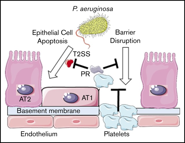
Platelets protect against lung injury from pathogen-secreted factors that promote host cell death even in the absence of overt infection.
Neutrophil airspace influx does not potentiate lung injury caused by pathogens in the susceptible host with thrombocytopenia.
Abstract
Thrombocytopenia is associated with worse outcomes in patients with acute respiratory distress syndrome, which is most commonly caused by infection and marked by alveolar–capillary barrier disruption. However, the mechanisms by which platelets protect the lung alveolar–capillary barrier during infectious injury remain unclear. We found that natively thrombocytopenic Mpl−/− mice deficient in the thrombopoietin receptor sustain severe lung injury marked by alveolar barrier disruption and hemorrhagic pneumonia with early mortality following acute intrapulmonary Pseudomonas aeruginosa (PA) infection; barrier disruption was attenuated by platelet reconstitution. Although PA infection was associated with a brisk neutrophil influx, depletion of airspace neutrophils failed to substantially mitigate PA-triggered alveolar barrier disruption in Mpl−/− mice. Rather, PA cell-free supernatant was sufficient to induce lung epithelial cell apoptosis in vitro and in vivo and alveolar barrier disruption in both platelet-depleted mice and Mpl−/− mice in vivo. Cell-free supernatant from PA with genetic deletion of the type 2 secretion system, but not the type 3 secretion system, mitigated lung epithelial cell death in vitro and lung injury in Mpl−/− mice. Moreover, platelet releasates reduced poly (ADP ribose) polymerase cleavage and lung injury in Mpl−/− mice, and boiling of platelet releasates, but not apyrase treatment, abrogated PA supernatant–induced lung epithelial cell cytotoxicity in vitro. These findings indicate that while neutrophil airspace influx does not potentiate infectious lung injury in the thrombocytopenic host, platelets and their factors protect against severe pulmonary complications from pathogen-secreted virulence factors that promote host cell death even in the absence of overt infection.
Visual Abstract
Introduction
Acute respiratory distress syndrome (ARDS) is estimated to underlie 10% of intensive care unit admissions worldwide and cause 74 500 deaths annually in the United States.1,2 The most common risk factor for ARDS is infection, primarily pneumonia and sepsis.1,3 Platelet deficiency has consistently been associated with worse outcomes in ARDS, and a missense genetic variant in LRRC16A/CARMIL1 that limits reductions in platelet counts during ARDS is associated with improved patient survival.4-8 However, it remains uncertain whether platelets can directly provide protection during lung injury.9,10 Platelets potentiate injury in mouse models of acid aspiration and antibody-mediated transfusion-related lung injury in part through platelet–neutrophil interactions.11,12 However, diffuse lung injury can occur even in severe neutropenia,13-15 and a major clinical trial failed to show significant benefit from aspirin therapy in patients with lung injury.16 In contrast to acid-aspiration and antibody-mediated injury models, platelets limit alveolar hemorrhage during bacterial lipopolysaccharide (LPS)–induced injury and reduce lung bleeding in pneumonia models.17,18 Alveolar hemorrhage frequently results from disruption of the alveolar–capillary barrier,19,20 which is a pathologic hallmark of lung injury.21
Invading pathogens, particularly those with toxins and protease virulence factors,22,23 can cause lung epithelial cell death.24,25 Pseudomonas aeruginosa (PA) is a common cause of hospital-associated pneumonia in critically ill patients and complication of ARDS.26,27 PA virulence is mediated in part by the type 2 secretion system (T2SS), which secretes products into the extracellular space, and the type 3 secretion system (T3SS), which forms a needle and pore system to directly inject toxins into the host cytoplasm.28 Notably, the T3SS is not required for virulence, as the PA T2SS causes lethal pneumonia in the absence of the T3SS.29 Further, T2SS products trigger lung cell death in vitro,24,30,31 and other non-T3SS products can cause hemorrhagic pneumonia.22,29,32
Lung epithelial cell death contributes to alveolar–capillary barrier disruption.33-36 Platelets have been shown to support lung microvascular integrity independent of their classic hemostatic pathways.17,37,38 Platelets possess numerous factors that may promote cell survival, support the endothelial barrier,39,40 and counter programmed cell death pathways.41-43 Importantly, platelets are not only present in the vascular space during lung inflammation but also enter the alveolar space during experimental lung injury.44,45 Therefore, we hypothesized that platelets protect against PA mediated lung injury in part by countering lung epithelial cell death.
Materials and methods
Animals
Mpl−/− mice were obtained from the Walter and Eliza Hall Institute in Melbourne, Australia.46 Mpl−/− mice were backcrossed 12 generations to C57Bl/6 mice at the Walter and Eliza Hall Institute and then backcrossed another generation to C57Bl/6J mice at the University of Pittsburgh before establishing a founder colony. C57Bl/6J mice were used for experiments when littermates were unavailable. All experimental procedures were performed in age- (8-16 weeks old) and sex-matched mice in randomized fashion. A technician blinded to the experimental hypothesis performed the in vivo experiments. Depending upon mouse availability, experiments were conducted with precalculated sample size as a single experiment or divided into independent experiments. The University of Pittsburgh Institutional Animal Care and Use Committee approved the animal protocol. Resilience studies were conducted separately from pneumonia studies, and mice were monitored carefully and euthanized when they met predefined criteria.
Experimental bacterial pneumonia and cell-free bacterial SN model
PA1447 was grown per protocol.48 In-frame deletion mutants PA14ΔexoTUY and PA14ΔxcpQ were used in select experiments.48,49 Intratracheal (IT) inoculations were performed as previously described.48,50,51 PA cell-free bacterial supernatant (SN) was prepared from pelleted PA by careful aspiration of the SN followed by passage through a 0.22 µm sterile filter. The absence of bacterial growth was confirmed by plating filtered SN directly on LB agar plates. Klebsiella pneumoniae (KP) strain 43816 serotype 2 (American Type Culture Collection) was handled as previously described.51
Mouse necropsies, MPO content, and lung tissue histology
Mice were euthanized 20 hours following PA inoculation with isoflurane overdose followed by exsanguination. Mouse necropsy, lung tissue processing, bronchoalveolar lavage (BAL), and myeloperoxidase (MPO) activity were performed as previously described.48,50,52,53 In dedicated experiments, hematoxylin and eosin staining was performed on lung specimens as previously described.50
BAL hemoglobin, platelet counts, OD540, and IgM measurements
BAL fluid was cataloged by digital photography and BAL optical density at 540 nm (OD540) was measured immediately using 100-µL aliquots. BAL hemoglobin and platelet counts were measured in 1 mL BAL fluid by Hemavet 950 (Drew Scientific) as described in a prior report.54 BAL total protein concentration was determined after centrifugation by Pierce BCA Protein Assay. BAL immunoglobulin M (IgM) was determined following 1:10 dilution according to the manufacturer’s instructions (#E90-101, Bethyl Labs).
Evans blue extravasation in the lungs
Pulmonary microvascular permeability was measured using the Evans blue dye extravasation technique55,56 by measuring the absorbance of the formamide extract of lung at 620 nm and 740 nm with correction for heme: corrected absorbance = OD620 − (1.426 × OD740 + 0.03).57,58
Antibody depletion of platelets and neutrophils
To deplete circulating neutrophils, Mpl−/− mice were administered 200 µg anti-Ly-6G antibody (clone 1A8 rat IgG2a, BioLegend) or isotype control (RTK2758 rat IgG2a, κ, BioLegend) by intraperitoneal injection 24 hours prior to bacterial exposure. Wild-type (WT) mice were administered 75 µg anti-GP1b-α (R300 Rat IgG, Emfret) or nonimmune IgG control (C301, Emfret) antibody by retro-orbital injection after isoflurane anesthesia to deplete circulating platelets 24 hours prior to bacterial SN inoculation. Blood counts were performed using Hemavet 950.
Immunofluorescence of mouse lungs
Mouse lungs were perfused with 5 mL normal saline via the right ventricle then inflated via the trachea with 2% paraformaldehyde at 20 cm H2O for 5 minutes prior to preservation. Frozen sections were stained with 4′,6-diamidino-2-phenylindole, terminal deoxynucleotidyltransferase-mediated dUTP nick end labeling (TUNEL), pro–surfactant protein C (pro-SPC), and CD31. Confocal images were collected using a Nikon A1R equipped with GAsP detectors and a 20× (0.75 numerical aperture) objective. TUNEL-positive cells were identified using segmentation analysis (NIS Elements).
Murine lung epithelial cells
MLE 12 cells (CRL-2110, ATCC) were grown in RPMI 1640 supplemented with 5% heat-inactivated fetal bovine serum, 1% penicillin-streptomycin, and 1% l-glutamine at 37°C in 5% CO2.59,60 Passages 4 to 10 were used for experiments. Cytotoxicity was measured by lactate dehydrogenase assay according to the manufacturer’s instructions (#JM-K313-500, MBL International). Manufacturer-supplied cell lysis buffer was used to establish 100% cytotoxicity. For cell death inhibition studies, cells were incubated with carbobenzoxy-valyl-alanyl-aspartyl-[O-methyl]-fluoromethylketone (Z-VAD-fmk) or Nec-1s (Millipore) for 1 hour prior to the addition of PA SN.
Live-cell imaging
MLE 12 cells cultured on 35-mm glass microwell dishes (MatTek) were imaged in a closed, thermo-controlled (37°C) incubator (Tokai Hit) atop the motorized stage of an inverted Nikon TiE fluorescent microscope (Nikon). Annexin V and propidium iodide dyes (V13241, ThermoFisher) were added with initiation of imaging. Dyes were excited using a diode-pumped light engine (SpectraX, Lumencor) and detected by ORCA-Flash 4.0 sCMOS camera (Hamamatsu Corporation) with Chroma Technology excitation/emission filters. Time-lapse large-area images (5 × 5 fields) were collected every 15 minutes using a 20× dry optic (0.75 numerical aperture) and stitched using NIS Elements (Nikon).
Flow cytometric–based caspase-3/7 cleavage assay
After experimental exposure, MLE 12 cells were incubated with a fluorogenic caspase-3/7 substrate (C10740, Invitrogen) per the manufacturer’s instructions. Flow cytometry was performed immediately on a FACSCalibur machine (BD Biosciences) and analyzed with FlowJo V10.
Western blots
MLE 12 cells were lysed with ice-cold Pierce cell lysis buffer with complete protease and phosphatase inhibitors (Roche). Lung tissues were lysed with ice-cold lysis buffer #6 (R&D Systems) and then homogenized and sonicated on ice. Cleaved caspase-3 (#9664), total caspase-3 (#9662), cleaved PARP (#94885), or β-actin (#4970) antibodies were purchased from Cell Signaling Technology. Membranes were developed with chemiluminescent substrate (34095, ThermoFisher) using an Amersham 600 imager (General Electric).
Isolation and transfusion of mouse platelets
Blood was collected into 10% vol/vol citrate phosphate dextrose mixed with 2 µg/mL prostaglandin I2 (PGI2)61 and allowed to sit for 10 minutes before centrifugation at 200 g for 5 minutes without brake to obtain platelet-rich plasma (PRP). PRP was gently transferred to a new tube using wide-orifice pipette tips and taking care to avoid the buffy coat. An additional 2 µg/mL PGI2 was added to PRP prior to centrifuging at 1000 g for 10 minutes (no brake) to pellet platelets, which were gently re-suspended in modified Tyrode’s buffer (137 mM NaCl, 0.3 mM Na2HPO4, 2 mM KCl, 12 mM NaHCO3, 5 mM N-2-hydroxyethylpiperazine-N′-2-ethanesulfonic acid, and 5 mM d-glucose, pH 7.27) with 0.35% vol/vol sterile-filtered endotoxin-free bovine serum albumin. Platelets were normalized to desired count in 100 µL buffer for transfusion via retro-orbital vein after isoflurane. All steps were performed at room temperature.
Platelet releasates
Platelet releasates (PR) were prepared from platelets isolated from WT mice or 4 healthy human volunteers (University of Pittsburgh #IRB0410173) as above without PGI2. Platelets were stimulated with 1 U/mL of thrombin, incubated at 37°C for 20 minutes, and centrifuged at 1000 g. The clear SN (releasates) was stored at −80°C.
Statistical analysis
A 2-tailed Mann-Whitney U test was used when comparing 2 groups, and a Kruskal-Wallis test with Dunn’s post hoc test were used when comparing >2 groups, unless otherwise indicated. P < .05 was considered significant. In one experiment, a linear bootstrap regression model was applied to identify a statistical outlier, which was removed from subsequent analysis. All statistics were performed using GraphPad Prism V7 (La Jolla, CA) and Stata V15 (College Station, TX).
Results
Thrombocytopenic Mpl−/− mice sustain severe lung injury after IT PA infection
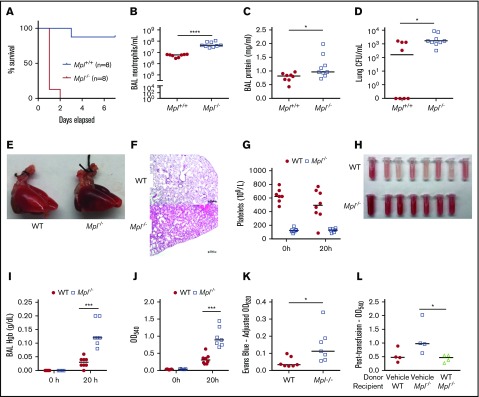
To evaluate the role of platelets during pathogen-triggered lung injury, we exposed natively thrombocytopenic Mpl−/− mice to acute lung infection with the reference PA strain, PA14. Mpl−/− mice exhibit normal erythrocyte and leukocyte counts in circulation,62 and no obvious defects in host defense have been reported.46,63 After IT inoculum of 106 colony-forming units (CFU) of PA, Mpl−/− mice demonstrated rapid mortality; 8 of 8 Mpl−/− mice died within 48 hours, whereas 7 of 8 littermate Mpl+/+ mice survived to 7 days (Figure 1A). Given the rapid mortality of Mpl−/− mice, we used a 20-hour time point to evaluate mechanisms of enhanced susceptibility in the acute PA lung infection model. Mpl−/− mice showed severe lung injury with brisk neutrophilic airspace influx (Figure 1B) and increased BAL protein concentrations (Figure 1C). Notably, platelets have roles in host defense against invading pathogens,18,64-66 and as others have shown,67 platelets appear to support control of lung bacterial burden in acute PA pneumonia (Figure 1D). However, we did not identify significant dissemination of PA to liver or spleen (supplemental Figure 1A-B) to suggest extrapulmonary sepsis that would account for early Mpl−/− mortality. Mpl−/− mice showed lung hepatization on gross examination (Figure 1E) and diffuse hemorrhage on representative hematoxylin and eosin–stained lung sections (Figure 1F). Mpl−/− mice exhibited ∼20% of the baseline circulating platelet counts of WT mice (Figure 1G; Mpl−/− median, 123 × 109 platelets/L; WT median, 621 × 109 platelets/L). We noted striking BAL hemorrhage, a hallmark of alveolar–capillary barrier disruption,19,20 after lung injury in Mpl−/− mice (Figure 1H) that was attenuated in platelet-sufficient WT mice. We found that BAL fluid OD540, a spectrophotometric peak of hemoglobin,68,69 strongly correlated (Spearman r = 0.98; supplemental Figure 1C) with BAL hemoglobin content (Figure 1I-J),54 supporting OD540 as an acceptable, rapid surrogate for BAL hemoglobin. In addition, Evans blue dye infusion confirmed increased lung microvascular permeability in Mpl−/− mice when compared with WT mice (Figure 1K). Moreover, adoptive transfer of platelets from WT mice into Mpl−/− mice 1 hour following acute PA infection mitigated lung injury, as demonstrated by reduction in BAL OD540 (Figure 1L). Therefore, Mpl−/− mice exposed to acute PA bacterial infection model demonstrate rapid mortality marked by lung injury and alveolar hemorrhage that is attenuated by partial platelet reconstitution.
Figure 1.
Thrombocytopenic Mpl−/−mice sustain severe lung injury after IT PA exposure. (A) Mpl−/− and Mpl+/+ mice survival following IT inoculation with PA (P < .0001, log-rank [Mantel-Cox] test). In separate experiments, BAL neutrophil counts/mL (B), BAL protein concentrations (mg/mL) (C), and lung bacterial CFU/mL (D) were measured 20 hours post-PA infection (n = 8 Mpl+/+ mice, n = 9 Mpl−/− mice). Whole-lung images (E) and hematoxylin and eosin–stained lung tissue sections (F; scale bars represent 250 μm) from WT and Mpl−/− mice 20 hours following PA infection. (G) Circulating platelet counts obtained from WT and Mpl−/− mice at 0 and 20 hours post-PA infection. Gross appearance of BAL fluid (H), BAL fluid hemoglobin (Hgb) concentration (g/dL) at 0 and 20 hours (I), BAL fluid OD540 measurements at 0 and 20 hours (J) (n = 5/group at 0 hours, n = 8/group at 20 hours, 1 death in the Mpl−/− group prior to 20 hours). (K) Evans blue dye measurements of lung tissue homogenates at OD620 (adjusted for hemoglobin; n = 7 mice/group). (L) BAL OD540 measurements taken at the 20-hour time point from WT or Mpl−/− mice transfused with either vehicle or WT platelets (109) 1 hour post-PA infection (n = 4 mice/group). For all graphs, each tube or point represents an individual mouse, and the group median is displayed. Statistical comparison by Mann-Whitney U test. *P < .05, ***P < .001, and ****P < .0001.
Pathogenic KP does not induce hemorrhagic lung injury, but the cell-free SN of PA is sufficient to induce neutrophil airspace influx and lung injury in thrombocytopenic Mpl−/− mice
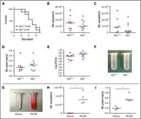
Alveolar hemorrhage after intrapulmonary LPS administration has been observed in models of severe antibody-mediated thrombocytopenia.37 Given the phenotype of hemorrhagic lung injury during acute PA pneumonia in Mpl−/− mice, we wondered whether another gram-negative bacteria, KP, caused a similar phenotype as LPS is a major KP virulence factor.70 We used KP serotype 2, a hypervirulent strain previously shown to cause extra-pulmonary dissemination and death in mice.50,51 Others have shown lung hemorrhage during acute KP pneumonia with platelet counts an order of magnitude lower than Mpl−/− mice.18 However, we did not observe gross alveolar hemorrhage in Mpl−/− mice nor significant differences in survival, BAL cell counts, BAL neutrophils (Mpl−/− group median, 2.4 × 10⁴ neutrophils/mL), BAL protein, or lung CFU counts between Mpl−/− and their platelet-sufficient WT littermates 48 hours following KP infection (Figure 2A-F), when KP serotype 2 is known to disseminate.50,51 Given the discrepancy between alveolar injury between PA and KP infection models, we wondered whether PA-secreted bacterial products may cause lung injury in the absence of live bacteria. Filtered cell-free PA SN was sufficient to cause alveolar–capillary barrier disruption in Mpl−/− mice compared with vehicle (LB broth), as demonstrated by gross alveolar hemorrhage (Figure 2G), BAL neutrophil influx (Figure 2H), and increased BAL protein (Figure 2I). Collectively, these findings suggest that platelet deficiency enhanced susceptibility to PA infection that is characterized by prominent alveolar injury,48 but not to KP infection characterized by extrapulmonary sepsis.50,51
Figure 2.
Pathogenic KP does not induce hemorrhagic lung injury, but the cell-free SN of PA is sufficient to induce neutrophil airspace influx and hemorrhagic injury in thrombocytopenic Mpl−/−mice. (A) Survival of Mpl−/− and Mpl+/+ mice following IT inoculation with pathogenic KP by log-rank (Mantel-Cox) test (n = 9 mice/group). In separate experiments, total BAL leukocytes/mL (B), BAL neutrophil counts/mL (C), BAL protein concentrations (mg/mL) (D), lung CFU/mL (E), and gross appearance of BAL fluid (F) (representative tube displayed; n = 9 mice/group) at the 48-hour time point. (G) Gross appearance of BAL fluid from representative Mpl−/− mice 20 hours after IT administration of either vehicle (LB) or PA cell-free SN with total BAL neutrophils/mL (H) and BAL protein concentrations (mg/mL) (I) (n = 4 mice/group). Each tube or point represents an individual mouse, and the group median is displayed. Statistical comparison by Mann-Whitney U test. *P < .05.
Neutrophil depletion does not significantly attenuate PA-triggered alveolar barrier disruption in thrombocytopenic Mpl−/− mice
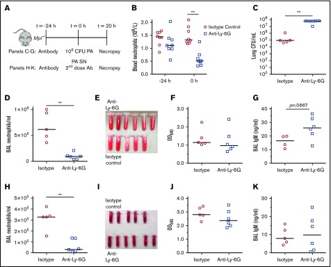
Prior reports have shown that platelets repair holes created by neutrophils exiting the vasculature during immune complex–mediated skin inflammation.38 We therefore examined whether brisk neutrophil airspace influx during acute PA pneumonia contributes to alveolar–capillary barrier disruption. We depleted circulating neutrophils in Mpl−/− mice with Ly-6G antibody 24 hours prior to IT PA inoculation (Figure 3A). The antibody-depletion strategy resulted in >60% fewer circulating neutrophils (Figure 3B) at the time of PA inoculation compared with isotype control IgG2a antibody. Given the essential role of neutrophils in host defense against PA, it was not surprising that neutrophil depletion increased lung bacterial burden (Figure 3C). Antibody depletion reduced neutrophil airspace recruitment by 85% compared with isotype-control during acute PA infection (Figure 3D; anti-Ly-6G group median, 3.2 × 10⁴ neutrophils/mL). Despite neutrophil reduction, there was no significant difference in the gross appearance of BAL fluid, BAL OD540, or BAL IgM, an independent measure of alveolar–capillary barrier disruption,57 in either live bacteria (Figure 3D-G) or bacterial SN (Figure 3H-K) models. Notably, there was a 90% reduction in neutrophil airspace recruitment with a second antibody treatment compared with isotype control (Figure 3H). These findings suggest that bacterial-secreted factors rather than neutrophil influx are the primary contributor to alveolar injury observed in thrombocytopenic Mpl−/− mice.
Figure 3.
Reduction of neutrophil airspace influx does not significantly attenuate alveolar barrier disruption in thrombocytopenic Mpl−/−mice following either live PA challenge or PA SN-induced sterile injury. (A) Schematic of experimental design: Mpl−/− mice were administered Ly-6G or isotype control antibody (Ab) at −24 hours and inoculated with PA at 0 hours, and necropsy was performed 20 hours after PA lung infection. (B) Reduction of circulating neutrophil counts (109/L blood) after anti-Ly6G. In separate experiments, mice were given either 106 live bacteria (C-G) or mice were given cell-free PA SN and a second dose of antibody at t = 0 hours (H-K). Following live bacterial challenge, lung CFU/mL (C), BAL neutrophil counts/mL (D), gross appearance of BAL fluid (E), BAL OD540 (F), and BAL IgM concentrations (ng/mL) (G) were obtained at necropsy 20 hours after 106 CFU IT PA. n = 6 mice/group. There was 1 death in the isotype control group prior to the 20-hour time point. Following PA SN-induced sterile injury, BAL neutrophil counts/mL (H), gross appearance of BAL fluid (I), BAL OD540 (J), and BAL IgM concentration (ng/mL) (K) were obtained at necropsy. n = 6 mice/group. There was 1 death in the isotype control group prior to the 20-hour time point. Each tube or point represents an individual mouse, and the group median is displayed. Statistical comparison by Mann-Whitney U test. **P < .01.
PA SN induces lung injury after antibody-mediated platelet depletion of WT mice, and PA SN-induced injury can be attenuated by partial reconstitution of platelet counts in thrombocytopenic Mpl−/− mice
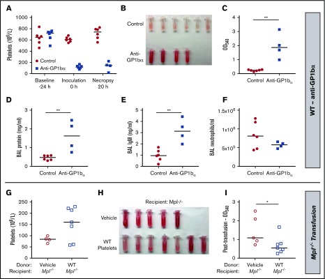
To mitigate concerns that compensatory defects in Mpl−/− mice caused enhanced susceptibility, we examined PA SN in a second model of thrombocytopenia by depleting platelets in WT C57Bl/6J mice using anti-glycoprotein1bα (GP1bα) antibody (Figure 4A; anti-GP1bα group median, 118 × 109 platelets/L; control group median, 607 × 109 platelets/L). PA SN administration recapitulated hemorrhagic lung injury in anti-GP1bα-treated WT mice as demonstrated by gross alveolar hemorrhage (Figure 4B) as well as significantly increased BAL OD540, BAL protein, and BAL IgM concentration (Figure 4C-E). Interestingly, BAL neutrophil counts did not significantly differ between groups (Figure 4F). We then returned to the Mpl−/− mouse model system to demonstrate that partial platelet reconstitution (Figure 4G; platelet recipient median, 160 × 109 platelets/L; vehicle recipient median, 83 × 109 platelets/L) mitigated PA SN-triggered alveolar–capillary barrier disruption (Figure 4H-I). We therefore concluded that platelets protect against secreted bacterial products that were sufficient to induce lung injury in platelet-deficient mice.
Figure 4.
PA SN induces lung injury after antibody-mediated platelet depletion of WT mice, and PA SN-induced injury can be attenuated by partial reconstitution of platelet counts in natively thrombocytopenic Mpl−/−mice. WT mice were administered anti-GP1bα or control antibody 24 hours prior to IT PA SN, and necropsy was performed at 20 hours with peripheral blood platelet counts (109/L) (A). At necropsy 20 hours after IT PA SN inoculation, gross appearance of BAL fluid (B), BAL OD540 measurements (C), BAL protein concentrations (mg/mL) (D), BAL IgM concentrations (ng/mL) (E), and BAL neutrophil counts/mL (F) are shown (n = 6 mice/group; 2 deaths occurred in the anti-GP1bα group prior to the 20-hour time point). In separate experiments, vehicle or 109 platelets from WT mice were transfused into Mpl−/− mice 1 hour following IT PA SN administration. Blood platelet counts (109/L) (G), gross appearance of BAL fluid (H), and BAL OD540 measurements (I) were obtained 20 hours post-PA infection. Each tube or point represents a single mouse (n = 5 for vehicle, n = 7 for the WT platelet group; peripheral blood was not successfully obtained from 1 mouse in the vehicle group), and the group median is displayed. Statistical comparison by Mann-Whitney U test. *P < .05 and **P < .01.
PA exoproducts are sufficient to induce lung epithelial cell apoptosis, in part through a secreted T2SS protein
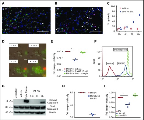
PA T2SS products have been shown to induce cell death in vitro,24,30,31 and lung epithelial cell death may lead to alveolar–capillary barrier disruption.33-35 We found increased TUNEL-positive cell counts in the alveolar space of Mpl−/− mice 6 hours after PA SN exposure compared with baseline (Figure 5A-B), suggesting that PA SN can cause lung cell death in vivo. Notably, TUNEL positivity frequently costained with pro-SPC, suggesting that type 2 alveolar epithelial cells are susceptible to PA SN-triggered programmed cell death. Therefore, we used a murine type 2 alveolar epithelial cell line (MLE 12) to better characterize PA SN-triggered cell death. We found that PA SN caused lung epithelial cell death in a time-dependent manner in vitro (Figure 5C), and live-cell imaging of MLE cells exposed to PA SN revealed cellular blebbing and early annexin V staining, consistent with apoptosis, that later transitioned to propidium iodide staining (Figure 5D). MLE cell death was inhibited by Z-VAD-fmk, a pan-caspase inhibitor, but not the RIP1-kinase inhibitor Nec-1s that targets necroptosis, even after prolonged PA SN exposure (Figure 5E). Our finding of apoptotic cell death was corroborated by flow cytometry demonstrating MLE caspase-3/7 activation (Figure 5F; supplemental Figure 1D) and by immunoblot of MLE cell lysates, which identified caspase-3 cleavage, following PA SN exposure (Figure 5G). We found that boiling PA SN prior to MLE exposure decreased cytotoxicity by a median of 87% (Figure 5H), suggesting that protein products in PA SN mediate its cytotoxic effect. We then compared lung epithelial cell death following exposure to SN from WT PA14, PA14 ΔxcpQ (deletion mutant lacking the outer membrane protein XcpQ that controls T2SS exoproduct efflux),28 or PA14 ΔexoTUY (deletion mutant lacking the T3SS exotoxins T, U, and Y).49 Deletion of the T2SS secretory protein XcpQ decreased lung epithelial cell death, whereas deletion of T3SS exotoxins T, U, and Y did not (Figure 5I). These findings indicate that PA SN can induce apoptotic lung epithelial cell death, in part through T2SS protein exoproducts.
Figure 5.
PA exoproducts are sufficient to induce apoptotic lung epithelial cell death, in part through a secreted T2SS protein. Single-plane confocal images showing Mpl−/− mouse lung tissue at 0 hours (A) and 6 hours (B) after IT PA SN. Tissue was labeled with 4′,6-diamidino-2-phenylindole (nuclei, blue), TUNEL (red), phalloidin (actin, white), and anti-pro-SPC (type 2 cells, green). There was an increase in the number of TUNEL-positive cells at 6 hours, the majority of which were also positive for pro-SPC (indicated by white arrows). Magenta arrows indicate TUNEL-positive cells likely to be endothelial cells based on separate staining for CD31 as well as the shape and size of the nucleus. Scale bars represent 100 μm. (C) Kinetics of cell death as measured by a lactate dehydrogenase assay in MLE cells incubated with vehicle or PA SN. (D) Live-cell imaging showing cellular blebbing and annexin V staining (green) followed by propidium iodide staining (red). Scale bars represent 10 μm. (E) Fold change in MLE cytotoxicity 16 hours following the addition of 10% PA SN, where cells were pretreated with vehicle (dimethyl sulfoxide), pan-caspase inhibitor (Z-VAD-fmk), or RIP1-inhibitor (Nec-1s). Each point represents the median cytotoxicity of 3 to 4 technical replicates from a single trial relative to PA SN and vehicle treatment, which is set at 1.0. (F) Flow cytometry of MLE cells positive for a fluorogenic caspase-3/7 substrate 6 hours after the addition of 10% PA SN. (G) Cleaved caspase-3, total caspase-3, and β-actin protein expression in MLE cells after the addition of 10% PA SN at the indicated time points. Staurosporine served as a positive control for caspase cleavage in panels F and G. (H) Fold change in MLE cytotoxicity after the addition of PA SN or equal parts PA SN denatured for 30 minutes at 100°C. (I) Fold change in MLE cytotoxicity after the addition of 50% PA SN from the PA14 parent strain, PA14ΔxcpQ, or PA14ΔexoTUY. Each point represents median cytotoxicity from a single trial as described above. Comparison by Kruskal-Wallis test with Dunn’s test for multiple comparisons (E; the line represents post hoc analysis) or Mann-Whitney U test (I). *P < .05.
Genetic deletion of PA T2SS, but not T3SS exotoxins, attenuates alveolar–capillary barrier disruption in thrombocytopenic Mpl−/− mice
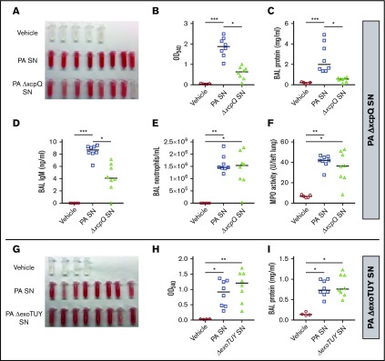
Given that lung epithelial cell death in vitro was attenuated after PA14ΔxcpQ SN exposure in contrast to PA14 ΔexoTUY SN, we wondered whether this phenomenon could account for alveolar–capillary barrier disruption in vivo. Mpl−/− mice administered PA ΔxcpQ SN showed attenuated lung injury compared with the vehicle-treated or parent PA SN-treated mice as evidenced by BAL fluid appearance, BAL OD540, BAL protein, and BAL IgM (Figure 6A-D). Notably, there were similar neutrophil counts in the airspaces and lungs of the SN-treated groups (Figure 6E-F), reinforcing the concept that neutrophils do not potentiate lung injury in this model. As expected, the cell-free SN of PA ΔexoTUY did not attenuate lung injury compared with vehicle-treated or PA SN-treated Mpl−/− mice as measured by BAL fluid appearance, BAL OD540, and BAL protein (Figure 6G-I). Therefore, platelets provide protection against lung injury mediated by bacterial secreted products of PA T2SS even in the absence of live pathogen or cell–cell contact and suggest a role for platelets as defenders against lung epithelial cell death.
Figure 6.
Genetic deletion of PA T2SS (ΔxcpQ), but not T3SS exotoxins (ΔexoTUY), attenuates alveolar–capillary barrier disruption in thrombocytopenic Mpl−/−mice. Gross appearance of BAL fluid (A), BAL OD540 measurements (B), BAL protein concentrations (mg/mL) (C), BAL IgM (ng/mL) (D), BAL neutrophil counts/mL (E), or lung MPO activity (U/left lung) (F) were obtained in Mpl−/− mice 20 hours following IT administration of vehicle or cell-free SN from PA14 or PA14ΔxcpQ strain (n = 5 mice in vehicle group, n = 8 mice in PA14 and PA14ΔxcpQ groups). In separate experiments, gross appearance of BAL fluid (G), BAL OD540 measurements (H), and BAL protein concentrations (mg/mL) (I) were obtained in Mpl−/− mice 20 hours following IT administration of vehicle or cell-free SN from PA14 or PA14ΔexoTUY (n = 4 mice in vehicle group, n = 8 mice in PA14 and PA14ΔexoTUY groups). Each tube or point represents an individual mouse, and the group median is displayed. Statistical comparison by Kruskal-Wallis test with Dunn’s test for multiple comparisons (lines represent post hoc analysis). *P < .05, **P < .01, and ***P < .001.
PR attenuates PA SN-triggered lung cell death and limits PA SN-mediated lung injury in thrombocytopenic Mpl−/− mice
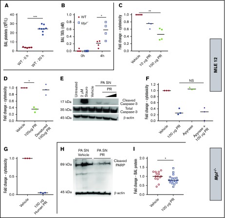
Several studies have suggested that platelets enter the alveolar space in response to experimental lung injury in mice,44,45,67 and platelet products have been noted in BAL fluid from patients with diffuse lung injury.71 We found platelet accumulation in the BAL fluid of WT mice following acute PA IT infection (Figure 7A). Thrombin–antithrombin complexes (TATc) have been noted in BAL from patients with ventilator-associated pneumonia,72 and TATc was increased in BAL fluid from both WT and Mpl−/− mice after PA SN exposure (Figure 7B). We next evaluated whether releasates from thrombin-stimulated platelets could counter PA SN-induced lung epithelial cell death in vitro. PR dose-dependently protected MLE 12 cells from PA SN-mediated cytotoxicity when compared with vehicle-treated cells (Figure 7C), and protection was markedly attenuated by boiling PR (Figure 7D), suggesting that platelet proteins mediate the cytoprotective effect. PR inhibited caspase-3 cleavage in PA SN-treated MLE 12 cells (Figure 7E). Notably, there was no significant impact on cytotoxicity from apyrase treatment (Figure 7F). Moreover, human PR also reduced MLE cytotoxicity by a median of 94% after PA SN (Figure 7F), suggesting conserved platelet proteins may underlie their protective effect. Finally, delivery of PR to Mpl−/− mice reduced cleavage of PARP, a marker of caspase-3 activation, in lung tissue and decreased BAL protein concentrations when compared with vehicle-treated Mpl−/− mice (Figure 7H-I). Taken together, platelet factors appear to play mitigating roles against infection-induced lung injury beyond hemostasis alone.
Figure 7.
PR attenuate PA SN-triggered lung cell death and limits PA SN-mediated lung injury in thrombocytopenic Mpl−/−mice. (A) WT mice BAL platelet counts (109/L) at baseline (0 hours, n = 5) and 20 hours after IT 106 CFU PA (n = 8). (B) BAL thrombin–antithrombin complexes (nM) in WT and Mpl−/− mice at 0 and 4 hours after IT PA SN (n = 4 each group). In separate experiments using MLE cells, fold change in MLE cytotoxicity 6 hours after PA SN in the presence or absence of pooled thrombin-stimulated PR from WT mice (C) (n = 4 trials, where 3 of the 4 trials included 10 µg group) or denatured PR (D). Cleaved caspase-3, total caspase-3, and β-actin expression in MLE cells treated with PA SN in the presence or absence of increasing PR (100 and 300 µg total protein) at 4 hours (E), fold change in MLE cytotoxicity 6 hours after PA SN in the presence or absence of apyrase (0.2 U) or PR from WT mice (F), or thrombin-stimulated platelets pooled from healthy volunteers (G) (n = 4). For panels C-D and F-G, each point represents the median cytotoxicity of 3 to 4 technical replicates from a single trial, relative to PA SN and vehicle treatment cytotoxicity, which is set at 1.0. In separate experiments with Mpl−/− mice 20 hours after IT PA SN with either vehicle or 100 µg PR, lung tissue expression of cleaved PARP and β-actin is shown, n = 3 in each group (H). Fold change in BAL protein concentration. n = 14 (vehicle) and n = 17 (PR-treated group) from 3 separate experiments (I). Each point represents an individual mouse, and the group median is displayed. Statistical comparison by Mann-Whitney U test (*P < .05 and ***P < .001) (A-B,I) or Kruskal-Wallis test with Dunn’s test for multiple comparisons (lines represent post hoc analysis; *P < .05 (C-D,F).
Discussion
The major finding of our study is that platelets attenuate pathogen-induced lung injury and provide protection against lung epithelial cell apoptosis. We show that platelet deficiency is associated with severe alveolar–capillary barrier disruption in both live bacteria and bacterial exoproduct models of acute PA intrapulmonary infection. Barrier disruption is attenuated by the partial reconstitution of platelet counts via transfusion. Based on our findings, we propose that in addition to their role in hemostasis at the alveolar–capillary barrier, platelets can also enter the airspace during infection and release protective factors that may contribute to host protection by limiting alveolar epithelial cell death and attenuating further lung injury. Significant work remains to characterize platelet factors that mitigate PA-induced lung injury in our model system. Our findings suggest conserved platelet products mediate the effect and candidate factors could include mitogens,43 thrombospondin-1,48 and defensins.73
The second major finding of our study is that neutrophils do not potentiate lung injury following acute PA pneumonia. Neutrophils have been implicated as mediators of acute lung injury,74 and this is supported by evidence from several animal models.10-12 Furthermore, neutrophil transmigration across the endothelium is critical to vascular injury in models of immune complex and UV light–mediated skin injury during thrombocytopenia.38,75 However, neutrophils are not required for acute lung injury in humans as neutropenic patients can develop ARDS.13-15,76,77 We show that antibody-mediated neutrophil depletion does not substantially protect against alveolar–capillary barrier disruption in Mpl−/− mice during both acute live bacteria and PA SN infection models. Furthermore, BAL neutrophil influx in our KP pneumonia model (without associated alveolar hemorrhage) was similar in magnitude to our anti-Ly-6G treatment. We further show that the cell-free SN of PA is sufficient to cause lung injury in thrombocytopenic mice, which can be attenuated by the genetic deletion of the T2SS protein XcpQ that enables secretion of several factors with cell-damaging properties.24,30,31,48 Interestingly, pathogens with cytotoxic and proteolytic virulence mechanisms, such as PA,14 cytomegalovirus,15 and invasive fungi such as Aspergillus and Mucormycosis,78 are common causes of diffuse lung injury in neutropenia. Notably, in patients diagnosed with idiopathic pneumonia syndrome who were later found to harbor pathogen, there was increased mortality in those patients with evidence of cytotoxic pathogens in their BAL.79 Therefore, we suggest that cytotoxic pathogen virulence factors can mediate lung injury independent of neutrophils and that this effect is accentuated in the thrombocytopenic host.
Finally, our study identifies a potential role for platelet granule factors in protection against pathogen-triggered lung injury.80 We show that PR attenuates lung epithelial cell death in vitro and disruption of the lung vascular barrier in platelet-deficient mice, independent of whole platelets. Consistent with our findings, others have shown that platelet granules are crucial to preventing inflammatory bleeding during thrombocytopenia.81 In contrast, the double knockout of platelet α and dense granule secretion did not modify lung vascular barrier function in an LPS model of lung inflammation in mice with normal platelet counts.82 However, invading pathogens may pose a distinct challenge to the alveolar–capillary barrier beyond the presence of LPS alone, and this challenge may be accentuated during platelet deficiency. Although we did not observe alveolar hemorrhage during acute KP pneumonia in the Mpl−/− mouse, others have shown significant lung hemorrhage during KP pneumonia after antibody-mediated reduction of platelet counts an order of magnitude lower than Mpl−/− mice.18 Therefore, platelet count may provide important context; we show that even partial reconstitution of platelet counts can significantly mitigate alveolar–capillary barrier disruption, which is consistent with another report.17 Therefore, both platelet number and the nature of the challenge posed by invading pathogens may influence the platelet role in safeguarding the lung vascular barrier. Platelet granule factors may be dispensable for alveolar–capillary barrier integrity during LPS injury if platelet counts are normal, yet they may be required to protect against cytotoxic and proteolytic damage mediated by invading lung pathogens, including during thrombocytopenia.
In conclusion, secreted bacterial products are capable of inducing lung epithelial cell apoptosis and fatal lung injury during acute PA pneumonia marked by severe breakdown of the alveolar–capillary barrier that can be attenuated by genetic deletion of the PA T2SS, partial reconstitution of platelet counts, or administration of platelet factors. Our findings enforce the concept that thrombocytopenia identifies a host susceptible to pathogen-related complications in the lung and that platelet factors can provide direct protection against lung epithelial cell death mediated by pathogen products even in the absence of live bacteria or cell–cell contact. Further study is warranted to identify the mechanisms by which platelet factors modulate infection-triggered lung epithelial injury and cell death pathways.
Supplementary Material
The full-text version of this article contains a data supplement.
Acknowledgments
The authors thank Warren Alexander and the Walter and Eliza Hall Institute (Melbourne, Australia) for the kind gift of Mpl−/− mice. The authors also thank Servier Medical Art (Les Laboratoires Servier SAS; https://smart.servier.com/) and Creative Commons (https://creativecommons.org/licenses/by/3.0/legalcode) for the use of graphics included in the visual abstract.
This work was supported by the National Heart, Lung, and Blood Institute, National Institutes of Health under award numbers 4T32 HL007563 and F32 HL142172 (W.B.), P01HL114453 (R.K.M. and P.R.), and R01 HL136143, HL086884, and HL142084 (J.S.L.); American Heart Association predoctoral fellowship 18PRE33960033 (T.O.); the Cystic Fibrosis Foundation Research Development Program (J.P. and J.S.L.); the Vascular Medicine Institute; the Hemophilia Center of Western Pennsylvania; and the Institute for Transfusion Medicine (W.B. and J.S.L.).
The content is solely the responsibility of the authors and does not necessarily represent the official views of the National Institutes of Health or any other sponsoring agency.
Footnotes
For original data, please contact J.S.L. (e-mail: leejs3@upmc.edu).
Authorship
Contribution: W.B. performed the experiments, analyzed the data, interpreted the data, and wrote the manuscript; T.O., M.Y., Y.Q., M.H., Z.X., and H.L. performed the experiments and analyzed the data; Z.C., R.M.Q.S., and C.S.C. provided critical reagents, interpreted the data, and revised the work for important intellectual content; M.N. provided critical statistical expertise, interpreted the data, and revised the work for important intellectual content; J.P., R.K.M., A.R., P.R., and R.L.S. interpreted the data and revised the work for important intellectual content; and J.S.L. conceived, designed, analyzed, and interpreted the data, and wrote the manuscript.
Conflict-of-interest disclosure: The authors declare no competing financial interests.
Correspondence: Janet S. Lee, Montefiore University Hospital NW628, 3459 Fifth Ave, Pittsburgh, PA 15213; e-mail: leejs3@upmc.edu.
References
- 1.Bellani G, Laffey JG, Pham T, et al. ; ESICM Trials Group. Epidemiology, patterns of care, and mortality for patients with acute respiratory distress syndrome in intensive care units in 50 countries. JAMA. 2016;315(8):788-800. [DOI] [PubMed] [Google Scholar]
- 2.Rubenfeld GD, Caldwell E, Peabody E, et al. . Incidence and outcomes of acute lung injury. N Engl J Med. 2005;353(16):1685-1693. [DOI] [PubMed] [Google Scholar]
- 3.Zilberberg MD, Epstein SK. Acute lung injury in the medical ICU: comorbid conditions, age, etiology, and hospital outcome. Am J Respir Crit Care Med. 1998;157(4 Pt 1):1159-1164. [DOI] [PubMed] [Google Scholar]
- 4.Fein AM, Lippmann M, Holtzman H, Eliraz A, Goldberg SK. The risk factors, incidence, and prognosis of ARDS following septicemia. Chest. 1983;83(1):40-42. [DOI] [PubMed] [Google Scholar]
- 5.Brogly N, Devos P, Boussekey N, Georges H, Chiche A, Leroy O. Impact of thrombocytopenia on outcome of patients admitted to ICU for severe community-acquired pneumonia. J Infect. 2007;55(2):136-140. [DOI] [PubMed] [Google Scholar]
- 6.Wang T, Liu Z, Wang Z, et al. . Thrombocytopenia is associated with acute respiratory distress syndrome mortality: an international study. PLoS One. 2014;9(4):e94124. [DOI] [PMC free article] [PubMed] [Google Scholar]
- 7.Claushuis TAM, van Vught LA, Scicluna BP, et al. ; Molecular Diagnosis and Risk Stratification of Sepsis Consortium. Thrombocytopenia is associated with a dysregulated host response in critically ill sepsis patients. Blood. 2016;127(24):3062-3072. [DOI] [PubMed] [Google Scholar]
- 8.Wei Y, Tejera P, Wang Z, et al. . A missense genetic variant in LRRC16A/CARMIL1 improves acute respiratory distress syndrome survival by attenuating platelet count decline. Am J Respir Crit Care Med. 2017;195(10):1353-1361. [DOI] [PMC free article] [PubMed] [Google Scholar]
- 9.Bozza FA, Shah AM, Weyrich AS, Zimmerman GA. Amicus or adversary: platelets in lung biology, acute injury, and inflammation. Am J Respir Cell Mol Biol. 2009;40(2):123-134. [DOI] [PMC free article] [PubMed] [Google Scholar]
- 10.Middleton EA, Rondina MT, Schwertz H, Zimmerman GA. Amicus or adversary revisited: platelets in acute lung injury & acute respiratory distress syndrome. Am J Respir Cell Mol Biol. 2018;59(1):18-35. [DOI] [PMC free article] [PubMed] [Google Scholar]
- 11.Zarbock A, Singbartl K, Ley K. Complete reversal of acid-induced acute lung injury by blocking of platelet-neutrophil aggregation. J Clin Invest. 2006;116(12):3211-3219. [DOI] [PMC free article] [PubMed] [Google Scholar]
- 12.Looney MR, Nguyen JX, Hu Y, Van Ziffle JA, Lowell CA, Matthay MA. Platelet depletion and aspirin treatment protect mice in a two-event model of transfusion-related acute lung injury. J Clin Invest. 2009;119(11):3450-3461. [DOI] [PMC free article] [PubMed] [Google Scholar]
- 13.Rinaldo JE, Rogers RM. Adult respiratory distress syndrome. N Engl J Med. 1986;315(9):578-580. [DOI] [PubMed] [Google Scholar]
- 14.Ognibene FP, Martin SE, Parker MM, et al. . Adult respiratory distress syndrome in patients with severe neutropenia. N Engl J Med. 1986;315(9):547-551. [DOI] [PubMed] [Google Scholar]
- 15.Braude S, Apperley J, Krausz T, Goldman JM, Royston D. Adult respiratory distress syndrome after allogeneic bone-marrow transplantation: evidence for a neutrophil-independent mechanism. Lancet. 1985;1(8440):1239-1242. [DOI] [PubMed] [Google Scholar]
- 16.Kor DJ, Carter RE, Park PK, et al. ; US Critical Illness and Injury Trials Group: Lung Injury Prevention with Aspirin Study Group (USCIITG: LIPS-A). Effect of aspirin on development of ARDS in at-risk patients presenting to the emergency department: the LIPS-A randomized clinical trial. JAMA. 2016;315(22):2406-2414. [DOI] [PMC free article] [PubMed] [Google Scholar]
- 17.Boulaftali Y, Hess PR, Getz TM, et al. . Platelet ITAM signaling is critical for vascular integrity in inflammation. J Clin Invest. 2013;123(2):908-916. [DOI] [PMC free article] [PubMed] [Google Scholar]
- 18.de Stoppelaar SF, van’t Veer C, Claushuis TA, Albersen BJ, Roelofs JJ, van der Poll T. Thrombocytopenia impairs host defense in gram-negative pneumonia-derived sepsis in mice. Blood. 2014;124(25):3781-3790. [DOI] [PMC free article] [PubMed] [Google Scholar]
- 19.Martin AM Jr, Simmons RL, Heisterkamp CA III. Respiratory insufficiency in combat casualties. I. Pathologic changes in the lungs of patients dying of wounds. Ann Surg. 1969;170(1):30-38. [DOI] [PMC free article] [PubMed] [Google Scholar]
- 20.Bachofen M, Weibel ER. Alterations of the gas exchange apparatus in adult respiratory insufficiency associated with septicemia. Am Rev Respir Dis. 1977;116(4):589-615. [DOI] [PubMed] [Google Scholar]
- 21.Matthay MA, Ware LB, Zimmerman GA. The acute respiratory distress syndrome. J Clin Invest. 2012;122(8):2731-2740. [DOI] [PMC free article] [PubMed] [Google Scholar]
- 22.Elsen S, Huber P, Bouillot S, et al. . A type III secretion negative clinical strain of Pseudomonas aeruginosa employs a two-partner secreted exolysin to induce hemorrhagic pneumonia. Cell Host Microbe. 2014;15(2):164-176. [DOI] [PubMed] [Google Scholar]
- 23.von Ranke FM, Zanetti G, Hochhegger B, Marchiori E. Infectious diseases causing diffuse alveolar hemorrhage in immunocompetent patients: a state-of-the-art review. Hai. 2013;191(1):9-18. [DOI] [PMC free article] [PubMed] [Google Scholar]
- 24.Beaufort N, Corvazier E, Mlanaoindrou S, de Bentzmann S, Pidard D. Disruption of the endothelial barrier by proteases from the bacterial pathogen Pseudomonas aeruginosa: implication of matrilysis and receptor cleavage. PLoS One. 2013;8(9):e75708. [DOI] [PMC free article] [PubMed] [Google Scholar]
- 25.Grassmé H, Kirschnek S, Riethmueller J, et al. . CD95/CD95 ligand interactions on epithelial cells in host defense to Pseudomonas aeruginosa. Science. 2000;290(5491):527-530. [DOI] [PubMed] [Google Scholar]
- 26.Markowicz P, Wolff M, Djedaïni K, et al. ; ARDS Study Group. Multicenter prospective study of ventilator-associated pneumonia during acute respiratory distress syndrome. Incidence, prognosis, and risk factors. Am J Respir Crit Care Med. 2000;161(6):1942-1948. [DOI] [PubMed] [Google Scholar]
- 27.Forel J-M, Voillet F, Pulina D, et al. . Ventilator-associated pneumonia and ICU mortality in severe ARDS patients ventilated according to a lung-protective strategy. Crit Care. 2012;16(2):R65. [DOI] [PMC free article] [PubMed] [Google Scholar]
- 28.Filloux A. Protein secretion systems in pseudomonas aeruginosa: an essay on diversity, evolution, and function. Front Microbiol. 2011;2:155. [DOI] [PMC free article] [PubMed] [Google Scholar]
- 29.Jyot J, Balloy V, Jouvion G, et al. . Type II secretion system of Pseudomonas aeruginosa: in vivo evidence of a significant role in death due to lung infection. J Infect Dis. 2011;203(10):1369-1377. [DOI] [PMC free article] [PubMed] [Google Scholar]
- 30.Plotkowski M-C, Póvoa HCC, Zahm J-M, et al. . Early mitochondrial dysfunction, superoxide anion production, and DNA degradation are associated with non-apoptotic death of human airway epithelial cells induced by Pseudomonas aeruginosa exotoxin A. Am J Respir Cell Mol Biol. 2002;26(5):617-626. [DOI] [PubMed] [Google Scholar]
- 31.Jenkins CE, Swiatoniowski A, Issekutz AC, Lin T-J. Pseudomonas aeruginosa exotoxin A induces human mast cell apoptosis by a caspase-8 and -3-dependent mechanism. J Biol Chem. 2004;279(35):37201-37207. [DOI] [PubMed] [Google Scholar]
- 32.Basso P, Ragno M, Elsen S, et al. . Pseudomonas aeruginosa pore-forming exolysin and type IV pili cooperate to induce host cell lysis. MBio. 2017;8(1):e02250-16. [DOI] [PMC free article] [PubMed] [Google Scholar]
- 33.Matute-Bello G, Liles WC, Steinberg KP, et al. . Soluble Fas ligand induces epithelial cell apoptosis in humans with acute lung injury (ARDS). J Immunol. 1999;163(4):2217-2225. [PubMed] [Google Scholar]
- 34.Lipke AB, Matute-Bello G, Herrero R, et al. . Febrile-range hyperthermia augments lipopolysaccharide-induced lung injury by a mechanism of enhanced alveolar epithelial apoptosis. J Immunol. 2010;184(7):3801-3813. [DOI] [PMC free article] [PubMed] [Google Scholar]
- 35.Albertine KH, Soulier MF, Wang Z, et al. . Fas and fas ligand are up-regulated in pulmonary edema fluid and lung tissue of patients with acute lung injury and the acute respiratory distress syndrome. Am J Pathol. 2002;161(5):1783-1796. [DOI] [PMC free article] [PubMed] [Google Scholar]
- 36.Matute-Bello G, Winn RK, Jonas M, Chi EY, Martin TR, Liles WC. Fas (CD95) induces alveolar epithelial cell apoptosis in vivo: implications for acute pulmonary inflammation. Am J Pathol. 2001;158(1):153-161. [DOI] [PMC free article] [PubMed] [Google Scholar]
- 37.Goerge T, Ho-Tin-Noe B, Carbo C, et al. . Inflammation induces hemorrhage in thrombocytopenia. Blood. 2008;111(10):4958-4964. [DOI] [PMC free article] [PubMed] [Google Scholar]
- 38.Gros A, Syvannarath V, Lamrani L, et al. . Single platelets seal neutrophil-induced vascular breaches via GPVI during immune-complex-mediated inflammation in mice. Blood. 2015;126(8):1017-1026. [DOI] [PubMed] [Google Scholar]
- 39.Kitchens CS, Pendergast JF. Human thrombocytopenia is associated with structural abnormalities of the endothelium that are ameliorated by glucocorticosteroid administration. Blood. 1986;67(1):203-206. [PubMed] [Google Scholar]
- 40.Mammoto T, Jiang A, Jiang E, Mammoto A. Platelet-rich plasma extract prevents pulmonary edema through angiopoietin-Tie2 signaling. Am J Respir Cell Mol Biol. 2015;52(1):56-64. [DOI] [PMC free article] [PubMed] [Google Scholar]
- 41.Hisakura K, Murata S, Takahashi K, et al. . Platelets prevent acute hepatitis induced by anti-fas antibody. J Gastroenterol Hepatol. 2011;26(2):348-355. [DOI] [PubMed] [Google Scholar]
- 42.Chatterjee M, von Ungern-Sternberg SNI, Seizer P, et al. . Platelet-derived CXCL12 regulates monocyte function, survival, differentiation into macrophages and foam cells through differential involvement of CXCR4-CXCR7. Cell Death Dis. 2015;6(11):e1989. [DOI] [PMC free article] [PubMed] [Google Scholar]
- 43.Au AEL, Sashindranath M, Borg RJ, et al. . Activated platelets rescue apoptotic cells via paracrine activation of EGFR and DNA-dependent protein kinase. Cell Death Dis. 2014;5(9):e1410. [DOI] [PMC free article] [PubMed] [Google Scholar]
- 44.Lax S, Rayes J, Wichaiyo S, et al. . Platelet CLEC-2 protects against lung injury via effects of its ligand podoplanin on inflammatory alveolar macrophages in the mouse. Am J Physiol Lung Cell Mol Physiol. 2017;313(6):L1016-L1029. [DOI] [PMC free article] [PubMed] [Google Scholar]
- 45.Ortiz-Muñoz G, Mallavia B, Bins A, Headley M, Krummel MF, Looney MR. Aspirin-triggered 15-epi-lipoxin A4 regulates neutrophil-platelet aggregation and attenuates acute lung injury in mice. Blood. 2014;124(17):2625-2634. [DOI] [PMC free article] [PubMed] [Google Scholar]
- 46.Alexander WS, Roberts AW, Nicola NA, Li R, Metcalf D. Deficiencies in progenitor cells of multiple hematopoietic lineages and defective megakaryocytopoiesis in mice lacking the thrombopoietic receptor c-Mpl. Blood. 1996;87(6):2162-2170. [PubMed] [Google Scholar]
- 47.Lee DG, Urbach JM, Wu G, et al. . Genomic analysis reveals that Pseudomonas aeruginosa virulence is combinatorial. Genome Biol. 2006;7(10):R90. [DOI] [PMC free article] [PubMed] [Google Scholar]
- 48.Qu Y, Olonisakin T, Bain W, et al. . Thrombospondin-1 protects against pathogen-induced lung injury by limiting extracellular matrix proteolysis. JCI Insight. 2018;3(3):96914. [DOI] [PMC free article] [PubMed] [Google Scholar]
- 49.Miyata S, Casey M, Frank DW, Ausubel FM, Drenkard E. Use of the Galleria mellonella caterpillar as a model host to study the role of the type III secretion system in Pseudomonas aeruginosa pathogenesis. Infect Immun. 2003;71(5):2404-2413. [DOI] [PMC free article] [PubMed] [Google Scholar]
- 50.Zhao Y, Olonisakin TF, Xiong Z, et al. . Thrombospondin-1 restrains neutrophil granule serine protease function and regulates the innate immune response during Klebsiella pneumoniae infection. Mucosal Immunol. 2015;8(4):896-905. [DOI] [PMC free article] [PubMed] [Google Scholar]
- 51.Olonisakin TF, Li H, Xiong Z, et al. . CD36 provides host protection against Klebsiella pneumoniae intrapulmonary infection by enhancing lipopolysaccharide responsiveness and macrophage phagocytosis. J Infect Dis. 2016;214(12):1865-1875. [DOI] [PMC free article] [PubMed] [Google Scholar]
- 52.Mangalmurti NS, Xiong Z, Hulver M, et al. . Loss of red cell chemokine scavenging promotes transfusion-related lung inflammation. Blood. 2009;113(5):1158-1166. [DOI] [PMC free article] [PubMed] [Google Scholar]
- 53.Lee JS, Frevert CW, Matute-Bello G, et al. . TLR-4 pathway mediates the inflammatory response but not bacterial elimination in E. coli pneumonia. Am J Physiol Lung Cell Mol Physiol. 2005;289(5):L731-L738. [DOI] [PubMed] [Google Scholar]
- 54.Antoniak S, Tatsumi K, Hisada Y, et al. . Tissue factor deficiency increases alveolar hemorrhage and death in influenza A virus-infected mice. J Thromb Haemost. 2016;14(6):1238-1248. [DOI] [PMC free article] [PubMed] [Google Scholar]
- 55.Camerer E, Regard JB, Cornelissen I, et al. . Sphingosine-1-phosphate in the plasma compartment regulates basal and inflammation-induced vascular leak in mice. J Clin Invest. 2009;119(7):1871-1879. [DOI] [PMC free article] [PubMed] [Google Scholar]
- 56.Zhao Y, Xiong Z, Lechner EJ, et al. . Thrombospondin-1 triggers macrophage IL-10 production and promotes resolution of experimental lung injury. Mucosal Immunol. 2014;7(2):440-448. [DOI] [PMC free article] [PubMed] [Google Scholar]
- 57.Matute-Bello G, Downey G, Moore BB, et al. ; Acute Lung Injury in Animals Study Group. An official American Thoracic Society workshop report: features and measurements of experimental acute lung injury in animals. Am J Respir Cell Mol Biol. 2011;44(5):725-738. [DOI] [PMC free article] [PubMed] [Google Scholar]
- 58.Xu Z, Castellino FJ, Ploplis VA. Plasminogen activator inhibitor-1 (PAI-1) is cardioprotective in mice by maintaining microvascular integrity and cardiac architecture. Blood. 2010;115(10):2038-2047. [DOI] [PMC free article] [PubMed] [Google Scholar]
- 59.Leiva M, Ruiz-Bravo A, Jimenez-Valera M. Effects of telithromycin in in vitro and in vivo models of lipopolysaccharide-induced airway inflammation. Chest. 2008;134(1):20-29. [DOI] [PubMed] [Google Scholar]
- 60.Herrlich A, Leitch V, King LS. Role of proneuregulin 1 cleavage and human epidermal growth factor receptor activation in hypertonic aquaporin induction. Proc Natl Acad Sci USA. 2004;101(44):15799-15804. [DOI] [PMC free article] [PubMed] [Google Scholar]
- 61.Podrez EA, Byzova TV, Febbraio M, et al. . Platelet CD36 links hyperlipidemia, oxidant stress and a prothrombotic phenotype. Nat Med. 2007;13(9):1086-1095. [DOI] [PMC free article] [PubMed] [Google Scholar]
- 62.Hirata S, Takayama N, Jono-Ohnishi R, et al. . Congenital amegakaryocytic thrombocytopenia iPS cells exhibit defective MPL-mediated signaling. J Clin Invest. 2013;123(9):3802-3814. [DOI] [PMC free article] [PubMed] [Google Scholar]
- 63.Gurney AL, Carver-Moore K, de Sauvage FJ, Moore MW. Thrombocytopenia in c-mpl-deficient mice. Science. 1994;265(5177):1445-1447. [DOI] [PubMed] [Google Scholar]
- 64.Yeaman MR. Platelets: at the nexus of antimicrobial defence. Nat Rev Microbiol. 2014;12(6):426-437. [DOI] [PubMed] [Google Scholar]
- 65.Wong CHY, Jenne CN, Petri B, Chrobok NL, Kubes P. Nucleation of platelets with blood-borne pathogens on Kupffer cells precedes other innate immunity and contributes to bacterial clearance. Nat Immunol. 2013;14(8):785-792. [DOI] [PMC free article] [PubMed] [Google Scholar]
- 66.Gaertner F, Ahmad Z, Rosenberger G, et al. . Migrating platelets are mechano-scavengers that collect and bundle bacteria. Cell. 2017;171(6):1368-1382.e23. [DOI] [PubMed] [Google Scholar]
- 67.Amison RT, O’Shaughnessy BG, Arnold S, et al. . Platelet depletion impairs host defense to pulmonary infection with Pseudomonas aeruginosa in mice. Am J Respir Cell Mol Biol. 2018;58(3):331-340. [DOI] [PubMed] [Google Scholar]
- 68.Robles FE, Chowdhury S, Wax A. Assessing hemoglobin concentration using spectroscopic optical coherence tomography for feasibility of tissue diagnostics. Biomed Opt Express. 2010;1(1):310-317. [DOI] [PMC free article] [PubMed] [Google Scholar]
- 69.Drabkin DL, Austin JH. Spectrophotometric studies: II. Preparations from washed blood cells; nitric oxide hemoglobin and sulfhemoglobin. J Biol Chem. 1935; 112(1):51. [Google Scholar]
- 70.Merino S, Camprubí S, Albertí S, Benedí VJ, Tomás JM. Mechanisms of Klebsiella pneumoniae resistance to complement-mediated killing. Infect Immun. 1992;60(6):2529-2535. [DOI] [PMC free article] [PubMed] [Google Scholar]
- 71.Idell S, Maunder R, Fein AM, et al. . Platelet-specific alpha-granule proteins and thrombospondin in bronchoalveolar lavage in the adult respiratory distress syndrome. Chest. 1989;96(5):1125-1132. [DOI] [PubMed] [Google Scholar]
- 72.Schultz MJ, Millo J, Levi M, et al. . Local activation of coagulation and inhibition of fibrinolysis in the lung during ventilator associated pneumonia. Thorax. 2004;59(2):130-135. [DOI] [PMC free article] [PubMed] [Google Scholar]
- 73.Kraemer BF, Campbell RA, Schwertz H, et al. . Novel anti-bacterial activities of β-defensin 1 in human platelets: suppression of pathogen growth and signaling of neutrophil extracellular trap formation. PLoS Pathog. 2011;7(11):e1002355. [DOI] [PMC free article] [PubMed] [Google Scholar]
- 74.Matthay MA, Zemans RL. The acute respiratory distress syndrome: pathogenesis and treatment. Annu Rev Pathol. 2011;6(1):147-163. [DOI] [PMC free article] [PubMed] [Google Scholar]
- 75.Hillgruber C, Pöppelmann B, Weishaupt C, et al. . Blocking neutrophil diapedesis prevents hemorrhage during thrombocytopenia. J Exp Med. 2015;212(8):1255-1266. [DOI] [PMC free article] [PubMed] [Google Scholar]
- 76.Maunder RJ, Hackman RC, Riff E, Albert RK, Springmeyer SC. Occurrence of the adult respiratory distress syndrome in neutropenic patients. Am Rev Respir Dis. 1986;133(2):313-316. [DOI] [PubMed] [Google Scholar]
- 77.Laufe MD, Simon RH, Flint A, Keller JB. Adult respiratory distress syndrome in neutropenic patients. Am J Med. 1986;80(6):1022-1026. [DOI] [PubMed] [Google Scholar]
- 78.Rinaldo JE, Borovetz H. Deterioration of oxygenation and abnormal lung microvascular permeability during resolution of leukopenia in patients with diffuse lung injury. Am Rev Respir Dis. 1985;131(4):579-583. [DOI] [PubMed] [Google Scholar]
- 79.Seo S, Renaud C, Kuypers JM, et al. . Idiopathic pneumonia syndrome after hematopoietic cell transplantation: evidence of occult infectious etiologies. Blood. 2015;125(24):3789-3797. [DOI] [PMC free article] [PubMed] [Google Scholar]
- 80.Ho-Tin-Noé B, Boulaftali Y, Camerer E. Platelets and vascular integrity: how platelets prevent bleeding in inflammation. Blood. 2018;131(3):277-288. [DOI] [PubMed] [Google Scholar]
- 81.Ho-Tin-Noé B, Goerge T, Cifuni SM, Duerschmied D, Wagner DD. Platelet granule secretion continuously prevents intratumor hemorrhage. Cancer Res. 2008;68(16):6851-6858. [DOI] [PMC free article] [PubMed] [Google Scholar]
- 82.Deppermann C, Kraft P, Volz J, et al. . Platelet secretion is crucial to prevent bleeding in the ischemic brain but not in the inflamed skin or lung in mice. Blood. 2017;129(12):1702-1706. [DOI] [PubMed] [Google Scholar]
Associated Data
This section collects any data citations, data availability statements, or supplementary materials included in this article.


