Abstract
Background
Keratin has shown promising outcomes as a biomaterial due to its inherent bioactivity, biocompatibility and regenerative effects. The effect of keratin on repair and regeneration of dental tissues has never been studied before. Current therapies to treat pulp tissues involve its replacement with inert, synthetic materials that do not have a proper biological function, leading to failure and tooth loss. This study aimed to develop a biocompatible keratin hydrogel (KH) suitable for pulp therapies.
Methods
Keratins extracted from sheep wool were isolated, quantified and reconstituted to form KH. Different concentrations of keratin gel suitable for dental application were characterized by rheological analysis. The optimized gel based on flow characteristics was studied further for microstructure including porosity, percentage swelling ratio and contact angle measurements, using analytical tools such as scanning electron microscopy (SEM), micro-computed tomography and goniometer. To assess both biocompatibility and pulpal response, KH was implanted into rat upper molar teeth following partial pulpotomy. After 28 days, the tissue sections were analyzed by histological and immunohistochemical methods to identify dentin matrix protein 1 (DMP-1) formation and compared with control (Ca(OH)2-treated) teeth.
Results
The results of the study demonstrated a viscous and injectable, porous, dimensionally stable, hydrophilic and biocompatible gel that allowed pulp healing to occur by a reparative response, with widespread DMP-1 expression.
Conclusions
The findings of this study indicate that keratins can be developed as a biomaterial source for alternate biological treatment options for pulp therapies.
Keywords: Biomaterial, Dental pulp, Keratin hydrogels, Polymers, Regenerative medicine
Introduction
Maintaining the vitality of dental pulp is a fundamental concept of dental treatment (1). Vital pulp therapy includes procedures performed to maintain normal healthy pulp on teeth affected by caries or trauma. The procedures involved include pulp capping (direct or indirect) and pulpotomy (partial or full) (2). For this purpose, many synthetic materials have been developed over the years. Among them, calcium hydroxide (Ca(OH)2) and mineral trioxide aggregate (MTA) have been popular pulp-dressing materials for both pulp capping and pulpotomy procedures (3, 4). Recently, Biodentine™, a calcium silicate–based material, has been proposed as being highly biocompatible (5).
Nevertheless, all the above-mentioned synthetic materials induce reparative dentinogenesis up to a limited extent, but do not allow full regeneration of the lost tissue. Hence, current investigations are based on developing treatments to initiate pulp and dentin regeneration of lost tissue.
A biological treatment approach, with the help of a scaffold that would provide the framework for cell growth and differentiation, might help to trigger the sequence of regenerative events. Several bioactive scaffolds loaded with transforming growth factor-betas (TGF-βs), bone morphogenic proteins (BMPs), fibroblast growth factor-2 (FGF-2), matrix extracellular phosphoglycoprotein (MEPE) alone or mixed with stem cells, are all being studied for their effect on dentin and pulp regeneration (6).
Keratin is a natural polymer, and its regenerative effect on tissues (7, 8) including bone (9) is well documented. Several in vivo animal studies have proved its biocompatibility due to its conserved structural sequence across species (10). The ease of availability, tissue compatibility and relatively slow degradability (due to lack of specific keratinase enzymes in mammals) makes it a biomaterial with good potential. A recent investigation has demonstrated that keratin enhanced both growth and differentiation behavior of odontoblast-like cells (11). The investigation of the pulp response to new biomaterials is vital for material development. With this in mind, keratin intermediate filament proteins were extracted from sheep wool, purified, quantified, isolated and reconstituted to form hydrogels as previously described (11). The aim of this study was to develop and characterize an injectable keratin hydrogel (KH) for clinical application and investigate its biocompatibility following 4 weeks implantation of KH in rat dental pulp, by histological and immunohistochemical methods.
Materials and Methods
Fabrication of keratin hydrogel
Keratin intermediate filament proteins (KIFPs) were extracted and characterized from sheep wool using an established extraction technique, as described previously (11, 12). The isolated proteins were milled to fine powder, reconstituted with deionized water at different concentrations (7%, 10%, 12%, 15%, 17% and 20% w/v with 3% glycerol), vortexed and centrifuged (3,200 g, 20 minutes, 23°C). Gels were formed by 10- to 12-hour incubation at 37°C. The determination of the final gel concentration was based on the studies of flow characteristics which are detailed below and its subsequent injectability through a 21-gauge needle.
KH characterization
Rheological analysis to assess flow characteristics
The AlphaTech system (Discovery HR-3; New Castle, USA) with cone plate geometry was used to assess flow characteristics based on changes in dynamic elastic/storage modulus (G′) and viscous/loss modulus (G″) of different gel concentrations subjected to frequency sweep (0.1-100 rads/s, 1% strain) at room temperature. Three concentrations – namely, 15%, 17% and 20% (w/v) gels – were further assessed for their flow characteristics following immersion in a minimal essential medium (MEM; Life Technologies/Gibco, CA, USA) for 10 days at 37°C. Following this, 20% (w/v) KH was sterilized by gamma radiation at a dose of 25-30 kGy, and changes in flow characteristics following irradiation were assessed.
Microstructure and porosity analysis of KH
The microstructure of 20% (w/v) KH was assessed by scanning electron microscopy (SEM; JEOL Ltd, Tokyo, Japan) to determine pore size and micro-computed tomography (micro-CT; Bruker, Belgium) for porosity. The KH was injected into cylindrical silicone tubing (60 × 10 mm) placed inside a Petri dish (60 × 15 mm; Sigma, St. Louis, MO, USA) at −20°C and lyophilized. Representative cylindrical scaffolds (cross-sectioned) were used for analysis. The pore size was determined by using the free hand tool of Image analysis software (ImageJ; National Institutes of Health, Bethesda, MD, USA).
Swelling percentage
To determine the swelling ratio of characterized KH (20% w/v keratin), the solutions were cast in silicone molds (∼9-mm diameter ×3 mm) prior to gelation, and placed in a Petri dish. The samples were weighed after gelation (W0), placed in a 6-well plate and then immersed in approximately 3 mL of sterile phosphate-buffered saline (PBS) at pH 7.4, followed by incubation at 37°C for 24 hours. At specific time intervals (0, 10, 20, 30, 40 and 50 minutes; 1, 3, 6 and 24 hours), the samples were weighed (Ww) after removal of the excess PBS with a tissue paper. The percentage swelling ratio was calculated using the formula: % Swelling ratio = [(W0−Ww)/W0] ×100. The measurements were done in triplicate at each time point.
Contact angle
The samples for contact angle measurements were made as described in the previous section. The gel samples were placed on a clean glass slide. A 2-µL water droplet was dispensed using a micropipette onto the center of each sample, and the static contact angle measured at room temperature using a goniometer (First Ten Angstroms [FTA32]; First Ten Angstroms Inc., Portsmouth, VA, USA). All measurements were taken immediately after the drop placement. The measurements were repeated with all of the replicated samples, and the mean value calculated (n = 6).
Animal surgery
All animal procedures were done with prior approval from the Otago University Animal Ethics Committee (AEC 90/13). Calcium hydroxide (Ca(OH)2) paste (Dycal®; Dentsply, UK) was used as a control material. Fourteen adult Sprague Dawley rats aged 6 weeks, weighing between 250 and 300 g were used for this investigation. Seven upper right first molars per group (n = 7), had their pulp exposed for the experimental (KH) and the control group. The rats were anesthetized with ketamine (75 mg/kg), dormitor (0.5 mg/kg) and atropine (0.06 mg/kg) subcutaneously. The rats were placed in the dorsal decubitus position, with the lower limbs affixed to a custom-built surgical table and with orthodontic wire retractors for better visualization. The teeth were cleaned and disinfected with chlorhexidine digluconate (0.2%) prior to tooth preparation. Under surgical loupes (×2.5; Keeler Ophthalmic Instruments, Broomall, PA, USA), an occlusal cavity was prepared in the upper right first molar with a sterile ¼-round dental bur at 3,500 rpm under copious irrigation. Subsequently, the roof of the pulp chamber was perforated, and a partial pulpotomy was done. Hemorrhage at the exposed site was controlled by compression using sterile cotton pellets. The cavity was then carefully rinsed, air-dried, and KH (Fig. 1B) or Ca(OH)2 was placed on the exposed pulp, followed by restoration with glass ionomer cement (GIC; Fuji IX, Japan). At the end of 28 days, the rats were euthanized using carbon dioxide inhalation, the maxillae were resected en bloc, fixed (10% formalin for 48 hours), decalcified (10% ethylenediaminetetraacetic acid) and processed for hematoxylin and eosin (H&E) staining and immunohistochemical staining for dentin matrix protein 1 (DMP-1) expression using rabbit polyclonal primary antibody (Sigma-Aldrich, St. Louis, MO, USA).
Fig. 1.

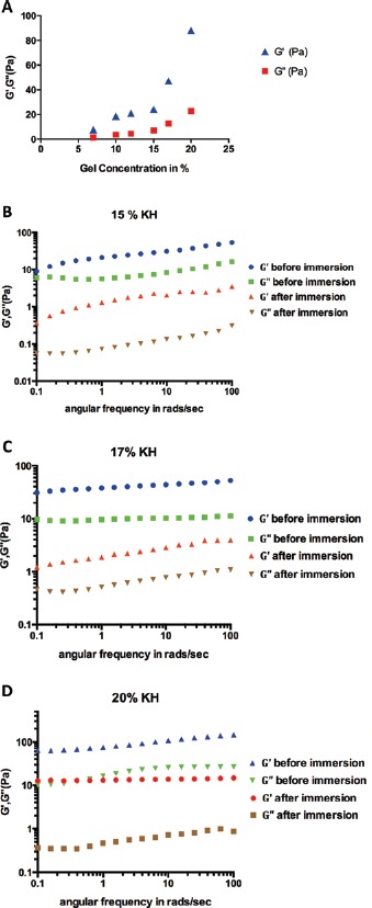
Flow characteristics of keratin hydrogel (KH) based on rheological analysis. (A) Graphical representation of dynamic elastic/storage modulus (G′), dynamic viscous/loss modulus (G″) of KH at different concentrations. (B-D) Graphical representation of changes in G′ and G″ of 3 KH concentrations (15%, 17% and 20%) following hydration at 37°C for 10 days.
The pulpal response was histologically evaluated in a semiquantitative manner. The scoring criteria are shown in Table I. Chronic inflammatory cells, primarily lymphocytes, were counted in experimental and control teeth using a calculating grid method (20 random squares, each square was 4,000 µm2, total area 80,000 µm2, at ×200 original magnification) and a blind scoring method (13, 14). Immunohistochemical stained slides were viewed using a light microscope, and the presence or absence of positive immunostaining (cytoplasmic and nuclear) indicated by a brown color was recorded in comparison with the IgG isotype control. The data were subjected to a nonparametric Mann-Whitney test to detect the significant differences between the groups with 95% confidence intervals, using GraphPad Prism version 6 (GraphPad Software, CA, USA).
Table I.
Histological assessment criteria
| I. Inflammatory cell response | |
| 0 | Absent |
| 1 | Mild (<25 cells): a few inflammatory cells scattered throughout the pulp |
| 2 | Moderate (between 25-125 cells): more than a few inflammatory cells scattered throughout the pulp |
| 3 | Severe (>125 cells): a dense collection of inflammatory cells scattered throughout the pulp |
| II. Necrosis | |
| 0 | Absence of necrotic tissue |
| 1 | Presence of necrotic tissue |
| III. Vitality | |
| 0 | No visible blood vessels |
| 1 | Presence of blood vessels |
| IV. Mineralization in the radicular pulp | |
| 0 | No trace of mineralization in the radicular pulp |
| 1 | Increased deposition of dentin beneath the restorative section and/or along the root of the remaining pulp tissue |
The scores were based on the number of cells per measured area.
Results
Flow characteristics
The dynamic elastic/storage modulus (G’) and dynamic viscous/loss modulus (G”) of different KH concentrations are shown in Figure 1A. There was a gradual increase in G’ from 7% to 20% (w/v) gel concentration, with the maximum G’ recorded for 20% (w/v) KH. There was a significant reduction (p<0.0001, t-test, n = 2) in G’ and G” following immersion in the medium for 10 days at all 3 concentrations tested (15%, 17% and 20% w/v) (Fig. 1B-C-D). The G’ value of irradiated 20% (w/v) KH was 96.01 ± 28.98 Pa, which was not different form that for nonirradiated samples (p>0.05, t-test, n = 2; data not shown).
Microstructure, swelling ratio and contact angle
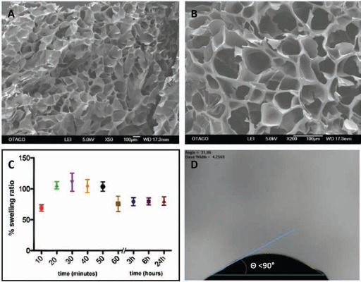
SEM images of lyophilized 20% (w/v) KH at different magnifications showed a branched, interconnected porous micro architecture (Fig. 2A, B). The average pore size was 163.5 ± 71.12 μm (n = 90). The average percentage porosity as determined by micro-CT analysis was approximately 82.80% ± 0.08%.
Fig. 2.

Microstructure, swelling behavior and wettability of 20% keratin hydrogel (KH). (A, B) Scanning electron microscopic images of 20% KH scaffolds at different magnifications. (C) Graphical representation of percentage swelling ratio after immersion in PBS at 37°C. Values are expressed as mean percentage degradation ± standard deviation. (D) Representative contact angle measurement of KH was 31.06° (i.e., Θ <90°).
The mean percentage swelling ratio at 10 minutes was 68.92 ± 5.171 which gradually increased and peaked at 30 minutes to 110.6 ± 14.53. At 60 minutes, the values were 75.67 ± 12.37 followed by 79.22 ± 6.4851, 79.54 ± 5.601 and 80.17 ± 6.732 at 3 hours, 6 hours and 24 hours, respectively (Fig. 2C). The percentage swelling ratio stabilized at the end of 1 hour with no significant difference between from 1 hour to 24 hours (p>0.05, ANOVA), with a mean value of 78.65 ± 2.025. The average contact angle measurement for KH (Fig. 2D) was found to be 35.52 ± 7.187 (in degrees, n = 6).
Biocompatibility
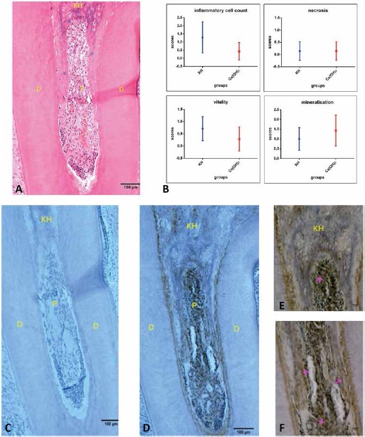
The effect of KH on pulpal response is shown in Figure 3A, with statistical comparisons in Figure 3B. The hydrogel appeared as a basophilic material that had spread throughout the root canals. The bulk of the hydrogel was present at the partial pulpotomy site with cellular infiltration. The underlying pulp tissue with the inflammatory infiltrate showed the presence of plump fibroblasts, mesenchymal cells interspersed with collagen fibers (Fig. 3A). A mild to moderate inflammatory response was recorded for KH and a mild response for the control group (Fig. 3B). One case of pulp tissue necrosis was reported in each group, with no significant difference (p>0.05) between the groups (Fig. 3B). The majority of pulps (n = 5) exposed to KH showed the presence of blood vessels, indicating a vital pulp with no difference compared with control (p>0.05) (Fig. 3B).
Fig. 3.

Biocompatibility assessment by histological and immunohistochemical staining. (A) Representative light microscopic microphotograph showing pulpal response at 28 days after keratin hydrogel (KH) placement following partial pulpotomy. The KH is visible as a basophilic material at the pulpotomy site, along the canal walls and pulpal space showing vital pulp with associated inflammatory cells. Reparative dentin-like deposits can be seen along the dentinal walls (arrow heads), which is more like tertiary dentin with more matrix, irregular tubules with entrapped cells. D = original dentin; P = pulp; (B) Graphical representation of the histological scores for the test and control groups at the end of 21 days. Data presented as means ± standard deviation (n = 7 for each group). There was no significant difference in the histology grading scores (p>0.05, nonparametric Mann-Whitney test) when compared with the Ca(OH)2. (C) Negative isotype control at ×100. (D) Positive dentin matrix protein 1 (DMP-1) expression as shown by brown 3,3’-diaminobencidine (DAB) staining at ×100. (E, F) Higher magnification (×400) images showing DMP expression, as indicated by pink arrows.
The mineralization evaluation of the KH group showed a heterogenous deposition of dentin-like material throughout the root canal in the majority of specimens (n = 5 of 7) suggestive of reparative dentinogenesis. In some samples, more organized, vertically arranged parallel tubules resembling tubular reparative dentin-like matrix were found along the dentinal walls projecting into the pulpal side (Fig. 3A). In addition, reparative dentin more like osteodentin with areas of cellular inclusions interspersed with areas resembling dentinoid, were also observed (Fig. 3A). The peripheral rat odontoblastic layer was continuous in some samples and interrupted in others. In both groups, there were root canals completely filled with osteodentin-like material. Statistical comparison of mineralization scores showed no significant difference (Fig. 3B, p>0.05).
The pattern of expression of DMP-1 was consistent, and correlated to dentin-like deposits found in H&E sections. A dark brown staining suggestive of intense DMP-1 signals was observed within the hydrogel at the pulpotomy site, as well as apical to the KH (Fig. 3C-D-E-F).
Discussion
The last 10 years have witnessed the flourishing of biomaterials and tissue engineering approaches for replacing existing treatment strategies. Different biomaterial scaffolds made of synthetic and natural polymers have been widely studied for this purpose. Poly(lactide-co-glycolide) (PLG) scaffolds and collagen gels with stem cells have been investigated for the regeneration of pulp tissue (15). Most of the published work has reported on ex vivo expanded pulp tissue placed in thin tooth slice models to evaluate de novo pulp regeneration. The role of keratin in various fields of regenerative medicine such as as a hemostatic agent, and in nerve, cardiac muscle and bone regeneration, is well documented (16).
The keratin intermediate filament proteins (kIFPs) isolated using the extraction technique have been previously characterized (11). The hydration of the lyophilized kIFPs, followed by incubation at 37°C, enabled the formation of a hydrogel at protein concentrations ≥70 mg/mL to ≤200 mg/mL. Although, for concentrations >200 mg/mL, the polymer formed heterogeneous lumps and failed to form a soluble mass. The gelation process mainly occurs through physical cross-linking (polymer chain entanglement). The elasticity (G’) of the hydrogels was shown to increase with biopolymer (keratin protein) concentration with a nearly constant G’ value at 20% that could be attributed to the proportional cross-link density.
In this study, the effects of hydration and temperature on the flow characteristics of KH (15%, 17% and 20% w/v) were evaluated. Though the recorded G’ value, at a KH concentration of 20%, was higher than for the 2 other tested formulations, there was a significant decrease in G’ following immersion in liquid medium for 10 days for all 3 groups, which is in accordance with the reported hydrolytic stability previously published (11). The 15% and 17% w/v KH with lower protein content were more susceptible to hydrolysis compared with the 20% gels with more alpha-keratin content (higher disulphide bonds, with more cross-links). In addition, sterilization of 20% KH with gamma radiation did not affect the flow characteristics, and 20% KH exhibited the ability to be injected for specific clinical applications, such as direct pulp capping.
Structural analysis of 20% KH scaffolds by SEM and micro-CT revealed a highly porous, interconnected internal architecture. This is similar to the results of other published studies that have reported an average pore size of 70-150 μm and a porosity of 70%-80% favorable for cell adhesion and proliferation (10, 17, 18). Similarly the swelling ratio was in accordance with that reported in the literature, which reports that keratin, derived from either wool or human hair, drastically swelled due to the stretching of polymeric chains (18, 19). The hydrophilic nature of the gel, along with a high swelling ratio, can be advantageous in increasing the pore size, surface area and movement of cells and nutrients, and thus improving cell infiltration. However, our lab group is presently conducting studies to control and establish a favorable swelling ratio. In addition, in most in vivo studies, 15%-20% w/v KHs were used, which is a concentration that can be adjusted according to the application envisioned (7, 10, 20). With this in mind, 20% (w/v) KH was selected for the in vivo study, based on the determined physicochemical properties of the gel.
This is the first study to investigate the effects of keratin on pulpal healing and response in an in vivo model. Many studies have been published using rat molar teeth to evaluate tissue reactions after pulp exposure and the effect of treatment – e.g., direct pulp capping or pulpotomy (21, 22). A rat molar tooth, with a Ca(OH)2 capping of caries-free teeth, is considered a standard model for studying the pulpal reaction to bioactive molecules (23-24-25-26-27-28). Therefore, this model was chosen to be used in this study, in comparison with other hydrogels that are still the subject of preclinical trials.
The major limitation faced when selecting rat tooth is the size; despite the aid of surgical personalized loupes, it is still difficult to work on rat molars. The cavity size was very small, so to place the test gel and seal the surface with restorative material was challenging. Moreover, the hydrophilic hydrogel swelled up once placed in the cavity. This made the placement and retention of the restorative material difficult, with a high chance of postoperative hyperocclusion. Hence, it was decided to do a partial pulpotomy and inject the KH into the space created. This allowed enough material to be placed and also enabled visual confirmation of the material prior to sealing with GIC. An experimental time period of 28 days was chosen for this study which was consistent with that in other published studies (29, 30).
At the end of 28 days, the bulk of the hydrogel found at the pulpotomy site was indicated by histological analysis to be showing slow biodegradation. The results of the histological analysis showed that there was a mild to moderate inflammatory response following the KH placement. A surgically induced inflammatory response was to be expected. However, there was no significant difference in the histology grading scores when compared with Ca(OH)2. The dentin-like tissue formed in the radicular pulp was more like tertiary dentin, characterized by more matrix, thus suggestive of reparative effects. The mechanism of reparative dentin formation is complex, involving several processes such as neovascularization, proliferation, migration and differentiation of progenitor cells residing in the pulp into odontoblast- or osteoblast-like cells (31). Hence, the presence of KH was nontoxic to pulp cells, allowing it to heal, with the resultant reparative response. Since this was an initial study, the effect of material per se on repair is not completely clear, and therefore it will be addressed in the future. In addition, at 28 days, although there were noticeable newly formed blood vessels suggestive of neovascularization, no definite pulp tissue regeneration was apparent in the coronal aspect of the pulp chamber or within the hydrogel. Most of the area beneath the amputation was filled with a heterogeneous mixture of poorly mineralized substances and/or hydrogel interspersed with pulp remnants.
The scattered, calcified tissues formed in the KH group were further studied by immunostaining for DMP-1 as a marker. DMP-1 is one of the noncollagenous, extracellular matrix proteins and is considered as a marker for dentin, odontoblasts and odontoblast-like cells (32). It has been demonstrated to induce differentiation of dental pulp stem cells into odontoblasts and, therefore, to initiate the formation of calcified tissues (33, 34). It is one of the regulators of hydroxyapatite deposition and helps in biomineralization of dentin and bone (35). The calcified tissues formed in the pulp undergoing repair showed intense DMP-1 signaling. Hence, it was clear from the positive immunostaining that implantation of KH resulted in formation of mineralized dentin-like deposits.
Future studies should be planned with a greater number of animals (a sample with adequate power), comparison with hydrogels derived from other polymers; different time periods and/or after longer healing periods; different counterstains or immunohistochemical staining, using specific markers to assess odontogenic, angiogenic and neurogenic potential; and 3-dimensional evaluation using micro-CT to identify dentinal bridge formation. From the material point of view, the material properties might to be further refined to effectively control biodegradation, and comparative studies with the KH encapsulating stem cells and/or additional biomolecules are also in mind.
Conclusion
The results of this study showed that the characterized KH (20% w/v) was a stable, elastic, semisolid injectable gel that was nontoxic and biocompatible to pulpal tissue. Within the constraints of this pilot study, KH allowed normal radicular pulp tissue organization with no adverse reactions and pulp healing to occur by a reparative response.
Disclosures
Financial support: This research was supported by the New Zealand Dental Association Research Fund.
Conflict of interest: The authors of this manuscript declare that they have no potential conflicts of interest with respect to the authorship and/or publication of this article.
References
- 1.Goldberg M Smith AJ Cells and extracellular matrices of dentin and pulp: a biological basis for repair and tissue engineering. Crit Rev Oral Biol Med 200415(1)13–27. [DOI] [PubMed] [Google Scholar]
- 2.Zhang W Yelick PC Vital pulp therapy: current progress of dental pulp regeneration and revascularization. Int J Dent 20102010(2010)856087. [DOI] [PMC free article] [PubMed] [Google Scholar]
- 3.Barrieshi-Nusair KM Qudeimat MA A prospective clinical study of mineral trioxide aggregate for partial pulpotomy in cariously exposed permanent teeth. J Endod 200632(8)731–735. [DOI] [PubMed] [Google Scholar]
- 4.Roberts HW Toth JM Berzins DW Charlton DG Mineral trioxide aggregate material use in endodontic treatment: a review of the literature. Dent Mater 200824(2)149–164. [DOI] [PubMed] [Google Scholar]
- 5.Mori GG Teixeira LM de Oliveira DL Jacomini LM da Silva SR Biocompatibility evaluation of biodentine in subcutaneous tissue of rats. J Endod 201440(9)1485–1488. [DOI] [PubMed] [Google Scholar]
- 6.Huang GT Pulp and dentin tissue engineering and regeneration: current progress. Regen Med 20094(5)697–707. [DOI] [PMC free article] [PubMed] [Google Scholar]
- 7.Sierpinski P Garrett J Ma J et al. The use of keratin biomaterials derived from human hair for the promotion of rapid regeneration of peripheral nerves. Biomaterials 200829(1)118–128. [DOI] [PubMed] [Google Scholar]
- 8.Aboushwareb T Eberli D Ward C et al. A keratin biomaterial gel hemostat derived from human hair: evaluation in a rabbit model of lethal liver injury. J Biomed Mater Res B Appl Biomater 200990(1)45–54. [DOI] [PubMed] [Google Scholar]
- 9.de Guzman RC Saul JM Ellenburg MD et al. Bone regeneration with BMP-2 delivered from keratose scaffolds. Biomaterials 201334(6)1644–1656. [DOI] [PubMed] [Google Scholar]
- 10.Hill P Brantley H Van Dyke M Some properties of keratin biomaterials: kerateines. Biomaterials 201031(4)585–593. [DOI] [PubMed] [Google Scholar]
- 11.Ajay Sharma L Ali MA Love RM Wilson MJ Dias GJ Novel keratin preparation supports growth and differentiation of odontoblast-like cells. Int Endod J 201649(5)471–482. [DOI] [PubMed] [Google Scholar]
- 12.Yamauchi K Yamauchi A Kusunoki T Kohda A Konishi Y Preparation of stable aqueous solution of keratins, and physiochemical and biodegradational properties of films. J Biomed Mater Res 199631(4)439–444. [DOI] [PubMed] [Google Scholar]
- 13.Yaltirik M Ozbas H Bilgic B Issever H Reactions of connective tissue to mineral trioxide aggregate and amalgam. J Endod 200430(2)95–99. [DOI] [PubMed] [Google Scholar]
- 14.Morse DR Wilcko JM Pullon PA Furst ML Passo SA A comparative tissue toxicity evaluation of the liquid components of gutta-percha root canal sealers. J Endod 19817(12)545–550. [DOI] [PubMed] [Google Scholar]
- 15.Galler KM DSouza RN Hartgerink JD Schmalz G Scaffolds for dental pulp tissue engineering. Adv Dent Res 201123(3)333–339. [DOI] [PubMed] [Google Scholar]
- 16.Rouse JG Van Dyke ME A review of keratin-based biomaterials for biomedical applications. Materials (Basel) 20103(2)999–1014. [Google Scholar]
- 17.Tachibana A Kaneko S Tanabe T Yamauchi K Rapid fabrication of keratin-hydroxyapatite hybrid sponges toward osteoblast cultivation and differentiation. Biomaterials 200526(3)297–302. [DOI] [PubMed] [Google Scholar]
- 18.Verma V Verma P Ray P Ray AR Preparation of scaffolds from human hair proteins for tissue-engineering applications. Biomed Mater 20083(2)025007. [DOI] [PubMed] [Google Scholar]
- 19.Tanabe T Okitsu N Yamauchi K Fabrication and characterization of chemically crosslinked keratin films. Mater Sci Eng C Mater Biol Appl 200424(3)441–446. [Google Scholar]
- 20.Apel PJ Garrett JP Sierpinski P et al. Peripheral nerve regeneration using a keratin-based scaffold: long-term functional and histological outcomes in a mouse model. J Hand Surg Am 200833(9)1541–1547. [DOI] [PubMed] [Google Scholar]
- 21.Sena M Yamashita Y Nakano Y et al. Octacalcium phosphate-based cement as a pulp-capping agent in rats. Oral Surg Oral Med Oral Pathol Oral Radiol Endod 200497(6)749–755. [DOI] [PubMed] [Google Scholar]
- 22.Takei E Shigetani Y Yoshiba K Hinata G Yoshiba N Okiji T Initial transient accumulation of M2 macrophage-associated molecule-expressing cells after pulpotomy with mineral trioxide aggregate in rat molars. J Endod 201440(12)1983–1988. [DOI] [PubMed] [Google Scholar]
- 23.Dammaschke T Rat molar teeth as a study model for direct pulp capping research in dentistry. Lab Anim 201044(1)1–6. [DOI] [PubMed] [Google Scholar]
- 24.Al-Hiyasat AS Barrieshi-Nusair KM Al-Omari MA The radiographic outcomes of direct pulp-capping procedures performed by dental students: a retrospective study. J Am Dent Assoc 2006137(12)1699–1705. [DOI] [PubMed] [Google Scholar]
- 25.Barthel CR Rosenkranz B Leuenberg A Roulet JF Pulp capping of carious exposures: treatment outcome after 5 and 10 years: a retrospective study. J Endod 200026(9)525–528. [DOI] [PubMed] [Google Scholar]
- 26.Baume LJ Holz J Long term clinical assessment of direct pulp capping. Int Dent J 198131(4)251–260. [PubMed] [Google Scholar]
- 27.Haskell EW Stanley HR Chellemi J Stringfellow H Direct pulp capping treatment: a long-term follow-up. J Am Dent Assoc 197897(4)607–612. [DOI] [PubMed] [Google Scholar]
- 28.Horsted P Sandergaard B Thylstrup A El Attar K Fejerskov O A retrospective study of direct pulp capping with calcium hydroxide compounds. Endod Dent Traumatol 19851(1)29–34. [DOI] [PubMed] [Google Scholar]
- 29.Chacko V Kurikose S Human pulpal response to mineral trioxide aggregate (MTA): a histologic study. J Clin Pediatr Dent 200630(3)203–209. [DOI] [PubMed] [Google Scholar]
- 30.Kaida H Hamachi T Anan H Maeda K Wound healing process of injured pulp tissues with emdogain gel. J Endod 200834(1)26–30. [DOI] [PubMed] [Google Scholar]
- 31.Six N Tompkins K Septier D Veis A Goldberg M Recruitment and characterization of the cells involved in reparative dentine formation in the exposed rat molar pulp after implantation of amelogenin gene splice products A 4 and A − 4. Oral Biosci Med 2004135–44. [Google Scholar]
- 32.Qin C DSouza R Feng JQ Dentin matrix protein 1 (DMP1): new and important roles for biomineralization and phosphate homeostasis. J Dent Res 200786(12)1134–1141. [DOI] [PubMed] [Google Scholar]
- 33.Narayanan K Srinivas R Ramachandran A Hao J Quinn B George A Differentiation of embryonic mesenchymal cells to odontoblast-like cells by overexpression of dentin matrix protein 1. Proc Natl Acad Sci USA 200198(8)4516–4521. [DOI] [PMC free article] [PubMed] [Google Scholar]
- 34.Almushayt A Narayanan K Zaki AE George A Dentin matrix protein 1 induces cytodifferentiation of dental pulp stem cells into odontoblasts. Gene Ther 200613(7)611–620. [DOI] [PubMed] [Google Scholar]
- 35.Zhao C Hosoya A Kurita H et al. Immunohistochemical study of hard tissue formation in the rat pulp cavity after tooth replantation. Arch Oral Biol 200752(10)945–953. [DOI] [PubMed] [Google Scholar]


