Abstract
Introduction
Pseudomonas aeruginosa pulmonary infections are the primary cause of morbi-mortality in patients with cystic fibrosis (CF). In this cohort study, the objective was to identify candidate biomarkers of P. aeruginosa infection within the airway microbiota.
Methods
A 3-year prospective multicentre study (PYOMUCO study) was conducted in Western France and included patients initially P. aeruginosa free for at least 1 year. A 16S-targeted metagenomics approach was applied on iterative sputum samples of a first set of patients (n=33). The composition of airway microbiota was compared according to their P. aeruginosa status at the end of the follow-up (colonised vs non-colonised), and biomarkers associated with P. aeruginosa were screened. In a second step, the distribution of a candidate biomarker according to the two groups of patients was verified by qPCR on a second set of patients (n=52) coming from the same cohort and its load quantified throughout the follow-up.
Results
Porphyromonas (mainly P. catoniae) was found to be an enriched phylotype in patients uninfected by P. aeruginosa (p<0.001). This result was confirmed by quantitative PCR. Conversely, in patients who became P. aeruginosa-positive, P. catoniae significantly decreased before P. aeruginosa acquisition (p=0.014).
Discussion
Further studies on replication cohorts are needed to validate this potential predictive biomarker, which may be relevant for the follow-up in the early years of patients with CF. The identification of infection candidate biomarkers may offer new strategies for CF precision medicine.
Keywords: cystic fibrosis, microbiota, biomarkers, pseudomonas aeruginosa, porphyromonas
Key messages.
Could the airway microbiota be a source of predictive biomarkers of early P seudomonas aeruginosa pulmonary infection?
The airway microbiota might be more or less permissive to P. aeruginosa in cystic fibrosis (CF) airways.
Based on a cohort study, we found the first candidate biomarker predictive of P. aeruginosa pulmonary infection in CF.
Introduction
Respiratory polymicrobial infections play a major role in cystic fibrosis (CF) progression and the acquisition of bacterial pathogens during the course of the disease is now well described. After ~25 years old, the establishment of CF pathogens is usually completed, P. aeruginosa being the most predominant species in CF lung. P. aeruginosa has a negative impact on pulmonary function promoting more frequent acute exacerbations. P. aeruginosa is linked to worsened prognosis for patients with CF; they have a decreased life expectancy and experience a more rapid decline in pulmonary function compared with non-colonised patients. At the stage of chronic infection, the eradication of the pathogen is impossible; this places all the hopes of treatment at the early stage of the infection. Indeed, the chances to successfully eradicate P. aeruginosa are the most important in the early stages of P. aeruginosa colonisation.
Improvement in the median survival of patients with CF is correlated with early antibiotic therapy in patients colonised with P. aeruginosa, the eradication success being essentially dependent on how early P. aeruginosa is detected.1 As early P. aeruginosa pulmonary infection is completely asymptomatic in most cases,2 the monitoring is based on systematic microbiological analysis. In this aim, P. aeruginosa quantitative PCR (qPCR) detection was shown to be relevant in patients’ follow-up.3 For the coming years, one challenge is to decipher the factors involved in the early onset of P. aeruginosa colonisation. Demographic and environmental factors were shown to increase the risk of P. aeruginosa acquisition, but are not precise enough to predict the risk of P. aeruginosa early colonisation.4
In the framework of the present study, we hypothesised that lung commensal microbiota could be associated with P. aeruginosa early colonisation in CF. In the precision medicine era, the aim of this study was to find biomarkers for providing close monitoring to CF patients more at risk of early P. aeruginosa colonisation and improving clinical benefit of successful early P. aeruginosa eradication.
Methods
Patient cohort, inclusion criteria and global data
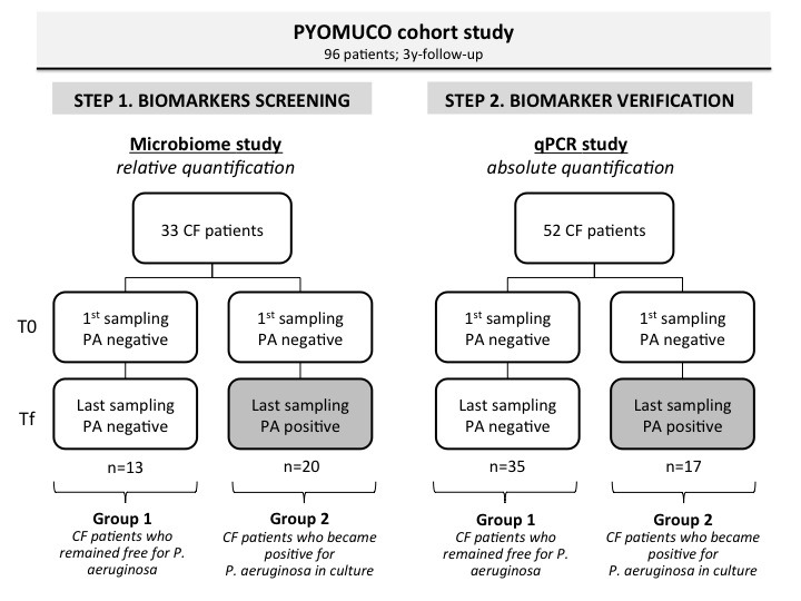
A 3-year prospective multicentre study (PYOMUCO study) was conducted in Western France to assess the time saved in the detection of P. aeruginosa in patients with CF by qPCR compared with culture detection methods.3 Only patients P. aeruginosa free for at least 1 year were included. The cohort was divided into two groups at the end of the follow-up; group 1 contained patients who remained free of P. aeruginosa while patients from group 2 became positive in culture for P. aeruginosa during the follow-up. For each patient, sputum was collected every 3 months up to the first P. aeruginosa positivity in culture. For this ancillary study, 33 patients with CF of the PYOMUCO cohort were selected as follows: 20 patients from the 36 who became P. aeruginosa-positive at the end of the follow-up, and 13 patients from the 28 who remained P. aeruginosa-negative both in qPCR and culture.3 The analysis of airway microbiota was performed retrospectively on spontaneous sputum samples collected at two time points: at enrolment (T0) and at the end of the follow-up (Tf) (figure 1). Overall, 75.7% of the samples originated from a paediatric population (<18 years old at sampling time). Clinical and biological data were collected at each sampling time (see online supplementary table S1). The majority of patients were homozygous (n=21, 63.6%) or heterozygous (n=12, 36.4%) for the F508del-CFTR mutation. Four clinical states were defined: baseline clinical state, pulmonary exacerbation, treatment for exacerbation and recovery (BETR categories). Sputum sample quality was verified by cytological examination as previously described.3 P. aeruginosa was quantified using qPCR and a culture-based method, as previously described.3 In order to confirm Porphyromonas distribution with respect to P. aeruginosa colonisation throughout the follow-up, P. catoniae absolute quantification was carried out with qPCR on 52 additional patients of the PYOMUCO cohort.
Figure 1.

Two-step approach of the study. Samples were issued from the PYOMUCO cohort study whose patients (n=96), initially (T0) all Pseudomonas aeruginosa (PA) free for at least 1 year, were separated into two groups (group 1 and group 2) according to their P. aeruginosa status at the end of the follow-up (Tf).3 Group 1 patients remained negative, whereas group 2 patients became positive. In a first step carried out in a first set of patients (n=33), bacterial biomarkers associated with P. aeruginosa were screened by 16S-targeted metagenomics; a candidate biomarker (Porphyromonas catoniae) was revealed. In a second step, distribution of the candidate biomarker according to the two groups of patients was verified by quantitative PCR (qPCR) on a second set of patients (n=52) coming from the same cohort. CF, cystic fibrosis.
bmjresp-2018-000374supp003.xlsx (51KB, xlsx)
Targeted metagenomics and P. catoniae absolute quantification in sputum samples
Total DNA was extracted using the QIAamp DNA Mini Kit (QIAGEN, Courtabœuf, France) as previously described.5 For bacterial diversity assessment, barcoded high-throughput 454 pyrosequencing was performed on the amplified V3 and V4 hypervariable regions of the 16S rRNA gene, and data analysed as previously described (Bioproject PRJNA445243).5 6 The absolute quantification of P. catoniae was performed using a validated qPCR scheme with the standard curve method. The qPCR was set up on the ABI 7500 Fast Real-Time PCR system (Applied Biosystems, Foster City, California, USA) with SYBR Green, and original primers (sense: 5′-GTGTCTTCGCCCAGCTTACT-3′; antisense: 5′-AGGATGCGGCGGGTTTCA-3′) targeting the rplb gene. PCR reactions were carried out in a total volume of 25 µL with 12.5 µL of Select Mastermix (Applied Biosystems), and a temperature profile of 50°C for 2 min, 95°C for 10 min, followed by 40 cycles at 95°C for 15 s, 60°C for 60 s, 95°C for 30 s and 60°C for 15 s.
Bioinformatics and statistical analyses
Sequences were analysed with the standard UPARSE pipeline according to Edgar’s instructions as previously described5 (see online supplementary file 1).
bmjresp-2018-000374supp001.htm (14.8KB, htm)
Statistical comparison between groups was performed with the Mann-Whitney U and Kruskal-Wallis tests, and linear discriminant analysis effect size (LEfSe) was used to elucidate bacterial taxa associated with group 1 or group 2 patients. The false discovery rate was calculated to correct for multiple hypothesis testing. Principal component analysis and clustering analysis were computed on different distance matrix to document the presence of enterotype-like clusters in airway CF microbiota.7 These clusters were named pulmotypes.
Results
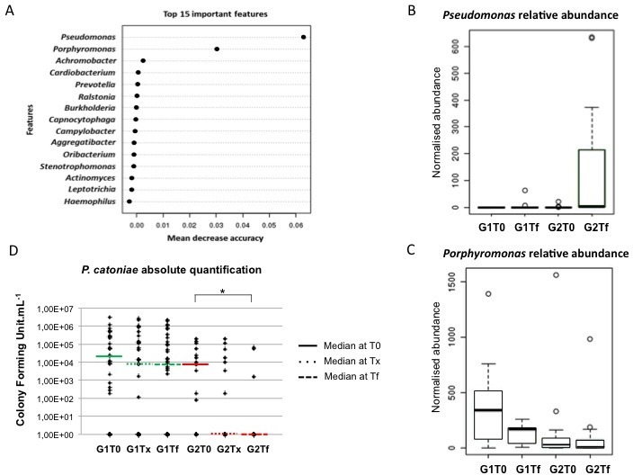
The cohort samples clustered into three pulmotypes (p=0.001, Kruskal-Wallis test) driven by the differences in relative abundance of three dominant genera, Streptococcus, Haemophilus and Staphylococcus, as well as other co-occurring genera (see online supplementary figure S1). Overall, 11 predominant genera (relative abundance ≥1%) were found, including Porphyromonas which abundance varied between 2.5% and 6.9% depending on the pulmotype. Random forest analysis revealed a significant connexion between relative abundance of Pseudomonas and Porphyromonas (figure 2A). Interestingly, in group 1 patients who remained uninfected by P. aeruginosa during the follow-up (figure 2B), Porphyromonas relative abundance at T0 was significantly higher than in group 2 patients (p<0.001, Mann-Whitney U test) (figure 2C).
Figure 2.

Inverse correlation between Pseudomonas aeruginosa and Porphyromonas revealed by both 16S rRNA microbiota study and quantitative PCR study in the two groups of patients with cystic fibrosis: group 1 patients (G1) remained P. aeruginosa-negative from the initial visit (T0) to the last one (Tf); group 2 patients (G2), initially negative at T0, became P. aeruginosa-positive at Tf. (A) Results of random forest analysis showing the 15 taxa that contributed the most to each group based on the measure of mean decrease in accuracy. (B) Normalised abundance of P. aeruginosa in each group at the two time points. (C) Normalised abundance of Porphyromonas in each group at the two time points. (D) Absolute quantification of P. catoniae in each group (G1, G2) at the two time points (T0, Tf); a third time point was added (Tx) that corresponded to the visit which preceded the last one (Tf).
bmjresp-2018-000374supp002.pdf (473.5KB, pdf)
As Porphyromonas reads mainly corresponded to P. catoniae species, we focused on this bacterial species. In order to check the distribution of P. catoniae according to the patient group, we quantified P. catoniae by qPCR in another set of patients from the PYOMUCO cohort. For group 1 patients, we did not observe any statistical difference in P. catoniae population between the first (T0) and last sample (Tf) (p=0.41, t-test). Conversely, group 2 patients showed a significant drop in P. catoniae population (p=0.039, t-test) (figure 2D). Then, we compared patients according to their P. catoniae population before P. aeruginosa colonisation. Group 1 had a significantly higher initial P. catoniae absolute quantity than group 2 (p=0.026). Finally, we tested the predictive power of P. catoniae. To do this, we analysed the susceptibility of patients with CF to acquire P. aeruginosa according to the presence or absence of P. catoniae in the penultimate sputum (Tx) (figure 2D). We observed that 40.7% of patients without P. catoniae developed a P. aeruginosa infection the visit after (3 months later), while only 24% of patients positive for P. catoniae developed the infection (table 1).
Table 1.
Table of contingency showing the distribution of patients with cystic fibrosis (n=52) according to their Porphyromonas catoniae colonisation status during the follow-up with respect to Pseudomonas aeruginosa status at the end of the follow-up
| Patient group* | P. catoniae colonisation† | |
| Negative | Positive | |
| Group 1 | 16 (59.3%) | 19 (46%) |
| Group 2 | 11 (40.7%) | 6 (24%) |
| Total | 27 | 25 |
*Group 1, patients who remained P. aeruginosa-negative; group 2, patients who became P. aeruginosa-positive; Hazard Ratio 1.7.
†Detection carried out on the penultimate sputum sample.
Discussion
Bacteria from the Porphyromonas genus are anaerobic commensals of the core pulmonary microbiota in healthy people8 and also described as part of the CF pulmonary core microbiota.5 6 9–12 Characterisation of bronchoalveolar lavages’ microbiota in infants with asymptomatic CF retrieved Porphyromonas as one of the six highest abundant taxa.10
Looking into details at the taxonomic affiliation of Porphyromonas reads, the vast majority of them were affiliated to P. catoniae, which is in agreement with other culture-dependent and culture-independent studies.11 12 Interestingly, in another study, the abundance of Porphyromonas was significantly lower in sputa of patients with chronic obstructive pulmonary disease as compared with healthy subjects.8 Moreover, a decrease in P. catoniae abundance was observed during exacerbations,13 and after antibiotic treatment, the abundance of P. catoniae returned to a baseline identical to that of the pre-exacerbation period.13 These results also echo a previous observation in patients with CF under the CFTR potentiator drug. Indeed, a sustained increase of Porphyromonas relative abundance after initiation of ivacaftor was stated, which was positively correlated with the percentage of predicted FEV1.6
The present study gave clues on the power of P. catoniae in predicting the risk of P. aeruginosa acquisition. Indeed, P. catoniae colonisation was associated with a lower risk of P. aeruginosa infection. Conversely, patients harbouring no P. catoniae within their airway microbiota showed 1.7-fold risk of acquiring P. aeruginosa later.
Taken together, these results suggest that P. catoniae may be considered as a favourable prognostic biomarker in CF. Further prospective studies on replication cohorts are needed to define the benefit provided by P. catoniae quantification in identifying patients with a higher risk of P. aeruginosa infection. In case of confirmation, we suggest to perform molecular quantifications of both P. catoniae and P. aeruginosa as part of the CF diagnosis toolbox in order to enlarge the ‘window of opportunity’ in the management of P. aeruginosa infection.
To conclude, this study showed the crucial importance of microbiota data in the management of patients with CF. In the personalised and precision medicine era, microbiota-based study could identify signatures that could be useful in predicting the CF progression. Identification of new bacteria of interest opens the possibility of using them as prognostic biomarkers or as companion diagnostic test. Further cohort studies are needed to validate these findings and to address the question of causality. The influence of the input microbiota on CF progression during early life has also to be investigated. In the not-too-distant future, study of both biochemical and microbial signatures will constitute new approaches to understand CF microbiology.
Acknowledgments
The authors gratefully acknowledge the patients with CF who participated in the study. The authors thank Iain MacGill for English revision of the manuscript.
Footnotes
Contributors: GH-A and JM conceived the study. SG, C-AG and MK performed DNA extraction, qPCR and pyrosequencing. MK, JM, PL and GB performed pyrosequencing data and statistical analysis. MK, C-AG, M-SF and SG performed multiplex PCR. GH-A, RLB, CF and GR contributed to data collection. JM, PL, SV and SM contributed to design. MK, GH-A, JM and PL wrote the manuscript. All authors participated in data analysis and editing the manuscript for final approval. GH-A and JM take overall responsibility for the content of the manuscript.
Funding: This work was supported by a grant from the French Cystic Fibrosis Associations ‘Vaincre la Mucoviscidose’ and ‘Grégory Lemarchal’ (contract no. RC20170501971).
Competing interests: None declared.
Patient consent for publication: Not required.
Ethics approval: Two review boards, the local Comité de Protection des Personnes VI-Ouest and the institutional review board of the Brest University Hospital Centre, approved the protocol.
Provenance and peer review: Not commissioned; externally peer reviewed.
Data sharing statement: No additional data are available.
Author note: The study was presented in part at the 41th European Cystic Fibrosis Conference, 8–11 June 2018, in Belgrade, Serbia. A European patent has been registered (no. EP17306297).
References
- 1. Marchetti F, Giglio L, Candusso M, et al. Early antibiotic treatment of Pseudomonas aeruginosa colonisation in cystic fibrosis: a critical review of the literature. Eur J Clin Pharmacol 2004;60:67–74. 10.1007/s00228-004-0735-2 [DOI] [PubMed] [Google Scholar]
- 2. ECFS The early cystic fibrosis years de Boeck and K. Southern, 2018. [Google Scholar]
- 3. Héry-Arnaud G, Nowak E, Caillon J, et al. Evaluation of quantitative PCR for early diagnosis of Pseudomonas aeruginosa infection in cystic fibrosis: a prospective cohort study. Clin Microbiol Infect 2017;23:203–7. 10.1016/j.cmi.2016.11.016 [DOI] [PubMed] [Google Scholar]
- 4. Maselli JH, Sontag MK, Norris JM, et al. Risk factors for initial acquisition of Pseudomonas aeruginosa in children with cystic fibrosis identified by newborn screening. Pediatr Pulmonol 2003;35:257–62. 10.1002/ppul.10230 [DOI] [PubMed] [Google Scholar]
- 5. Keravec M, Mounier J, Prestat E, et al. Insights into the respiratory tract microbiota of patients with cystic fibrosis during early Pseudomonas aeruginosa colonization. Springerplus 2015;4:1–8. 10.1186/s40064-015-1207-0 [DOI] [PMC free article] [PubMed] [Google Scholar]
- 6. Bernarde C, Keravec M, Mounier J, et al. Impact of the CFTR-potentiator ivacaftor on airway microbiota in cystic fibrosis patients carrying a G551D mutation. PLoS One 2015;10:e0124124 10.1371/journal.pone.0124124 [DOI] [PMC free article] [PubMed] [Google Scholar]
- 7. Arumugam M, Raes J, Pelletier E, et al. Enterotypes of the human gut microbiome. Nature 2011;473:174–80. 10.1038/nature09944 [DOI] [PMC free article] [PubMed] [Google Scholar]
- 8. Erb-Downward JR, Thompson DL, Han MK, et al. Analysis of the lung microbiome in the "healthy" smoker and in COPD. PLoS One 2011;6:e16384 10.1371/journal.pone.0016384 [DOI] [PMC free article] [PubMed] [Google Scholar]
- 9. Quinn RA, Whiteson K, Lim YW, et al. Ecological networking of cystic fibrosis lung infections. NPJ Biofilms Microbiomes 2016;2 10.1038/s41522-016-0002-1 [DOI] [PMC free article] [PubMed] [Google Scholar]
- 10. Laguna TA, Wagner BD, Williams CB, et al. Airway microbiota in bronchoalveolar lavage fluid from clinically well infants with cystic fibrosis. PLoS One 2016;11:e0167649 10.1371/journal.pone.0167649 [DOI] [PMC free article] [PubMed] [Google Scholar]
- 11. van der Gast CJ, Walker AW, Stressmann FA, et al. Partitioning core and satellite taxa from within cystic fibrosis lung bacterial communities. Isme J 2011;5:780–91. 10.1038/ismej.2010.175 [DOI] [PMC free article] [PubMed] [Google Scholar]
- 12. Muhlebach MS, Hatch JE, Einarsson GG, et al. Anaerobic bacteria cultured from cystic fibrosis airways correlate to milder disease: a multisite study. Eur Respir J 2018;52 10.1183/13993003.00242-2018. [Epub ahead of print: 11 Jul 2018]. [DOI] [PMC free article] [PubMed] [Google Scholar]
- 13. Cuthbertson L, Rogers GB, Walker AW, et al. Respiratory microbiota resistance and resilience to pulmonary exacerbation and subsequent antimicrobial intervention. Isme J 2016;10:1081–91. 10.1038/ismej.2015.198 [DOI] [PMC free article] [PubMed] [Google Scholar]
Associated Data
This section collects any data citations, data availability statements, or supplementary materials included in this article.
Supplementary Materials
bmjresp-2018-000374supp003.xlsx (51KB, xlsx)
bmjresp-2018-000374supp001.htm (14.8KB, htm)
bmjresp-2018-000374supp002.pdf (473.5KB, pdf)


