Abstract
The biosynthetic gene clusters for herbicidins (hbc) and aureonuclemycin (anm) were identified in Streptomyces sp. KIB-027 and Streptomyces aureus, respectively. The roles of genes possibly involved in post-core-assembly steps in herbicidin biosynthesis in these clusters and a related her cluster were studied. Through systematic gene deletions, structural elucidation of the accumulated intermediates in the mutants, and in vitro verification of the encoded enzymes, the peripheral modification pathway for herbicidin biosynthesis is now fully established.
Graphical Abstract
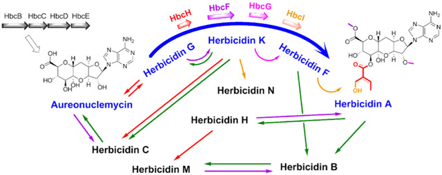
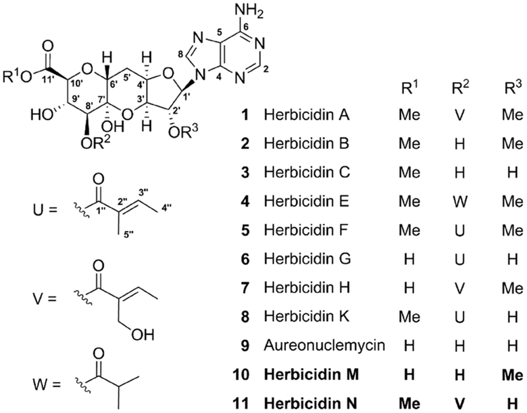
Herbicidins (HBCs, 1–8) are undecose nucleoside antibiotics isolated from different strains of Streptomyces and exhibit a wide range of biological activities, including herbicidal, antibacterial, antifungal, and antiparasitic activities.1–9 They share a common core but differ in their methylation patterns at C2′-OH and C11′-OH and the acylation mode at C8′-OH (Figure 1). In contrast, aureonuclemycin (ANM, 9) isolated from Streptomyces aureus var. suzhoueusis is unique because it bears only the bare nucleoside core of 1–8 (Figure 1).10
Figure 1.
Structures of HBCs. Compounds 1, 2, 7, and 10 were isolated from Streptomyces sp. KIB-027. 3, 5, 6, 8, and 11 were isolated from the mutants. 9 was isolated from Streptomyces lividans anmBCDE.
Because of their complex chemical structures and diverse biological activities, HBCs have attracted considerable attention as targets for chemical synthesis.11–13 However, the biosynthetic pathways for HBCs were unknown until a recent report that described the identification of the biosynthetic gene cluster (BGC) her in Streptomyces sp. L-9-10 and the initial characterization of the biosynthetic pathway for HBC A (1).14 Further investigation of the assembly of the tricyclic undecose core may reveal new mechanisms for C–C bond formation,15 and elucidation of the peripheral modifications is crucial for diversification of HBCs through pathway engineering. Herein, two BGCs for HBCs (hbc) and ANM (anm) were identified from Streptomyces sp. KIB-027 and S. aureus, respectively. The entire peripheral modification pathway of HBCs was identified by gene-deletion experiments, structural elucidation of accumulated intermediates, in vitro characterizations of the expressed enzymes, and biochemical verification of their catalytic functions.
Streptomyces sp. KIB-027 was isolated from soil samples collected at Gaoligong Mountain, Yunnan province, China. Through LC–MS analysis of its fermentation broth, four products with UV spectra and molecular weights identical to those of the known HBCs were detected (Figure S1). Subsequent large scale fermentation of KIB-027 led to the isolation of three known HBC congeners, HBC A (1), HBC B (2), and HBC H (7), and one new compound, HBC M (10) (Figure 1). The structures of 1, 2, and 7 were confirmed by comparing the NMR data with those reported previously (Figures S2–S13). The structure of 10 was determined by analysis of its HRMS, one-dimensional, and two-dimensional NMR data (Table S1 and Figures S14–S22).
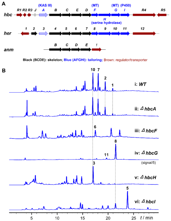
Next, the genome of KIB-027 was sequenced and a BGC (hbc) similar to the her cluster found in Streptomyce sp. L-9-1014 was located. The hbc cluster contains genes encoding two methyltransferases (MTs, HbcF and HbcG) and a cytochrome P450 monooxygenase (HbcI) that may be responsible for the late-stage decoration of 1. The cluster also contains 12 other genes encoding a ketoacyl-ACP synthase III (KAS III, HbcA), an S-adenosylhomocysteine (SAH) hydrolase (HbcB), two NAD-binding oxidoreductases (HbcC and HbcD), a B12-dependent radical S-adenosyl-l-methionine (SAM) enzyme (HbcE), a serine hydrolase (HbcH), a short-chain dehydrogenase (incomplete, HbcJ), and five regulatory proteins (HbcR1–R5) (Figure 2A and Table S2).
Figure 2.
(A) Organizations of the hbc, her, and anm biosynthetic gene clusters. (B) HPLC analyses of fermentations with Streptomyces sp. KIB-027 the wild type strain and the mutants.
Meanwhile, genome mining for an analogous hbc/her cluster in S. aureus, which produces only 9, was also conducted to identify the essential genes for core assembly. As a result, a putative ANM gene cluster (anm) containing four genes (anmB/C/D/E) homologous to both hbcB/C/D/E and her4/5/7/6 was found (Figure 2A and Table S2). Interestingly, BLAST analysis using hbcB/C/D/E as a query showed the existence of more than 20 hbc/her/anm-like clusters in bacteria (Figure S23). To verify the biosynthetic ability of the anm cluster, the anmB–E locus was cloned into pSET152 downstream to an ermEp* promoter for heterologous expression in S. lividans. LC–MS analysis showed that the anm-expressing strain S. lividans anmBCDE indeed produced ANM (9) as indicated by LC–MS (Figure S24) and confirmed by NMR analysis of the isolated product (Figures S25 and S26).12
Comparison of the results presented above helped to distinguish the enzymes catalyzing skeleton formation (such as those in anm) from those involved in post-core-assembly modifications. It was thus hypothesized that the remaining genes hbcA/F/G/H/I in the hbc cluster and their counterparts her3/8/10/9/11 in the her cluster are responsible for the peripheral modifications of HBCs. These genes in KIB-027 were each knocked out by in-frame deletion to study their functions. While KAS III, HbcA was originally thought to catalyze the esterification reaction at C8′-OH,16–18 the hbcA-deletion mutant (ΔhbcA) still produced HBCs with yields comparable to those generated by the wild type strain (Figure 2B, i and ii). Hence, hbcA is unlikely to be involved in the biosynthesis of HBCs. Another functionally unattributed enzyme, the serine hydrolase HbcH (Her9), may instead be responsible for C8′-OH acylation.
As predicted, the production of 1, 2, 7, and 10 was completely abolished when hbcH was deleted (ΔhbcH). Similar outcomes were also observed for other gene knockout mutants (ΔhbcF, ΔhbcG, and ΔhbcI). Interestingly, some new metabolites, which are likely HBC congeners, were produced by these mutants (Figure 2B, iii–vi). All of these compounds were isolated and structurally characterized by HRMS and NMR analysis. Specifically, the ΔhbcF mutant produced HBC G (6) with a tiglyl group at C8′ (see Figure 2B, iii, Table S3, and Figures S27-S34); the ΔhbcG mutant mainly produced HBC K (8) having a tiglyl group at C8′ and a methoxyl group at C11′ (Figure 2B, iv, Table S4, and Figures S35–S42); the ΔhbcH mutant produced HBC C (3) bearing a methoxyl group on C11′ but lacking an acyl group at C8′ (Figure 2B, v, Table S5, and Figures S43–S50); and the ΔhbcI mutant produced HBC F (5), containing a tiglyl group at C8′ and two methoxyl groups at C2′ and C11′ (Table S6 and Figures S51–S58), and a small amount of 8 (Figure 2B, vi). Collectively, these results indicated that HbcF and HbcG are responsible for the methylation of C11′-OH (6 → 8) and C2′-OH (8 → 5), respectively, HbcH is associated with the uploading of the tiglyl group at C8′ (9 → 6), and HbcI catalyzes the hydroxylation of the tiglyl group of 5 to give 1.
In addition to HBC K (8), the ΔhbcG mutant also produced another minor product, 11 (Figure 2B, iv), whose molecular weight is greater than that of 8 by 16 Da. Compound 11 was later determined to be a new HBC K (8) analogue carrying a hydroxyl group at C5″ of the tiglyl moiety and was designated as HBC N (Figure 1, Table S7, and Figures S59–S66). Further in vivo experiments showed that the double-deletion mutant of hbcG and hbcI (ΔhbcGI) produced only 8 (Figure S67). Thus, in addition to its primary function of catalyzing the hydroxylation of 5 (5 → 1), HbcI can also process 8 (8 → 11), albeit less effectively. On the contrary, compound 6 is not a substrate of HbcI because no hydroxylated product of 6 was produced by the ΔhbcF mutant. Hence, methylation of the C11′ hydroxyl group of 6 (6 → 8) appears to be a prerequisite for HbcI-catalyzed hydroxylation. Further methylation of the C2′ hydroxyl group of 8 (8 → 5) renders it a better substrate for HbcI.
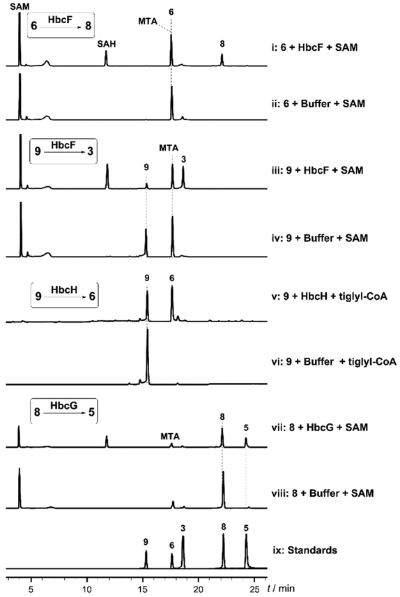
On the basis of the in vivo studies mentioned above, HbcH/Her9-mediated esterification (9 → 6) and HbcF/Her8-catalyzed methylation (6 → 8) are expected to occur early in the post-core-assembly process. However, it was puzzling that neither the ΔhbcF mutant nor the ΔhbcH mutant produced the presumed precursor 9. To address such obscurity, these reactions were then investigated in vitro. HbcF/G/H and the HbcH homologue Her9 were each overexpressed in Escherichia coli and purified as N-His6-tagged proteins (Figures S68 and S82). The Her8/10 proteins were obtained as previously described,14 but an attempt to obtain soluble HbcI was unsuccessful. The methylation activity of HbcF and Her8 was tested using 6, and conversion to 8 was observed in both cases (Figure 3 , i and ii, Figure S69 for a different HPLC method, and Figure S86). HbcF could also methylate 9 to give 3 (Figure 3, iii and iv). The capability of HbcF to convert 9 to 3 could explain why 9 was not detected in the fermentation of the ΔhbcH mutant. Interestingly, the HbcF homologue Her8 has a more strict substrate specificity and shows no methylation activity on 9 (Figure S83).
Figure 3.
HPLC analyses of the enzymatic reactions involved in the HBC tailoring pathway. HbcF-catalyzed reaction with (i) 6 or (iii) 9 as the substrate. (v) HbcH-mediated reaction with 9 as the substrate. (vii) HbcG-catalyzed reaction with 8 as the substrate. (ii, iv, vi, and viii) Control reactions. (ix) Authentic standards. The results obtained with the Her enzymes are shown in Figures S83, S84, S86, and S87.
With the role of HbcF/Her8 having been established, efforts were directed to verify HbcH/Her9 as the enzyme responsible for the acylation of 9. The acyl donor tiglyl-CoA was chemically prepared from tiglic acid and HSCoA (Figure S71). Enzymatic assays indicated that HbcH could catalyze the loading of the tiglyl group onto 9 to form 6 (Figure 3, v and vi). The same was also observed for HbcH homologue Her9 (Figure S84). However, when 3 was used as the acceptor, only a trace amount of tiglylated 8 was observed (Figure S70, v and vi). Hence, the reaction of HbcH/Her9 most likely occurs prior to any methylation of 9. In addition, the previously reported 3 may only be a shunt product, but not an intermediate, in HBC biosynthesis.
Next, the HbcG/Her10-catalyzed methylation taking place after the HbcH/Her9 and HbcF/Her8 reactions was studied in vitro. Though the HbcG protein tends to precipitate in the reaction system, its ability to catalyze the methylation at C2′-OH of 8 to form 5 could still be demonstrated (Figure 3, vii and viii). HbcG has stringent substrate selectivity because it could not methylate other substrates with free C2′-OH (9, 6, and 3) (Figure S72). These results also showed that C8′-O-acylation and C11′-O-methylation are prerequisites for HbcG reaction. The same conclusion was also reached for Her10 based on the results of in vivo her10 deletion (Figure S81) and the in vitro incubation of Her10 with compounds 9, 6, and 8 (Figures S83, S86, and S87). Lastly, the activity of Her11, a homologue of HbcI, was tested by feeding 5 to a herbicidin nonproducing strain Streptomyces albus J1074 overexpressing Her11. The result revealed 5 was successfully converted to 1 by Her11 in vivo (Figure S88).
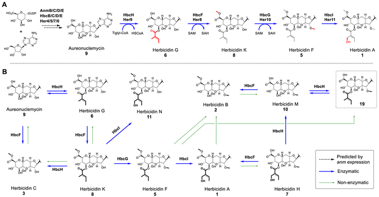
Taken together, a complete tailoring pathway of 1 can be proposed as depicted in Figure 4A. Unlike HbcG, HbcF can catalyze not only the methylation of 6 and 9 but also the conversion of other compounds containing C11′-COOH (7 and 10) to their methyl ester forms (1 and 2, respectively) (Figure S73, i–iv). When 3 and 8 containing C11′-COOMe were used as substrates, no methylation of other hydroxyl groups by HbcF was noted (Figure S73). Instead, 3 and 8 were found to undergo spontaneous hydrolysis of the C11′-methyl ester producing a small amount of 9 and 6, respectively, which could be remethylated in the presence of HbcF and SAM (Figure S73, v–viii). The substrate-tolerant HbcF may play a role in ensuring methyl esterification of the carboxylic acid of HBCs.
Figure 4.
Summary of the biosynthesis of HBCs. (A) Proposed herbicidin A biosynthetic pathway. (B) Proposed biosynthetic network for decorated HBCs derived from 9.
The substrate spectrum of acyltransferase HbcH/Her9 was also investigated with compounds containing a bare C8′-OH as substrates and tiglyl-CoA as the acyl donor. It was found that 2 was inactive but 10 was tiglylated to yield a new compound 19 (Figures S70, vii–xi, and S85). In combination with the results of the acylation of 9 and 3 by HbcH (Figure 3, iii and iv, and Figure S84), the preferred acyl acceptors for HbcH/Her9 reaction are likely the C11′-carboxyl HBCs. In addition, we tested the catalytic activity of HbcH using 9 as the acceptor toward various acyl-CoAs donors, including acetyl-, propionyl-, butyryl-, isobutyryl-, isovaleryl-, benzoyl-, and malonyl-CoA and the chemically synthesized angelyl-CoA, a cis isomer of tiglyl-CoA. It was found that HbcH could catalyze the transfer of these acyl groups, except malonyl-CoA, to form a series of derivatives of 9 (Figures S70, i and ii, S74, and S75), a feature useful for generating new HBC analogues.
The fact that HbcH is homologous to serine hydrolase also prompted us to investigate whether HbcH has hydrolytic activity for HBCs 1 and 5–8, which all contain a tiglyl or a 5″-hydroxytiglyl ester at C8′. In these experiments, spontaneous hydrolysis of the methyl ester at C11′ and the acyl ester group at C8′ was observed for 1, 5, and 8 after overnight reaction, and the presence of HbcH further promoted the hydrolysis of the C8′ tiglyl group of 8 but not others (Figures S76–S78). In addition, HbcH could facilitate hydrolysis of an otherwise stable acyl group at C8′ of 6 and 7. These findings demonstrated that the C8′ acyl ester in C11′-OMe HBCs can undergo hydrolysis spontaneously in vitro, while the same activity requires HbcH participation for C11′-OH HBCs. When HSCoA was present in the reaction mixture, the hydrolytic activity of HbcH was significantly inhibited (Figures S78–S80). The fact that HbcH has both acyl loading and hydrolysis activity suggests that some HBC homologues such as HBC B (2) may be produced by spontaneous or enzymatic hydrolysis of HBC A (1) as previously suggested.14,19 The data presented above allow the full constitution of the biosynthetic network for the generation of all HBC congeners, in which HbcH, HbcF, and spontaneous hydrolysis contribute to the diversity of HBCs (Figure 4B).
In summary, we have characterized the complete tailoring pathway of herbicidin A (1), including the serine hydrolase-catalyzed tiglyl loading, two steps of SAM-dependent methylation, and the P450-catalyzed hydroxylation reaction. To the best of our knowledge, HbcH and Her9 are the first enzymes found in bacteria to catalyze the transfer of a tiglyl group from tiglyl-CoA, a primary catabolite of isoleucine. In addition, this study placed most of the reported ANM-derived HBCs in the HBC modification network and suggested that more HBC analogues could be generated using the newly characterized tailoring enzymes.
Supplementary Material
ACKNOWLEDGMENTS
The authors thank Prof. Li-Ming Tao of East China University of Science and Technology for providing the ANM-producing strain. This work was supported by grants from the NNSFC (81473124) and the CAS (Youth Innovation Promotion Association 2016235, XDB20000000, QYZDJ-SSW-SLH037, and K. C. Wong Education Foundation) to H.-X.P. or G.-L.T. and from the National Institutes of Health (GM040541) and Welch Foundation (F-1511) to H.-w.L.
Footnotes
Supporting Information
The Supporting Information is available free of charge on the ACS Publications website at DOI: 10.1021/acs.or-glett.9b00066.
Materials and methods and supplementary tables and figures (PDF)
Notes
The authors declare no competing financial interest.
REFERENCES
- (1).Arai M; Haneishi T; Kitahara N; Enokita R; Kawakubo K; Kondo YJ Antibiot. 1976, 29, 863. [DOI] [PubMed] [Google Scholar]
- (2).Haneishi T; Terahara A; Kayamori H; Yabe J; Arai MJ Antibiot. 1976, 29, 870. [DOI] [PubMed] [Google Scholar]
- (3).Takiguchi Y; Yoshikawa H; Terahara A; Torikata A; Terao MJ Antibiot. 1979, 32, 862. [DOI] [PubMed] [Google Scholar]
- (4).Takiguchi Y; Yoshikawa H; Terahara A; Torikata A; Terao MJ Antibiot. 1979, 32, 857. [DOI] [PubMed] [Google Scholar]
- (5).Terahara A; Haneishi T; Arai M; Hata T; Kuwano H; Tamura CJ Antibiot. 1982, 35, 1711. [DOI] [PubMed] [Google Scholar]
- (6).Choi CW; Choi JS; Ko YK; Kim CJ; Kim YH; Oh JS; Ryu SY; Yon GHB Bull. Korean Chem. Soc 2014, 35, 1215. [Google Scholar]
- (7).Chai X; Youn UJ; Sun D; Dai J; Williams P; Kondratyuk TP; Borris RP; Davies J; Villanueva IG; Pezzuto JM; Chang LC J. Nat. Prod 2014, 77, 227. [DOI] [PMC free article] [PubMed] [Google Scholar]
- (8).Zhang JC; Yang YB; Chen GY; Li XZ; Hu M; Wang BY; Ruan BH; Zhou H; Zhao LX; Ding ZT J. Antibiot. 2017, 70, 313. [DOI] [PubMed] [Google Scholar]
- (9).Chen JJ; Rateb ME; Love MS; Xu Z; Yang D; Zhu X; Huang Y; Zhao LX; Jiang Y; Duan Y; McNamara CW; Shen BJ Nat. Prod 2018, 81, 791. [DOI] [PubMed] [Google Scholar]
- (10).Dai X; Li G; Wu Z; Lu D; Wang H; Li Z; Zhou L; Chen X; Chen W Chem. Abstr 1989, 111, 230661f. [Google Scholar]
- (11).Hager D; Paulitz C; Tiebes J; Mayer P; Trauner DJ Org. Chem. 2013, 78, 10784. [DOI] [PubMed] [Google Scholar]
- (12).Hager D; Mayer P; Paulitz C; Tiebes J; Trauner D Angew. Chem., Int. Ed 2012, 51, 6525. [DOI] [PubMed] [Google Scholar]
- (13).Ichikawa S; Shuto S; Matsuda AJ Am. Chem. Soc 1999, 121, 10270. [Google Scholar]
- (14).Lin GM; Romo AJ; Liem PH; Chen Z; Liu HW J. Am. Chem. Soc 2017, 139, 16450. [DOI] [PMC free article] [PubMed] [Google Scholar]
- (15).Wyszynski FJ; Lee SS; Yabe T; Wang H; Gomez-Escribano JP; Bibb MJ; Lee SJ; Davies GJ; Davis BG Nat. Chem 2012, 4, 539. [DOI] [PubMed] [Google Scholar]
- (16).Bretschneider T; Zocher G; Unger M; Scherlach K; Stehle T; Hertweck C Nat. Chem. Biol 2012, 8, 154. [DOI] [PubMed] [Google Scholar]
- (17).Li JN; Xie ZJ; Wang M; Ai GM; Chen YH PLoS One 2015, 10, e0120542. [DOI] [PMC free article] [PubMed] [Google Scholar]
- (18).Pickens LB; Kim W; Wang P; Zhou H; Watanabe K; Gomi S; Tang YJ Am. Chem. Soc 2009, 131, 17677. [DOI] [PMC free article] [PubMed] [Google Scholar]
- (19).Yoshikawa H; Takiguchi Y; Terao MJ Antibiot. 1983, 36, 30. [DOI] [PubMed] [Google Scholar]
Associated Data
This section collects any data citations, data availability statements, or supplementary materials included in this article.


