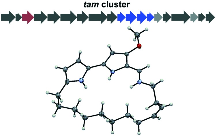
 Identification of a macrocyclic tambjamine natural product, tambjamine MYP1, from a marine bacterium that may enhance bioactivity by restraining bond rotation.
Abstract
Tambjamines are natural products that consist of a conserved bipyrrole core functionalized with different imines giving rise to many derivatives. The core structure of tambjamines allows ion coordination through the nitrogen atoms, which is a key aspect in many of their observed antimicrobial, anticancer, and antimalarial bioactivities. Minor variances in the compound structure have a considerable impact on the potency of these activities, so identifying new analogues is valuable for maximizing tambjamine biological potential. In this work, we describe the isolation and structure elucidation of the first naturally occurring macrocyclized tambjamine, tambjamine MYP1, from the marine microbe Pseudoalteromonas citrea. We also compare the apparent pKa of cyclic and linear tambjamine analogues and discuss how structural strain may effect the compound's ion coordination abilities.
Introduction
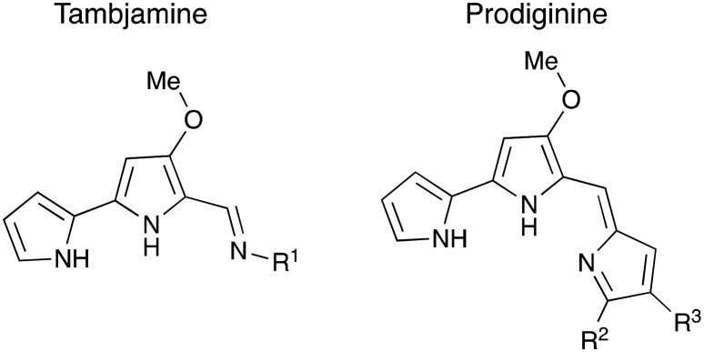
Tambjamines are a family of yellow-pigmented alkaloid natural products that possess a number of useful properties including antimicrobial, antifungal, antimalarial, and anti-tumour activities.1–3 Compounds in this family contain a methoxy-bipyrrole core with a variable imine moiety, consisting of decarboxylated amino acid side chains or linear alkyl chains (general structure in Fig. 1).4–8 Numerous tambjamines have been isolated from marine and terrestrial organisms and many have also been produced synthetically.1,4,6–11 Identifying new tambjamine natural products is appealing due to their exceptional bioactivity profile, much of which arises from the bipyrrole core, a structural feature shared with the well-studied prodiginine family.12–14 This rigid, aromatic bipyrrole structure allows for DNA intercalation and the formation of ion coordination complexes.12,15,16
Fig. 1. General tambjamine and prodiginine structures.
The ability to coordinate ions depends heavily on the orientation of the compound; in prodiginine compounds, rotation around the methenyl bond produces an α (E) and a β (Z) rotamer.17,18 This bond restriction also exists for tambjamines, thus similar rotameric forms are possible (Fig. 2). The β rotamer is believed to be the more bioactive and pharmaceutically relevant conformation for both molecule classes. Its ability to coordinate ions is thought to be the source of multiple bioactivities, including chloride transmembrane transport to induce apoptosis through cytosol acidification and copper dependent oxidative cleavage of DNA.12,16,18,19
Fig. 2. General tambjamine structure in the α and β rotamer conformations.
Stabilizing the β rotamer by introducing bulky imine moieties to restrict bond rotation is a tactic used to enhance the bioactivity of these compounds; this strategy has been effectively employed to increase antimalarial potency of synthetic tambjamine analogues.2 The lipophilicity of the imine sidechain also influences the anion affinity and transmembrane transport of tambjamines.20 Manipulating these structural characteristics of tambjamines can easily be done synthetically to interrogate structure–activity relationships, but it is also valuable to consider the naturally occurring derivatives that have come about through evolution.
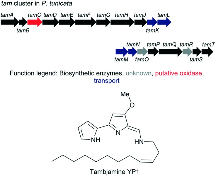
Most naturally derived tambjamines have been isolated from marine invertebrates such as bryozoans and nudibranchs, however it is postulated that these compounds are actually produced by microbial symbionts.4–7 Thus far, only two tambjamine analogues have been identified from bacteria; BE-18591 from the terrestrial Streptomyces sp. BA 18591, and tambjamine YP1 from marine bacterium Pseudoalteromonas tunicata.8,9 In 2007, Burke et al. linked tambjamine YP1 to its biosynthetic gene cluster in P. tunicata (Fig. 3), and assigned putative functions to all biosynthetic enzymes based on similarity to prodiginine biosynthetic genes and domain homology to known proteins (functional annotation can be found in Table S1†).21 This annotation allowed us to identify a very similar cluster in Pseudoalteromonas citrea using the antiSMASH cluster comparison tool.22P. citrea displays characteristics indicative of tambjamine production, such as a lemon-yellow colouring and anti-fouling properties, making it a likely producer.23,24
Fig. 3. The tam cluster in P. tunicata and the product tambjamine YP1.9,21 .
Interestingly, the tam cluster in both P. tunicata and P. citrea contains a predicted oxidase enzyme, TamC.21,22 This enzyme displays a high degree of similarity (34% identity) to the Rieske oxygenase, RedG, responsible for the cyclization of streptorubin B in Streptomyces coelicolor A3(2).25,26 RedG catalyzes a carbon–carbon bond formation between the pyrrole ring C of undecylprodigiosin and an internal CH2 unit in the alkyl chain.25,27 The presence of TamC in the tam cluster suggests the possible construction of a cyclized product. In the initial identification of tambjamine YP1 from P. tunicata, a potential cyclized variant was briefly mentioned by Burke et al., but to date no structural data has been reported.9,21
In this work, we have extracted and purified the predicted cyclized tambjamine YP1, herein called tambjamine MYP1, from P. citrea, and elucidated the macrocyclic structure using NMR spectroscopy and X-ray crystallography. We also compare the apparent pKa of our cyclic compound with a linear counterpart to investigate the implications this unique structural feature may have on ion coordination due to its rotational restraint and cage-like structure.
Results and discussion
Optimization studies on the growth and extraction of P. citrea determined that the bacteria displayed the highest intensity of yellow-pigmentation when grown on a solid marine agar rather than in a liquid culture. The isolation of the putative tambjamine was successfully completed from 3.2 L of solid media cultures of P. citrea extracted using ethyl acetate (EtOAc). The concentrated crude extract was purified by flash Si-chromatography and subsequent RP-HPLC using a C8 column, to obtain a yellow solid with a final yield of 0. 03% purified tambjamine from the dry cell mass.
High-resolution mass spectrometry using FTMS-ESI in positive ion mode generated an [M + H]+ ion at m/z = 354.25532 corresponding to a molecular formula of C22H32ON3 (calc 354.25399, Δ = 3.74 ppm). The reported [M + H]+ of tambjamine YP1 is m/z 356.27,9 the difference of approximately 2 mass units suggesting that the extracted tambjamine has one additional degree of unsaturation compared to tambjamine YP1. This mass difference is a good indication that a cyclic tambjamine compound is produced by P. citrea.
To determine the structure of the new tambjamine and particularly the points of cyclization, we performed a number of 1-D and 2-D NMR experiments in chloroform-d (CDCl3). Based on the NMR data we have proposed the structure shown in Fig. 4 and all 1H and 13C chemical shifts measured are displayed in Table 1. When we compare shifts for our new molecule to corresponding literature values for the linear tambjamine YP1 variant we can confirm the two molecules have very similar structures.9 However, there are key differences; no proton signal was detected at the 5′ pyrrole position of the putative cyclic tambjamine marking this as one point of cyclization. The proton and carbon atoms at position 19 have a large downfield shift from the linear tambjamine YP1 chemical shift values, and the 1H signal only integrates for two protons. These pieces of evidence give the second connection point for cyclization. The remaining spectral data remain consistent with the values for the linear compound, except the 13C chemical shifts for quaternary carbons 5′ and 2′ could not be identified with certainty due to the small quantity of purified material.
Fig. 4. Structure of tambjamine MYP1 and selected 2D NMR correlations.
Table 1. 1H and 13C NMR data for tambjamine YP1 and its novel cyclized variant in CDCl3.
| Position | Tambjamine MYP1 |
Tambjamine YP1
a
|
||
| δ H (J in Hz) | δ C | δ H | δ C | |
| 1′ (NH) | ||||
| 2′ | — | — | 123.5 | |
| 3′ | 6.63, s* | 113.5 | 6.70 | 113.0 |
| 4′ | 5.99, s* | 109.7 | 6.25 | 110.5 |
| 5′ | — | 7.06 | 124.1 | |
| 1 (NH) | ||||
| 2 | — | 142.1 | — | 143.4 |
| 3 | 5.87, s | 90.4 | 5.95 | 91.6 |
| 4 | — | 163.0 | — | 163.9 |
| OMe | 3.92, s | 58.5 | 3.90 | 58.8 |
| 5 | — | 110.1 | — | 111.3 |
| 6 | 7.14, d (14.77) | 140.1 | 7.29 | 141.1 |
| 7 (NH) | 9.29, br d | |||
| 8 | 3.54, br | 51.4 | 3.46 | 51.2 |
| 9 | 2.50, m | 27.8 | 2.46 | 28.8 |
| 10 | 5.22, dt (10.89, 7.70) | 123.2 | 5.35 | 124.1 |
| 11 | 5.55, dt (10.89, 7.20) | 137.1 | 5.55 | 134.5 |
| 12 | 2.06, m (7.20) | 27.4 | 2.01 | 27.8 |
| 13 | 1.22, m | 28.6–31.8 c | 1.21–1.70 d | 26.5–27.7 d |
| 14 | 0.85–1.29 b | |||
| 15 | ||||
| 16 | ||||
| 17 | ||||
| 18 | 1.82, m | 27.7 | ||
| 19 | 2.75, br t (6.10) | 27.2 | 0.86 | 14.5 |
aData from ref. 9.
bValue ranges for positions 14–17.
cValue ranges for positions 13–17.
dValue ranges for positions 13–18.
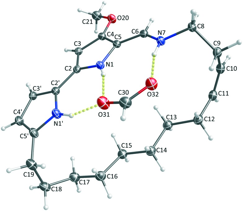
To confirm the NMR structure and cyclization points and to visualize the overall conformation of the cyclic tambjamine YP1 compound, we carried out X-ray crystallography. The purified compound readily co-crystallized in a complex with a formate anion and a formic acid molecule, stabilized through hydrogen-bonding interactions (Fig. S6†). For clarity, the formic acid molecule is omitted from the thermal ellipsoid drawing in Fig. 5. The full asymmetric unit, all refinement parameters, and additional view angles can be found in Fig. S7 and Table S2.†
Fig. 5. X-ray structure of tambjamine MYP1. Thermal ellipsoids are drawn to 30% probability (crystal structure was deposited in the Cambridge Structural Database (CSD), deposition number 1892787).
The solved crystal structure agrees with our NMR-based structure. The interatomic distance between C5′ and C19 is 1.502 ± 0.004 Å; the approximate distance of a C–C bond, validating this point of macrocyclization. Additionally, the crystal structure confirms the stereochemistry of the cis-alkene at C10–C11 with a torsional angle between C9–C10–C11–C12 of 1.3 ± 0.4°.
It is interesting to note that the linear compound, tambjamine YP1, originally isolated from P. tunicata was not detected from P. citrea in this work. This is surprising as it appears both P. citrea and P. tunicata produce the same cyclic tambjamine.9,21
This discrepancy in production leads to questions about the potential differences in biological and chemical properties of the analogues produced by very similar gene clusters. One such difference is the rotameric conformation and by extension ion binding properties of the linear and cyclic compounds.
Due to chemical and bioactivity similarities between the tambjamine and prodiginine families of molecules, we turned to the abundance of literature characterizing prodiginine structural features for guidance. In the prodiginine family it is known that the α rotamer is formed at low pH, due to favourable hydrogen bonding between the pyrrolium ion and the methoxy resulting in a high pKa of 8.23 ± 0.03 for one family member.17,28,29 Whereas, the β rotamer of the same molecule has a lower pKa of 5.4 ± 0.2 resulting from hydrogen bonding between pyrrole rings and is stabilized at a high pH.17,28,29 The β rotamer can also be formed at low pH when an anion such as chloride is present, raising the pKa to facilitate ionic interactions.12,28 Because the two rotamers have quite different pKa values, a molecule's apparent pKa reflects the contributions of each rotamer to the overall population.28 The propensity to form each rotamer can be estimated with the apparent pKa of the molecule, with higher pKas suggesting a preference for α rotamer and lower pKas suggesting a preference for the β rotamer.28 One caveat that should be noted is binding with anions such as chloride ions may cause shifts between the rotamer populations.28 As stated in the introduction, the β rotamer of prodiginines appears to be more biologically active and so variants that prefer taking this conformation are more likely to display strong bioactivity profiles.12,16,18,19
Far less is known about the tambjamine system, however, it is also likely able to form both the α and β rotamers. Experimental evidence of the β rotamer has been reported and although no pKa values have been determined for a specific rotamer conformation,8,9 an apparent pKa of 10.06 has been measured for tambjamine E.15 By analogy to the prodiginine system we would expect tambjamines that preferentially form the β rotamer to possess greater bioactivities. To investigate the potential biological impact of the macrocyclization present in tambjamine MYP1, we compared the apparent pKa of tambjamine MYP1 with the linear and unsaturated derivative, tambjamine BE-18591.
Tambjamine BE-18591 was synthesized from 4-methoxy-2,2′-carbaldehyde and dodecylamine following previously described protocols.30 The protonation state of each compound was monitored using their absorption spectra in 1 : 1 (v/v) MeCN/H2O and 0.1 M NaCl following the same approach as Melvin et al.18 The absorbance maxima of each compound changes depending on its protonation, allowing the apparent pKa to be measured by the absorbance shift as the pH increases. BE-18591 shifts from 400 nm in low pH to 368 nm as the pH increases, whereas tambjamine MYP1 shifts from a maximum at 420 nm to 385 nm as the pH increases (spectra can be found in Fig. S8†). As we do not know the population of each rotamer contributing to the overall pKa, the value measured is the apparent pKa of each compound.
The pKa of tambjamine MYP1 was calculated to be 10.4 ± 0.3, in comparison to a pKa of 9.9 ± 0.2 for the linear tambjamine BE-18591 (calculations in eqn S1†). These values are similar to those measured for tambjamine E, as described above. In our experiments, there appears to be a slight increase in pKa for the cyclic molecule, however the difference is small. Perhaps both molecules have very similar populations of the two rotamers and so cyclization may not cause a large increase in the β conformation population. Additionally, the macrocycle might form a cage-like structure around the chloride ion, similar to what we see in the crystal structure around formate (Fig. 5) restricting anion disassociation as the pH increases and favouring imine protonation. Due to the large size of the macrocycle the strain associated with cyclization may not be pronounced, thus it would be interesting to investigate the properties of synthetic tambjamines with smaller macrocycles. If the existing macrocycle is large enough to rotate to the α form it may be able to assume a helical conformation, which could have intriguing structural and biological implications. Further work will be needed to probe the bioactivity of macrocyclic tambjamine structures and their limitations.
The macrocyclization at the 5′ position bears resemblance to cyclizations seen in prodiginine analogues including streptorubin B, metacycloprodigiosin, and the marineosins.14,25,27 The cyclization of these compounds are all catalyzed by Rieske non-heme iron-dependent oxygenases, which use radical chemistry to form C–C bonds.25,27 In all of the characterized examples of prodiginine cyclization, the radical is formed on an internal CH2 of an alkyl chain, this instigates bond formation with either a pyrrole carbon or the methenyl carbon.25,27 In our cyclized tambjamine the C–C bond is initiated at the terminal methyl of the alkyl chain, forming a much less stable radical. This means that the putative oxidase, TamC, catalyzes a very difficult proton abstraction from the alkyl chain to produce the radical. Similar cyclization connectivity is also present in the compound cyclononylprodiginine isolated from Actinomadura madurae,31 however the biosynthesis of this compound is yet to be studied. To our knowledge, there are no known Rieske oxygenases that catalyze a comparable reaction, thus TamC presents an intriguing target for future studies in this family of enzymes.
Conclusion
We have described the isolation, crystallization, and identification of tambjamine MYP1 from Pseudoalteromonas citrea; this is the first macrocyclic tambjamine compound identified from nature. To probe if the macrocyclic structure favours formation of the putatively more bioactive β rotamer we determined the apparent pKa of the cyclic tambjamine and compared it that of a linear analogue. The small difference between the two pKa values suggests the rotamer populations of the cyclic and acyclic molecules are very similar and that this quite large macrocycle does not drastically change the conformational preference. Future investigations could focus on properties of tambjamines with smaller, more strained macrocycles and characterization of the proposed cyclase enzyme (TamC) could shed light on the functional capacity of the Rieske oxygenase enzyme family.
Experimental
Molecule extraction
Pseudoalteromonas citrea DSM 8771 (GenBank accession GCA_000238375.3) was grown as a starter liquid culture in marine media (5 g L–1 peptone, 3 g L–1 yeast extract, 35 g L–1 Instant Ocean), this was left to grow overnight at room temperature, non-shaking. Large-scale solid-support cultures of P. citrea were cultivated on four 33 × 23 cm casserole dishes holding 200 mL marine media agar inoculated using 5 mL of the liquid starter culture each and then grown for 24 h at 21 °C. The cells were removed from the agar by scraping and soaked in ∼500 mL EtOAc per 1.5 g of cell mass for 30 min. Cells were removed by filtration and the organic extract was concentrated in vacuo to yield 65 mg crude. This was further purified by Si-chromatography with a stepwise gradient from 100 : 0 to 50 : 50 hexanes/EtOAc. All yellow-pigmented fractions were collected and pooled for in vacuo concentration, resulting in 18 mg of yellow oil. The sample was further purified on a Waters Alliance HPLC, with an XBridge® BEH C8 OBD™ prep column. The method began with a 2 min wash using a solvent composition of 30 : 60 : 10 H2O/MeOH/1% (v/v) formic acid, followed by a 13 min gradient to 5 : 85 : 10 H2O/MeOH/1% (v/v) formic acid. Monitoring was carried out at a wavelength of 420 nm, and tambjamine MYP1 had a retention time of 7.5 min. Ultimately a yield of 0.5 mg dark yellow solid was obtained from 1.5 g cell mass.
Mass spectrometry
Samples for high-resolution mass spectrometry (HRMS) analysis were prepared in MeOH. HRMS was performed using a ThermoFisher Scientific Orbitrap Velos Pro instrument using Fourier-transform mass spectrometry (FTMS) and electrospray ionization in positive mode (ESI+). All spectra were analyzed using Xcalibur™ 2.2 SPI software.
NMR
NMR spectra were acquired in CDCl3 using either a Bruker NEO-500 (500 MHz 1H, 125 MHz 13C) or NEO-700 (700 MHz 1H, 175 Hz 13C) instrument. One-dimensional 1H and 13C NMR, and two-dimensional 1H–1H correlated spectroscopy (COSY), 1H–1H total correlated spectroscopy (TOCSY), 1H–1H nuclear Overhauser effect spectroscopy (NOESY), 1H–13C heteronuclear single quantum coherence (HSQC), and 1H–13C heteronuclear multiple bond coherence (HMBC) experiments were performed.
X-ray crystallography
The purified compound was co-crystallized with formic acid by evaporation in vacuo using a solvent mixture of 70% methanol and 0.1% formic acid in water. Once evaporated, the sample was left to rest for 14 days at –20 °C. Two crystals of diffraction quality were grown; they appeared plate-like and yellow.
A crystal was mounted onto a Micromount™ aperture (MiTeGen – Microtechnologies for Structural Genomics; USA) using paratone 8277 oil (Exxon). The mounted crystal was put under a nitrogen gas stream at –93.16 °C produced by the Oxford Cryostream 800. Diffraction data were collected, compiled, and reduced, and the crystal structure was solved and refined. Thermal ellipsoid images were produced using Mercury 3.10.2 software by the Cambridge Crystallographic Data Centre.32
Diffraction data was collected on a Bruker AXS D8 Venture Duo diffractometer, and recorded with a Bruker AXS PHOTON II Charge-Integrating Pixel Array Detector. The data were collected and reduced using the APEX3 software package v2018.1-0 by Bruker AXS,33 and the structure was solved and refined using SHELXT-2014/5 and SHELXL-2017/1.34,35
pKa determination
UV-vis absorption spectra were recorded on an Implen NanoPhotometer® NP80 Spectrophotometer. The pH measurements were obtained at 22 °C on an Orion Star™ A111 pH Benchtop Meter using an Orion Triode™ gel-filled electrode (9107BN). Calibration was done using commercial buffers (Orion ROSS™, pH 4.01, 7.00, and 10.01). Solutions (10 mL total volume) were prepared in 50 : 50 H2O/MeCN (v/v) at pH ∼12, containing 0.1 M NaCl. The mixture was gradually made acidic by addition of 0.1–0.01 M HCl. Following each addition of acid (10–20 μL), UV-vis spectra were recorded in the wavelength range of 200–900 nm, and the pH of the solution was measured. Apparent pKa values were determined using the method of Patterson.36
Conflicts of interest
There are no conflicts to declare.
Supplementary Material
Acknowledgments
We thank Dr. Paul Lummis for advice on the crystallization of our molecule. Financial support for this research was provided by the Natural Sciences and Engineering Research Council of Canada (NSERC Discovery Grant to ACR, NSERC-Canada Graduate Scholarship-Masters to KJP and JAD, and NSERC- Undergraduate Student Research Award to EMD).
Footnotes
†Electronic supplementary information (ESI) available: NMR and X-ray crystallography data, compound synthesis, and pKa calculations. CCDC 1892787. For ESI and crystallographic data in CIF or other electronic format see DOI: 10.1039/c9md00061e
References
- Pinkerton D. M., Banwell M. G., Garson M. J., Kumar N., De Moraes M. O., Cavalcanti B. C., Barros F. W. A., Pessoa C. Chem. Biodiversity. 2010;7:1311–1324. doi: 10.1002/cbdv.201000030. [DOI] [PubMed] [Google Scholar]
- Kancharla P., Kelly J. X., Reynolds K. A. J. Med. Chem. 2015;58:7286–7309. doi: 10.1021/acs.jmedchem.5b00560. [DOI] [PMC free article] [PubMed] [Google Scholar]
- Franks A., Egan S., Holmström C., James S., Lappin-Scott H., Kjelleberg S. Appl. Environ. Microbiol. 2006;72:6079–6087. doi: 10.1128/AEM.00559-06. [DOI] [PMC free article] [PubMed] [Google Scholar]
- Lindquist N., Fenical W. Experientia. 1991;47:504–506. [Google Scholar]
- Carbone M., Irace C., Costagliola F., Castelluccio F., Villani G., Calado G., Padula V., Cimino G., Cervera J. L., Santamaria R., Gavagnin M. Bioorg. Med. Chem. Lett. 2010;20:2668–2670. doi: 10.1016/j.bmcl.2010.02.020. [DOI] [PubMed] [Google Scholar]
- Paul V. J., Lindquist N., Fenical W. Mar. Ecol.: Prog. Ser. 1990;59:109–118. [Google Scholar]
- Carté B., Faulkner D. J. J. Org. Chem. 1983;48:2314–2318. [Google Scholar]
- Nakajima S., Kojiri K., Suda H. J. Antibiot. 1993;2881:1894–1896. doi: 10.7164/antibiotics.46.1894. [DOI] [PubMed] [Google Scholar]
- Franks A., Haywood P., Holmström C., Egan S., Kjelleberg S., Kumar N. Molecules. 2005;10:1286–1291. doi: 10.3390/10101286. [DOI] [PMC free article] [PubMed] [Google Scholar]
- Blackman A. J., Li C. Aust. J. Chem. 1994;47:1625–1629. [Google Scholar]
- Aldrich L. N., Stoops S. L., Crews B. C., Marnett L. J., Lindsley C. W. Bioorg. Med. Chem. Lett. 2010;20:5207–5211. doi: 10.1016/j.bmcl.2010.06.154. [DOI] [PubMed] [Google Scholar]
- Hernando E., Soto-cerrato V., Cortés-Arroyo S., Pérez-Tomás R., Quesada R. Org. Biomol. Chem. 2014;12:1771–1778. doi: 10.1039/c3ob42341g. [DOI] [PubMed] [Google Scholar]
- Darshan N., Manonmani H. K. J. Food Sci. Technol. 2015;52:5393–5407. doi: 10.1007/s13197-015-1740-4. [DOI] [PMC free article] [PubMed] [Google Scholar]
- Hu D. X., Withall D. M., Challis G. L., Thomson R. J. Chem. Rev. 2016;116:7818–7853. doi: 10.1021/acs.chemrev.6b00024. [DOI] [PMC free article] [PubMed] [Google Scholar]
- Melvin M. S., Ferguson D. C., Lindquist N., Manderville R. A. J. Org. Chem. 1999;64:6861–6869. doi: 10.1021/jo990944a. [DOI] [PubMed] [Google Scholar]
- Melvin M. S., Wooton K. E., Rich C. C., Saluta G. R., Kucera G. L., Lindquist N., Manderville R. A. J. Inorg. Biochem. 2001;87:129–135. doi: 10.1016/s0162-0134(01)00338-5. [DOI] [PubMed] [Google Scholar]
- Garcia-Valverde M., Alfonso I., Quinonero D., Quesada R. J. Org. Chem. 2012;77:6538–6544. doi: 10.1021/jo301008c. [DOI] [PubMed] [Google Scholar]
- Melvin M. S., Tomlinson J. T., Park G., Day C. S., Saluta G. R., Kucera G. L., Manderville R. A. Chem. Res. Toxicol. 2002;15:734–741. doi: 10.1021/tx025507x. [DOI] [PubMed] [Google Scholar]
- Borah S., Melvin M. S., Lindquist N., Manderville R. A. J. Am. Chem. Soc. 1998;120:4557–4562. [Google Scholar]
- Knight N. J., Hernando E., Haynes C. J. E., Busschaert N., Clarke H. J., Takimoto K., Garcia-Valverde M., Frey J. G., Quesada R., Gale P. A. Chem. Sci. 2016;7:1600–1608. doi: 10.1039/c5sc03932k. [DOI] [PMC free article] [PubMed] [Google Scholar]
- Burke C., Thomas T., Egan S., Kjelleberg S. Environ. Microbiol. 2007;9:814–818. doi: 10.1111/j.1462-2920.2006.01177.x. [DOI] [PubMed] [Google Scholar]
- Weber T., Blin K., Duddela S., Krug D., Kim H. U., Bruccoleri R., Lee S. Y., Fischbach M. A., Müller R., Wohlleben W., Breitling R., Takano E., Medema M. H. Nucleic Acids Res. 2015;43:W237–W243. doi: 10.1093/nar/gkv437. [DOI] [PMC free article] [PubMed] [Google Scholar]
- Gauthier M. J. Int. J. Syst. Bacteriol. 1977;27:349–354. [Google Scholar]
- Holmstrom C., Egan S., Franks A., McCloy S., Kjelleberg S. FEMS Microbiol. Ecol. 2002;41:47–58. doi: 10.1111/j.1574-6941.2002.tb00965.x. [DOI] [PubMed] [Google Scholar]
- Withall D. M., Haynes S. W., Challis G. L. J. Am. Chem. Soc. 2015;137:7889–7897. doi: 10.1021/jacs.5b03994. [DOI] [PubMed] [Google Scholar]
- Altschul S. F., Gish W., Miller W., Myers E. W., Lipman D. J. J. Mol. Biol. 1990;215:403–410. doi: 10.1016/S0022-2836(05)80360-2. [DOI] [PubMed] [Google Scholar]
- Perry C., Santos E. L. C. D. L., Alkhalaf L. M., Challis G. L. Nat. Prod. Rep. 2018;35:622–632. doi: 10.1039/c8np00004b. [DOI] [PubMed] [Google Scholar]
- Rizzo V., Morelli A., Pinciroli V., Sciangula D., D'Alessio R. J. Pharm. Sci. 1999;88:73–78. doi: 10.1021/js980225w. [DOI] [PubMed] [Google Scholar]
- Chawrai S. R., Williamson N. R., Mahendiran T., Salmond G. P. C., Leeper F. J. Chem. Sci. 2012;3:447–454. [Google Scholar]
- Saggiomo V., Otto S., Marques I., Felix V., Torroba T., Quesada R. Chem. Commun. 2012;48:5274–5276. doi: 10.1039/c2cc31825c. [DOI] [PubMed] [Google Scholar]
- Gerber N. N. Appl. Microbiol. 1969;18:1–3. doi: 10.1128/am.18.1.1-3.1969. [DOI] [PMC free article] [PubMed] [Google Scholar]
- Macrae C. F., Edgington P. R., McCabe P., Pidcock E., Shields G. P., Taylor R., Towler M., van de Streek J. J. Appl. Crystallogr. 2006;39:453–457. [Google Scholar]
- Bruker AXS Inc .
- Sheldrick G. M. Acta Crystallogr., Sect. A: Found. Crystallogr. 2008;64:112–122. doi: 10.1107/S0108767307043930. [DOI] [PubMed] [Google Scholar]
- Sheldrick G. M. Acta Crystallogr., Sect. A: Found. Adv. 2015;71:3–8. doi: 10.1107/S2053273314026370. [DOI] [PMC free article] [PubMed] [Google Scholar]
- Patterson G. S. J. Chem. Educ. 1999;76:395–398. [Google Scholar]
Associated Data
This section collects any data citations, data availability statements, or supplementary materials included in this article.


