Abstract
Background
A significant number of women are diagnosed with minor cytological abnormalities on cervical screening. Many authorities recommend surveillance as spontaneous regression might occur. However, attendance for cytological follow‐up decreases with time and might put some women at risk of developing invasive disease.
Objectives
To assess the optimum management strategy for women with minor cervical cytological abnormalities (atypical squamous cells of undetermined significance ‐ ASCUS or low‐grade squamous intra‐epithelial lesions ‐ LSIL) at primary screening in the absence of HPV (human papillomavirus) DNA test.
Search methods
We searched the following electronic databases: Cochrane Central Register of Controlled Trials (CENTRAL Issue 4, 2016), MEDLINE (1946 to April week 2 2016) and Embase (1980 to 2016 week 16).
Selection criteria
We included randomised controlled trials (RCTs) comparing immediate colposcopy to cytological surveillance in women with atypical squamous cells of undetermined significance (ASCUS/borderline) or low‐grade squamous intra‐epithelial lesions (LSIL/mild dyskaryosis).
Data collection and analysis
The primary outcome measure studied was the occurrence of cervical intra‐epithelial neoplasia (CIN). The secondary outcome measures studied included default rate, clinically significant anxiety and depression, and other self‐reported adverse effects.
We classified studies according to period of surveillance, at 6, 12, 24 or 36 months, as well as at 18 months, excluding a possible exit‐examination. We calculated pooled risk ratios (RR) and 95% confidence intervals (CI) using a random‐effects model with inverse variance weighting. Inter‐study heterogeneity was assessed with I2 statistics.
Main results
We identified five RCTs with 11,466 participants that fulfilled the inclusion criteria. There were 18 cases of invasive cervical cancer, seven in the immediate colposcopy and 11 in the cytological surveillance groups, respectively. Although immediate colposcopy detects CIN2+ and CIN3+ earlier than cytology, the differences were no longer observed at 24 months (CIN2+: 3 studies, 4331 women; 17.9% versus 18.3%, RR 1.14, CI 0.66 to 1.97; CIN3+: 3 studies, 4331 women; 10.3% versus 11.9%, RR 1.02, CI 0.53 to 1.97). The inter‐study heterogeneity was considerable (I2 greater than 90%). Furthermore, the inclusion of the results of the exit examinations at 24 months, which could inflate the CIN detection rate of cytological surveillance, may have led to study design‐derived bias; we therefore considered the evidence to be of low quality.
When we excluded the exit examination, the detection rate of high‐grade lesions at the 18‐month follow‐up was higher after immediate colposcopy (CIN2+: 2 studies, 4028 women; 14.3% versus 10.1%, RR 1.50, CI 1.12 to 2.01; CIN3+: 2 studies, 4028 women, 7.8% versus 6.9%, RR 1.24, CI 0.77 to 1.98) both had substantial inter‐study heterogeneity (I2 greater than 60%) and we considered the evidence to be of moderate quality).
The meta‐analysis revealed that immediate referral to colposcopy significantly increased the detection of clinically insignificant cervical abnormalities, as opposed to repeat cytology after 24 months of surveillance (occurrence of koilocytosis: 2 studies, 656 women; 32% versus 21%, RR 1.49, 95% CI 1.17 to 1.90; moderate‐quality evidence) incidence of any CIN: 2 studies, 656 women; 64% versus 32%, RR 2.02, 95% CI 1.33 to 3.08, low‐quality evidence; incidence of CIN1: 2 studies, 656 women; 21% versus 8%, RR 2.58, 95% CI 1.69 to 3.94, moderate‐quality evidence).
Due to differences in trial designs and settings, there was large variation in default rates between the included studies. The risk for default was higher for the repeat cytology group, with a four‐fold increase at 6 months, a six‐fold at 12 and a 19‐fold at 24 months (6 months: 3 studies, 5117 women; 6.3% versus 13.3%, RR 3.85, 95% CI 1.27 to 11.63, moderate‐quality evidence; 12 months: 3 studies, 5115 women; 6.3% versus 14.8%, RR 6.39, 95% CI 1.49 to 29.29, moderate‐quality evidence; 24 months: 3 studies, 4331 women; 0.9% versus 16.1%, RR 19.1, 95% CI 9.02 to 40.43, moderate‐quality evidence).
Authors' conclusions
Based on low‐ or moderate‐quality evidence using the GRADE approach and generally low risk of bias, the detection rate of CIN2+ or CIN3+ after two years does not appear to differ between immediate colposcopy and cytological surveillance in the absence of HPV testing, although women may default from follow‐up. Immediate colposcopy probably leads to earlier detection of high‐grade lesions, but also detects more clinically insignificant low‐grade lesions. Colposcopy may therefore be the first choice when good compliance is not assured. These results emphasize the need for an accurate reflex HPV triage test to distinguish women who need diagnostic follow‐up from those who can return safely to routine recall.
Plain language summary
Management of minor cytological abnormalities identified on cervical screening
The issue Cervical screening programmes reduce the risk of cervical cancer, through the use of cervical cytology (smear tests), which aim to detect and treat any precancerous changes which might put some women at risk of developing invasive disease (invasive cervical cancer) in the future. Usually only severe precancerous changes require treatment, however, there is some discrepancy in how to manage women with minor cytological changes (atypical squamous cells of undetermined significance (ASCUS/borderline) or low‐grade squamous intra‐epithelial lesions (LSIL/mild dyskaryosis) if HPV (human papillomavirus) testing is not routinely available.
The aim of the review We aimed to assess whether immediate colposcopy or 'watchful waiting', with repeat cervical cytology, was better for women with minor cervical cytological abnormalities.
What are the main findings? We included 5 randomised controlled trials including 11,466 participants with minor abnormalities on cervical cytology, treated either with immediate colposcopy or repetitive cytology. The included studies assessed differences in occurrence of cervical precancerous lesions between the two treatments.
The results suggested that women attending immediate colposcopy after a single low‐grade abnormal cervical cytology test were more likely to have clinically insignificant findings detected than women who were managed with 'watchful waiting'.
There were 18 cases of invasive cervical cancer, seven in the immediate colposcopy and 11 in the cytological surveillance groups. The detection rate of clinically insignificant low‐grade lesions was higher in the immediate colposcopy group, as was the detection rate of clinically more significant high‐grade precancerous lesions (CIN2 or CIN2 or worse) at 18 months, but not by 24 months.
The risk of non‐compliance was significantly greater for the repeat cytology arm and increased with the length of the follow‐up.
What is the quality of the evidence? We graded the evidence as low to moderate quality.
What are the conclusions? HPV DNA testing has been shown to be an effective triage tool for women with minor cervical cytology abnormalities. However, this test is not currently routinely available globally. Therefore, if HPV DNA testing is not available, immediate colposcopy is likely to detect more precancerous lesions earlier than cytological surveillance, but after two years there does not seem to be a difference between the two approaches. Women could be referred for immediate colposcopy after a single low‐grade abnormal or borderline cervical cytology test, if compliance with cytological surveillance is expected to be poor. When follow‐up compliance is expected to be good, repeat cervical cytology may be offered, as this may reduce the risk of over‐diagnosis and over‐treatment.
Summary of findings
Summary of findings 1. Summary of findings: occurrence of CIN and default rates.
| Immediate colposcopy compared with cytological surveillance for minor cervical cytological abnormalities: occurrence of different grades CIN in histology according to follow‐up time and default rates | ||||||
|
Patient or population: women with ASCUS or LSIL Settings: colposcopy clinic Intervention: immediate colposcopy Comparison: cytological surveillance | ||||||
| Outcomes | Illustrative comparative risks* (95% CI) | Relative effect (95% CI) | No of participants (studies) | Quality of the evidence (GRADE) | Comments | |
| Assumed risk | Corresponding risk | |||||
| Risk with cytological surveillance | Risk with immediate colposcopy | |||||
| Occurrence of CIN2+ in histology at 18 months | 101 per 1000 | 151 per 1000 (113 to 203) | RR 1.50 (1.12 to 2.01) | 4028 (2 studies) | ⊕⊕⊕⊝ moderate1 | |
| Occurrence of CIN2+ in histology at 24 months | 183 per 1000 | 209 per 1000 (121 to 361) | RR 1.14 (0.66 to 1.97) | 4331 (3 studies) | ⊕⊕⊝⊝ low2,3 | |
| Occurrence of CIN3+ in histology at 18 months | 69 per 1000 | 86 per 1000 (53 to 137) | RR 1.24 (0.77 to 1.98) | 4028 (2 studies) | ⊕⊕⊕⊝ moderate4 | |
| Occurrence of CIN3+ in histology at 24 months | 119 per 1000 | 121 per 1000 (63 to 234) | RR 1.02 (0.53 to 1.97) | 4331 (3 studies) | ⊕⊕⊝⊝ low3,5 | |
| Occurrence of any CIN in histology at 24 months | 316 per 1000 | 639 per 1000 (420 to 974) | RR 2.02 (1.33 to 3.08) | 656 (2 studies) | ⊕⊕⊝⊝ low3,8 | |
| Default rates at 6 months | 63 per 1000 | 241 per 1000 (80 to 728) | RR 3.85 (1.27 to 11.63) | 5117 (3 study) | ⊕⊕⊕⊝ moderate6 |
|
| Default rates at 12 months | 63 per 1000 | 413 per 1000 (93 to 1000) | RR 6.60 (1.49 to 29.29 | 5115 (3 studies) | ⊕⊕⊕⊝ moderate7 |
|
| *The basis for the assumed risk (e.g. the median control group risk across studies) is provided in footnotes. The corresponding risk (and its 95% confidence interval) is based on the assumed risk in the comparison group and the relative effect of the intervention (and its 95% CI). For default rates the relative effect is calculated between cytological surveillance versus immediate colposcopy. For histology the relative effect is calculated between immediate colposcopy versus cytological surveillance. ASCUS: atypical squamous cells of undetermined significance CI: Confidence interval; CIN: cervical intraepithelial neoplasia; LSIL: low‐grade squamous intra‐epithelial lesions; RR: Risk Ratio | ||||||
| GRADE Working Group grades of evidence High quality: Further research is very unlikely to change our confidence in the estimate of effect. Moderate quality: Further research is likely to have an important impact on our confidence in the estimate of effect and may change the estimate. Low quality: Further research is very likely to have an important impact on our confidence in the estimate of effect and is likely to change the estimate. Very low quality: We are very uncertain about the estimate. | ||||||
1 Downgraded to moderate due to substantial inter‐study heterogeneity (P = 0.08, I2= 61%). 2 Downgraded to low due to considerable inter‐study heterogeneity (P < 0.00001, I2= 94%). 3 Downgraded due to presence of the other possible bias resulting in falsely high CIN detection rate in the cytological surveillance arm. 4 Downgraded to moderate due to substantial inter‐study heterogeneity (P = 0.02, I2= 75%). 5 Downgraded to low due to considerable inter‐study heterogeneity (P < 0.00001, I2= 93%). 6 Downgraded to moderate due to considerable inter‐study heterogeneity (P = 0.02, I2= 76%). 7 Downgraded to moderate due to considerable inter‐study heterogeneity (P = 0.0004, I2= 87%). 8 Downgraded to low due to substantial inter‐study heterogeneity (P = 0.02, I2 = 82%).
Background
Cervical cancer is largely preventable through screening and treatment of screen‐detected cervical lesions. Despite this, cervical cancer remains the most common female malignancy in virtually all low‐ and middle‐income countries, and the third most common in women worldwide (GLOBOCAN 2013). Of all cervical cancer, 83% occurs in low‐income countries. Cervical cancer still remains an important public health issue in Europe with more than 66,000 new cases and 29,000 deaths annually. The majority of these cases are diagnosed in Eastern European countries, which reflects the absence of a screening programme (Arbyn 2007). A woman's risk of developing cervical cancer by age 75 years ranges from 0.9% in high‐income countries to 1.9% in low‐ and middle‐income countries (Arbyn 2011). In Europe, about 60% of women with cervical cancer are alive five years after diagnosis (EUROCARE 2003). The disease primarily affects younger women and therefore, the total years‐of‐life lost is proportionately higher than that for most other cancers, which often have a later onset. The purpose of cervical cytology screening programmes using cytology (also known as Pap smear, named after Dr Papanicolaou (Papanicolaou 1941), is the early detection and treatment of pre‐invasive lesions and, ultimately, reduction in both the incidence and mortality from cervical cancer. Screening programmes have proven their value and efficacy in reducing both the incidence and mortality from cervical cancer in countries where they have been widely applied, including the UK. In countries with an established screening programme, there are different challenges: improving coverage and accuracy of screening, as well as the selection and better management of women with lesions of true malignant potential that require intervention. Without doubt, the most significant advance globally has been the realisation that persistent infection with oncogenic human papillomavirus (HPV) is causally associated with cervical cancer, as well as the development of prophylactic vaccines. The HPV DNA test that aims to detect the viral genome has also been developed and has potential clinical applications in primary screening, in the triage of minor cytological abnormalities, and in follow‐up after treatment (Arbyn 2004; Bulkmans 2007; Koliopoulos 2007; Naucler 2007; Paraskevaidis 2004).
Description of the condition
Cervical cytology may be classified according to the Bethesda system (Solomon 2002), or the British Society of Cervical Cytology (BSCC) terminology (NHSCSP 2000), and can be reported in order of severity as: (1) normal; (2) atypical squamous cells of undetermined significance (ASCUS) (Bethesda)/borderline (BSCC); (3) low‐grade squamous intra‐epithelial lesion (LSIL) (Bethesda)/mild dyskaryosis (BSCC); (4) high‐grade squamous intraepithelial lesion (HSIL) (Bethesda)/high‐grade dyskaryosis (either favours moderate or severe) (BSCC). Cervical cytology classified as high‐grade (moderate and severe dyskaryosis in the UK) occurs in roughly 1% to 3% of the screened population. A high‐grade lesion (cervical intra‐epithelial neoplasia 2+ (CIN2+)) is confirmed by histology in greater than 50% to 60% of these cases. The risk of subsequent progression to malignancy is approximately 30%, although a significant proportion (20% to 30%) have regressive CIN.
Although it is widely accepted that women with high grade cytological abnormalities should be referred immediately for a colposcopic examination or subsequent treatment, or both, uncertainty exists regarding the optimum way of managing those with low‐grade findings.
Women with cytology classified as ASCUS or LSIL (Bethesda classification) (Solomon 2002), or their British Society of Cervical Cytology (BSCC) terminology (NHSCSP 2000) equivalents of borderline and mild dyskaryosis, comprise approximately 7% of all the smears performed in the UK every year (Department of Health 2006). These minor abnormalities, with unknown or low malignant potential, are more common in younger women and present a difficult problem with regards to their management; the implications are important as they consume a disproportionate amount of clinical resources, with their significance still debatable. However, despite a low‐grade cytological smear, a considerable proportion of these women (15% to 20%) still have an underlying histological high‐grade lesion (CIN2+) (Bolger 1988; Contreras‐Melendez 1992; Flannelly 1997; Giles 1989; Paraskevaidis 2002; Soutter 1986; Walker 1986) and therefore, are at risk of developing invasive disease.
The HPV test appears to have a role in the triage of those women who need referral to colposcopy. Evidence in the literature reports a significantly better sensitivity and similar specificity for the HPV test in comparison to repeat cytology for the detection of high‐grade lesions for initial ASCUS/borderline cytology (55% positivity). The introduction of the HPV DNA test in cases with ASCUS cytologic findings could enhance the detection of those women with underlying high‐grade CIN who should be referred to colposcopy or returned to routine recall instead of repeat cytology (Arbyn 2004; Arbyn 2012; Kelly 2011). A survey amongst 43 European countries in 2013 showed that the majority (90%) of the countries have introduced HPV testing for ASCUS triage, and 62% use the test in the triage of LSIL as well (Arbyn 2015).
However, this does not appear to be true for LSIL/mild dyskaryosis lesions or women under 30 to 40 years, as the high‐positivity rate of HPV in this group (85%), does not support its use as a triage tool (Arbyn 2005; Arbyn 2013a; TOMBOLA 2009). Furthermore the HPV test is not yet widely available, reinforcing the need for clear recommendations in settings without access to these triage tests (Cuzick 2008).
Description of the intervention
Until new, markedly reliable triage markers develop, the management options of LSIL cytology remain either immediate referral to colposcopy (examination of the cervix and vagina using magnifying light microscope, colposcope, and acetic acid) or cytological surveillance with repeat smears.
A repeat cytology sample at six months may identify women with persistent lesions who require referral to colposcopy.
The management after immediate referral to colposcopy will depend on the colposcopic findings. If they are suggestive of a high‐grade lesion, multiple punch biopsies may be appropriate, particularly in young women of reproductive age. If the colposcopic findings are consistent with a low‐grade lesion, surveillance every six months and treatment, only if the abnormality persists beyond two years, may be justified, particularly in young women, as a significant proportion of these lesions may regress. The rate of regression decreases significantly with increasing age, and in the presence of a high‐risk HPV subtype and increasing duration of presence of the lesion (Kyrgiou 2010).
How the intervention might work
Optimising management of women with LSIL cervical cytology is difficult.
A policy of immediate referral to colposcopy could potentially result, not only in increased numbers of referrals, thus overloading colposcopy clinics and increasing costs, but also in over‐intervention or over‐treatment, or both, due to subtle colposcopic findings. Many young women of reproductive age might be exposed to the physical and psychological sequelae of unnecessary interventions and treatment, which can also be associated with long‐term, pregnancy‐related, morbidity (Arbyn 2008; Founta 2010; Kyrgiou 2006; Kyrgiou 2012a; Kyrgiou 2014; Kyrgiou 2016a; Paraskevaidis 2007).
On the other hand, triage with repeat cytology, and referral to colposcopy, only if the abnormality persists, may result in a potential reduction of the number of unnecessary referrals, but carries risks of missing high‐grade lesions and increased non‐attendance rates (Shafi 1997). Default from screening is known to put women with equivocal smears and occult high‐grade disease at risk of developing invasive cancer.
The TOMBOLA study in the UK showed that, compared with cytological surveillance, a policy of immediate colposcopy does detect more high‐grade lesions, but might lead to over‐treatment. To reduce this, the trial authors suggested that a policy of targeted punch biopsies with subsequent treatment for CIN2 and CIN3, and cytological surveillance for CIN1 or less, provides the best balance between benefit and harm for the management of these women. Immediate loop excision resulted in over‐treatment and more adverse effects, and was therefore not be recommended (TOMBOLA 2009).
Why it is important to do this review
This review aimed to appraise the current evidence from RCTs on cytological surveillance versus immediate colposcopy in the absence of HPV‐testing in order to make a decision on optimum evidence based practice, as currently this is an area of uncertainty.
A list of abbreviations used in the text can be found in Appendix 1.
Objectives
To assess the optimum management strategy for women with minor cervical cytological abnormalities (atypical squamous cells of undetermined significance ‐ ASCUS or low‐grade squamous intra‐epithelial lesions ‐ LSIL) at primary screening in the absence of HPV (human papillomavirus) DNA test.
Methods
Criteria for considering studies for this review
Types of studies
We included all RCTs comparing immediate referral to colposcopy versus cytological surveillance in women with ASCUS or LSIL cytological abnormalities at primary cervical screening in the absence of HPV DNA testing. We only included studies reporting combined data for minor abnormalities with more severe grades (high‐grade SIL, moderate and severe dyskaryosis), if separate data according to the grade were available. In case of overlap or duplicate reports, we extracted all relevant outcomes from all the publications of each trial. We included trials with multiple arms if at least two arms addressed an eligible comparison; non‐eligible arms were excluded. We excluded non‐randomised studies and pseudo‐randomised trials with alternate allocation of subjects; meeting and conference abstracts; and studies comparing HPV DNA test to colposcopy or repeat cytology.
Types of participants
We included studies with adult women (greater than 18 years old) with minor cytological cervical abnormalities (ASCUS/borderline dyskaryosis and LSIL/mild dyskaryosis). From included studies we excluded women with high‐grade squamous intra‐epithelial lesions (HSIL)/moderate and severe dyskaryosis, when separate data for LSIL abnormalities were available.
Types of interventions
Surveillance with repeat cytology in any setting versus immediate colposcopy.
The gold standard was histological diagnosis at colposcopy or at the end of the surveillance period in the form of punch biopsies or excisional treatment. Excisional treatment included large loop excision of the transformation zone (LLETZ or LEEP), laser conization (LC), cold knife conization (CKC) and needle or straight wire excision of the transformation zone (NETZ/SWETZ). We excluded trials that did not report on histological results (gold standard) (for example, in the form of punch biopsies or excisional treatment).
Types of outcome measures
Primary outcomes
The primary outcomes were the cumulative incidence of cervical intra‐epithelial neoplasia grade 2 or worse (CIN2+) and grade 3 or worse (CIN3+) from histology assessment.
Secondary outcomes
The secondary outcomes included:
cumulative incidence at other histological thresholds, including HPV‐associated morphological findings and CIN1;
default rates from repeat cytology or colposcopy clinic appointments;
anxiety and depression scores (based on validated questionnaires);
short‐term adverse effects of management (pain, bleeding, and discharge, together with the duration (in days) and severity; and
treatment rates in both groups.
Search methods for identification of studies
We searched for papers in all languages.
We searched the literature from 1946, when the conservative methods of treatment for CIN were first introduced into clinical practice, and included references published up to the present day.
We identified the RCTs comparing these alternative strategies for the management of low‐grade cervical abnormalities by a computerised literature search, by tracing references listed in relevant articles and by a manual search of appropriate journals.
Electronic searches
See: Cochrane Gynaecological Cancer Group methods used in reviews. We searched the following electronic databases.
The Cochrane Central Register of Controlled Trials (CENTRAL, 2016, Issue 4)
MEDLINE (Ovid) (1946 to April week 2, 2016)
Embase (Ovid) (1980 to 2016, week 16)
We used the following main keywords: ‘randomised controlled trials’ ‘cervical intraepithelial neoplasia (CIN)’, ‘cervical cancer’, ‘ASCUS’, ‘borderline’, ‘LSIL’, ‘low‐grade squamous intra‐epithelial lesion’, ‘mild dyskaryosis’, ‘colposcopy’, ‘smear’, ‘cytology’.
We used the 'related articles' feature in MEDLINE to retrieve additional references. For databases other than MEDLINE, we adapted the search strategy accordingly. The search strategies for all the databases are available in Appendix 2, Appendix 3 and Appendix 4.
Searching other resources
We searched the metaRegister of controlled trials (www.isrctn.com/page/mrct), the National Cancer Institute's database (www.cancer.gov/clinicaltrials), Physicians Data Query (www.cancer.gov/publications/pdq), Current Controlled Trials (www.controlled-trials.com), and ClinicalTrials.gov (www.clinicaltrials.gov) for ongoing studies.
We searched conference proceedings and abstracts through Zetoc (http://zetoc.mimas.ac.uk) and WorldCat Dissertations (www.oclc.org/support/documentation/firstsearch/databases/dbdetails/details/WorldCatDissertations.htm). We also searched reports of conferences in the following sources.
Annual Meetings of the British Society of Colposcopy and Cervical Pathology.
Triannual Meetings of the International Federation of Cervical Pathology and Colposcopy.
Triannual Meetings of the European Federation of Colposcopy.
Annual Meetings of the American Society of Colposcopy and Cervical Pathology.
We checked the citation lists of included studies and contacted experts in the field, Presidents of the British, European, American and International Societies of Colposcopy and Cervical Pathology to identify further reports of studies.
We intended to include both published and unpublished studies, if they met the inclusion criteria for the review.
Data collection and analysis
Selection of studies
We downloaded all titles and abstracts retrieved by electronic searching into the reference management database Endnote. We removed duplicates and two review authors (MK, IK) independently examined the remaining references. We excluded those trials which clearly did not meet the inclusion criteria, and obtained copies of the full text of potentially relevant references. We (MK, IK) independently assessed the eligibility of the retrieved papers. We then compared the results and resolved any disagreements by discussion. If necessary, we involved a third review author (MA) to reach consensus. We documented the reasons for exclusion.
Data extraction and management
We (MK, IK) extracted the relevant data from the trials identified. This included study population, population characteristics, sample size, study methods, method of randomisation, methodological quality, follow‐up and dropout, assessment of outcomes, and results).
We classified trials according to the length of cytological surveillance (6, 12, 18, 24, 30, and 36 months) and analysed them in different groups.
We extracted or computed from all included studies, the total number of women included and incidence of all grades of histological diagnoses, default rates, depression, anxiety scores, after‐effects and other patient‐reported outcomes in both groups, whenever available. We contacted trial authors to obtain separate data when data on LSILs were merged with HSIL lesions.
In addition, we collected data on the length of period of surveillance and the type of histology used at the end of surveillance or at colposcopy (gold standard: punch biopsies or excisional treatment).
We also documented the diagnostic criteria used for the interpretation of cytology and histology in each study, where these were read, and whether there was a central review in each study in order to assess if the above had significant variation that may have impacted on the value of the review.
For included trials, we extracted the following data.
Author, year of publication, journal and language
Country
Setting where the trial was conducted
Inclusion and exclusion criteria
Trial design, methodology
-
Trial population
total number enrolled and number included in each group
participant characteristics (such as age and other demographic and socioeconomic characteristics ‐ risk factors for cervical cancer)
index cytology, history of previous cytological lesions
type of cytology (conventional or liquid‐based)
-
Intervention details
surveillance with repeat cytology in primary care and number of follow‐up smears
immediate colposcopy and related interventions (i.e. punch biopsies and/or treatment and type)
the type of gold standard used (histology at colposcopy or at the end of the surveillance period): punch biopsies or excisional treatment. We planned to report the type of excisional treatment: LLETZ or LEEP, LC, CKC and NETZ or SWETZ.
Risk of bias (See Assessment of risk of bias in included studies)
-
Outcomes reported in each trial
-
Primary outcomes:
the cumulative incidence of CIN2+ and CIN3+ at histology
-
Secondary outcomes:
cumulative incidence at other histological thresholds HPV‐associated morphological findings and CIN1
default rates from repeat cytology or colposcopy clinic appointments
treatment rates in both groups
anxiety scores
depression scores
short‐term after‐effects of management (rates and types), specifically pain, bleeding, and discharge, together with the duration (in number of days) and severity
other self‐reported outcomes
-
-
Details of outcomes reported:
for each outcome: outcome definition (with diagnostic criteria if relevant)
unit of measurement (if relevant)
for scales: upper and lower limits, and whether high or low score is good
results: number of participants allocated to each intervention group
for each outcome of interest: sample size; missing participants
the time points at which outcomes were collected and reported
-
We extracted data on outcomes as below.
For dichotomous outcomes (all outcomes were reported as dichotomous), we extracted the number of women in each treatment arm who experienced the outcome of interest and the number of women assessed at endpoint, in order to estimate a risk ratio (RR).
We (MK, IK) independently extracted data in a data extraction form specially designed for the review. We resolved differences between review authors by discussion or by appeal to a third review author (MA), if necessary.
Assessment of risk of bias in included studies
We used the Cochrane tool for assessing risk of bias (Higgins 2011). We (MK, IK) independently assessed the risk of bias within each included study based on the following six domains, with our judgements presented as 'low risk of bias'; 'high risk of bias', and 'unclear'.
For RCTs, if identified, we included assessment of:
sequence generation;
allocation concealment;
blinding (of participants, healthcare providers and outcome assessors);
-
incomplete outcome data:
-
we recorded the proportion of participants whose outcomes were not reported at the end of the study; we noted if loss to follow‐up was not reported. We coded the satisfactory level of loss to follow‐up for each outcome as:
'low risk of bias', if fewer than 20% of participants were lost to follow‐up, and reasons for loss to follow‐up were similar in both treatment arms
'high risk of bias', if more than 20% of participants were lost to follow‐up, or reasons for loss to follow‐up differed between treatment arms
'unclear' if loss to follow‐up was not reported;
-
selective reporting of outcomes; and
other possible sources of bias.
Measures of treatment effect
For all (dichotomous) outcomes, we used the risk ratio (RR).
Dealing with missing data
We contacted one study author to obtain data stratified by grade of index cytology (Flannelly 1994). Otherwise, all relevant data were available from the original publications. We did not impute any missing outcome data.
Assessment of heterogeneity
We assessed inter‐study heterogeneity with the Cochran Q test (Cochran 1954), by visual inspection of forest plots (Deeks 2011), by estimation of the percentage of heterogeneity between studies which cannot be ascribed to sampling variation (I2 statistic) (Higgins 2003), and by a formal test of the significance for heterogeneity (Deeks 2001). If there was evidence of substantial heterogeneity, we investigated and reported the possible reasons for it.
Assessment of reporting biases
We explored potential publication bias graphically in the funnel plot (Sterne 2011).
Data synthesis
When sufficient clinically similar studies were available, we pooled their results in meta‐analyses. We calculated the risk ratio (RR) and 95% confidence intervals (95% CI) for each reported outcome in the immediate colposcopy versus repeat cytology arm for dichotomous outcomes using the Cochrane Review Manager 5 software (RevMan 2014). We used random‐effects models with inverse variance weighting for all meta‐analyses (Dersimonian 1986). If data were not of suitable quality for meta‐analysis, we presented the data in tables and discussed the data in the text of the review. We analysed data according to different lengths of surveillance: 6, 12, 18, 24, 30, 36 or more months.
Subgroup analysis and investigation of heterogeneity
We performed a subgroup analysis and analysed the data separately for ASCUS/borderline and LSIL/mild dyskaryosis smears, wherever possible.
Sensitivity analysis
We performed a sensitivity analysis excluding one trial (ALTS 2003) that showed opposite evidence compared to other included studies. No additional sensitivity or subgroup analyses were possible due to the small number of studies in each meta‐analysis.
Quality of evidence
We presented the overall quality of the evidence for each outcome according to the GRADE approach, which takes into account issues not only related to internal validity (risk of bias, inconsistency, imprecision, publication bias) but also to external validity such as directness of results (Langendam 2013). We created a 'Summary of findings' table based on the methods described the Cochrane Handbook for Systematic Reviews of Interventions (Schünemann 2011) and using GRADEpro GDT. We used the GRADE checklist and GRADE Working Group quality of evidence definitions (Meader 2014).
High quality: Further research is very unlikely to change our confidence in the estimate of effect.
Moderate quality: Further research is likely to have an important impact on our confidence in the estimate of effect and may change the estimate.
Low quality: Further research is very likely to have an important impact on our confidence in the estimate of effect and is likely to change the estimate.
Very low quality: We are very uncertain about the estimate.
Results
Description of studies
The characteristics of the included and excluded studies and the outcomes examined are described in the Characteristics of included studies and Characteristics of excluded studies tables.
Results of the search
The searches identified 5176 records (4715 after de‐duplication). We screened the abstracts and excluded 4699 records. We identified 16 potentially eligible full‐text articles for further assessment. We did not identify any unpublished studies. Altogether eight of the 16 identified full‐text articles were supporting reports of the same large trial (TOMBOLA 2009) and two full‐text articles reported the same outcomes for one trial (ALTS 2003), but separately, depending on the initial histology of ASCUS or LSIL. We excluded a further two studies after full text review: one that was not a RCT (De Bie 2011), and one study that did not report required outcomes (Elit 2011). After combinations and exclusions, five unique studies fulfilled the inclusion criteria of this review (ALTS 2003; Flannelly 1994; Kitchener 2004; Shafi 1997; TOMBOLA 2009). More details of the literature search and the reasons for exclusion are presented in the PRISMA flowchart (Moher 2009) (Figure 1).
1.
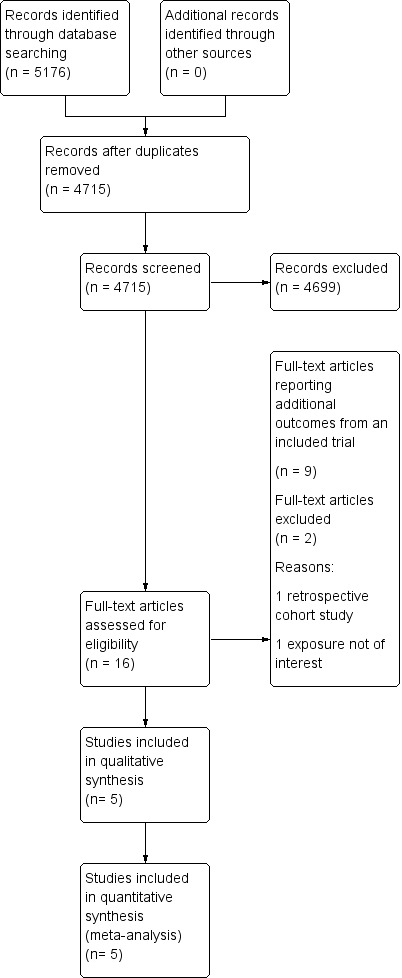
Study flow diagram
Included studies
The detailed characteristics of the included studies are shown in Characteristics of included studies and individual outcomes in Table 2. The five studies included 11,466 participants. All studies were, one conducted in the USA (ALTS 2003) and four in the UK (Flannelly 1994; Kitchener 2004; Shafi 1997; TOMBOLA 2009). Four studies explored the two management strategies in women with either ASCUS (borderline) or LSIL (mild dyskaryosis) (ALTS 2003; Kitchener 2004; Shafi 1997; TOMBOLA 2009), and one study looked at women with mild or moderate dyskaryosis (Flannelly 1994). We obtained separate data for women referred with mild or moderate dyskaryosis from the study authors (Flannelly 1994) and only included women with mild dyskaryosis in the analysis. All studies assessed non‐attendance and the rates for different CIN grades at histology for the immediate colposcopy group, as compared to the histological diagnosis at the end of the surveillance period. Two studies (Kitchener 2004; TOMBOLA 2009) assessed the psychological morbidity of the different approaches (i.e. anxiety, depression and other self‐reported outcomes). The length of the surveillance period varied between studies. Three of the included RCTs followed up women for up to 24 months (ALTS 2003; Flannelly 1994; Shafi 1997), one for 12 months (Kitchener 2004) and another for 36 months (TOMBOLA 2009). One study had four arms with different lengths of follow‐up (immediate colposcopy versus surveillance for 6, 12, or 24 months) (Flannelly 1994). Three studies provided interim histological results (ALTS 2003; Shafi 1997; TOMBOLA 2009), while two of the studies provided results only at the end of the surveillance period (Flannelly 1994; Kitchener 2004). The largest trial randomised 5060 women (ALTS 2003) and the smallest 353 (Shafi 1997).
1. Reported individual outcomes in the included studies.
| Study | Outcomes |
Immediate colposcopy n/N (%) |
Cytological surveillance n/N (%) |
RR + 95% CI |
| ALTS 2003(ASCUS) | Histology at 18 monthsa | |||
| CIN 2 | 61/1163 (5.2) | 26/1164 (2.2) | 2.35 [1.49, 3.69] | |
| CIN 2+ | 119/1163 (10.2) | 92/1164 (7.9) | 1.29 [1.00, 1.68] | |
| CIN 3+ | 58/1163 (5.0) | 66/1164 (5.7) | 0.88 [0.62, 1.24] | |
| Histology at 24 months | ||||
| CIN 2 | 61/1163 (5.2) | 60/1164 (5.2) | 1.02 [0.72,1.44] | |
| CIN 2+ | 119/1163 (10.2) | 168/1164 (14.4) | 0.71 [0.57, 0.88] | |
| CIN 3+ | 58/1163 (5.0) | 108/1164 (9.3) | 0.54 [0.39, 0.73] | |
| Default rates: | ||||
| Default rate at 24 months | 15/1163 (1.3) | 165/1164 (14.2) | 10.99 [6.52, 18.53] | |
| ALTS 2003(LSIL) | Histology at 18 monthsa | |||
| CIN 2 | 63/673 (9.4) | 36/675 (5.3) | 1.76 [1.18, 2.61] | |
| CIN 2+ | 127/673 (18.9) | 95/675 (14.1) | 1.34 [1.05, 1.71] | |
| CIN 3+ | 64/673 (9.5) | 59/675 (8.7) | 1.09 [0.78, 1.52] | |
| Histology at 24 months | ||||
| CIN 2 | 63/673 (9.4) | 58/675 (8.6) | 1.09 [0.78,1.53] | |
| CIN 2+ | 127/673 (18.9) | 151/675 (22.4) | 0.84 [0.68, 1.04] | |
| CIN 3+ | 64/673 (9.5) | 93/675 (13.8) | 0.69 [0.51, 0.93] | |
| Default rates: | ||||
| Default rate at 24 months | 4/673 (0.6) | 110/675 (16.3) | 27.42 [10.17, 73.93] | |
| Flannelly 1994 | Histology at 6 months | |||
| HPV / Koilocytic atypia | 13/145 (9.0) | 28/160 (17.5) | 0.52 [0.28, 0.95] | |
| Any CIN | 121/145 (83.4) | 86/160 (53.8) | 1.55 [1.32, 1.82] | |
| CIN 1 | 23/145 (15.9) | 27/160 (16.9) | 0.94 [0.57, 1.56] | |
| CIN 2 | 32/145 (22.1) | 26/160 (16.3) | 1.36 [0.85, 2.16] | |
| CIN 2+ | 98/145 (67.6) | 59/160 (36.9) | 1.83 [1.45, 2.31] | |
| CIN 3+ | 66/145 (45.5) | 33/160 (20.6) | 2.21 [1.55, 3.14] | |
| Histology at 12 months | ||||
| HPV / Koilocytic atypia | 13/145 (9.0) | 18/158 (11.4) | 0.79 [0.40, 1.55] | |
| Any CIN | 121/145 (83.4) | 96/158 (60.8) | 1.37 [1.19, 1.59] | |
| CIN 1 | 23/145 (15.9) | 25/158 (15.8) | 1.00 [0.60, 1.69] | |
| CIN 2 | 32/145 (22.1) | 26/158 (16.5) | 1.34 [0.84, 2.14] | |
| CIN 1 / 2 | 55/145 (37.9) | 51/158 (32.3) | 1.18 [0.86, 1.60] | |
| CIN 2+ | 98/145 (67.6) | 71/158 (44.9) | 1.50 [1.22, 1.85] | |
| CIN 3+ | 66/145 (45.5) | 45/158 (28.5) | 1.60 [1.18, 2.17] | |
| Histology at 24 months | ||||
| HPV / Koilocytic atypia | 13/145 (9.0) | 11/158 (7.0) | 1.29 [0.60, 2.78] | |
| Any CIN | 121/145 (83.4) | 53/158 (33.5) | 2.49 [1.97, 3.13] | |
| CIN 1 | 23/145 (15.9) | 9/158 (5.7) | 2.78 [1.33, 5.82] | |
| CIN 2 | 32/145 (22.1) | 12/158 (7.6) | 2.91 [1.56, 5.42] | |
| CIN 2+ | 98/145 (67.6) | 44/158 (27.8) | 2.43 [1.84, 3.20] | |
| CIN 3+ | 66/145 (45.5) | 32/158 (20.3) | 2.25 [1.57, 3.21] | |
| Default rates: | ||||
| Default rate at 6 months | 0/145 (0) | 19/160 (11.9) | 35.37 [2.15, 580.52] | |
| Default rate at 12 months | 0/145 (0) | 23/158 (14.6) | 43.16 [2.65, 704.13] | |
| Default rate at 24 months | 0/145 (0) | 38/158 (24.1) | 70.70 [4.38, 1140.47] | |
| Kitchener 2004 | Histology at 12 months | |||
| Any CIN | 83/130 (63.8) | 71/243 (29.2) | 2.19 [1.73, 2.76] | |
| CIN 1 / 2 | 61/130 (46.9) | 47/243 (19.3) | 2.43 [1.77, 3.32] | |
| CIN 3+ | 22/130 (16.9) | 24/243 (9.9) | 1.71 [1.00, 2.93] | |
| Default rates: | ||||
| Default rate at 6 months | 5/130 (3.8) | 46/243 (18.9) | 4.92 [2.01, 12.08] | |
| Default rate at 12 months | 5/130 (3.8) | 95/243 (39.1) | 10.16 [4.24, 24.35] | |
| GHQ casenessb | Choicec | No choicec | ||
| Baseline | 134/233 (58) | 119/241 (49) | 1.16 [0.98, 1.38] | |
| 6 months (pre visit) | 71/183 (39) | 77/190 (41) | 0.96 [0.75, 1.23] | |
| 6 months (post visit) | 59/175 (34) | 66/177 (37) | 0.90 [0.68, 1.20] | |
| 12 months | 40/135 (29) | 35/127 (28) | 1.08 [0.73, 1.58] | |
| Shafi 1997 | Histology at 18 monthsa | |||
| CIN 2 | 8/182(4.4) | 2/171 (1.1) | 3.76 [0.81, 17.45] | |
| CIN 2+ | 43/182 (23.6) | 16/171 (9.4) | 2.53 [1.48, 4.31] | |
| CIN 3+ | 35/182 (19.2) | 14/171(8.2) | 2.35 [1.31, 2.45] | |
| Histology at 24 months | ||||
| HPV / Koilocytic atypia | 92/182 (50.5) | 57/171 (33.3) | 1.52 [1.17, 1.96] | |
| Any CIN | 88/182 (48.4) | 51/171 (29.8) | 1.62 [1.23, 2.13] | |
| CIN 1 | 45/182 (24.7) | 17/171 (9.9) | 2.49 [1.48, 4.17] | |
| CIN 2 | 8/182 (4.4) | 10/171 (5.8) | 0.75 [0.30, 1.86] | |
| CIN 2+ | 43/182 (23.6) | 34/171 (19.9) | 1.19 [0.80, 1.77] | |
| CIN 3+ | 35/182 (19.2) | 24/171 (14.0) | 1.37 [0.85, 2.20] | |
| Default rates: | ||||
| Default rate at 24 months | 1/182 (0.5) | 36/171 (21.1) | 38.32 [5.31, 276.40] | |
| Tombola 2009 | Histology at 30 monthsa | |||
| CIN 2 | 181/2216 (8.2) | 101/2223 (4.5) | 1.80 [1.42, 2.28] | |
| CIN 2+ | 369/2216 (16.7) | 269/2223 (12.1) | 1.38 [1.19, 1.59] | |
| CIN 3+ | 188/2216 (8.5) | 168/2223 (7.6) | 1.12 [0.92, 1.37] | |
| Histology at 36 months | ||||
| CIN 2 | 181/2216 (8.2) | 157/2223 (7.1) | 1.16 [0.94, 1.42] | |
| CIN 2+ | 369/2216 (16.7) | 350/2223 (15.7) | 1.06 [0.93, 1.21] | |
| CIN 3+ | 188/2216 (8.5) | 193/2223 (8.7) | 0.98 [0.81, 1.18] | |
| Default rates: | ||||
| Default rate at 6 months | 151/2216 (6.8) | 285/2223 (12.8) | 1.88 [1.56, 2.27] | |
| Default rate at 12 months | 151/2216 (6.8) | 327/2223 (14.7) | 2.16 [1.80, 2.59] | |
| Paind | ||||
| Any pain | 304/782 (38.9) | 145/968 (15.0) | 2.60 [2.18, 3.09] | |
| Moderate or more severe | 144/774 (18.6) | 56/965 (5.8) | 3.21 [2.39, 4.30] | |
| Bleedingd | ||||
| Any bleeding | 366/781 (46.9) | 166/967 (17.2) | 2.73 [2.33, 3.19] | |
| Moderate or more severe | 144/772 (18.6) | 16/961 (1.7) | 11.20 [6.74, 18.61] | |
| Discharged | ||||
| Any discharge | 267/780 (34.2) | 83/964 (8.6) | 3.98 [3.17, 4.99] | |
| Moderate or more severe | 133/777 (17.1) | 36/962 (3.7) | 4.57 [3.20, 6.53] | |
| Anxietye | ||||
| 6 weeks | 59/751 (7.9) | 121/900 (13.4) | 0.58 [0.43, 0.79] | |
| 12 months | 190/1161 (16.4) | 218/1130 (19.3) | 0.85 [0.71, 1.01] | |
| 18 months | 162/1050 (15.4) | 177/1008 (17.6) | 0.88 [0.72, 1.07] | |
| 24 months | 179/1001 (17.9) | 177/962 (18.4) | 0.97 [0.81, 1.17] | |
| 30 months | 146/949 (15.4) | 143/887 (16.1) | 0.95 [0.77, 1.18] | |
| Depressionf | ||||
| 6 weeks | 50/757 (6.6) | 68/902 (7.5) | 0.88 [0.62, 1.25] | |
| 12 months | 110/1162 (9.5) | 132/1136 (11.6) | 0.81 [0.64, 1.04] | |
| 18 months | 106/1052 (10.1) | 114/1016 (11.2) | 0.90 [0.70, 1.15] | |
| 24 months | 111/1001 (11.1) | 104/964 (10.8) | 1.03 [0.80, 1.32] | |
| 30 months | 101/948 (10.7) | 108/887 (12.2) | 0.88 [0.68, 1.13] |
For Immediate colposcopy, n = n at immediate colposcopy visit, possible follow‐up excluded.
a Cumulative incidence during follow‐up, excluding the exit examination or deferred treatment.
b GHQ caseness = GHQ (General Health Questionnaire) score ≥ 4.
c Analysis for this outcome between the original randomization groups.
d Based on Questionnaire 6 weeks after immediate colposcopy or first cytological surveillance visit.
e ≥ 11 on hospital anxiety and depression anxiety subscale
f ≥ 8 on hospital anxiety and depression subscale
The design, management protocol and exit examination somewhat varied for different studies. Flannelly 1994 randomly allocated women into four groups: immediate colposcopy, or 6, 12 and 24 months' surveillance. All recruited women underwent LLETZ at the conclusion of their study arm that was preceded by a directed punch biopsy, if a distinct lesion was seen. Women were withdrawn if there were severe cytological abnormalities or colposcopic impression of possible microinvasion. Shafi 1997 had a similar design and treated all women immediately on the day of recruitment or at the completion of 24 months of surveillance. Women were treated during follow‐up, if the follow‐up smear showed severe dyskaryosis or worse.
Kitchener 2004 had a different design and randomised women into two principal groups: cytological surveillance for 12 months (with colposcopy and treatment when necessary at 6 or 12 months, if either follow‐up smear was abnormal) versus the choice to have surveillance or immediate colposcopy and treatment when necessary. Results on attendance rates were presented for 6 and 12 months' surveillance. In TOMBOLA 2009 (the Trial Of Management of Borderline and Other Low grade Abnormal smears) women were followed up at 6‐month intervals for 36 months in the cytological surveillance arm. The follow‐up, as well as the immediate colposcopy, were done within a routine nationwide healthcare practice. Moderate dyskaryosis or worse, or three consecutive inadequate results led to referral to colposcopy, whereas women who reverted to normal were discharged to "routine 3‐yearly recall" after three consecutive normal smears. Women allocated to immediate colposcopy were further randomised to either immediate treatment (LLETZ) or directed biopsies with selective recall for treatment, if the colposcopy was adequate and showed abnormalities (no additional procedures were carried out if the colposcopy was normal). Women from both arms were recalled for exit examination at 36 months that included colposcopy and LLETZ if the transformation zone was abnormal.
In the ALTS 2003 (ASCUS‐LSIL Triage Study) trial, all women after randomisation had a repeat liquid‐based cytology sample (LBC) for cytology and HPV testing at enrolment. Women in the cytological surveillance arm were followed up for 24 months at 6‐month intervals with pelvic examination, cervical smear, HPV‐test and cervicography taken at each visit. If the cytology suggested HSIL or worse at enrolment or during surveillance, women were referred to colposcopy with treatment if CIN2 or worse was detected on a biopsy. The immediate‐colposcopy‐arm women underwent a colposcopic examination on the same day or within 3 weeks from recruitment and received treatment if CIN2 or 3 was detected at biopsy. Women from both arms were recalled for exit colposcopy at 24 months; treatment was performed if CIN2 or 3 was detected on biopsy and for women with CIN1 with history of LSIL or HPV+ ASCUS during at least one of the previous two visits.
Excluded studies
Studies excluded during full‐text review are described in the Characteristics of excluded studies table. The reasons for exclusion were non‐randomised design in one (De Bie 2011) and in one study the exposure of interest was low‐grade histological changes (CIN1 to 2) instead of cytological changes (Elit 2011).
Risk of bias in included studies
Generally we considered the risk of selection and reporting biases across included trials to be low. The risk of performance bias and other possible sources of bias we considered high in all included trials. Risk of detection bias (TOMBOLA 2009), as well as attrition bias (ALTS 2003) were each considered low in only one trial. We have presented the quality assessment of the included RCTs with the Cochrane tool in Risk of bias in included studies and in Figure 2.
2.
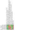
Risk of bias summary: review authors' judgements about each risk of bias item for each included study
Allocation
We considered the random sequence generation appropriate in all studies and the allocation concealment in all but one study (Flannelly 1994), which did not describe the allocation concealment process in full.
Blinding
Blinding of participants and personnel was not possible given the nature of the intervention. There was no blinding of the outcome in two studies (Kitchener 2004; Shafi 1997), while the risk of detection bias was low in TOMBOLA 2009 and unclear in the remaining studies (ALTS 2003; Flannelly 1994). Kitchener 2004 and Shafi 1997 stated that the outcome assessors were not blinded. Flannelly 1994 described no details of the blinding. ALTS 2003 reported that all clinical information were unmasked during the exit examination, but included no description of the blinding of the assessors during the follow‐up period. TOMBOLA 2009 described that the colposcopists performing the exit examination were blinded to the initial cytology results, the randomisation arm and the clinical events that occurred after randomisation, although it is unclear whether the personnel were blinded during surveillance.
Incomplete outcome data
Loss to follow‐up rates were high overall. The highest rates were seen in Kitchener 2004 and TOMBOLA 2009 (23% and 42% respectively), while the impact was judged as unclear in another two (Flannelly 1994; Shafi 1997). ALTS 2003 had the lowest rates of losses to follow‐up (14% to 17%).
Selective reporting
We considered the risk of selective reporting low in all trials, although only TOMBOLA 2009 and ALTS 2003 had pre published protocols.
Other potential sources of bias
Other potential sources of bias were considered to be high in all trials. One trial used Zelen randomisation, where participants themselves in one randomisation group (choice‐group) chose whether they preferred an immediate colposcopy or cytological surveillance, which might have created bias towards either direction in terms of later incidence of CIN2+ or CIN3+ lesions (Kitchener 2004). All other trials included either a deferred treatment (Shafi 1997), a colposcopy in addition to cytological surveillance on all follow‐up visits (Flannelly 1994) or had an exit examination with colposcopy regardless of the results of the cytological surveillance at the end of trial (ALTS 2003; TOMBOLA 2009). This could well have introduced detection bias and over inflated the CIN detection rate in the cytological surveillance arms. We further accounted for this possible source of bias by presenting and meta‐analysing the results without the exit‐examination when possible (18‐month follow‐up window).
Effects of interventions
See: Table 1
Primary outcomes
Occurrence of CIN2+
In the majority of the included studies that presented data on more than one time point (ALTS 2003; Flannelly 1994; Shafi 1997; TOMBOLA 2009), the incidence of CIN2+ was higher for the immediate colposcopy group at the first time point (before the exit examination at the time of completion), but showed no difference with time (at the time of the trial’s completion) (Table 2). Flannelly 1994 revealed differences between the intervention groups for all time points assessed.
The meta‐analysis at 18 months (without the exit examination) revealed a difference in the detection of CIN2+ between the two groups (14.3% versus 10.0%, RR 1.50, CI 1.12 to 2.01) with some heterogeneity (P = 0.08, I2 = 61%), (moderate‐quality evidence). There was no difference at 24 months (17.9% versus 18.3%, RR 1.14, 95% CI 0.66 to 1.97), but there was high inter‐study heterogeneity (P < 0.00001, I2 = 94%) and presence of other possible bias, resulting in a falsely high CIN detection rate in the cytological surveillance arm, and we considered this to be low‐quality evidence (Analysis 1.1; Figure 3; Table 1).
1.1. Analysis.
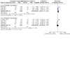
Comparison 1: Occurence of CIN2+ at different lengths of follow‐up: immediate colposcopy versus cytological surveillance, Outcome 1: Occurrence of CIN2+
3.
Forest plot of comparison: occurrence of CIN2+ at different lengths of follow‐up
Occurrence of CIN3+
All the studies assessed the detection rate of CIN3 or worse (CIN3+), although the time point assessed varied. The studies showed conflicting results, as some studies showed an increase in the CIN3+ detection with immediate colposcopy (Flannelly 1994; Kitchener 2004), one found improved detection at 18 months (without the exit examination) but not at 24 months (Shafi 1997), TOMBOLA 2009 found no difference, while ALTS 2003 found no difference at 18 months (without the exit examination), and a higher CIN3+ detection rate for cytological surveillance at 24 months (Table 2).
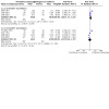
The cumulative incidence of CIN3+ was increased after immediate colposcopy when compared to cytological surveillance at 12 months (32% versus 14.0%, RR 2.07, 95% CI 1.54 to 2.79, P = 0.41, I2 = 0%), but due to other possible bias resulted in a falsely high CIN detection rate in the cytological surveillance arm, although at 18 months (without the exit examination) (7.8% versus 6.9%, (moderate‐quality evidence), 95% CI 0.77 to 1.98) and 24 months (10.3% versus 11.9%, RR 1.02, CI 0.53 to 1.97), this difference was no longer observed. This was mainly driven by the opposing results of a large trial (ALTS 2003). There was high inter‐study heterogeneity for the comparison at 18 months (P < 0.02, I2 = 75%) (moderate‐quality evidence), and at 24 months (P < 0.00001, I2 = 93%). In addition to high heterogeneity the presence of other possible bias probably resulting in a falsely high CIN detection rate in the cytological surveillance arm, meant that we regarded this as low‐quality evidence (Analysis 2.1; Figure 4; Table 1).
2.1. Analysis.
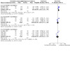
Comparison 2: Occurence of CIN3+ at different lengths of follow‐up: immediate colposcopy versus cytological surveillance, Outcome 1: Occurrence of CIN3+
4.
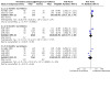
Forest plot of comparison: occurence of CIN3+ at different lengths of follow‐up
Secondary outcomes
Incidence of other CIN grades at histology
Occurrence of histologically proven HPV or koilocytic atypia
One study assessed this at 6, 12 and 24 months (Flannelly 1994) and another at 24 months of surveillance (Shafi 1997). Flannelly 1994 reported a lower rate of HPV‐associated histological changes for immediate colposcopy as compared to six‐month cytological surveillance (9% versus 17%, RR 0.52, 95% CI 0.28 to 0.95) but no difference was observed at 12 or 24 months. Interestingly, Shafi 1997 identified a higher rate for women attending for immediate colposcopy, as opposed to repeat cytology (50.5% versus 33.3%, RR 1.52, 95% CI 1.17 to 1.96) (Table 2).
The meta‐analysis revealed that immediate referral to colposcopy increased the detection of clinically non‐significant infections as opposed to repeat cytology at 24 months (32% versus 21%, RR 1.49, 95% CI 1.17 to 1.90) (Analysis 4.1). There was no evident inter‐study heterogeneity (P = 0.69, I2 = 0%), but due to other possible bias resulting in a falsely high CIN detection rate in the cytological surveillance arm, we regarded this as moderate‐quality evidence.
4.1. Analysis.
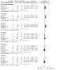
Comparison 4: Histology at 24 months: immediate colposcopy versus cytological surveillance, Outcome 1: Histology at 24 months
Occurrence of any CIN
Three studies provided data on the rates of any CIN grade (Flannelly 1994; Kitchener 2004; Shafi 1997). Flannelly 1994 provided data for cytological surveillance at 6, 12 and 24 months, Kitchener 2004 at 12 months and Shafi 1997 at 24 months. All individual studies demonstrated that women undergoing immediate colposcopy had a higher rate of detected CIN of any grade (Table 2).
The meta‐analysis confirmed that the cumulative incidence of any grade of CIN was higher among women referred to immediate colposcopy (12 months: RR 1.72, 95% CI 1.09 to 2.70; 24 months: RR 2.02, 95% CI 1.33 to 3.08). Considerable heterogeneity was present at both time points (P = 0.001, I2 = 91% and P = 0.02, I2 = 82%, respectively) and due to other possible bias resulting in falsely high CIN detection rates in the cytological surveillance arm, we regarded this as low‐quality evidence (Analysis 3.1; Analysis 4.1; Table 1).
3.1. Analysis.

Comparison 3: Histology at 12 months: immediate colposcopy versus cytological surveillance, Outcome 1: Presence of any CIN in histology at 12 months
Occurrence of CIN1
One study that assessed the rate of CIN1 at six and 12 months suggested no difference between immediate colposcopy and cytological surveillance (Flannelly 1994) (Table 2). Two studies assessed the occurrence of CIN1 rate at 24 months (Flannelly 1994; Shafi 1997), both of which suggested a higher rate of detection of low‐grade disease in the immediate colposcopy group (Table 2).
The meta‐analysis revealed at least double the rate of CIN1 detection when women underwent colposcopy immediately as opposed to repeat cytology (21% versus 8%, RR 2.58, 95% CI 1.69 to 3.94), suggesting that immediate colposcopy may have increased the detection of clinically insignificant lesions (Analysis 4.1). There was no evident inter‐study heterogeneity (P = 0.81, I2 = 0%), but due to other possible bias resulting in a falsely high CIN detection rate in the cytological surveillance arm we regarded this as moderate‐quality evidence.
Occurrence of CIN2
Four studies assessed the incidence of CIN2 (ALTS 2003; Flannelly 1994; Shafi 1997; TOMBOLA 2009); one reported results at 6 and 12 months (Flannelly 1994), two (ALTS 2003; Shafi 1997) at 18 months (without the exit examination), three (ALTS 2003; Flannelly 1994; Shafi 1997) at 24 months and one (TOMBOLA 2009) at 30 and 36 months. The rate of CIN2 was higher in the immediate colposcopy group in the majority of the studies, apart from one (Shafi 1997). The difference in CIN2 rate detection was higher at 18 months rather than at 24 months in ALTS 2003 and similarly at 30 months as opposed to 36 months in TOMBOLA 2009 (Table 2).
The overall rate of CIN2 in the meta‐analysis was higher for the immediate colposcopy group at 18 months (6.5% versus 3.2%, RR 2.04, CI 1.52 to 2.73) with low heterogeneity (P = 0.46, I2= 0%); high‐quality evidence (Analysis 5.1; Figure 5). There was no evident difference at 24 months of surveillance (7.6% versus 5.4%, RR 1.45, 95% CI 0.87 to 2.40); there was considerable inter‐study heterogeneity (P = 0.009, I2= 74%), and additionally, due to the presence of other possible bias resulting in a falsely high CIN detection rate in the cytological surveillance arm, this was regarded as low‐quality evidence.
5.1. Analysis.
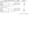
Comparison 5: Occurence of CIN2 at different lengths of follow‐up: Immediate colposcopy versus cytological surveillance, Outcome 1: Occurrence of CIN2
5.
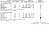
Forest plot of comparison: occurence of CIN2 at different lengths of follow‐up
In total there were 18 cases of invasive cervical cancer, seven in immediate colposcopy and 11 in cytological surveillance groups: one case of stage 1A1 cervical cancer in the deferred treatment group detected by cytological smear in Shafi 1997, three cases of invasive cervical cancer in both immediate colposcopy and cytological surveillance groups in ALTS 2003 and four cases of invasive disease in immediate colposcopy and seven in the cytological surveillance group in TOMBOLA 2009. There were no cases of invasive cancer in the two other included trials (Flannelly 1994; Kitchener 2004).
Default rates
All five included trials reported on the non‐compliance rates for immediate colposcopy versus cytological surveillance (Table 2). Two reported non‐attendance rates at six and 12 months (Kitchener 2004; TOMBOLA 2009), two at 24 months (ALTS 2003; Shafi 1997) and one for all the time points (6, 12 and 24 months) (Flannelly 1994). The risk of non‐compliance was significantly greater for the repeat cytology arm and increased with the length of the follow‐up. TOMBOLA 2009, the largest study and one with high methodological quality, reported an almost double risk of default for women followed up in the community when compared to those referred to immediate colposcopy (6 months: RR 1.88, 95% CI 1.56 to 2.27; 12 months: RR 2.16, 95% CI 1.80 to 2.59) (Table 2). The magnitude of effect was much greater in the remaining trials. In ALTS 2003 the RR for cumulative risk of default at 24 months was up to 27.42 (95% CI 10.17 to 73.93) for the surveillance arm at the exit exam, as opposed to the immediate colposcopy group (Table 2). The study setting and protocols varied greatly between the included trials, which could well explain the observed differences in default rates between studies.
The meta‐analysis suggested that the risk for non‐compliance was higher for the repeat cytology group with a four‐fold increase at 6 months, a six‐fold increase at 12 months and a 19‐fold increase at 24 months (6 months: RR 3.85, 95% CI 1.27 to 11.63; 12 months: RR 6.60, 95% CI 1.49 to 29.29; 24 months: RR 19.1 95% CI 9.02 to 40.43). The inter‐study heterogeneity was considerable for the analysis at six and 12 months (P = 0.02, I2 = 76% and P < 0.0004, I2 = 87%, respectively), and we therefore regarded this as moderate‐quality evidence, whereas we considered the evidence to be of high quality at 24 months (Analysis 6.1; Figure 6; Table 1).
6.1. Analysis.
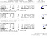
Comparison 6: Default rates: immediate colposcopy versus cytological surveillance, Outcome 1: Default rates
6.
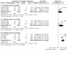
Forest plot of comparison: default rates at different lengths of follow‐up
Presence of after‐effects
TOMBOLA 2009 was the only study that reported on the incidence of after‐effects based on a questionnaire completed six weeks after immediate colposcopy or the first cytological follow‐up smear. Pain, bleeding, or vaginal discharge were all more common after immediate colposcopy than after cytology (Table 2).
Presence of anxiety, distress or depression
Both TOMBOLA 2009 and Kitchener 2004 reported incidence of psychosocial morbidity in the randomisation groups (Table 2). In TOMBOLA 2009, the authors reported the incidence of anxiety and depression at six weeks, and thereafter at six‐month intervals until 30 months of surveillance. Anxiety, measured as score of 11 or more on the Hospital Anxiety and Depression anxiety sub scale, was less common in the immediate colposcopy group compared to the cytological surveillance group at six weeks (RR 0.58, 95% CI 0.43 to 0.79). There was no difference between the randomisation groups thereafter. Incidence of depression, defined as a score of 8 or more on the Hospital Anxiety and Depression Scale (HADS) (Zigmond 1983), did not differ between the two groups throughout the follow‐up period. Kitchener 2004 used the General Health Questionnaire (GHQ)‐caseness (Bridges 1986) (defined as a score of 4 or above) to measure the presence of anxiety or distress. There were no differences between choice and no‐choice arms at baseline or after six and 12 months of surveillance. Given the differences in the comparison groups and the assessment methods, a meta‐analysis was not possible.
Subgroup analyses
The optimum management option may be different for women found to have ASCUS (borderline) or LSIL cytology (mild dyskaryosis) in the baseline smear. We were unable to perform a subgroup analysis for women with ASCUS cytology as only one study (ALTS 2003) presented this separately. We could extract separate data for women referred with LSIL cytology only from two studies (ALTS 2003; Flannelly 1994). These studies reported on the incidence of CIN2, CIN2+ and CIN3+ or worse at 24 months.
The incidence of the above histological outcomes was higher for immediate colposcopy as opposed to cytology, although based on small sample size (CIN2: 8.4% versus 11.6%, RR 1.72, 95% CI 0.66 to 4.48; CIN2+: 23.4% versus 27.5%, RR 1.43, 95% CI 0.51 to 4.01: CIN3+: 15% versus 15.9%, RR 1.24, 95% CI 0.39 to 3.94); all analyses demonstrated inter‐study heterogeneity (P < 0.01, I2 > 75%) and presented moderate‐quality evidence (Analysis 7.1).
7.1. Analysis.
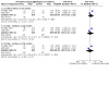
Comparison 7: Histology at 24 months: immediate colposcopy versus cytological surveillance, LSIL/mild dyskaryosis only, Outcome 1: CIN incidence at 24 months, after LSIL/mild dyskaryosis at baseline
ALTS 2003 revealed opposing direction of effect to the other included studies in some of the outcomes assessed. We performed a subgroup analysis that excluded this trial after 24 months of surveillance and found little or no difference in the rate of CIN2 and CIN2+ between the immediate colposcopy and cytological surveillance groups: CIN2 (RR 1.54, 95% CI 0.41 to 5.78, P = 0.02, I2= 83%) and for CIN2+ (RR 1.72, 95% CI 0.86 to 3.47, P = 0.004, I2 = 88%; moderate‐quality evidence). Immediate colposcopy increased the detection of CIN3+ as opposed to cytological surveillance (30.9% versus 17%, RR 1.80, 95% CI 1.11 to 2.92, heterogeneity: P = 0.10, I2= 62%; high‐quality evidence) (Analysis 8.1; Analysis 8.2; Analysis 8.3).
8.1. Analysis.
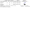
Comparison 8: Histology at 24 months: immediate colposcopy versus cytological surveillance, excluding 1 trial, Outcome 1: Presence of CIN2 in histology at 24 months
8.2. Analysis.
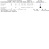
Comparison 8: Histology at 24 months: immediate colposcopy versus cytological surveillance, excluding 1 trial, Outcome 2: Presence of CIN2+ in histology at 24 months
8.3. Analysis.

Comparison 8: Histology at 24 months: immediate colposcopy versus cytological surveillance, excluding 1 trial, Outcome 3: Presence of CIN3+ in histology at 24 months
Discussion
Summary of main results
The results of this meta‐analysis suggest that the difference in the CIN2 or CIN2+ rate detection was higher for the immediate colposcopy group at 18 months (without the exit examination), but this was no longer the case at 24 months. Although this suggests that colposcopy may allow earlier diagnosis of CIN2 lesions that cytology would require longer to detect, it may also be explained by the trials’ design. All participants in the cytological surveillance arms underwent exit examination, a colposcopy with or without treatment, irrespective of their cytological status in all trials. This ‘artificial’ detection of cases of high‐grade disease at completion would plausibly be lower in a ‘real‐life’ setting in the cytological surveillance arm; this may over inflate the reliability of repeat cytology to detect high‐grade disease.
Although the evidence suggests that there may be little or no difference in the rate of CIN3+ detection at 24 months between the two arms, this result was mainly driven by a large RCT (ALTS 2003) with almost opposing results to the remaining RCTs (Flannelly 1994; Kitchener 2004; Shafi 1997; TOMBOLA 2009). The ALTS study had a different design in that all women randomised and analysed in the cytological surveillance arm had a repeat smear that was assessed by an expert cytopathologist at recruitment. Almost 40% of CIN3+ lesions in the cytology arm were detected during this repeat cytology smear and this may have inflated the ability of cytology to detect disease when compared to immediate colposcopy (ALTS 2003). After exclusion of this trial, the difference of CIN3+ detection favoured immediate colposcopy.
Women attending for immediate colposcopy after a single LSIL or ASCUS smear were 50% more likely to have clinically insignificant HPV infections detected. The rates of histologically detected lesions associated with HPV infection were lower for the repeat cytology group, possibly explained by spontaneous regression of clinically insignificant lesions during cytological surveillance. Similarly, there was an increase in the detection of CIN1 lesions, supporting the perception that immediate colposcopy may increase the risk of over‐intervention and over‐treatment, by detection of insignificant lesions that would otherwise spontaneously revert to normal over time.
The evidence suggests that the risk for default to follow‐up was probably higher for the repeat cytology group at 6, 12 and 24 months. The difference between the two groups changed from four‐fold at 6 months and six‐fold at 12 months up to 19‐fold at 24 months. The point estimate at 24 months could well be biased and over‐inflated due to selection of studies at that follow‐up window, that is, lack of data from one study (TOMBOLA 2009) and inclusion of another with a very different trial setting (ALTS 2003). Compliance with follow‐up still appeared to decline with time and women may be at increased risk of invasion due to underlying occult high‐grade disease. The inter‐study heterogeneity was significant, possibly due to differences in the magnitude of the default rates in the immediate colposcopy group between different trials. These differences may be explained by differences in the design and randomisation process that could increase (TOMBOLA 2009: randomisation and admittance to either colposcopy or cytological surveillance after HPV test results) or decrease (ALTS 2003: immediate colposcopy preferably on the same day) the risk of non‐compliance in the immediate colposcopy arm. Only one study reported on the after‐effects ‐ pain, bleeding or vaginal discharge ‐ and found that these were significantly more common after immediate colposcopy (TOMBOLA 2009). Meta‐analysis of the after‐effects and anxiety and depression was not possible.
Overall completeness and applicability of evidence
This is the first meta‐analysis that comprehensively investigates the value of immediate colposcopy versus cytological surveillance in women with lesions of equivocal significance in the absence of HPV test. A previous systematic review only included three small RCTs (Kyrgiou 2007a). Two large trials, one from the US (ALTS 2003) and the other from the UK (TOMBOLA 2009) addressed this comparison comprehensively with large sample sizes. Because all included studies were conducted in high‐income setting, the applicability of the evidence to lower income settings is not clear. This systematic review still provides a comprehensive review of the existing literature and meta‐analytical pooling that will allow clinicians to evaluate the trade‐off between the risk of detection of insignificant lesions versus the risk of poor compliance and non‐detection of occult high‐grade disease.
Quality of the evidence
All of the included studies were RCTs, however, our analysis has several limitations and the results should be interpreted with caution. There are only a small number of included studies, two of which included a comparatively large number of participants (ALTS 2003; TOMBOLA 2009), which may account for some of the heterogeneity that was observed and which was considerable or substantial in many of the comparisons. It was this high heterogeneity, as well as the presence of bias from study design possibly resulting in falsely high CIN detection rate in the cytological surveillance arm in the 24‐month follow‐up window, that accounted for the down‐grading of the evidence to 'moderate' or 'low' in several cases following the GRADE assessment. Several of the included studies varied in their design and comparison groups, with the presence of exit examination and the length of follow‐up. Kitchener 2004, for example, had a choice and non‐choice arm, ALTS 2003 performed a repeat cytology at recruitment in the cytology surveillance arm, while one of the older studies (Shafi 1997) performed a treatment for all participants at completion. For many of the explored outcomes and comparisons, there was substantial heterogeneity, possibly explained by the difference in the studies’ design.
The results presented in the individual trials are those derived from women who completed the given period of surveillance as the presence or absence of disease cannot be verified in those women who defaulted from surveillance. Furthermore, all the included studies, apart from Kitchener 2004, performed exit examinations at the time of the study completion for both arms, some of which included an excision of the transformation zone for all subjects (Shafi 1997). The comparison of the rates of different grades of disease at the time of the study completion may represent an ‘artificial’ detection of lesions that would not have been detected in the cytology arm in a ‘real‐life’ setting, with the risk to inflate the ability of repeat cytology to detect disease. We therefore considered the quality of evidence in the 24‐month follow‐up window to be low.
Most of the included studies did not assess the impact of age of the population in the preferred management approach. Only TOMBOLA 2009 assessed the effect of age on histological outcomes and concluded that the difference in cumulative incidence of CIN2+ between the trial arms was more pronounced amongst younger women (20 to 39 years), as opposed to older women (40 to 59 years). Furthermore, in younger women (age 20 to 39 years) the risk ratios for CIN2 or worse were markedly higher than CIN3 or worse.
Potential biases in the review process
All of the included studies were RCTs. It was not possible to blind participants and personnel given the nature of the intervention. The random sequence generation was considered appropriate in all studies and the allocation concealment in all but one study (Flannelly 1994), where the allocation concealment process was not described in full. Only one study (TOMBOLA 2009) described blinding of assessors; the colposcopist was blinded at exit examination only with regards to the women's cytology status, randomisation category and any clinical outcomes. The lack of assessor blinding in the other included studies (ALTS 2003; Flannelly 1994; Kitchener 2004; Shafi 1997) may have introduced a degree of detection bias. Loss to follow‐up rates were high overall ranging from 14% to 42%, which may have introduced a degree of attrition bias. The risk of selective reporting was considered low in all trials.
In order to minimise bias whilst undertaking the review, two review authors (MK and IK) independently reviewed the retrieved citations and the extracted data. There was no discrepancy in the included studies; some minor discrepancies in the data extraction were resolved with discussion and the involvement of a third reviewer (MA) if necessary.
Agreements and disagreements with other studies or reviews
Almost one in ten women screened in the UK will have a minor lesion detected in her smear. Several authorities recommend conservative surveillance in the primary care setting, as spontaneous regression of most of these lesions will occur (Kyrgiou 2007a). Referral to colposcopy may increase psychological morbidity for these women who experience similar levels of anxiety to those referred with high‐grade disease (TOMBOLA 2009). This increase in anxiety appears to be transient and is no different after six weeks. Furthermore, further investigation of lesions that were deemed to regress without intervention increases the risk of over‐intervention and over‐treatment with subsequent possible adverse reproductive sequale (Arbyn 2008; Founta 2010; Kyrgiou 2006;Kyrgiou 2012a; Kyrgiou 2014;Kyrgiou 2015; Kyrgiou 2016a). The opponents supporting immediate assessment with colposcopy emphasise that protracted attendance for repeat cytology decreases with time (Soutter 2012) and this was also confirmed in this analysis. The evidence suggests that approximately one third of women referred with borderline or low‐grade cytological abnormalities have an occult high‐grade disease and non‐compliance with surveillance may put some women at risk of developing invasive disease (Bolger 1988; Contreras‐Melendez 1992; Giles 1989; Paraskevaidis 2002; Soutter 1986; Walker 1986). The participants lost to follow‐up due to poor compliance in these trials could result in an underestimation of the true number of high‐grade lesions that were missed in the cytology group.
The financial implications of the two approaches to health services should also be considered. Cost‐effectiveness analysis in some of the earlier studies suggested that referral to colposcopy is a more cost‐effective alternative to repeat cytology (Flannelly 1997; Johnson 1993). The cost‐effectiveness analysis of the TOMBOLA trial suggested no difference amongst the two approaches although cytological surveillance was less costly (TOMBOLA 2009). The ALTS trial suggested that HPV‐triage or repeat cytology of a single repeat smear was cost‐effective, although there was no difference in cost‐effectiveness if two or more smears were performed (ALTS 2003). The cost‐effectiveness of the two approaches largely depends on the policy of subsequent surveillance in the case of negative colposcopy; this was not addressed in any of the included studies.
Strong evidence supports the preferential use of HPV test particularly in the triage of women with ASCUS (borderline) cytology (Arbyn 2004; Arbyn 2012; Arbyn 2013a) that would benefit from colposcopy as a more accurate and cost‐effective modality. Its value is less pronounced in younger women below the age of 30 and women with LSIL (mild dyskaryosis) (Arbyn 2009; Arbyn 2013a). However, this test is not always readily available, particularly in low‐resource settings. In Europe alone, one in ten women with ASCUS cytology and four in ten women with LSIL will not have a triage HPV test and clear recommendations are required to manage such cases. Newer markers such as mRNA or p16/Ki67 immunostaining have more recently shown promising results claiming an equal sensitivity to that of the DNA test but with improved specificity in this setting (Arbyn 2013b; Nasioutziki 2011; Tsoumpou 2009; Tsoumpou 2011). Further combinations of novel biomarkers and Clinical Decision Support Scoring Systems (DSSS) based on mathematical modelling that will create user‐friendly tools have the potential to further improve the management of this population (Bountris 2014; Karakitsos 2011; Karakitsos 2012; Kyrgiou 2016b).
Authors' conclusions
Implications for practice.
In the absence of HPV testing, women could be referred for immediate colposcopy after a single low‐grade or borderline cervical smear, if compliance with cytological surveillance is expected to be poor. When follow‐up compliance is expected to be good, repeated cytology may be preferred. This latter approach may reduce the risk of over‐diagnosis and over‐treatment (diagnosis and treatment of clinically insignificant lesions). Clinicians should be cautious and intervene only in women that are found to have high‐grade disease at colposcopy, in order to minimise the risk of over‐treatment. There is a clear need for a reflex triage method that can make distinction between women needing diagnostic work‐up and those who can be released safely to routine screening.
Implications for research.
Immediate colposcopy may not increase the detection of high‐grade disease, compared to continued cytological surveillance over two years. Compliance, however, declines over time and is probably increased for women under cytological surveillance. In the absence of HPV test, a general policy could be immediate colposcopy after a single low‐grade or borderline cervical smear when poor compliance is anticipated.
What's new
| Date | Event | Description |
|---|---|---|
| 2 July 2020 | Review declared as stable | No studies identified for inclusion after a scoping search up to 30 Jan 2020. Review marked as ‘No longer for update’ as the intervention has been superseded by HPV testing. See Cytology versus HPV testing for cervical cancer screening in the general population |
History
Protocol first published: Issue 5, 2012 Review first published: Issue 1, 2017
| Date | Event | Description |
|---|---|---|
| 30 January 2017 | Amended | Minor corrections made to the summary of findings table. |
Acknowledgements
The authors wish to acknowledge Jo Morrison for her clinical and editorial advice, Jane Hayes for designing the search strategy and Gail Quinn, Clare Jess and Tracey Bishop for their contribution to the editorial process.
This project was supported by the National Institute for Health Research, via Cochrane Infrastructure funding to the Cochrane Gynaecological, Neuro‐oncology and Orphan Cancer Group. The views and opinions expressed therein are those of the authors and do not necessarily reflect those of the Systematic Reviews Programme, NIHR, NHS or the Department of Health.
Appendices
Appendix 1. Abbreviations
ASCUS: atypical squamous cells of undetermined significance CI: confidence Interval CIN: cervical intra‐epithelial lesion CKC: cold knife conization HSIL: high‐grade squamous intra‐epithelial lesion LC: laser conization LEEP: loop electrosurgical excisional procedure LLETZ: large loop excision of the transformation zone LSIL: low‐grade squamous intra‐epithelial lesion NETZ: needle excision of the transformation zone RCT: randomised controlled trial RR: risk ratio SWETZ: straight wire excision of the transformation zone
Appendix 2. CENTRAL search strategy
#1 MeSH descriptor Uterine Cervical Dysplasia, this term only #2 MeSH descriptor Cervical Intraepithelial Neoplasiaexplode all trees #3 cervi* near/5 (dysplasia or neoplasia)
#4 CIN* #5 dyskaryosis or dyskaryosis #6 LGSIL or LSIL or ASCUS #7 (#1 OR #2 OR #3 OR #4 OR #5) #8 MeSH descriptor Precancerous Conditions, this term only #9 MeSH descriptor Early Detection of Cancer, this term only #10 MeSH descriptor Carcinoma, Squamous Cell, this term only #11 MeSH descriptor Neoplasms, Squamous Cell, this term only #12 squamous near/5 lesion* #13 precancer* or pre‐cancer* or premalignan* or pre‐malignan* or preneoplas* or pre‐neoplas* #14 (#8 OR #9 OR #10 OR #11 OR ( #12 AND #13 )) #15 MeSH descriptor Cervix Uteri, this term only #16 cervi* #17 (#15 OR #16) #18 (#14 AND #17) #19 MeSH descriptor Vaginal Smears, this term only #20 (cervi* or vagina*) and (smear* or screen* or test*) #21 (#19 OR #20) #22 (#7 OR #18 OR #21) #23 MeSH descriptor Colposcopy, this term only #24 colposcop* #25 LLETZ or LEEP or NETZ or SWETZ or LC or CKC #26 MeSH descriptor Biopsy explode all trees #27 biops* #28 transformation zone #29 MeSH descriptor Conization explode all trees #30 conization or conization #31 excis* #32 (#23 OR #24 OR #25 OR #26 OR #27 OR #28 OR #29 OR #30 OR #31) #33 (#22 AND #32)
Appendix 3. MEDLINE search strategy
MEDLINE Ovid
1 Uterine Cervical Dysplasia/ 2 Cervical Intraepithelial Neoplasia/ 3 (cervi* adj5 (dysplasia or neoplasia)).mp. 4 CIN*.mp. 5 (dyskaryosis or dyskariosis).mp. 6 (LGSIL or LSIL or ASCUS).mp. 7 1 or 2 or 3 or 4 or 5 or 6 8 Precancerous Conditions/ 9 Early Detection of Cancer/ 10 Carcinoma, Squamous Cell/ 11 Neoplasms, Squamous Cell/ 12 (squamous adj5 lesion*).mp. 13 (precancer* or pre‐cancer* or premalignan* or pre‐malignan* or preneoplas* or pre‐neoplas*).mp. 14 8 or 9 or 10 or 11 or 12 or 13 15 Cervix Uteri/ or cervi*.mp. 16 14 and 15 17 Vaginal Smear/ or ((cervi* or vagina*) and (smear* or screen* or test*)).mp. 18 7 or 16 or 17 19 Colposcopy/ 20 colposcop*.mp. 21 (LLETZ or LEEP or NETZ or SWETZ or LC or CKC).mp. 22 exp Biopsy/ 23 biops*.mp. 24 transformation zone.mp. 25 Conization/ 26 (conization or conization).mp. 27 excis*.mp. 28 19 or 20 or 21 or 22 or 23 or 24 or 25 or 26 or 27 29 18 and 28 30 randomized controlled trial.pt. 31 controlled clinical trial.pt. 32 randomized.ab. 33 placebo.ab. 34 clinical trials as topic.sh. 35 randomly.ab. 36 trial.ti. 37 30 or 31 or 32 or 33 or 34 or 35 or 36 38 29 and 37 39 exp animals/ not humans.sh. 40 38 not 39
key: mp=protocol supplementary concept, rare disease supplementary concept, title, original title, abstract, name of substance word, subject heading word, unique identifier pt=publication type ab=abstract ti=title sh=subject heading
Appendix 4. Embase search strategy
1 uterine cervix dysplasia/ 2 uterine cervix carcinoma in situ/ 3 (cervi* adj5 (dysplasia or neoplasia)).mp. 4 CIN*.mp. 5 (dyskaryosis or dyskariosis).mp. 6 (LGSIL or LSIL or ASCUS).mp. 7 1 or 2 or 3 or 4 or 5 or 6 8 precancer/ 9 early diagnosis/ 10 squamous cell carcinoma/ 11 (squamous adj5 lesion*).mp. 12 (precancer* or pre‐cancer* or premalignan* or pre‐malignan* or preneoplas* or pre‐neoplas*).mp. 13 8 or 9 or 10 or 11 or 12 14 exp uterine cervix/ or cervi*.mp. 15 13 and 14 16 vagina smear/ or ((cervi* or vagina*) and (smear* or screen* or test*)).mp. 17 7 or 15 or 16 18 colposcopy/ 19 colposcop*.mp. 20 (LLETZ or LEEP or NETZ or SWETZ or LC or CKC).mp. 21 exp biopsy/ 22 biops*.mp. 23 transformation zone.mp. 24 uterine cervix conization/ 25 (conization or conization).mp. 26 excis*.mp. 27 18 or 19 or 20 or 21 or 22 or 23 or 24 or 25 or 26 28 17 and 27 29 crossover procedure/ 30 double‐blind procedure/ 31 randomized controlled trial/ 32 single‐blind procedure/ 33 random*.mp. 34 factorial*.mp. 35 (crossover* or cross over* or cross‐over*).mp. 36 placebo*.mp. 37 (double* adj blind*).mp. 38 (singl* adj blind*).mp. 39 assign*.mp. 40 allocat*.mp. 41 volunteer*.mp. 42 29 or 30 or 31 or 32 or 33 or 34 or 35 or 36 or 37 or 38 or 39 or 40 or 41 43 28 and 42 44 (exp Animal/ or Nonhuman/ or exp Animal Experiment/) not Human/ 45 43 not 44
key: mp=title, abstract, subject headings, heading word, drug trade name, original title, device manufacturer, drug manufacturer, device trade name, keyword
Data and analyses
Comparison 1. Occurence of CIN2+ at different lengths of follow‐up: immediate colposcopy versus cytological surveillance.
| Outcome or subgroup title | No. of studies | No. of participants | Statistical method | Effect size |
|---|---|---|---|---|
| 1.1 Occurrence of CIN2+ | 3 | Risk Ratio (IV, Random, 95% CI) | Subtotals only | |
| 1.1.1 18 months' surveillance | 2 | 4028 | Risk Ratio (IV, Random, 95% CI) | 1.50 [1.12, 2.01] |
| 1.1.2 24 months' surveillance | 3 | 4331 | Risk Ratio (IV, Random, 95% CI) | 1.14 [0.66, 1.97] |
Comparison 2. Occurence of CIN3+ at different lengths of follow‐up: immediate colposcopy versus cytological surveillance.
| Outcome or subgroup title | No. of studies | No. of participants | Statistical method | Effect size |
|---|---|---|---|---|
| 2.1 Occurrence of CIN3+ | 4 | Risk Ratio (IV, Random, 95% CI) | Subtotals only | |
| 2.1.1 12 months' surveillance | 2 | 676 | Risk Ratio (IV, Random, 95% CI) | 2.07 [1.54, 2.79] |
| 2.1.2 18 months' surveillance | 2 | 4028 | Risk Ratio (IV, Random, 95% CI) | 1.24 [0.77, 1.98] |
| 2.1.3 24 months' surveillance | 3 | 4331 | Risk Ratio (IV, Random, 95% CI) | 1.02 [0.53, 1.97] |
Comparison 3. Histology at 12 months: immediate colposcopy versus cytological surveillance.
| Outcome or subgroup title | No. of studies | No. of participants | Statistical method | Effect size |
|---|---|---|---|---|
| 3.1 Presence of any CIN in histology at 12 months | 2 | 676 | Risk Ratio (IV, Random, 95% CI) | 1.72 [1.09, 2.70] |
| 3.2 Presence of CIN1/2 in histology at 12 months | 2 | 676 | Risk Ratio (IV, Random, 95% CI) | 1.69 [0.83, 3.43] |
| 3.3 Presence of CIN3+ in histology at 12 months | 2 | 676 | Risk Ratio (IV, Random, 95% CI) | 1.63 [1.25, 2.12] |
3.2. Analysis.

Comparison 3: Histology at 12 months: immediate colposcopy versus cytological surveillance, Outcome 2: Presence of CIN1/2 in histology at 12 months
3.3. Analysis.
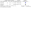
Comparison 3: Histology at 12 months: immediate colposcopy versus cytological surveillance, Outcome 3: Presence of CIN3+ in histology at 12 months
Comparison 4. Histology at 24 months: immediate colposcopy versus cytological surveillance.
| Outcome or subgroup title | No. of studies | No. of participants | Statistical method | Effect size |
|---|---|---|---|---|
| 4.1 Histology at 24 months | 3 | Risk Ratio (IV, Random, 95% CI) | Subtotals only | |
| 4.1.1 HPV/Koilocytic atypia | 2 | 656 | Risk Ratio (IV, Random, 95% CI) | 1.49 [1.17, 1.90] |
| 4.1.2 Any CIN | 2 | 656 | Risk Ratio (IV, Random, 95% CI) | 2.02 [1.33, 3.08] |
| 4.1.3 CIN1 | 2 | 656 | Risk Ratio (IV, Random, 95% CI) | 2.58 [1.69, 3.94] |
| 4.1.4 CIN2 | 3 | 4331 | Risk Ratio (IV, Random, 95% CI) | 1.25 [0.80, 1.96] |
| 4.1.5 CIN2+ | 3 | 4331 | Risk Ratio (IV, Random, 95% CI) | 1.14 [0.66, 1.97] |
| 4.1.6 CIN3+ | 3 | 4331 | Risk Ratio (IV, Random, 95% CI) | 1.02 [0.53, 1.97] |
Comparison 5. Occurence of CIN2 at different lengths of follow‐up: Immediate colposcopy versus cytological surveillance.
| Outcome or subgroup title | No. of studies | No. of participants | Statistical method | Effect size |
|---|---|---|---|---|
| 5.1 Occurrence of CIN2 | 3 | Risk Ratio (IV, Random, 95% CI) | Subtotals only | |
| 5.1.1 18 months' surveillance | 2 | 4028 | Risk Ratio (IV, Random, 95% CI) | 2.04 [1.52, 2.73] |
| 5.1.2 24 months' surveillance | 3 | 4331 | Risk Ratio (IV, Random, 95% CI) | 1.45 [0.87, 2.40] |
Comparison 6. Default rates: immediate colposcopy versus cytological surveillance.
| Outcome or subgroup title | No. of studies | No. of participants | Statistical method | Effect size |
|---|---|---|---|---|
| 6.1 Default rates | 5 | Risk Ratio (IV, Random, 95% CI) | Subtotals only | |
| 6.1.1 Default rates at 6 months | 3 | 5117 | Risk Ratio (IV, Random, 95% CI) | 3.85 [1.27, 11.63] |
| 6.1.2 Default rates at 12 months | 3 | 5115 | Risk Ratio (IV, Random, 95% CI) | 6.60 [1.49, 29.29] |
| 6.1.3 Default rates at 24 months | 3 | 4331 | Risk Ratio (IV, Random, 95% CI) | 19.10 [9.02, 40.43] |
Comparison 7. Histology at 24 months: immediate colposcopy versus cytological surveillance, LSIL/mild dyskaryosis only.
| Outcome or subgroup title | No. of studies | No. of participants | Statistical method | Effect size |
|---|---|---|---|---|
| 7.1 CIN incidence at 24 months, after LSIL/mild dyskaryosis at baseline | 2 | Risk Ratio (IV, Random, 95% CI) | Subtotals only | |
| 7.1.1 CIN2 incidence at 24 months | 2 | 1651 | Risk Ratio (IV, Random, 95% CI) | 1.72 [0.66, 4.48] |
| 7.1.2 CIN2+ incidence at 24 months | 2 | 1651 | Risk Ratio (IV, Random, 95% CI) | 1.43 [0.51, 4.01] |
| 7.1.3 CIN3+ incidence at 24 months | 2 | 1651 | Risk Ratio (IV, Random, 95% CI) | 1.24 [0.39, 3.94] |
Comparison 8. Histology at 24 months: immediate colposcopy versus cytological surveillance, excluding 1 trial.
| Outcome or subgroup title | No. of studies | No. of participants | Statistical method | Effect size |
|---|---|---|---|---|
| 8.1 Presence of CIN2 in histology at 24 months | 2 | 656 | Risk Ratio (IV, Random, 95% CI) | 1.54 [0.41, 5.78] |
| 8.2 Presence of CIN2+ in histology at 24 months | 2 | 656 | Risk Ratio (IV, Random, 95% CI) | 1.72 [0.86, 3.47] |
| 8.3 Presence of CIN3+ in histology at 24 months | 2 | 656 | Risk Ratio (IV, Random, 95% CI) | 1.80 [1.11, 2.92] |
Characteristics of studies
Characteristics of included studies [ordered by study ID]
ALTS 2003.
| Study characteristics | ||
| Methods | 3‐arm RCT | |
| Participants | 5060 women (3488 women, ASCUS smear and 1572 women, LSIL smear) Study arms:
Inclusion criteria:
Exclusion criteria:
|
|
| Interventions |
All
Colposcopy indications: 1. Immediate colposcopy at the date of recruitment or within 3 weeks. Treatment if CIN2 or 3 in biopsy. 2. Referral to colposcopy if the HPV test positive or missing or if enrolment cytology HSIL. Treatment if CIN2 or 3 in biopsy 3. Referral to colposcopy if cytology HSIL. Treatment if CIN2 or 3 in biopsy LSIL only
|
|
| Outcomes |
|
|
| Notes | ||
| Risk of bias | ||
| Bias | Authors' judgement | Support for judgement |
| Random sequence generation (selection bias) | Low risk | Telephone‐based central randomisation service used. Sequence generation not described in detail, but not estimated to induce bias |
| Allocation concealment (selection bias) | Low risk | Allocation done from central point |
| Blinding of participants and personnel (performance bias) All outcomes | High risk | Blinding of participants was not possible and personnel were not blinded of allocation. |
| Blinding of outcome assessment (detection bias) All outcomes | Unclear risk | Blinding of investigators undertaking follow‐up assessments was not described in detail. "all available clinical information was unmasked and provided to the clinician conducting the exit pelvic examination and colposcopy." |
| Incomplete outcome data (attrition bias) All outcomes | Low risk | Losses to follow‐up were disclosed (ASCUS: 14.2% at 24 months in the surveillance group, LSIL: 16.3% at 24 months in the surveillance group) and the analyses were conducted as specified a priori. Loss to follow‐up might have introduced only small bias to histology results. |
| Selective reporting (reporting bias) | Low risk | Pre‐published trial protocol available, outcome reporting done according to protocol |
| Other bias | High risk | Source of funding reported; competing interests reported. Ethical approval & informed consent from participants obtained. Repeat smear at randomisation visit and exit colposcopy at 24 months, both could potentially introduce detection bias and over inflate the detection rate of pre‐invasive lesions in the cytological surveillance arm at 24 months. |
Flannelly 1994.
| Study characteristics | ||
| Methods | 4‐arm RCT | |
| Participants | 902 women, presenting with mildly or moderately dyskaryotic smear for the first time. Study arms:
Inclusion criteria:
Exclusion criteria:
|
|
| Interventions | Group 1. Immediate colposcopy with biopsies +/‐ treatment (n = 227) Group 2. 6‐month surveillance (n = 225):
Group 3. 12‐month surveillance (n = 223):
Group 4. 24‐month surveillance (n = 227):
|
|
| Outcomes |
|
|
| Notes | ||
| Risk of bias | ||
| Bias | Authors' judgement | Support for judgement |
| Random sequence generation (selection bias) | Low risk | "...all eligible women randomised by serial allocation to one of four groups." |
| Allocation concealment (selection bias) | Unclear risk | Method of concealment not described in sufficient detail |
| Blinding of participants and personnel (performance bias) All outcomes | High risk | Blinding of participants was not possible and blinding of personnel was not reported. |
| Blinding of outcome assessment (detection bias) All outcomes | Unclear risk | Blinding of outcome assessment not reported |
| Incomplete outcome data (attrition bias) All outcomes | Unclear risk | 109/675 women in the surveillance arms defaulted from study, rates being higher with longer follow‐up (9.8% at 6 months, 15.2% at 12 months, and 23.3% at 24 months). More specific reasons not available. Significant default rates might bias histology results |
| Selective reporting (reporting bias) | Low risk | Protocol not available. Results still reported comprehensively and reporting considered not to introduce bias |
| Other bias | High risk | Conflict of interest not reported; source of funding not reported; details of ethical approval not stated. Missing information still considered not to introduce bias All women in the surveillance arm had colposcopy in addition to smear, which could introduce bias and over inflate the detection rates in the surveillance arm at 6, 12 and 24 months of surveillance |
Kitchener 2004.
| Study characteristics | ||
| Methods | 2‐arm RCT with Zelen randomisation | |
| Participants | 712 women pre‐randomised, with mild dyskaryosis for the first time or recurrent borderline change on routine cervical screening in primary care. Aged 20 to 60, not pregnant, no abnormal vaginal bleeding Of 712 pre‐randomised women, 476 decided to participate in the study. Study arms:
Inclusion criteria:
Exclusion criteria:
|
|
| Interventions | Group 1. No‐choice arm (n = 243)
Group 2. Choice arm (n = 233) Women were given the opportunity to choose between a) and b):
|
|
| Outcomes |
|
|
| Notes | ||
| Risk of bias | ||
| Bias | Authors' judgement | Support for judgement |
| Random sequence generation (selection bias) | Low risk | Randomisation into two study arms prior to consent using computer‐generated random numbers |
| Allocation concealment (selection bias) | Low risk | Central allocation used. In one of the study arms the participants themselves chose the intervention group. |
| Blinding of participants and personnel (performance bias) All outcomes | High risk | Participants and investigators were not blinded. |
| Blinding of outcome assessment (detection bias) All outcomes | High risk | Outcome assessment was not blinded. |
| Incomplete outcome data (attrition bias) All outcomes | High risk | 712 pre‐randomised, 476 participated. Reasons for non‐participation reported, no clear differences between randomisation groups 172/476 were lost to follow‐up, no specific reasons stated. Adequate sample size might not have been achieved for all outcomes. |
| Selective reporting (reporting bias) | Low risk | Pre‐trial protocol was not available. Results still reported comprehensively |
| Other bias | High risk | Project funding reported; conflicts of interest not stated. Local ethical approval and informed consent from participants obtained. Missing information still considered not to introduce bias. Used Zelen randomisation, where some participants were able to choose whether to have immediate colposcopy or cytological surveillance, which might well bias the results included here. |
Shafi 1997.
| Study characteristics | ||
| Methods | 2‐arm RCT | |
| Participants | 353 women with borderline or mild dyskaryotic smear Study arms:
Inclusion criteria:
Exclusion criteria:
|
|
| Interventions | Group 1. Immediate treatment (n = 182)
Group 2. Deferred treatment (n = 171)
|
|
| Outcomes |
|
|
| Notes | ||
| Risk of bias | ||
| Bias | Authors' judgement | Support for judgement |
| Random sequence generation (selection bias) | Low risk | Randomised using a sequence of random numbers generated by computer. |
| Allocation concealment (selection bias) | Low risk | Total allocation concealment as randomisation performed individually from a central point. |
| Blinding of participants and personnel (performance bias) All outcomes | High risk | Blinding participants was not possible and personnel were not blinded to allocation. |
| Blinding of outcome assessment (detection bias) All outcomes | High risk | Investigators undertaking follow‐up were not blinded. |
| Incomplete outcome data (attrition bias) All outcomes | Unclear risk | 36/171 lost to follow‐up in deferred treatment group, 1/181 in the immediate colposcopy group. More specific reasons not reported. The 20% lost to follow‐up in other arm might introduce bias to histology results |
| Selective reporting (reporting bias) | Low risk | Pre‐trial protocol was not available. Results still reported comprehensively |
| Other bias | High risk | Individual sources of funding reported, conflicts of interests not declared. Local ethical approval and informed consent from participants obtained. Missing information still considered not to introduce bias. All women in the surveillance arm had deferred treatment at 24 months regardless of cytology, which could introduce bias and over inflate the CIN detection rate in the surveillance‐arm at 24 months. |
TOMBOLA 2009.
| Study characteristics | ||
| Methods | 2‐arm RCT | |
| Participants | 4439 women, aged 20‐59, with cytological result showing: Study arms:
Inclusion criteria:
Exclusion criteria
|
|
| Interventions | Group 1. Surveillance arm (n = 2223)
Group 2. Immediate colposcopy arm (n = 2216), second randomisation at colposcopy to a) or b) a) Biopsy and selective recall for LLETZ based on histology on biopsy
b) Immediate treatment (LLETZ)
|
|
| Outcomes |
|
|
| Notes | ||
| Risk of bias | ||
| Bias | Authors' judgement | Support for judgement |
| Random sequence generation (selection bias) | Low risk | Touch‐telephone stratified randomisation used |
| Allocation concealment (selection bias) | Low risk | Allocation done from central point |
| Blinding of participants and personnel (performance bias) All outcomes | High risk | Participants not possible to blind. Blinding of personnel not described |
| Blinding of outcome assessment (detection bias) All outcomes | Low risk | At exit examination the colposcopist was blinded to the women's initial cytology status, her randomisation and any clinical outcomes. Blinding of colposcopists or pathologists at other stages was not reported. |
| Incomplete outcome data (attrition bias) All outcomes | High risk | Before the first examination 107/2223 in the surveillance arm, 155/2216 in the colposcopy arm were lost to follow‐up. 1296 (58.3%) women in the surveillance arm and 1389 (62.7%) in the colposcopy arm attended the exit examination. Power calculations were based on 4500 participants. The significant loss to follow‐up and differences between arms might introduce bias to histology results. |
| Selective reporting (reporting bias) | Low risk | Pre‐published trial protocol available, outcome reporting according to protocol. |
| Other bias | High risk | Source of funding reported; Competing interests reported. Ethical approval & informed consent from participants obtained. All women were invited to an exit examination at 36 months, where colposcopy was performed regardless of smear results. This could introduce bias and over inflate the CIN detection rates in surveillance arm at 36 months. |
Characteristics of excluded studies [ordered by study ID]
| Study | Reason for exclusion |
|---|---|
| De Bie 2011 | Not RCT (retrospective cohort study) |
| Elit 2011 | Exposure not of interest (natural history of CIN 1) |
Differences between protocol and review
The title has changed from 'Management of low‐grade squamous intra‐epithelial lesions of the uterine cervix repeat cytology versus immediate referral to colposcopy' to 'Immediate referral to colposcopy versus cytological surveillance for minor cervical cytological abnormalities in the absence of HPV test' in order to better describe the applicability of the evidence only in the absence of HPV test.
None of outcomes included used continuous outcome measures and the methods described in the protocol to be applied on continuous outcomes were not needed and included in the review. If in a future update continuous outcomes are identified the following methodology will be used. For continuous outcomes (e.g. anxiety, depression scores), we will extract the final value and standard deviation (SD) of the outcome of interest and the number of patients assessed at endpoint in each treatment arm at the end of follow‐up, in order to estimate the mean difference (MD) (if trials measured outcomes on the same scale) or standardised mean differences (SMDs) (if trials measured outcomes on different scales) between treatment arms and its standard error (SE).
Surveillance or immediate colposcopy after ASCUS or borderline dyskaryosis was also included. These lesions,which constitute the majority of women with low‐grade smear, have hence been included in most trials and represent a major proportion of the women upon whom these results are applicable.
We decided to accept cytological surveillance in any setting, not only in primary care as eligible. Most of the included studies used other than primary care setting for follow‐up and we considered it appropriate to include them.
Subgroup analyses were not performed based on continent, study type, study quality and inclusion and exclusion criteria. We were able to include only a few studies in each meta‐analysis and were hence not able to conduct all planned subgroup analyses.
We used GRADE to assess the quality of evidence instead of just removing unpublished and low‐quality studies from sensitivity analyses due to GRADE being introduced only after the publication of the protocol.
Contributions of authors
The study was conceived and designed by MK, MA and EP. The data was acquired and collated by MK, IK, AM, PMH, MA and analysed by MK, IK, AM, PMH, MA and EP. The manuscript was drafted and revised critically for important intellectual content by all authors (MK, IK, AM, CF, SGM, PMH, MC, MA, EP). All authors gave final approval of the version to be published and have contributed to the manuscript. MC was not involved in application of the inclusion and exclusion criteria, in data extraction or in data analysis.
Sources of support
Internal sources
-
Cochrane Gynaecological, Neuro‐oncology and Orphan Cancers, UK
Marc Arbyn received financial support from Cochrane Gynaecological, Neuro‐oncology and Orphan Cancers
External sources
-
European Commission, Other
Marc Arbyn received financial support from the European Commission through the COHEAHR Network, coordinated by the Free University of Amsterdam (The Netherlands), funded by the 7th Framework programme of DG Research (Brussels, Belgium), and through the ECCG (European Co‐operation on Development and Implementation of Cancer Screening and Prevention Guidelines, via IARC, Lyon, France), funded by the Directorate of SANCO (Luxembourg, Grand‐Duchy of Luxembourg)
-
Belgian Foundation Against Cancer, Belgium
Marc Arbyn received financial support from the Belgian Foundation Against Cancer, Brussels, Belgium
-
British Society of Colposcopy Cervical Pathology Jordan/Singer Award, UK
Maria Kyrgiou received financial support from British Society of Colposcopy Cervical Pathology (Grant reference: P47773)
-
Imperial College Healthcare Charity, UK
Anita Mitra and Maria Kyrgiou received financial support from Imperial College Healthcare Charity (Grant reference: P47907)
-
Imperial Healthcare NHS Trust NIHR Biomedical Research Centre, UK
Maria Kyrgiou received financial support from Imperial Healthcare NHS Trust NIHR Biomedical Research Centre (Grant reference: P45272)
-
Genesis Research Trust, UK
Maria Kyrgiou received financial support from Genesis Research Trust (Grant reference: P55549)
-
Sigrid Jusélius Foundation, Finland
Ilkka Kalliala received postdoctoral fellowship under Dr Kyrgiou's group at Imperial College London from Sigrid Jusélius Foundation (Grant reference: P52483)
Declarations of interest
Maria Kyrgiou: None known Ilkka Kalliala: None known Anita Mitra: None known Christina Fotopolou: None known Sadaf Ghaem‐Maghami: None known Pierre Martin‐HIrcsh: None known Margaret Cruickshank: author of one of the included studies (TOMBOLA 2009): MC was not involved in application of the inclusion and exclusion criteria, in data extraction or in data analysis. Marc Arbyn: None known Evangelos Paraskevaidis: None known
Joint first author
Joint first author
Joint senior author
Joint senior author
Stable (no update expected for reasons given in 'What's new')
References
References to studies included in this review
ALTS 2003 {published data only}
- Kulasingam SL, Kim JJ, Lawrence WF, Mandelblatt JS, Myers ER, Schiffman M, et al. Cost-effectiveness analysis based on the atypical squamous cells of undetermined significance/low-grade squamous intraepithelial lesion Triage Study (ALTS). Journal of the National Cancer Institute 2006;98:92-100. [DOI] [PubMed] [Google Scholar]
- The ASCUS-LSIL Triage Study (ALTS) Group. A randomized trial on the management of low-grade squamous intraepithelial lesion cytology interpretations. American Journal of Obstetrics and Gynecology 2003;188(6):1393-400. [DOI] [PubMed] [Google Scholar]
- The ASCUS-LSIL Triage Study (ALTS) Group. Results of a randomized trial on the management of cytology interpretations of atypical squamous cells of undetermined significance. American Journal of Obstetrics and Gynecology 2003;188(6):1383-92. [DOI] [PubMed] [Google Scholar]
Flannelly 1994 {published data only}
- Flannelly G, Anderson D, Kitchener HC, Mann EM, Campbell M, Fisher P, et al. Management of women with mild and moderate cervical dyskaryosis. BMJ 1994;308(6941):1399-403. [DOI] [PMC free article] [PubMed] [Google Scholar]
Kitchener 2004 {published data only}
- Kitchener HC, Burns S, Nelson L, Myers AJ, Fletcher I, Desai M, et al. A randomised controlled trial of cytological surveillance versus patient choice between surveillance and colposcopy in managing mildly abnormal cervical smears. British Journal of Obstetrics and Gynaecology 2004;11(1):63-70. [DOI] [PubMed] [Google Scholar]
Shafi 1997 {published data only}
- Shafi MI, Luesley DM, Jordan JA, Dunn JA, Rollason TP, Yates M. Randomised trial of immediate versus deferred treatment strategies for the management of minor cervical cytological abnormalities. British Journal of Obstetrics and Gynaecology 1997;104(5):590-4. [DOI] [PubMed] [Google Scholar]
TOMBOLA 2009 {published data only}
- Cotton S, Sharp L, Little J, Cruickshank M, Seth R, Smart L, et al, Trial Of Management of Borderline and Other Low/grade Abnormal Smears Group. The role of human papillomavirus testing in the management of women with low/grade abnormalities: multicentre randomised controlled trial. BJOG: An International Journal of Obstetrics and Gynaecology 2010;117(6):645-59. [DOI] [PubMed] [Google Scholar]
- Cotton SC, Sharp L, Little J, Duncan I, Alexander L, Cruickshank ME, et al for TOMBOLA group. Trial of management of borderline and other low-grade abnormal smears (TOMBOLA): trial design. Contemporary Clinical Trials 2006;27(5):449-71. [DOI] [PubMed] [Google Scholar]
- Gurumurthy M, Cotton SC, Sharp L, Smart L, Little J, Waugh N, et al. Postcolposcopy management of women with histologically proven CIN 1: results from TOMBOLA. Journal of Lower Genital Tract Disease 2014;18(3):203-9. [DOI] [PubMed] [Google Scholar]
- Sharp L, Cotton S, Carsin AE, Gray N, Thornton A, Cruickshank M, et al for TOMBOLA Group. Factors associated with psychological distress following colposcopy among women with low-grade abnormal cervical cytology: a prospective study within the Trial Of Management of Borderline and Other Low-grade Abnormal smears (TOMBOLA). Psycho-oncology 2013;22(2):368-80. [DOI] [PubMed] [Google Scholar]
- Sharp L, Cotton S, Cruickshank M, Gray NM, Harrild K, Smart L, et al. The unintended consequences of cervical screening: distress in women undergoing cytologic surveillance. Journal of Lower Genital Tract Disease 2014;18(2):142-50. [DOI] [PubMed] [Google Scholar]
- Sharp L, Cotton S, Little J, Gray NM, Cruickshank M, Smart L, et al. Psychosocial impact of alternative management policies for low-grade cervical abnormalities: results from the tombola randomised controlled trial. PloS one 2013;8(12):e80092. [DOI] [PMC free article] [PubMed] [Google Scholar]
- Sharp L, Cotton S, Thornton A, Gray N, Cruickshank M, Whynes D, et al. Who defaults from colposcopy? A multi-centre, population-based, prospective cohort study of predictors of non-attendance for follow-up among women with low-grade abnormal cervical cytology. European Journal of Obstetrics, Gynecology, and Reproductive Biology 2012;165(2):318-25. [DOI] [PubMed] [Google Scholar]
- TOMBOLA Group. Biopsy and selective recall compared with immediate large loop excision in management of women with low grade abnormal cervical cytology referred for colposcopy: multicentre randomised controlled trial. BMJ 2009;339:b2548. [DOI] [PMC free article] [PubMed] [Google Scholar]
- TOMBOLA Group. Cytological surveillance compared with immediate referral for colposcopy in management of women with low grade cervical abnormalities: multicentre randomised controlled trial. BMJ 2009;339:2546. [DOI] [PMC free article] [PubMed] [Google Scholar]
- TOMBOLA Group. Options for managing low grade cervical abnormalities detected at screening: cost effectiveness study. BMJ 2009;339:b2549. [DOI] [PMC free article] [PubMed] [Google Scholar]
- Whynes DK, Woolley C, Philips Z, for TOMBOLA Group. Management of low-grade cervical abnormalities detected at screening: which method do women prefer? Cytopathology 2008;19(6):355-62. [DOI] [PubMed] [Google Scholar]
References to studies excluded from this review
De Bie 2011 {published data only}
- De Bie RP, Massuger LFAG, Dongen RAJM, Snijders MPML, Bulten J, Melchers WJG, et al. To treat or not to treat; the clinical dilemma of atypical squamous cells of undetermined significance (ASC-US). Acta Obstetrics and Gynecology Scandinavica 2011;90(4):313-8. [DOI] [PubMed] [Google Scholar]
Elit 2011 {published data only}
- Elit L, Levine MN, Julian JA, Sellors JW, Lytwyn A, Chong S, et al. Expectant management versus immediate treatment for low-grade cervical intraepithelial neoplasia: a randomized trial in Canada and Brazil. Cancer 2011;117(7):1438-45. [DOI] [PubMed] [Google Scholar]
Additional references
Arbyn 2004
- Arbyn M, Buntinx F, Van Ranst M, Paraskevaidis E, Martin-HirschJ, Dillner J. Virologic versus cytologic triage of women with equivocal Pap smears: a meta-analysis of the accuracy to detect high-grade intraepithelial neoplasia. Journal of the National Cancer Institute 2004;96(4):280-93. [DOI] [PubMed] [Google Scholar]
Arbyn 2005
- Arbyn M, Paraskevaidis E, Martin-Hirsch P, Prendiville W, Dillner J. Clinical utility of HPV-DNA detection: triage of minor cervical lesions, follow-up of women treated for high grade CIN: an update of pooled evidence. Gynecologic Oncology 2005;99(3 Suppl 1):S7-S11. [DOI] [PubMed] [Google Scholar]
Arbyn 2007
- Arbyn M, Primic-Zakelj M, Raifu AO, Grce M, Paraskevaidis E, Diakomanolis E, et al. The burden of cervical cancer in south-east Europe at the beginning of the 21st century. Collegium Antropologicum 2007;31(Suppl 2):7-10. [PubMed] [Google Scholar]
Arbyn 2008
- Arbyn M, Kyrgiou M, Simoens C, Raifu AO, Koliopoulos G, Martin-Hirsch P, et al. Perinatal mortality and other severe adverse pregnancy outcomes associated with treatment of cervical intraepithelial neoplasia: meta-analysis. BMJ 2008;337:a1284. [DOI] [PMC free article] [PubMed] [Google Scholar]
Arbyn 2009
- Arbyn M, Martin-Hirsch P, Buntinx F, Van Ranst M, Paraskevaidis E, Dillner J. Triage of women with equivocal or low-grade cervical cytology results: a meta-analysis of the HPV test positivity rate. Journal of Cellular and Molecular Medicine 2009;13:648-59. [DOI] [PMC free article] [PubMed] [Google Scholar]
Arbyn 2011
- Arbyn M, Castellsague X, De Sanjose S, Bruni L, Saraiya M, Bray F, et al. Worldwide burden of cervical cancer in 2008. Annals of Oncology 2011;22(12):2675-86. [DOI] [PubMed] [Google Scholar]
Arbyn 2012
- Arbyn M, Ronco G, Anttila A, Meijer CJ, Poljak M, Ogilvie G, et al. Evidence regarding human papillomavirus testing in secondary prevention of cervical cancer. Vaccine 2012;30 Suppl 5:F88-99. [DOI] [PubMed] [Google Scholar]
Arbyn 2013a
- Arbyn M, Roelens J, Simoens C, Buntinx F, Paraskevaidis E, Martin-Hirsch PPL, et al. Human papillomavirus testing versus repeat cytology for triage of minor cytological cervical lesions. Cochrane Database of Systematic Reviews 2013, Issue 3. [DOI: 10.1002/14651858.CD008054.pub2] [DOI] [PMC free article] [PubMed] [Google Scholar]
Arbyn 2013b
- Arbyn M, Roelens J, Cuschieri K, Cuzick J, Szarewski A, Ratnam S, et al. The APTIMA HPV assay versus the Hybrid Capture 2 test in triage of women with ASC-US or LSIL cervical cytology: a meta-analysis of the diagnostic accuracy. International Journal of Cancer 2013;132:101-8. [DOI] [PubMed] [Google Scholar]
Arbyn 2015
- Arbyn M, Snijders PJ, Meijer CJ, Berkhof J, Cuschieri K, Kocjan BJ, et al. Which high-risk HPV assays fulfil criteria for use in primary cervical cancer screening? Clinical Microbiology and Infection 2015;21:817-26. [DOI] [PubMed] [Google Scholar]
Bolger 1988
- Bolger BS, Lewis BV. A prospective study of colposcopy in women with mild dyskaryosis or koilocytosis. British Journal of Obstetrics and Gynaecology 1988;95(11):1117-9. [DOI] [PubMed] [Google Scholar]
Bountris 2014
- Bountris P, Haritou M, Pouliakis A, Margari N, Kyrgiou M, Spathis A, et al. An intelligent clinical decision support system for patient-specific predictions to improve cervical intraepithelial neoplasia detection. BioMed Research International 2014;2014:341483. [DOI] [PMC free article] [PubMed] [Google Scholar]
Bridges 1986
- Bridges KW, Goldberg DP. The validation of the GHQ-28 and the use of the MMSE in neurological in-patients. British Journal of Psychiatry 1986;148:548-53. [DOI] [PubMed] [Google Scholar]
Bulkmans 2007
- Bulkmans NW, Berkhof J, Rozendaal L, Van Kemenade FJ, Boeke AJ, Bulk S, et al. Human papillomavirus DNA testing for the detection of cervical intraepithelial neoplasia grade 3 and cancer: 5-year follow-up of a randomised controlled implementation trial. Lancet 2007;370(9601):1764-72. [DOI] [PubMed] [Google Scholar]
Cochran 1954
- Cochran WG. The combination of estimates from different experiments. Biometrics 1954;10:101-29. [Google Scholar]
Contreras‐Melendez 1992
- Contreras-Melendez L, Herbert A, Millward-Sadler GH, Moore IE, Masson GM, Camillieri AP, et al. Assessment of the accuracy of cytology in women referred for colposcopy and biopsy: the results of a 1 year audit. Cytopathology 1992;3(5):267-74. [DOI] [PubMed] [Google Scholar]
Cuzick 2008
- Cuzick J, Arbyn M, Sankaranarayanan R, Tsu V, Ronco G, Mayrand M H, et al. Overview of human papillomavirus-based and other novel options for cervical cancer screening in developed and developing countries. Vaccine 2008;26(Suppl 10):K29–41. [DOI] [PubMed] [Google Scholar]
Deeks 2001
- Deeks JJ, Altman DG, Bradburn MJ. Statistical methods for examining heterogeneity and combining results from several studies in meta-analysis. In: Egger M, Davey Smith G, Altman DG, editors(s). Systematic Reviews in Health Care: Meta-Analysis in Context. 2nd edition. London: BMJ Publication Group, 2001. [Google Scholar]
Deeks 2011
- Deeks JJ, Higgins JPT, Altman DG (editors). Chapter 9: Analysing data and undertaking meta-analyses. In: Higgins JPT, Green S (editors). Cochrane Handbook for Systematic Reviews of Interventions Version 5.1.0 (updated March 2011). The Cochrane Collaboration, 2011. Available from handbook.cochrane.org.
Department of Health 2006
- Department of Health. Cervical Screening Programme, England: 2005–6. www.cancerscreening.nhs.uk/cervical/cervical-statistics-bulletin-2005-06.pdf (accessed 20 March 2012).
Dersimonian 1986
- Dersimonian R, Laird NM. Meta-analysis in clinical trials. Controlled Clinical Trials 1986;7:177–88. [DOI] [PubMed] [Google Scholar]
EUROCARE 2003
- Micheli A, Coebergh JW, Mugno E, Massimiliani E, Sant M, Oberaigner W, et al, EUROCARE. European health systems and cancer care. Annals of Oncology 2003;14(Suppl 5):v41-60. [DOI] [PubMed] [Google Scholar]
Flannelly 1997
- Flannelly G, Campbell MK, Meldrum P, Torgerson DJ, Templeton A, Kitchener HC. Immediate colposcopy or cytological surveillance for women with mild dyskaryosis: a cost effectiveness analysis. Journal of Public Health Medicine 1997;19(4):419-23. [DOI] [PubMed] [Google Scholar]
Founta 2010
- Founta C, Arbyn M, Valasoulis G, Kyrgiou M, Tsili A, Martin-Hirsch P, et al. Proportion of excision and cervical healing after large loop excision of the transformation zone for cervical intraepithelial neoplasia. BJOG: An International Journal of Obstetrics and Gynaecology 2010;117:1468-74. [DOI] [PubMed] [Google Scholar]
Giles 1989
- Giles JA, Deery A, Crow J, Walker P. The accuracy of repeat cytology in women with mildly dyskaryotic smears. British Journal of Obstetrics and Gynaecology 1989;96(9):1067-70. [DOI] [PubMed] [Google Scholar]
GLOBOCAN 2013
- Ferlay J, Soerjomataram I, Ervik M, Dikshit R, Eser S, Mathers C et al, International Agency for Research on Cancer. GLOBOCAN 2012 v1.0, Cancer Incidence and Mortality Worldwide: IARC CancerBase No. 11 [Internet]. Lyon, France: International Agency for Research on Cancer; 2013. globocan.iarc.fr accessed on 04/01/2017.
Higgins 2003
- Higgins JP, Thompson SG, Deeks JJ, Altman DG. Measuring inconsistency in meta-analyses. BMJ 2003;327(7414):557–60. [DOI] [PMC free article] [PubMed] [Google Scholar]
Higgins 2011
- Higgins JPT, Altman DG, Sterne JAC (editors). Chapter 8: Assessing risk of bias in included studies. In: Higgins JPT, Green S (editors). Cochrane Handbook for Systematic Reviews of Interventions Version 5.1.0 (updated March 2011). The Cochrane Collaboration, 2011. Available from handbook.cochrane.org.
Johnson 1993
- Johnson N, Sutton J, Thornton JG, Lilford RJ, Johnson VA, Peel KR. Decision analysis for best management of mildly dyskaryotic smear. Lancet 1993;342:91-6. [DOI] [PubMed] [Google Scholar]
Karakitsos 2011
- Karakitsos P, Pouliakis A, Meristoudis C, Margari N, Kassanos D, Kyrgiou M, et al. A preliminary study of the potential of tree classifiers in triage of high-grade squamous intraepithelial lesions. Analytical and quantitative cytology and histology/The International Academy of Cytology [and] American Society of Cytology 2011;33:132-40. [PubMed] [Google Scholar]
Karakitsos 2012
- Karakitsos P, Chrelias C, Pouliakis A, Koliopoulos G, Spathis A, Kyrgiou M, et al. Identification of women for referral to colposcopy by neural networks: a preliminary study based on LBC and molecular biomarkers. Journal of Biomedicine & Biotechnology 2012;2012:303192. [DOI] [PMC free article] [PubMed] [Google Scholar]
Kelly 2011
- Kelly RS, Patnick J, Kitchener HC, Moss SM, NHSCSP HPV Special Interest Group. HPV testing as a triage for borderline or mild dyskaryosis on cervical cytology: results from the Sentinel Sites study. British Journal of Cancer 2011;105(7):983-8. [DOI] [PMC free article] [PubMed] [Google Scholar]
Koliopoulos 2007
- Koliopoulos G, Arbyn M, Martin-Hirsch P, Kyrgiou M, Prendiville W, Paraskevaidis E. Diagnostic accuracy of human papillomavirus testing in primary cervical screening: a systematic review and meta-analysis of non-randomized studies. Gynecologic Oncology 2007;104(1):232-46. [DOI] [PubMed] [Google Scholar]
Kyrgiou 2006
- Kyrgiou M, Koliopoulos G, Martin-Hirsch P, Arbyn M, Prendiville E, Paraskevaidis E. Obstetric outcomes after conservative treatment for intra-epithelial or early invasive cervical lesions: a systematic review and meta-analysis of the literature. Lancet 2006;367(9509):489-98. [DOI] [PubMed] [Google Scholar]
Kyrgiou 2007a
- Kyrgiou M, Koliopoulos G, Martin-Hirsch P, Kehoe S, Flannelly G, Mitrou S, et al. Management of minor cervical cytological abnormalities: a systematic review and a meta-analysis of the literature. Cancer Treatment Reviews 2007;33:514-20. [DOI] [PubMed] [Google Scholar]
Kyrgiou 2010
- Kyrgiou M, Valasoulis G, Founta C, Koliopoulos G, Karakitsos P, Nasioutziki M, et al. Clinical management of HPV-related disease of the lower genital tract. Annals of the New York Academy of Sciences 2010;1205:57-68. [DOI] [PubMed] [Google Scholar]
Kyrgiou 2012a
- Kyrgiou M, Arbyn M, Martin-Hirsch P, Paraskevaidis E. Increased risk of preterm birth after treatment for CIN. BMJ 2012;345:e5847. [DOI] [PubMed] [Google Scholar]
Kyrgiou 2014
- Kyrgiou M, Mitra A, Arbyn M, Stasinou SM, Martin-Hirsch P, Bennett P, et al. Fertility and early pregnancy outcomes after treatment for cervical intraepithelial neoplasia: systematic review and meta-analysis. BMJ 2014;349:g6192. [DOI] [PMC free article] [PubMed] [Google Scholar]
Kyrgiou 2015
- Kyrgiou M, Valasoulis G, Stasinou SM, Founta C, Athanasiou A, Bennett P, et al. Proportion of cervical excision for cervical intraepithelial neoplasia as a predictor of pregnancy outcomes. International Journal of Gynaecology and Obstetrics: The Official Organ of the International Federation of Gynaecology and Obstetrics 2015;128:141-7. [DOI] [PubMed] [Google Scholar]
Kyrgiou 2016a
- Kyrgiou M, Athanasiou A, Paraskevaidi M, Mitra A, Kalliala I, Martin-Hirsch P, et al. Adverse obstetric outcomes after local treatment for cervical preinvasive and early invasive disease according to cone depth: systematic review and meta-analysis. BMJ 2016;354:i3633. [DOI] [PMC free article] [PubMed] [Google Scholar]
Kyrgiou 2016b
- Kyrgiou M, Pouliakis A, Panayiotides JG, Margari N, Bountris P, Valasoulis G, et al. Personalised management of women with cervical abnormalities using a clinical decision support scoring system. Gynecological Oncology 2016;141:29–35. [DOI] [PubMed] [Google Scholar]
Langendam 2013
- Langendam MW, Akl EA, Dahm P, Glasziou P, Guyatt G, Schunemann HJ. Assessing and presenting summaries of evidence in Cochrane Reviews. Systematic Reviews 2013;23(2):81. [DOI] [PMC free article] [PubMed] [Google Scholar]
Meader 2014
- Meader N, King K, Llewellyn A, Norman G, Brown J, Rodgers M, et al. A checklist designed to aid consistency and reproducibility of GRADE assessments: development and pilot validation. Systematic Reviews 2014;3:82. [DOI] [PMC free article] [PubMed] [Google Scholar]
Moher 2009
- Moher D, Liberati A, Tetzlaff J, Altman DG, The PRISMA Group. Preferred reporting items for systematic reviews and meta-analyses: the PRISMA Statement. Open medicine: a peer-reviewed, independent, open-access journal 2009;3:e123-30. [PMC free article] [PubMed] [Google Scholar]
Nasioutziki 2011
- Nasioutziki M, Daniilidis A, Dinas K, Kyrgiou M, Valasoulis G, Loufopoulos PD, et al. The evaluation of p16INK4a immunoexpression/immunostaining and human papillomavirus DNA test in cervical liquid-based cytological samples. International Journal of Gynecological Cancer 2011;21:79-85. [DOI] [PubMed] [Google Scholar]
Naucler 2007
- Naucler P, Ryd W, Törnberg S, Strand A, Wadell G, Elfgren K, et al. Human papillomavirus and Papanicolaou tests to screen for cervical cancer. New England Journal of Medicine 2007;357(16):1589-97. [DOI] [PubMed] [Google Scholar]
NHSCSP 2000
- National Health Service Cervical Screening Programme. Achievable Standards, Benchmarks for Reporting, and Criteria for Evaluating Cervical Cytopathology. In: NHSCSP publication. 2nd edition. Vol. 1. Sheffield: NHS Cancer Screening Programmes, 2000:1-36. [Google Scholar]
Papanicolaou 1941
- Papanicolaou GN, Traut HF. The diagnostic value of vaginal smears in carcinoma of the uterus. American Journal of Obstetrics and Gynecology 1941;42:193-205. [Google Scholar]
Paraskevaidis 2002
- Paraskevaidis E, Kaponis A, Malamou-Mitsi V, Davidson EJ, Hirsch PM, Koliopoulos G, et al. The natural history of HPV infection of the uterine cervix. Long-term observational and histological data. Anticancer Research 2002;22(2B):1177-81. [PubMed] [Google Scholar]
Paraskevaidis 2004
- Paraskevaidis E, Arbyn M, Sotiriadis A, Diakomanolis E, Martin-Hirsch P, Koliopoulos G, et al. The role of HPV DNA testing in the follow-up period after treatment for CIN: a systematic review of the literature. Cancer Treatment Reviews 2004;30(2):205-11. [DOI] [PubMed] [Google Scholar]
Paraskevaidis 2007
- Paraskevaidis E, Kyrgiou M, Martin-Hirsch P. Have we dismissed ablative treatment too soon in colposcopy practice? BJOG: An International Journal of Obstetrics and Gynaecology 2007;114(1):3-4. [DOI] [PubMed] [Google Scholar]
RevMan 2014 [Computer program]
- Nordic Cochrane Centre, The Cochrane Collaboration Review Manager 5 (RevMan 5). Version 5.3. Copenhagen: Nordic Cochrane Centre, The Cochrane Collaboration, 2014.
Schünemann 2011
- Schünemann HJ, Oxman AD, Higgins JPT, Vist GE, Glasziou P, Guyatt GH. Chapter 11: Presenting results and ‘Summary of findings' tables. In: Higgins JPT, Green S (editors), Cochrane Handbook for Systematic Reviews of Interventions Version 5.1.0 (updated March 2011). The Cochrane Collaboration, 2011. Available from handbook.cochrane.org.
Solomon 2002
- Solomon D, Davey D, Kurman R, Moriarty A, O'Connor D, Prey M, et al: Forum Group Members. The 2001 Bethesda System: terminology for reporting results of cervical cytology. JAMA 2002;287(16):2114-9. [DOI] [PubMed] [Google Scholar]
Soutter 1986
- Soutter WP, Wisdom S, Brough AK, Monaghan JM. Should patients with mild atypia in a cervical smear be referred for colposcopy? British Journal of Obstetrics and Gynaecology 1986;93(1):70–4. [DOI] [PubMed] [Google Scholar]
Soutter 2012
- Soutter WP, Moss B, Perryman K, Kyrgiou M, Papakonstantinou K, Ghaem-Maghami S. Long-term compliance with follow-up after treatment for cervical intra-epithelial neoplasia. Acta Obstetrics and Gynecology Scandinavica 2012;91:1103-8. [DOI] [PubMed] [Google Scholar]
Sterne 2011
- Sterne JAC, Egger M, Moher D (editors). Chapter 10: Addressing reporting biases. In: Higgins JPT, Green S (editors). Cochrane Handbook for Systematic Reviews of Intervention. Version 5.1.0 (updated March 2011). The Cochrane Collaboration, 2011. Available from handbook.cochrane.org.
Tsoumpou 2009
- Tsoumpou I, Arbyn M, Kyrgiou M, Wentzensen N, Koliopoulos G, Martin-Hirsch P, et al. p16(INK4a) immunostaining in cytological and histological specimens from the uterine cervix: a systematic review and meta-analysis. Cancer Treatment Reviews 2009;35:210-20. [DOI] [PMC free article] [PubMed] [Google Scholar]
Tsoumpou 2011
- Tsoumpou I, Valasoulis G, Founta C, Kyrgiou M, Nasioutziki M, Daponte A, et al. High-risk human papillomavirus DNA test and p16(INK4a) in the triage of LSIL: a prospective diagnostic study. Gynecologic Oncology 2011;121(1):49-53. [DOI] [PubMed] [Google Scholar]
Walker 1986
- Walker EM, Dodgson J, Duncan ID. Does mild atypia on a cervical smear warrant further investigation? Lancet 1986;2(8508):672-3. [DOI] [PubMed] [Google Scholar]
Zigmond 1983
- Zigmond AS, Snaith RP. The hospital anxiety and depression scale. Acta Psychiatrica Scandinavia 1983;67:361-70. [DOI] [PubMed] [Google Scholar]
References to other published versions of this review
Kyrgiou 2007b
- Kyrgiou M, Koliopoulos G, Martin-Hirsch P, Kehoe S, Flannelly G, Mitrou S, et al. Management of minor cervical cytological abnormalities: a systematic review and a meta-analysis of the literature. Cancer Treatment Reviews 2007;33:514-20. [DOI] [PubMed] [Google Scholar]
Kyrgiou 2012b
- Kyrgiou M, Stasinou SM, Arbyn M, Valasoulis G, Ghaem-Maghami S, Martin-Hirsch PPL, et al. Management of low-grade squamous intra-epithelial lesions of the uterine cervix: repeat cytology versus immediate referral to colposcopy. Cochrane Database of Systematic Reviews 2012, Issue 5. [DOI: 10.1002/14651858.CD009836] [DOI] [Google Scholar]
Kyrgiou 2016c
- Kyrgiou M, Kalliala I, Mitra A, Ng KYB, Raglan O, Fotopoulou C, et al. Immediate referral to colposcopy versus cytological surveillance for low-grade cervical cytological abnormalities in the absence of HPV test: a systematic review and a meta-analysis of the literature. International Journal of Cancer 2016 [Epub ahead of print]. [DOI: 10.1002/ijc.30419] [DOI] [PubMed]


