Abstract
‘Cryptic’ species are an emerging biological problem that is broadly discussed in the present study. Recently, a cryptic species definition was suggested for those species which manifest low morphological, but considerable genetic, disparity. As a case study we present unique material from a charismatic group of nudibranch molluscs of the genus Trinchesia from European waters to reveal three new species and demonstrate that they show a dual nature: on one hand, they can be considered a ‘cryptic’ species complex due to their overall similarity, but on the other hand, stable morphological differences as well as molecular differences are demonstrated for every species in that complex. Thus, this species complex can equally be named ‘cryptic’, ‘pseudocryptic’ or ‘non-cryptic’. We also present evidence for an extremely rapid speciation rate in this species complex and link the species problem with epigenetics. Available metazoan-wide data, which are broadly discussed in the present study, show the unsuitability of a ‘cryptic’ species concept because the degree of crypticity represents a continuum when a finer multilevel morphological and molecular scale is applied to uncover more narrowly defined species making the ‘cryptic’ addition to ‘species’ redundant. Morphological and molecular methods should be applied in concordance to form a fine-scale multilevel taxonomic framework, and not necessarily implying only an a posteriori transformation of exclusively molecular-based ‘cryptic’ species into morphologically-defined ‘pseudocryptic’ ones. Implications of the present study have importance for many fields, including conservation biology and fine-scale biodiversity assessments.
Subject terms: Phylogenetics, Speciation, DNA sequencing, Zoology, Biodiversity
Introduction
The ‘cryptic species’ concept is widely used in modern biodiversity studies1 and implies that there are morphologically indistinguishable species that can be recognized only by molecular data2,3. The term ‘cryptic species’ became popular relatively recently4,5 and has supplanted the term ‘sibling species’ which was commonly used in previous cases with difficult-to-distinguish species6. Despite the fact that the ‘cryptic species’ concept has received considerable attention and is now used in various applications, it is intrinsically contradictory and might also be confused with other uses of the word ‘cryptic’. For example, the exact same ‘cryptic species’ term is used in ecology to denote that some species are very well camouflaged on some substrates7.
Recently, the difficulty of delineating the ‘cryptic species’ concept from the basic biological species definition was highlighted, and it was suggested that this concept should be used with care and only as a temporary formalization for taxonomic complexes for which a robust morphological framework is not yet established8. In a further discussion, Struck et al.9 rather disagreed and attempted to build a definition of ‘cryptic’ species on a lower degree of phenotypic (morphological) disparity than non-cryptic relatives. However, to define an exact degree of morphological disparity is very difficult, if it is possible at all, so Heethoff10 further concluded that the current ‘cryptic species’ concept represents either conceptual or terminological chaos. Additional complications arise from the most recent practice in different animal groups such as crustaceans11, insects12 or echinoderms13 when the term ‘cryptic’ species is used for morphologically diagnosable species.
The ‘cryptic’ species problem is not merely a theoretical problem. The strong trend to distinguish ‘cryptic’ and ‘non-cryptic’ species, and the explosive growth of exploration and description of ‘cryptic’ diversity, without a clearly defined terminological ground, is already resulting in recent outstanding controversies in important fields such as conservation biology - particularly regarding suggestions for special regulations of species names, that will affect the core of taxonomy14,15. The term ‘cryptic species’ is also commonly vaguely applied, making its definition uncertain10, which prompts the necessity of its revision. The present case is of special importance since it links a taxonomic problem in a particular organism group with the most general biological problem of the species concept, using broad-scope material on marine animals. Here, we show that a very colourful species of nudibranch mollusc Trinchesia caerulea (Montagu, 1804), which was originally described from the UK, but common throughout European waters (including Norway, Sweden and the Mediterranean region), is revealed to actually be a complex of four species (three of which are new). According to the present study, these species are robustly distinguished by their molecular data and were never taxonomically separated previously and thus fit within the ‘cryptic species’ concept and its updated definition including “low morphological disparity9”. However, the apparent cryptic morphology for that complex had previously been noted and questioned16,17 and in the present study those morphological distinctions have been supported by novel molecular data. Thus, these ‘cryptic’ species can be distinguished morphologically (and hence can be defined as ‘pseudo-cryptic’ species) not only a posteriori after molecular study, as was commonly suggested and has become a notable modern tendency18,19, but by using multilevel morphological information as a primary source. This adds a new perspective to the understanding of the core biological species problem, which should not just be to adhere to a putative cryptic notion, but methods should rather follow the idea “from cryptic to obvious species” with the rapid progress of various molecular analyses and aids to morphological differentiation20,21 in order to reveal an immense hidden multilevel biological diversity.
Materials and Methods
Material for this study was obtained by scuba diving at widely separated locations across Europe: from the United Kingdom, Ireland, Norway, Sweden, Spain, Italy, France, Croatia and Russia. The specimens were deposited in the Zoological Museum of Lomonosov Moscow State University (ZMMU), in the Gothenburg Natural History Museum (GNM), National Museums Northern Ireland (BELUM.Mn), and the Department of Science of the Roma Tre University (RM3). Integration of molecular and morphological data as well as phylogenetic and biogeographical patterns were used. The external and internal morphology of the 28 specimens was studied using digital cameras, under a stereomicroscope and scanning electron microscope. For molecular analysis 17 specimens were successfully sequenced for the mitochondrial genes cytochrome c oxidase subunit I (COI), 16S rRNA, and the nuclear genes Histone 3 (H3). The DNA extraction procedure, PCR amplification options, and sequence gathering are described in detail in previous studies8,22–25. Additional molecular data for 13 specimens of nudibranchs were obtained from GenBank (see supplementary information Table S1). Outgroup selection was based on previous studies23,25–27. Two different phylogenetic methods, Bayesian Inference (BI) and Maximum Likelihood (ML), were used to infer evolutionary relationships. To evaluate the genetic distribution of the different haplotypes, a haplotype network for the COI molecular data was reconstructed using Population Analysis with Reticulate Trees (PopART, http://popart.otago.ac.nz). Also, the minimum uncorrected p-distances between all the sequences as well as maximum intra- and minimum intergroup genetic distances were examined. Automatic Barcode Gap Discovery (ABGD)28 was used to define species. See supplementary information Text for methods in detail.
Results
Molecular phylogenetic relationships and morphological data of a nudibranch species complex
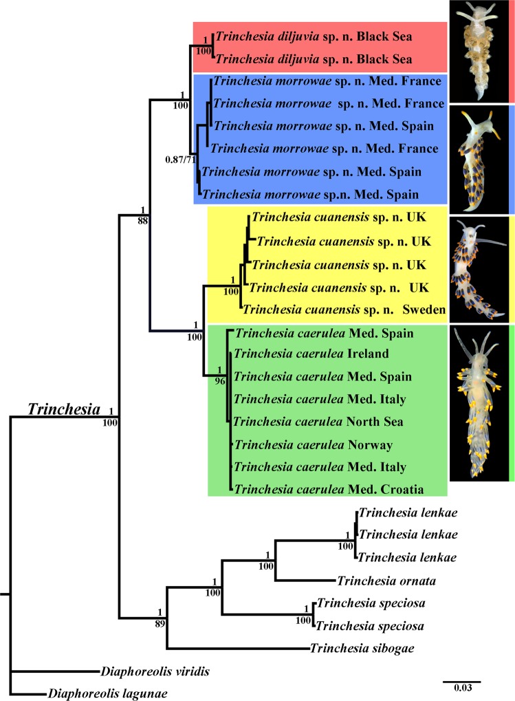
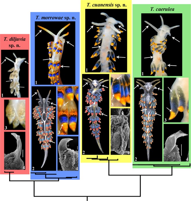
Phylogenetic analysis was performed using 28 specimens of Trinchesia, including data for 21 Trinchesia caerulea species complex specimens, four other congener species of the genus Trinchesia and two outgroup species. All supposedly morphologically cryptic morphs of Trinchesia caerulea illustrated in Thompson & Brown (1984)16 were collected in European waters and examined. In addition, specimens collected in the Black Sea that are taxonomically close to T. caerulea were also included in the study. We apply the concept ‘Trinchesia caerulea species complex’ here as the designation for all these studied specimens. The dataset consisted of 80 nucleotide sequences including mitochondrial COI and 16S, and the nuclear H3 genes. The SYM + G model was chosen for the concatenated dataset. Bayesian Inference (BI) and Maximum Likelihood (ML) analyses based on the combined dataset for the COI, 16S and H3 genes yielded similar results (Fig. 1). To define species, we use an integrative approach25,29 including phylogenetic tree topologies, ABGD analysis, pairwise distances and the haplotype network using PopART (Fig. 2, see also supplementary information Tables S1–S5). The results of the integrative study clearly identified four species in the Trinchesia caerulea species complex: T. caerulea (neotype is designated here, ZMMU Op-646), T. cuanensis sp. n. (holotype ZMMU Op-650, ZooBank registration: urn:lsid:zoobank.org:act: 77629514-DDB5-4757-86FE-78DDD7672563), T. morrowae sp. n. (holotype ZMMU Op-651, ZooBank registration: urn:lsid:zoobank.org:act: D7DB7FFB-F6B1-4A67-8DF4-A1D0F05963A7), and T. diljuvia sp. n. (holotype ZMMU Op-642, ZooBank registration: urn:lsid:zoobank.org:act: C9228E8B-DF2C-46A0-9E78-038ED6B87D7B). ZooBank registration for the paper is: urn:lsid:zoobank.org:act: 4B5F968F-69B6-4FB0-BB7B-43ABC4B09EBB. Information about the taxonomy of these four species can be found in the descriptions below, in the Figs 3–11 and in the supplementary information, Table S5.
Figure 1.
Phylogenetic relationships of nudibranchs Trinchesia based on COI + 16S + H3 concatenated dataset inferred by Bayesian Inference (BI). Numbers above branches represent posterior probabilities from Bayesian Inference. Numbers below branches indicate bootstrap values for Maximum Likelihood. A comparison of the external morphology is indicated by arrows. Abbreviations: Med. – The Mediterranean Sea; UK – United Kingdom. Photos by B.P. and T.K.
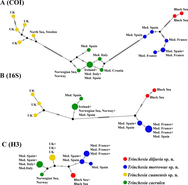
Figure 2.
The haplotype network based on (A) cytochrome c oxidase subunit I (COI), (B) 16S rRNA, and (C) –Histone 3 (H3) molecular data showing genetic mutations occurring within Trinchesia caerulea complex species.
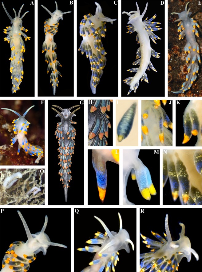
Figure 3.
Trinchesia caerulea (Montagu, 1804). External views of living specimens and comparison with data from Thompson & Brown (1984). (A) Neotype, Ireland, Mayo, Killary Harbour, Rusheen Point (ZMMU Op-646). (B) Specimen from Wales, Pembrokeshire, Martin’s Haven (BELUM.Mn2018.1). (C) Specimen from Norway, Gulen Dive Resort (ZMMU Op-622). (D) Same specimen, ventral view. (E) Specimen from Italy. (F) Specimen from Spain, Girona, L’Estartit (ZMMU Op-648). (G) Specimen from Wales, Pembrokeshire, Skomer Is., depicted in Thompson & Brown (1984: pl 30, c). (H) Same, details of cerata. (I) Details of ceras from original description of T. caerulea in Montagu (1804), specimen from UK (Devon). (J). Details of cerata of specimen from Ireland. (K) Details of cerata of specimen from Wales. (L) Details of ceras of specimen from Spain. (M) Details of cerata of specimen from Italy. (N) Details of cerata of specimen from Norway. (O) Egg mass from Italy. (P) Details of anterior part of specimen from Wales. (Q) Details of anterior part of specimen from Italy. (R) Details of anterior part of specimen from Norway. Photographs: Bernard Picton, (a), (b), (d), (e), (j), (k), (p); Tatiana Korshunova: (c), (n), (r); Miquel Pontes: (f), (l), (q); Giulia Furfaro: (o). Reproduction of figures from Thompson & Brown (1984) with permission of Gregory Brown, original artist and copyright holder of the images.
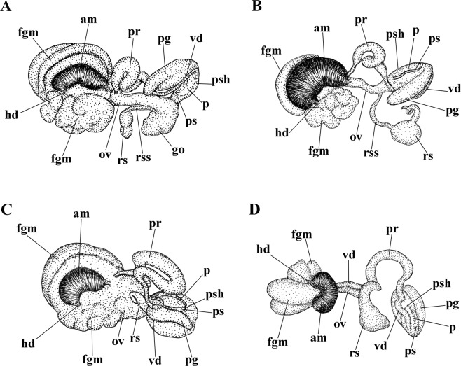
Figure 11.
Reproductive systems, schemes. (A) Trinchesia caerulea. (B) Trinchesia cuanensis sp. n. (C) Trinchesia morrowae sp. n. (D). Trinchesia diljuvia sp. n. Schemes by Tatiana Korshunova. Abbreviations: am, ampulla; fgm, female gland mass; hd, hermaphroditic duct; ov, oviduct; p, penis; pg, “penial” (supplementary) gland; pr, prostate; ps, penial stylet; psh, penial sheath; rs, receptaculum seminis; rss, stalk of receptaulum seminis; vd, vas deferens.
The molecular phylogenetic analysis also supported the presence of these four species in the Trinchesia caerulea species complex: T. caerulea (PP = 1, BS = 96%), T. cuanensis sp. n. (PP = 1, BS = 100%),T. morrowae sp. n. (PP = 0.87, BS = 71%), and T. diljuvia sp. n. (PP = 1, BS = 100%). The maximum genetic distance values within the T. caerulea group are 1.25% for COI, 0.25% for 16S, and 0.66% for H3 markers. The maximum genetic distance values within the T. cuanensis sp. n. group are 1.88% for COI, 0.46% for 16S, and 0% for H3 markers. The maximum genetic distance values within the T. morrowae sp. n. group are 2.18% for COI, 0.23% for 16S, and 0.65% for H3 markers. The maximum genetic distance values within the T. diljuvia sp. n. group are 0.16% for COI, 0.46% for 16S, and 0% for H3 markers. Regarding the supposedly fast-evolving COI marker, minimum genetic distance values between the T. caerulea and the T. cuanensis sp. n., T. morrowae sp. n., and T. diljuvia sp. n. groups are 7.20%, 10.95% and 11.89% respectively. The minimal COI p-distance (2.98%) was found between T. morrowae sp. n. and T. diljuvia sp. n.; the maximal COI p-distance (12.99%) was found between T. cuanensis sp. n. and T. diljuvia sp. n. The minimal 16S p-distance (1.39%) was found between T. caerulea and T. cuanensis sp. n.; the maximal 16S p-distance (6.47%) was found between T. caerulea and T. diljuvia sp. n. The minimal nuclear H3 p-distance (0.98%) was found between T. caerulea and T. cuanensis sp. n. and between T. morrowae sp. n. and T. diljuvia sp. n.; the maximal nuclear H3 p-distance (1.63%) was found between T. caerulea and T. morrowae sp. n. and T. caerulea and T. diljuvia sp. n and between T. cuanensis sp. n. and T. morrowae sp. n. All minimum intergroup genetic distances for every marker are larger than the maximum intragroup distances. (see supplementary information, Tables S2–S4). Results obtained by PopART showed a network of haplotypes that clearly clustered into four groups coincident with T. caerulea, T. cuanensis sp. n., T. morrowae sp. n., and T. diljuvia sp. n. (Fig. 2). The ABGD analysis of the COI data set run with two different models with the initial approach revealed seven potential species of the genus Trinchesia: T. caerulea, T. cuanensis sp. n., T. lenkae, T. ornata, T. sibogae, T. speciosa, and T. morrowae sp. n. In the recursive approach, the additional species T. diljuvia sp. n. is recognized. The ABGD analysis of the 16S data set run with two different models with the initial approach revealed eight potential species: T. diljuvia sp. n., T. caerulea, T. cuanensis sp. n., T. lenkae, T. morrowae sp. n., T. ornata, T. sibogae, and T. speciosa. Detailed morphological investigation was carried out based on molecular phylogenetic delimitation.
This particular case is very relevant for the ongoing general ‘cryptic’ species discussion8–10,30 because the European nudibranch fauna is one of the best studied in the world8,16,17,31–34, but the three new species of Trinchesia presented here were never described before despite not only significant molecular divergence (Figs 1 and 2) but also multilevel morphological differences clearly defined and linked to the molecular phylogenetic data for the first time in this study (Fig. 12). For further comparison of morphological data with the molecular results see the detailed descriptions below, Figs 3–11, and the Discussion.
Figure 12.
Multilevel integrative presentation of molecular (based on the molecular phylogenetic tree, Fig. 1) and morphological data for four species of the Trinchesia caerulea complex. For each species [T. caerulea (Montagu, 1804), T. cuanensis sp. n., T. morrowae sp. n., T. diljuvia sp. n.] external view of living specimen (1) (photos by B.P. and T.K.) is coupled with appropriate Fig. (2) of T. caerulea morphs from Thompson & Brown monograph (reproduced with permission of Gregory Brown, original artist and copyright holder of the images), without taxonomic status at that time (1984), and also supplied with taxonomically crucial distinguishing details of colour of dorsal cerata (3, photos by B.P. and T.K.) and scanning electron images of stylet of copulative organ (4, by A.M.). Key external distinguishing features between all four species are indicated by arrows (see Results, Discussion and supplementary information for details). Scale bars 10 μm.
Systematics
Phylum Mollusca
Order Nudibranchia Cuvier, 1817
Family Trinchesiidae Nordsieck, 1972
Genus Trinchesia Ihering, 1879
Type species Doris caerulea Montagu, 1804
Trinchesia caerulea (Montagu, 1804)
(Figures 1–4, 11, 12, Tables S1–S5)
Figure 4.
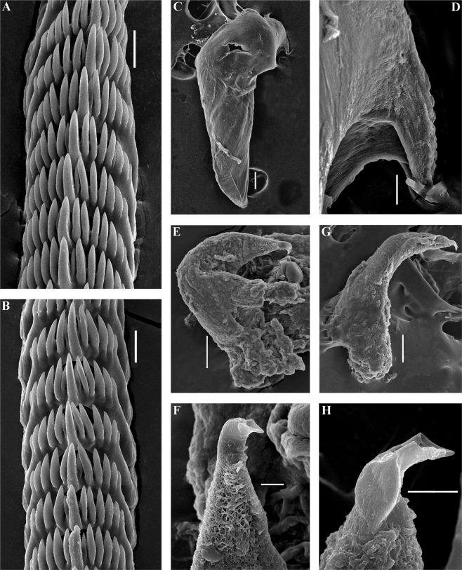
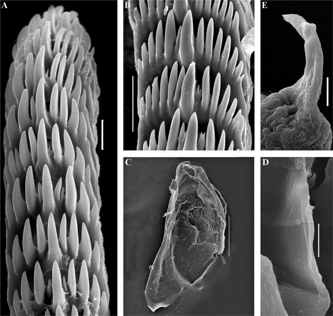
Trinchesia caerulea (Montagu, 1804). Internal morphology, scanning electron microscopy. (A) Posterior part of radula of specimen from Spain, L’Estartit, Girona (ZMMU Op-648). (B) Posterior part of radula of specimen from Norway, Gulen Dive Resort (ZMMU Op-622). (C) Jaw of specimen from Norway (Gulen). (D) Details of masticatory processes of jaws, same specimen. (E) Copulative organ with stylet of specimen from Norway. (F) Stylet, details, same specimen. (G) Copulative organ with stylet of specimen from Spain. (H) Stylet details, same specimen. Scale bars: a, b, d −20 μm, c −100 μm, e, g −50 μm, f, h −10 μm. SEM micrographs: Alexander Martynov.
Synonymy:
Doris caerulea Montagu, 180435: 78, pl. 7, Figs 4, 5
Figure 5.
Trinchesia cuanensis sp. n. External views of living specimens and comparison with morphs included by Thompson & Brown (1984) as a single species “Trinchesia caerulea”. (A). Holotype, Down, Strangford Lough, Northern Ireland, United Kingdom (ZMMU Op-650), dorsal view. (B) Same specimen, left lateral view. (C) Paratype from Bohuslän, Smögen, Sweden (GNM-9243), right lateral view. (D) Paratype from Antrim, Portrush, United Kingdom, dorsal view (BELUM.Mn.2018.2). (E) Paratype from Down, Strangford Lough, dorsal view (ZMMU Op-655). (F) Specimen from Isle of Man, Port Erin, depicted in Thompson & Brown (1984: pl 30, c), dorsal view. (G) Same, details of cerata. (H). Details of cerata of the holotype. (I) Details of cerata, of paratype from Down, Strangford Lough. (J) Details of cerata of paratype from Sweden. (K) Details of cerata, of paratype from Antrim, Portrush. (L) Details of cerata, of paratype from Strangford Lough. (M) Details of anterior part of holotype from Strangford Lough. (N) Details of anterior part of paratype from Strangford Lough. Photographs: Bernard Picton, (a), (b), (d), (e), (h), (i), (k), (l), (m), (n); Klas Malmberg, (c), (j). Reproduction of figures from Thompson & Brown (1984) with permission Gregory Brown, original artist and copyright holder of the images.
Eolidia bassi Vérany, 184636: 27
Eolis deaurata Dalyell, 185337: 301, pl 44, Figs 8–10
Figure 8.
Trinchesia morrowae sp. n. Internal morphology, scanning electron microscopy. (A) Posterior part of radula of paratype from Spain, L’Estartit (ZMMU Op-652). (B) Posterior part of radula of paratype from France, Banyuls-sur-Mer. (C) Jaw of specimen from Spain (L’Estartit, Girona). (D) Details of masticatory processes of jaws, same specimen. (E) Copulative organ with stylet of specimen from France, Banyuls-sur-Mer (ZMMU Op-653). (F) Details of stylet, paratype. Scale bars: (a) −20 μm; (b), (d), (f) −10 μm; (c) −100 μm; (e) −50 μm. SEM micrographs: Alexander Martynov.
Figure 10.
Trinchesia diljuvia sp. n. Internal morphology, scanning electron microscopy. (A) Posterior part of radula of paratype ZMMU Op-645; (B). Posterior part of radula (specimen ZMMU Op-643); (C). Jaw, paratype ZMMU Op-643; (D). Details of masticatory processes of jaws, same specimen; (E). Details of copulative organ with stylet, same specimen (ZMMU Op-645). Scale bars: (a), (d), (e) −10 μm; (b) −20 μm; (c) −120 μm; SEM micrographs: Alexander Martynov.
Eolis glotensis Alder & Hancock, 184638: 293
Eolis molios Herdman, 188139: 28–29, pl 1, Figs 1–3
Non Cuthona caerulea sensu Schmekel & Portmann (1982)34, Thompson & Brown, 198416 (mixture of several species).
Material examined
Neotype
NE Atlantic, Mayo, Killary Harbour, Rusheen Point, Ireland (53° 37′ 31″ N 9° 51′ 26″ W), 10–20 m depth, stones with hydroids, 30.04.2017, coll. M. Larsson & B.E. Picton (ZMMU Op-646, 17 mm in length, live, preserved length 8 mm).
Other specimens
NE Atlantic, Wales, Pembrokeshire, Martin’s Haven, United Kingdom (51°44′15.57″ N 5°14′43.37″ W), 10–20 m depth, rocky reef, feeding on Sertularella polyzonias, with spawn, 02.07.2013, coll. B.E. Picton, one specimen (BELUM.Mn2018.1, 15 mm in length, live), 02.07.2013, coll. B.E. Picton. NE Atlantic, Gulen Dive Center, Norway (60° 57′27.11″ N 5° 07′ 47.10″ E), depth 15–20 m, stones, collectors T.A. Korshunova, A.V. Martynov, 05.03.2017, one specimen (ZMMU Op-622, 15 mm in length, live, ca. 9 mm in length, preserved). Mediterranean Sea, Ugljan Island, Karantun, Croatia (44° 04′ N 15° 09′ E), depth 15–20 m, collectors Đ. Iglić, A. Petani, 21.01.2018, 2 specimens (ZMMU Op-647, 21 mm live, ca. 10 mm in length, preserved). Mediterranean Sea, Ugljan Island, Karantun, Croatia (44° 04′ N 15° 09′ E), depth 15–20 m, collectors Đ. Iglić, A. Petani, 21.01.2018, one specimen (GNM Gastropoda – 9792, 15 mm live, 6 mm in length, preserved). Mediterranean Sea, Tor Paterno, Latium, Italy, one specimen (41° 40′ N 3° 12′ 20″ E), 25 m depth, collector G. Furfaro, 26.04.2013 (RM3 333). Mediterranean Sea, Tor Paterno, Latium, Italy, one specimen (41° 40′ N 3° 12′ 20″ E), 25 m depth, collector G. Furfaro, 01.05.2013 (RM3 860). Mediterranean Sea, Tor Paterno, Latium, Italy, one specimen (41° 40′ N 3° 12′ 20″ E), 25 m depth, collector G. Furfaro, 01.05.2013 (RM3 861). Mediterranean Sea, Tor Paterno, Latium, Italy, one specimen (41° 40′ N 3° 12′ 20″ E), 25 m depth, collector G. Furfaro, 26.04.2013 (RM3 309). Mediterranean Sea, Catalonia, L’Estartit, Girona, Spain (42° 02′ 32″ N 3° 13′ 38″ E), depth 16 m, stones, collector M. Pontes, 22.04.2017, one specimen (ZMMU Op-648, 20 mm live, 7 mm in length, preserved). Mediterranean Sea, Catalonia, L’Estartit, Girona, Spain (42° 02′ 32″ N 3° 13′ 38″ E), depth 16 m, stones, collector M. Pontes, 22.04.2017, one specimen (ZMMU Op-649, 9 mm live, 3 mm in length, preserved).
Diagnosis
Body up to 21 mm with long foot corners; cerata with distinct colour zones, digestive gland basally greenish to light grayish occasionally with a narrow or diffuse dotted yellow band at the surface, an upper blue broad band, and towards the top of cerata a broad orange or yellow band; up to 12 rows of cerata, commonly four anterior rows; radular formula 57–63 × 0.1.0, penial stylet relatively short and bent at the top, seminal receptacle is 8-shaped without a long convoluted additional chamber, egg mass spiral with several whorls.
Description
External morphology
The live length of the neotype is 17 mm (Fig. 3A,J). The length of adult specimens may reach 21 mm. The body is narrow. The rhinophores are smooth and 1.5–2 times longer than the oral tentacles. The cerata are relatively short, spindle-shaped. Ceratal formula of the neotype: right (2,3,4,5; anus, 5,4,3,3,2) left (1,3,4,5; anus, 5,4,4,3,2). The foot is narrow anteriorly with relatively long foot corners.
Colour
The basal colour is whitish to light greenish, occasionally yellow, never forming any continuous broad white line dorsally (Fig. 3). Commonly, the cerata are basally light grayish to greenish, sometimes with a narrow dotted light yellowish or orange band, then a dark to light blue broad band, and towards the top of cerata there is broad orange or yellow band. The tips of the cerata are clear with a translucent cnidosac. The tips of the rhinophores and oral tentacles sometimes have diffuse white or light yellowish or yellow pigment, but no intense orange colour. In Croatia a population of ca. 30 specimens has been observed mostly without blue pigment on the cerata, but within a few hours after their collection almost all the white pigmentation changed to blue, which varied from relatively dark to very light.
Anatomy
Digestive system
The jaws are triangularly ovoid (Fig. 4C). The masticatory processes of the jaws bear a single row of conspicuous low denticles (Fig. 4D). The radular formula in two studied specimens is 57–63 × 0.1.0. The radular teeth are yellowish. The central tooth is broad, with low cusp and 6–8 lateral denticles, including smaller intercalated denticles that may occur in different parts of the tooth (Fig. 4A,B).
Reproductive system
(Fig. 11A). The ampulla is massive and swollen (Fig. 11A, am). The prostate is a convoluted tube (Fig. 11A, pr). The prostate transits to a penial sheath, which contains a conical penis with a relatively short, chitinous stylet, strongly curved at the top (Figs 4E–H, 11A, ps). A supplementary (“penial”) gland inserts into the base of the penis and is attached to the penial sheath along of its most length (Fig. 11A, pg). The seminal receptacle is an 8-shaped structure, comprising two reservoirs partly inserted into each other (Fig. 11A, rs). The female gland mass includes mucous and capsular glands (Fig. 11A, fgm).
Description of egg masses
The egg mass is a white or very pale pinkish spiral cord forming about 3 whorls or an irregular cord. It may contain 1000 eggs or more.
Distribution and habitats
Reliably confirmed from Britain and Ireland, Norway, and the Mediterranean Sea (Croatia, Italy and Spain). Found in stony, relatively shallow areas, often covered with various algae, usually at 10–25 m depth, occasionally at only 1–2 m. Feeds on hydrozoans of the genus Sertularella (S. polyzonias (L., 1758)) but also on S. crassicaulis (Heller, 1868) and S. gayi (Lamouroux, 1821) for which it has a strong preference40, and it is also reported feeding on Eudendrium racemosum (Cavolini, 1785)41, Hydrallmania falcata (L., 1758) and Halecium halecinum (L., 1758)16. We provide references to the potential food sources here, but this information should be used carefully since food associated with T. caerulea may now be the food preference of one of the other T. caerulea complex species. In the French Mediterranean, T. caerulea was recorded as feeding on S. crassicaulis. In the present study T. caerulea was confidently found commonly in L’Estartit (Catalonia) area as associated with S. polyzonias and much less frequently on Eudendrium racemosum.
Remarks
Morphological differences
T. caerulea can be distinguished from the partly sympatric (in the NE Atlantic) T. cuanensis sp. n. by this set of morphological data: 1). Greenish to light grayish basis of the cerata, not blackish to dark grayish as in T. cuanensis sp. n. 2). Absence on the cerata of a narrow blackish band above the broad blue band, which is present in T. cuanensis sp. n. 3). Long anterior foot corners, not short ones as in T. cuanensis sp. n.; 4). Relatively short penial stylet, whereas T. cuanensis sp. n. has an extraordinarily long stylet. T. caerulea can be distinguished from the partly sympatric, predominantly Mediterranean species, T. morrowae sp. n., by this set of morphological data: 1). Greenish to light grayish basis of cerata, not light grayish to yellowish as in T. morrowae sp. n.; 2). Absence of a distinct white dorsal line and thin white lateral lines, which are instead always present in T. morrowae sp. n.; 3). White opaque or light yellowish tips of rhinophores and oral tentacles, but not orange ones as in T. morrowae sp. n. 4). Long anterior foot corners, not just angular projections as in T. morrowae sp. n.; 5). The penial stylet is curved towards the tip, whereas in T. morrowae sp. n. it is curved rather basally; 6). T. caerulea has up to two times the adult body size of T. morrowae sp. n. T. caerulea can be distinguished from the exclusively Black Sea species T. diljuvia sp. n. by this set of morphological data: 1). Presence of distinct colour zones on the cerata, which are absent in T. diljuvia sp. n.; 2). T. caerulea has up to five times larger adult body size compared to T. diljuvia sp. n.; 3). Absence of a white dorsal line, which is always present in T. diljuvia sp. n.; 3). Long anterior foot corners, which are completely absent in T. diljuvia sp. n.; 5). Precise shape of the penial stylet is different between T. caerulea and T. diljuvia sp. n.
Molecular differences
Minimum uncorrected COI p-distances between the T. caerulea neotype specimen and T. cuanensis sp. n., T. morrowae sp. n. and T. diljuvia sp. n. specimens are 7.36%, 11.11%, and 12.05% respectively. See also Discussion, Fig. 12 for integration of molecular phylogenetic and morphological data, and supplementary information, Tables S1–S5.
Trinchesia cuanensis sp. n.
(Figures 1, 2, 5, 6 and 11, 12, Tables S1–S5).
Figure 6.
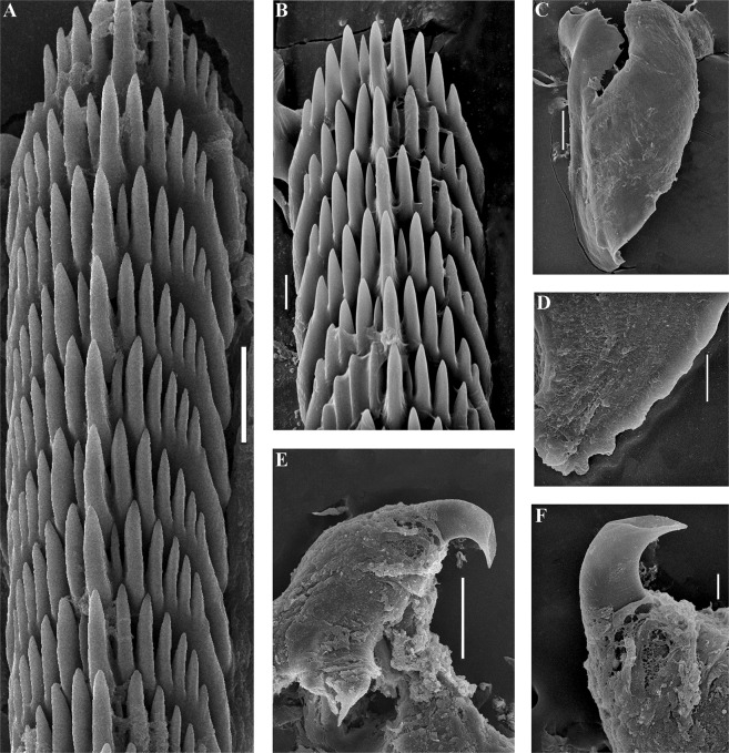
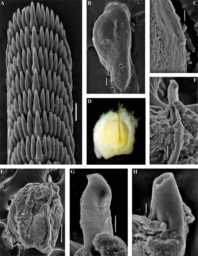
Trinchesia cuanensis sp. n. Internal morphology, scanning and light electron microscopy. (A) Posterior part of radula of holotype from Northern Ireland (ZMMU Op-650). (B) Jaw, holotype. (C) Details of masticatory processes of jaws, holotype. (D) Copulative organ with stylet of paratype from Northern Ireland (GNM Gastropoda – 9054), light microscopy. (E) Same, scanning electron microscopy. (F) Details of stylet, same paratype. (G) Details of apical part of stylet, same specimen. (H) Cross section of basal part of stylet, showing channel inside, holotype. Scale bars: a, f −20 μm, b, e −100 μm, c, h, g −10 μm. SEM micrographs: Alexander Martynov.
Synonymy
Cuthona caerulea sensu lato auct., e.g. Thompson & Brown (1984)16, Schmeckel & Portmann (1982)34 (mixed up with Doris caerulea Montagu, 1804).
Material examined
Holotype
NE Atlantic, Strangford Lough, Down, Ballyhenry, Northern Ireland, United Kingdom (54°23′22.2″ N 5°34′23.4″ W), 15–20 m depth, coll. B.E. Picton, 24.05.2014 (ZMMU Op-650, 15 mm in length, live, 7 mm in length, preserved). Paratypes. NE Atlantic, Strangford Lough, Portaferry, Northern Ireland, United Kingdom (54° 22,0′ N 05°32,0′ W), ca. 10 m depth, coll. B.E. Picton, 22.05.2014, one specimen (GNM Gastropoda – 9054, ca. 9 mm in length, preserved). NE Atlantic, Strangford Lough, Down, Ballyhenry, Northern Ireland, United Kingdom (54°23′22.2″ N 5°34′ 23.4″ W), 10–25 m depth, coll. B.E. Picton, 24.05.2014, one specimen (ZMMU Op-655, 15 mm in length, live, 8 mm in length, preserved). NE Atlantic, Portrush, Antrim, S of Little Skerry, Northern Ireland, United Kingdom (55°′12′59.238″ N 6° 38′39.292″ W), 24.3 m max depth, coll. B.E. Picton, 23.08.2006, three specimens (BELUM.Mn.2018.2, 13 mm in length, live, 5 mm in length, preserved). Bohuslän, outside of Smögen, Sweden (58° 21,00′ N 11°12,00′ E), 10–20 m depth, coll. Klas Malmberg, 02.05.2015, one specimen (GNM Gastropoda – 9243, 2 mm in length, preserved).
Zoobank registration
urn:lsid:zoobank.org:act: 77629514-DDB5-4757-86FE-78DDD7672563.
Etymology
After Lough Cuan, an alternative name for Strangford Lough in Northern Ireland, where many marine biological studies have been undertaken over the past 100 years.
Diagnosis
Body up to 15 mm (live), cerata with distinct colour zones, digestive gland basally dark grayish to blackish with commonly a narrow or reduced dotted orange or yellowish band, then narrower black zone, then a blue broad band, and towards the top of cerata there is broad orange or yellow band, up to 10 rows of cerata, commonly four anterior ceratal rows, radular formula 64 × 0.1.0, penial stylet extraordinarily long, rounded seminal receptacle with a long convoluted additional chamber, egg mass spiral with several whorls.
Description
External morphology
The length of the preserved holotype is 7 mm (Fig. 5). The live length up to 15 mm. The body is narrow. The rhinophores are smooth and 1.5–2.5 times longer than the oral tentacles. The cerata are relatively short, spindle-shaped. Ceratal formula of the holotype: right (3,4,5,6; anus, 7,5,4,3,3) left (2,4,5,6; anus, 8,5,5,3,3). The foot is narrow, anteriorly with short foot corners.
Colour
The basal colour is whitish to light greenish, sometimes with some diffuse pigment of a similar colour scattered over the body, but never forming any continuous broad white line dorsally (Fig. 5). The cerata are basally blackish to dark grayish (no green colour) with a narrow dotted orange or a yellowish band, then a dark blue broad band, followed by a narrow dark band, and towards the top of cerata there is broad orange or yellow band. The tips of the cerata are clear with a translucent cnidosac. The tips of the rhinophores and oral tentacles have diffuse white pigment, which is sometimes yellowish, but never of an intense orange colour.
Anatomy
Digestive system
The jaws are triangularly ovoid (Fig. 6B). The masticatory processes of the jaws bear a single row of inconspicuous low denticles (Fig. 6C). The radular formula is 64 × 0.1.0. The radular teeth are yellowish. The central tooth is broad, with low cusp and 7–8 lateral denticles, including smaller intercalated denticles near the cusp (Fig. 6A).
Reproductive system
(Fig. 11B). The ampulla is massive and swollen (Fig. 11B, am). The prostate is a highly convoluted tube (Fig. 11B, pr). The prostate transits to a penial sheath, which contains a conical penis with an extremely long, chitinous stylet, slightly curved at the top (Figs 6D–H, 11B, ps). A supplementary (“penial”) gland inserts into the base of the penis and attaches to the penial sheath along most of its length (Fig. 11B, pg). The seminal receptacle is a complex structure with long narrow stalk, large rounded reservoir and long convoluted narrow special supplementary reservoir inserted into large rounded one (Fig. 11B, rs). The female gland mass includes mucous and capsular glands (Fig. 11B, fgm).
Description of egg masses
The egg mass is a spiral cord forming a minimum of 2 whorls, or an irregular cord. Number of eggs is commonly more than 300.
Distribution and habitats
Confirmed records in Ireland, United Kingdom and Swedish west coast. Found in stony, relatively shallow areas, at 10–20 m depth. Feeds on hydrozoans Sertularella polyzonias, and possibly other Sertularella species.
Remarks
Morphological differences
T. cuanensis sp. n. can be distinguished from the partly sympatric (both are common in the North Atlantic) type species of the genus, T. caerulea, by this set of morphological data: 1). Blackish to dark grayish basis of the cerata, not greenish to light grayish as in T. caerulea; 2). Presence on the cerata of a narrow blackish band above the broad blue band, but not in T. caerulea; 3). Short anterior foot corners, not long as in T. caerulea; 4). Extraordinarily long penial stylet (not normal for the family Trinchesiidae), whereas T. caerulea has a normal, short one. T. cuanensis sp. n. can be distinguished from the partly sympatric, predominantly Mediterranean species, T. morrowae sp. n., by this set of morphological data: 1). Blackish to dark grayish basis of the cerata, not light grayish to yellowish as in T. morrowae sp. n.; 2). Presence on the cerata of a narrow blackish band above the broad blue band, not present in T. morrowae sp. n.; 3). Absence of a white dorsal line and thin white lateral lines, which are instead always present in T. morrowae sp. n.; 4). Short anterior foot corners, not just angular projections as in T. morrowae sp. n.; 5). Extraordinarily long penial stylet, whereas T. morrowae sp. n. has a normal short one. T. cuanensis sp. n. can be distinguished from the exclusively Black Sea species T. diljuvia sp. n. by this set of morphological data: 1). Presence of distinct colour zones on the cerata, which are absent in T. diljuvia sp. n.; 2). Absence of a white dorsal line, which is always present in T. diljuvia sp. n.; 3). Short anterior foot corners, which are completely absent in T. diljuvia sp. n.; 4). Extraordinarily long penial stylet, whereas T. diljuvia sp. n. has a relatively short one.
Molecular differences
Minimum uncorrected COI p-distances between T. cuanensis sp. n. type specimen and T. caerulea, T. morrowae sp. n. and T. diljuvia sp. n. specimens are 7.98%, 12.05%, and 13.77% respectively. See also Discussion, Fig. 12 for integration of molecular phylogenetic and morphological data, and supplementary information, Tables S1–S5.
Trinchesia morrowae sp. n.
(Figures 1, 2, 7, 8, 11, 12, Tables S1–S5).
Figure 7.
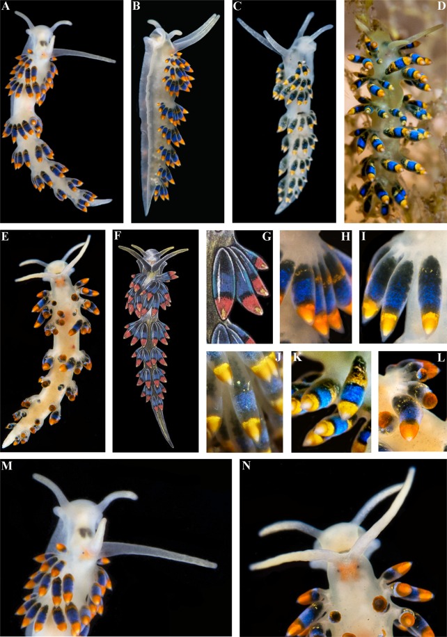
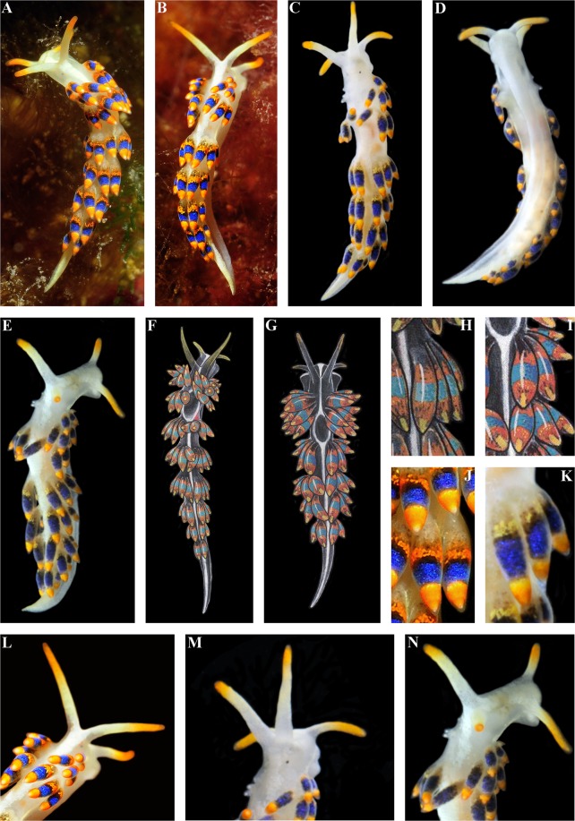
Trinchesia morrowae sp. n. External views of living specimens and comparison with morphs included in Thompson & Brown (1984) as the single species “Trinchesia caerulea”. (A) Holotype from Spain, Girona, L’Estartit, dorsal view (ZMMU Op-651). (B) Holotype from same location, dorso-lateral view (right side). (C) Paratype from France, Banyuls-sur-Mer (ZMMU Op-653), dorsal view. (D) Same paratype, ventral view. (E) Same paratype, dorso-lateral view (right side). (F). Specimen from UK (Lundy Island) depicted in Thompson & Brown (1984: pl 30, a), dorsal view. (G) Specimen from UK (Scilly Isles) depicted in Thompson & Brown (1984: pl 30, d), dorsal view. (H) Details of cerata of specimen from UK (Lundy Island) depicted in Thompson & Brown (1984: pl 30, a). (I) Details of cerata of specimen from UK (Scilly Isles) depicted in Thompson & Brown (1984: pl 30, d). (J) Details of cerata of holotype. (K) Details of cerata of specimen from France (ZMMU Op-653). (L) Details of anterior part of holotype. (M) Details of paratype from France. (N) Details of anterior part of the same paratype. Photographs: Miquel Pontes, (a), (b), (j); Tatiana Korshunova, (c), (d), (k), (m), (n). Reproduction of figures from Thompson & Brown (1984) with permission of Gregory Brown, original artist and copyright holder of the images.
Synonymy
Cuthona caerulea auct., e.g. Thompson & Brown (1984)16, Schmeckel & Portmann (1982)34, Picton & Morrow (1994)31 non Doris caerulea Montagu, 1804.
Material examined
Holotype
Mediterranean Sea, Catalonia, L’Estartit, Girona, Spain (42° 02′ 32″ N 3° 13′ 38″ E), depth 16 m, stones, collector M. Pontes, 22.04.2017 (ZMMU Op-651, 9 mm live, 3.2 mm in length, preserved). Paratypes. Mediterranean Sea, Banyuls-sur-Mer, France (42° 28′ 53.0″ N 3° 08’ 17″ E), depth 5–12 m, stones, collector T.A. Korshunova, A.V. Martynov, 08.09.2010, two specimens (ZMMU Op-653, ca. 8 mm live, ca. 3 mm in length, preserved). Mediterranean Sea, Banyuls-sur-Mer, France (42° 28′ 53.0″ N 3° 08′ 17″ E), depth 5–12 m, rocks, stones, collector T.A. Korshunova, A.V. Martynov, 10.09.2010, one specimen (ZMMU Op-535, 5 mm live, 3.5 mm in length, preserved). Mediterranean Sea, Banyuls-sur-Mer, France (42° 28′ 53.0″ N 3° 08′ 17″ E), depth 5–12 m, rocks, stones, collector T.A. Korshunova, A.V. Martynov, 09.09.2010, one specimen (ZMMU Op-656, 7 mm live, 2.5 mm in length, preserved). Mediterranean Sea, Catalonia, L’Estartit, Girona, Spain (42° 02′ 32″ N 3° 13′ 38″ E), depth 16 m, stones, collector M. Pontes, 22.04.2017, one specimen (ZMMU Op-652, 9 mm live, 3.2 mm in length, preserved).
Zoobank registration
urn:lsid:zoobank.org:act: D7DB7FFB-F6B1-4A67-8DF4-A1D0F05963A7.
Etymology
After Christine Morrow, co-author of “A Field Guide to the Nudibranchs of the British Isles” (1994), who sequenced a specimen of Trinchesia cuanensis which first revealed the presence of two species of this complex in the NE Atlantic.
Diagnosis
Body up to ca. 10 mm, white dorsal line, cerata with distinct colour zones, digestive gland basally light grayish to yellowish, then a usually broad distinct dotted orange band, then narrower black zone, then a blue broad band, then, compared to T. cuanensis sp. n., there is no narrow black band and towards the top of cerata there is broad orange band, three, rarely four anterior ceratal rows, radular formula 55–57 × 0.1.0, penial stylet short and considerably curved, seminal receptacle is oval without additional chamber, egg mass a short spiral.
Description
External morphology
The length of the holotype is 9 mm (live, Fig. 7). The length of adults may be up to 11 mm. The body is narrow. The rhinophores are smooth and ca. 1.5 times longer than the oral tentacles. The cerata are relatively short, spindle-shaped. The ceratal formula of the holotype: right (2,3,3; anus, 3,3,2,2,1) left (2,3,4; 4,3,2,2,1). The foot is narrow anteriorly without real foot corners, only angular processes.
Colour
The basal colour is whitish and a distinct thick white line runs dorsally, thinner white lines run laterally (Fig. 7). The cerata are basally light grayish to yellowish with a broad dotted orange band, then a narrower black zone, then a blue broad band, and towards the top of cerata there is broad orange band (there is no distinct black line in between the blue and orange upper zones). The tips of the cerata are clear with a translucent cnidosac. The tips of the rhinophores and oral tentacles have an intense orange pigment.
Anatomy
Digestive system
The jaws are triangularly ovoid (Fig. 8C) The masticatory processes of the jaws bear a single row of low, inconspicuous denticles (Fig. 8D). The radular formula in the holotype is 55–57 × 0.1.0. The radular teeth are yellowish. The central tooth is broad, with a low cusp and 4–7 lateral denticles, including smaller intercalated denticles (Fig. 8A,B).
Reproductive system
(Fig. 11C). The ampulla is massive and swollen (Fig. 11C, am). The prostate is a highly convoluted tube (Fig. 11C, pr). The prostate transits to a penial sheath, which contains a conical penis with a short chitinous stylet, slightly curved at the top (Figs 8E,F, 11C, ps). A supplementary (“penial”) gland inserts into the base of the penis and attaches to the penial sheath along most of its length (Fig. 11C, pg). The seminal receptacle is a relatively simple structure with a widened stalk and a small bent reservoir (Fig. 11C, rs). The female gland mass includes mucous and capsular glands (Fig. 11C, fgm).
Description of egg masses
The egg mass is a cream coloured spiral cord of at least 2–2.5 whorls, or a regular cord. Number of eggs less than 100.
Distribution and habitats
Predominantly known from various locations in the Mediterranean Sea, from Greece to the Western basin, but may also reach the southern part of the UK. Occurs in all the Iberian Peninsula coastal areas, including both Portugal and Spain. Found in stony, relatively shallow areas, from 1 to 25 m depth, also in Posidonia oceanica (L.) meadows. Feeds on Sertularella spp. hydroids, possibly on Sertularia perpusilla Stechow, 1919, and also Stylactis inermis Allman, 1872.
Remarks
Morphological differences
T. morrowae sp. n. can be distinguished from the partly sympatric (both are common in the North Atlantic and in the Mediterranean Sea) type species of the genus, T. caerulea, by this set of morphological data: 1). Light grayish- to yellow basis of the cerata, not greenish to light grayish as in T. caerulea; 2). Presence of a distinct lower orange band on the cerata, not a commonly reduced and diffuse one as in T. caerulea; 3). Presence of a thick white dorsal line and thin white lateral lines, which are always absent in T. caerulea; 4). Presence of commonly three, rarely four, anterior ceratal rows in T. morrowae sp. n., whereas there are commonly four in T. caerulea; 5). Angular anterior projections of the foot, not long foot corners as in T. caerulea; 6). Shape of the penial stylet, which is bent at the top in T. caerulea, but bent at the basis or middle in T. morrowae sp. n. 7). T. morrowae sp. n. has an adult body size up to half as large as T. caerulea.T. morrowae sp. n. can be distinguished from the partly sympatric (present only in the North Atlantic) T. cuanensis sp. n. by this set of morphological data: 1). Light grayish to yellowish basis of the cerata, not blackish to dark grayish as in T. cuanensis sp. n.; 2). Presence of a distinct lower orange band on the cerata, not a commonly diffuse reduced one as in T. cuanensis sp. n. 3). Presence of a thick white dorsal line and thin white lateral lines, which is always absent in T. cuanensis sp. n.; 4). Presence of commonly three, rarely four, anterior ceratal rows in T. morrowae sp. n., whereas there are commonly four in T. cuanensis sp. n.; 5). Angular anterior projections of the foot, not short foot corners as in T. cuanensis sp. n.; 6). Length of the penial stylet, which is short in T. morrowae sp. nov., but is extremely long in T. cuanensis. 7). T. morrowae sp. n. has an adult body size up to 2/3 that of T. cuanensis sp. n. T. morrowae sp. n. can be distinguished from the exclusively Black Sea species T. diljuvia sp. n. by this set of morphological data: 1). Presence of distinct colour zones on the cerata, which are absent in T. diljuvia sp. n.; 2). Presence of commonly three, rarely four, anterior ceratal rows inT. morrowae sp. n., whereas there are commonly two in T. diljuvia sp. n.; 3). Short anterior foot corners, which are completely absent in T. diljuvia sp. n.; 4). Shape and relative length of the penial stylet, which is short inT. morrowae sp. n. and relatively longer in T. diljuvia sp. n.
Molecular differences
Minimum uncorrected COI p-distances between the T. morrowae sp. n. type specimen and T. caerulea., and T. cuanensis sp. n specimens are 11.27%, and 11.42% respectively. COI p-distances between T. morrowae sp. n. and T. diljuvia sp. n. ranged from 2.98–4.23%, whereas intraspecific divergence ranged from 0.16 - 2.18% in T. morrowae sp. n. and 0.16% in T. diljuvia sp. n. which is smaller than the interspecific differences between these species. Intraspecific divergence in T. morrowae sp. n. may indicate that T. morrowae sp. n. is still undergoing the process of evolutionary divergence. This supports the scenario of an extremely rapid evolution of the Mediterranean species T. morrowae sp. n. (or its closely related ancestral species) in T. diljuvia sp. n. See also Discussion, Fig. 12 for integration of molecular phylogenetic and morphological data, and supplementary information, Tables S1–S5.
Trinchesia diljuvia sp. n.
(Figures 1, 2, 9–12, Tables S1–S5).
Figure 9.
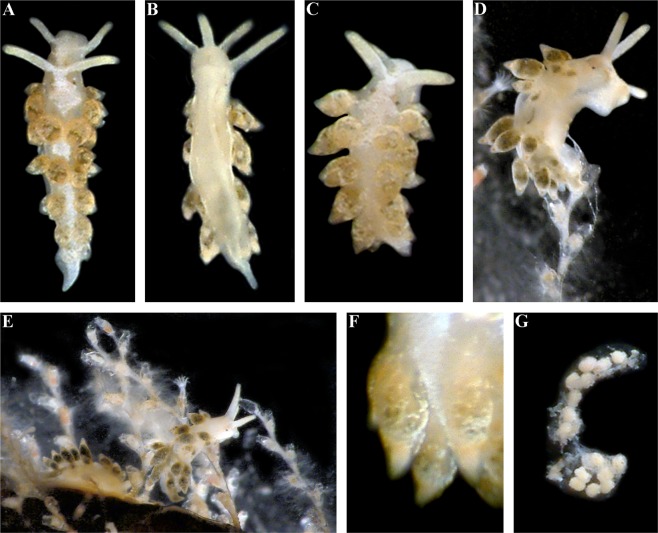
Trinchesia diljuvia sp. n. External views of living specimens. (A) Holotype, Black Sea, Yalta region (ZMMU Op-642) dorsal view; (B). Holotype, ventral view; (C). Paratype, Black Sea, Yalta region, dorsal view (ZMMU Op-645); (D). Paratype, Black Sea, Yalta region (ZMMU Op-643), lateral right view; (E). Living specimens from same location on food sources, hydroids; (F). Details cerata; (G). Egg mass from specimens from the same locality. Photographs: Tatiana Korshunova, Alexander Martynov.
Material examined
Holotype
Black Sea, Yalta region, Russia (44° 29′ N 34° 10′ E), depth 1–2 m, stones, collector T.A. Korshunova, A.V. Martynov, 24–26.07.2004, (ZMMU Op-642, 4.5 mm live, ca. 2 mm in length, preserved). Paratypes. Black Sea, Yalta region, Russia (44° 29′ N 34° 10′ E), depth 1–2 m, stones, collector T.A. Korshunova, A.V. Martynov, 28.07.2004, 1 specimen, dissected (ZMMU Op-643, ca. 3 mm live, ca. 1.5 mm in length, preserved). Black Sea, Yalta region, Russia (44° 29′ N 34° 10′ E), depth 1–2 m, stones, collector T.A. Korshunova, A.V. Martynov, 31.07.2004, 1 specimen, dissected (ZMMU Op-645, ca. 3.5 mm live, ca. 2 mm in length, preserved).
Zoobank registration
urn:lsid:zoobank.org:act: C9228E8B-DF2C-46A0-9E78-038ED6B87D7B.
Diagnosis
Body up to 4.5 mm, white dorsal line, no colour zones on light brownish (with traces of bluish pigment) cerata, commonly two, maximum three, anterior ceratal rows, radular formula 27–34 × 0.1.0, penial stylet short slightly curved, seminal receptacle is rounded without any additional chamber, egg mass short semi-spiral.
Etymology
Combination of Latin, diluviim, deluge, inundation and juvenilis, youthful, alluding to the paedomorphic appearance of the new species and speciation during the formation of the modern Black Sea after the Holocene meltwater events (see Discussion).
Description
External morphology
The length of the holotype is 4.5 mm (Fig. 9). The length of adults reaches 4.5 mm. The body is narrow. The rhinophores are smooth and 1.5 times longer than the oral tentacles. The cerata are short, distinctly spindle-shaped. Ceratal formula of the holotype: right (1,2,3; anus, 3,2,2,1) left (1,2,3; 3,2,2,1). The foot is narrow anteriorly without foot corners or angular processes.
Colour
The basal colour is whitish; a distinct thick white line runs dorsally with median expansion (Fig. 9). The cerata and digestive gland lack distinct orange and bright blue colour zones as seen in the previous three species. Most parts of the cerata are dark grayish to light brownish or light greenish and covered with white or sometimes with light bluish dispersed pigment. Sometimes towards the top of the cerata there is a thin darker band between the brownish zone and white covering. The tips of the cerata are clear with a pinkish cnidosac. The tips of rhinophores and oral tentacles are encrusted with dense white-yellowish to greenish pigment.
Anatomy
Digestive system
The jaws are triangularly ovoid (Fig. 10C) The masticatory processes of the jaws bear a single row of low, inconspicuous denticles (Fig. 10D). The radular formula in the holotype is 27–34 × 0.1.0. The radular teeth are yellowish. The central tooth is narrow, elongated, with low cusp and 4–7 lateral denticles, including smaller intercalated denticles) (Fig. 10A,B).
Reproductive system
(Fig. 11D). The ampulla is moderately short and swollen (Fig. 11D, am). The prostate is a slightly convoluted tube (Fig. 11D, pr). The prostate transits to a penial sheath, which contains a conical penis with a basally slightly curved chitinous stylet (Figs 10E, 11D, ps). A supplementary (“penial”) gland inserts into base of the penis (Fig. 11D, pg). The seminal receptacle is small, rounded, and on a stalk (Fig. 11D, rs). The female gland mass includes mucous and capsular glands (Fig. 11D, fgm).
Description of egg masses
The egg mass is a small semi-spiral cord with only a few eggs (ca. 20) (Fig. 9G).
Distribution and habitats
Black Sea, Yalta region only, not found in the Mediterranean or other regions. Found in stony, shallow areas, around 1–2 m depth, on Sertularella spp. hydroids.
Remarks
Morphological differences
T. diljuvia sp. n. can be distinguished from the North Atlantic and Mediterranean species T. caerulea by this set of morphological data: 1). Absence of distinct colour zones on the cerata, which are present in T. caerulea; 2). Presence of a white dorsal line in T. diljuvia sp. n., which is absent in T. caerulea; 3). Presence of commonly two, rarely three, anterior ceratal rows inT. diljuvia sp. n., whereas there are commonly four in T. caerulea; 4). Absence of anterior foot corners inT. diljuvia sp. n., which are present and long in T. caerulea; 5). Shape and relative length of the penial stylet, which is relatively longer in T. diljuvia sp. n. and relatively shorter in T. caerulea. T. diljuvia sp. n. can be distinguished from the exclusively North Atlantic species T. cuanensis by this set of morphological data: 1). Absence of distinct colour zones on the cerata, which are present in T. cuanensis sp. n.; 2). Presence of a white dorsal line in T. diljuvia sp. n. which is absent in T. cuanensis sp. n.; 3). Presence of commonly two, rarely three, anterior ceratal rows in T. diljuvia sp. n., whereas there are commonly four in T. cuanensis sp. n.; 4). Absence of anterior foot corners in T. diljuvia sp. n., which are present and short in T. cuanensis sp. n.; 5). Shape and relative length of the penial stylet, which is relatively very short in T. diljuvia sp. n. compared to the extremely long stylet in T. cuanesis sp. n. T. diljuvia sp. n. can be distinguished from the predominantly Mediterranean species T. morrowae sp. n. by this set of morphological data: 1). Absence of distinct colour zones on the cerata, which are present in T. morrowae sp. n.; 2). Presence of commonly two, rarely three, anterior ceratal rows in T. diljuvia sp. n., whereas there are commonly three in T. morrowae sp. n.; 3). Absence of anterior foot corners in T. diljuvia sp. n., whereas distinct angular projections are present inT. morrowae sp. n..; 4). Shape and relative length of the penial stylet, which is relatively longer in T. diljuvia sp. n. and relatively shorter in T. morrowae sp. n.
Molecular differences
Minimum uncorrected COI p-distances between the T. diljuvia sp. n. type specimen and T. caerulea, T. cuanensis sp. n. and T. morrowae sp. n., specimens are 11.89%, 12.99%, and 2.98% respectively. See also Discussion, Fig. 12 for integration of molecular phylogenetic and morphological data, and supplementary information, Tables S1–S5.
Discussion
The particular case of a nudibranch species complex and the general ‘cryptic species’ problem
This case of a colourful nudibranch species that represents an apparently very cryptic T. caerulea complex from Britain and Ireland through continental Europe and the Mediterranean to the Black Sea, i.e., with a putative low degree of morphological disparity in the sense of Struck et al.9, in turn reveals both stable external and internal features (Figs 1–12, Table S5, where external and internal distinguishing features for all four species are summarized). Prior to this study (T. caerulea was described initially from the UK more than 200 years ago), these putative morphs within the single T. caerulea species were always claimed to be “internally the same/similar”16,34, thus fulfilling the current definition9 of a ‘cryptic’ species.
To challenge this notion that ‘cryptic’ species can only be defined a posteriori by the molecular data18,19 we specifically included the illustration from Thompson & Brown (1984)16 (Fig. 12) to show that the separate species described here are recognizable even from the external morphological features (Figs 1, 12), but were considered at that time to be just a variation of T. caerulea, i.e., in this case this species complex could be regarded as ‘pseudocryptic’ prior to a molecular study. In this respect, it could be argued that we are confident about the separate species status of new species because we now have molecular data, whereas previous authors did not. However, the two common sympatric British and Irish species involved in this study, though they have a putatively similar/identical colour pattern, show very different characters at another level [i.e., penial stylets (Fig. 12)] which were overlooked in all previous studies but this trait implies a great obstacle during potential inter-species copulation. If these substantial differences in penial stylets would have been detected during previous morphological studies16, the question about their separate species status would have been raised much earlier. These data immediately contradict the updated definition of ‘cryptic species’9 which requires ‘cryptic species’ to be genetically isolated but morphologically non-distinguishable. Despite that this species complex was potentially morphologically distinguishable using fine details even in the pre-molecular era16 (and hence can be classified a ‘pseudocryptic’ species18, or ‘normal’ species not a posteriori to the molecular data but a priori), it remained almost absolutely ‘cryptic’ until recently because smaller distinguishing taxonomic units were not defined at that time and researchers decided they could not plausibly grant them a separate species status under what, at the time, was a dominant morphological “lumping” taxonomic framework. Therefore, this species complex can be considered ‘cryptic’, ‘pseudocryptic’ and ‘non-cryptic’ at the same time. The novel data on multilevel diversity from a particular species complex presented here, therefore, confirm that the general ‘cryptic species’ concept is far from being a satisfying solution and that the commonly used ‘cryptic species’ concept needs to be reconsidered.
The term ‘cryptic species’ has a vague definition and includes non-cryptic diversity
Struck et al.9 attempted to provide a stricter definition of cryptic species and proposed two main components of an updated definition: “diverged genotypic clusters of individuals (reflecting reproductive isolation) that do not form diagnostic morphological clusters” and the temporal dimension of “lower degrees of phenotypic (or more specifically morphological) disparity than non-cryptic relatives”. The central proposal of the new updated ‘cryptic’ species definition9 has a deficiency in a heavy dependence on a detailed study of a species/organism and an existing taxonomic framework of a particular group. For example, most of the European nudibranch species of the genus Dendronotus were just a single, very complicated and non-diagnosable ‘cryptic’ species, even for experts, but a recent study revealed that it is possible to provide not only molecular values, but also morphological differences for every species in the complex8. Furthermore, the requirements9 for a cryptic species to be morphologically non-distinguishable but genetically isolated is also controversial, since there are many examples where genetically separate species either are similar morphologically (as is the case for the blue mussel species complex) or considerably different as is the case for dolphins and whales, but in both groups numerous cases of genetic introgression and/or presence of fertile hybrids have been demonstrated42,43. As an answer to a recent proposal to consider ‘cryptic’ species rather as a temporary term8 some authors continued to argue for validity of the cryptic species9 concept and mentioned previous studies of such different animal groups as lizards, annelids, molluscs, cnidarians, and mammals as examples of putatively “uniform” morphology among different species, which, however, under scrutiny may demonstrate diagnosable morphological taxa. Notably, in the example with an annelid species44 it was indicated that “the cryptic species complex of Stygocapitella subterranea is not as cryptic as assumed as the Australian populations are morphologically and genetically different”.
If we claim that we cannot find any morphologically diagnosable structures in a ‘cryptic’ species complex, this would imply that we have exhaustively studied organism features and are able to unambiguously present all existing ‘macromorphological’ and ‘micromorphological’ features for a given organism. This is obviously impossible at the current level of our technology and knowledge. The putative strict distinction between morphological and molecular levels of organisms, on which the cryptic species concept is largely relying, loses clarity when we proceed to the tissue or cellular ‘micromorphological’ level. To the utmost degree, morphologically absolutely identical “body doubles”3 can be formed only on the basis of zygote clones that automatically imply its identical genetic structure. If we instead discovered putative “species clones” but with some considerable genetic differences, this most likely suggests that we have missed some morphological differences during a study. Therefore, it is biologically impossible for species to be “physically indistinguishable from each other”1,18,19 since even clones can be different from each other because of epigenetic mechanisms45–48.
In practice there is an array of various cases, from very highly similar external species, for which authors reported that they have not yet found confident morphological differences, e.g., in American lizards49, Japanese microsnails50, coryphellid nudibranchs of the genus Gulenia25 to species which are externally difficult to distinguish (but still possible) but internally demonstrate stable distinct characters, e.g., in the European nudibranch species of the genus Dendronotus8. This series continues with species which demonstrate some overall similarity and appear cryptic but under more detailed study turn out to demonstrate reliable and stable external and internal differences, like a polychaete species complex44 and the present case of the four Trinchesia species complex. Finally, there are the cases when the term ‘cryptic’ species was applied to externally diagnosable species (e.g. in the echinoderm ophiuroids13). Thus, a considerable amount of non-cryptic diversity is still currently included under the ‘cryptic species’ concept. It is therefore likely that if we really would like to make a distinction between ‘species’ and ‘cryptic’ species we will need a much more differentiated system of terminology than this simplistic pair ‘cryptic’ vs. ‘non-cryptic’. If we use the term ‘cryptic species’ conditionally and for the purpose of this discussion only, for example, additional terms might be ‘true cryptic species’ (morphological differences have not yet been found), ‘semi-cryptic species’ (morphological differences are very difficult to present), ‘quasi-cryptic species’ (morphological differences are relatively easy to present) and finally a ‘false cryptic species’ (morphological differences are obvious, but for some reason were missed or not highlighted in previous studies). However, while we can use such terminology for a preliminary sorting of multilevel diversity, quite obviously, the number of such various intermediate terms would grow exponentially, and it may be difficult to apply to other taxa since many cases may represent their own unique combination of genetic divergence/uniformity and morphological external and internal similarity/disparity. Remarkably, the last putative term in this series, ‘false cryptic species’ actually does not differ fundamentally from the basic term ‘species’, and the term ‘pseudocryptic species’ was previously applied to cases where it is possible to show at least some morphological differences4,18. It has already been noted that the line between cryptic and pseudocryptic “is not sharp”51, but these terms have been loosely applied until the meaning is lost52. Notably, the term ‘cryptic species’ has been applied53 to evaluate a nudibranch species revealed in a previous study54 to be “very similar to Limacia clavigera” yet with “consistent morphological and external differences between the two species examined” but without any corresponding molecular data for the morphologically-only defined “cryptic” Limacia iberica Caballer et al., 201654 species. Yet a previous proposal19 noted that cryptic species are, by definition, molecularly different species that can only turn out to be morphologically distinguishable ‘pseudocryptic’ species a posteriori. Thus, such highly inconsistent usage of the term ‘cryptic’ species confirms the most recent notion of Heethoff10 that the ‘cryptic’ species concept currently represents both conceptual and terminological chaos.
Taking also into consideration that one of the united definitions of the term ‘species’ defines it as only being separately evolved metapopulations, which are not required to be morphologically different or even reproductively isolated55, this in turn implies that any species at its origin is already completely ‘cryptic’. Indeed, many so called ‘cryptic’ species represent recently diverged lines and therefore their overall similarity may rely on significant similarity in the developmental genes responsible for the formation of the morphological structures, rather than on some mutations that may occur in the housekeeping genes which are commonly used for taxonomic purposes. More wide applications of full genomic/transcriptomic comparison may therefore potentially contribute to finding not only morphological but also high genetic similarity within recently diverged closely related lineages. Hence the term ‘super-cryptic’ species would be needed for the species without morphological, genetic or ecological differences which though probable, is only theoretically possible.
Therefore, these apparent ‘cryptic’ species with “low morphological disparity” instead turned out to be a species which previously was “insufficiently studied” at different levels of morphological organisation. Thus, at a given time technological and scientific achievements allow us to understand some species and present their descriptions in more and more detail using the dominant paradigm at the time for species descriptions that may easily lead to some well-distinguishable features being missed. As a result, when a new study uncovers new details, a species can be easily considered as not ‘cryptic’ or ‘pseudocryptic’ anymore, but just a normally diagnosable species. This is the exact situation of the present discovery of well-diagnosable morphological patterns at different levels in the genus Trinchesia, so that what was a totally cryptic and non-diagnosable complex a few years ago is now four well-diagnosable and non-cryptic separate species. Even if complexes in other organism groups that are apparently even more ‘cryptic’ than the present case25,49,50,56 still exist, and potentially can be discovered further within that nudibranch species complex, this does not mean that after some time we will not be able to study their morphological structures using, for example, more advanced micro CT or a similar technique available to future generations, including potential detailed scanning of living animals, and also discovering fine details of their ecology, etc., and will be able therefore to elaborate and define well-diagnosable taxonomic units, as we have presented here for the British and Mediterranean Trinchesia species. Another nudibranch species that previously was incorrectly identified as “T. caerulea” from the Caribbean Sea was shown to be considerably different from the true T. caerulea both by morphological and molecular data57. Morphologically distinguishing characters, therefore, should not be identified only a posteriori18 but should have at least similar weight with the molecular ones. A molecular study may promote discovery of a hidden diversity within a group, but a robust taxonomic framework can be built only in an agreement with morphological data, even if such a task is difficult and requires a more detailed study such as those undertaken for the other nudibranchs8,58, when a considerable amount of external interspecific variability is present and some species were externally nearly identical to other related but different species. In turn, in a completely opposite situation, the presence of morphologically well-recognized species which have a very low degree of molecular divergence from related species1,59,60 clearly indicates the necessity of restoring the significance of morphology to the diagnoses of species in the form of detailed morphological data.
Fine-scale multilevel framework for studying species
When we discovered several new species within a complex traditionally assigned to a single species, we created a new morphological and molecular framework that defined much smaller taxonomically distinguishable units than were previously used. Because life is hierarchically organized61 there is a multilevel diversity in character evolution8, and correspondingly, further smaller taxonomically relevant morphological and molecular concordant units can be discovered within previously selected larger taxonomic groups. This process can be potentially quite endless, or at least take a considerable amount of time, while we set an increasingly narrower scale for such morphological and molecular taxonomic units (those we evaluate as species under each given taxonomic framework), until we arrive at very small and narrowly defined species units, which may still include some amount of variability and the ability to hybridize with closely related, narrowly defined species. During the processes of decreasing the degree of disparity of morphological and molecular taxonomic units, the degree of their ‘crypticity’ is at first greatly increased because we are further attempting to find distinguishing features within increasingly smaller taxonomic units.
Therefore, instead of the very problematic division of this multilevel continuum into ‘non- cryptic’ and ‘cryptic’ components, we conclude that almost every species potentially hides numerous smaller and smaller units that need careful detection and description using an ever greater differentiating system of morphological and molecular characters, as was presented here for this case of ‘cryptic’ morphs within a nudibranch species complex (see detailed morphological comparative remarks after description of every species above). The problem was not in the absence of “physically distinguishable morphological characters”, but in the absence of enough finely differentiated units in the taxonomic framework that would have allowed species distinction at a much finer level.
Using the new framework established here for the T. caerulea species complex (Figs 1–12; Tables S1–S5), which comprises smaller and more differentiated taxonomic levels, we can now start to further explore the taxonomic diversity within that complex, with the additional possibility that we will detect new, even smaller and more finely differentiated morphological and molecular taxonomic levels. So, the possible new taxonomically distinguishing characters of potentially (not yet discovered) smaller levels of taxonomic diversity within T. caerulea (i.e. not yet discovered potential new species in that complex) would be very ‘cryptic’ and would have inevitably been considered just small variations within the previous framework, but within the new morphological and molecular framework, they will be ‘less cryptic’ than before. This general consideration and the remarkable practical case in the present study clearly show that it is meaningless to use this simplistic ‘cryptic-pseudocryptic’ species pairing while dealing with the enormous continuum of highly differentiated diversity at smaller and smaller levels, each of which is notably more and more ‘cryptic’ than the previous one. Instead, we are proposing two interlinked central implications of the present study: “from ‘cryptic’ to obvious species” within more and more finely differentiated numerous taxonomic levels.
Furthermore, because underestimation of the epigenetic mechanisms in taxonomic practice, the ‘cryptic’ species label may prevent the discovery of such an important evolutionary phenomenon as paedomorphosis, which is now recognized in different groups23,26,62,63. While we were analysing integrative morphological and molecular data for this nudibranch species complex we discovered a new Black Sea species, Trinchesia diljuvia sp. n., that is externally similar to the Mediterranean T. morrowae sp. n. (from which it is molecularly considerably diverged) and thus can be classified under the “low morphological disparity” general paradigm9 as morphologically uniform, but actually demonstrates stable and clear paedomorphic features (Figs 1, 9 and 12). The formation of the modern Black Sea, approximately 8000–9000 years ago64, makes the present example of paedomorphosis-related speciation in the new nudibranch species share the top spot for the most rapid animal speciation in the world. Only one recent comparable study on a marine fish suggested a very rapid degree of speciation of similar age (ca. 6000-8000 years ago)65.
Under this fine-scale multilevel species framework, morphological and molecular data, as well the other biological data, should equally contribute to the definition and description of any species. Most recent data show that taxonomists are gradually moving in this direction. Among 55 species of the putative ‘cryptic’ species of the amphipod crustacean which were recently investigated by integrative methods, biological differences (other than molecular differences) were not yet found for only four species5. A study on a polychaete family recently appeared which does not refute the possibility of finding morphologically distinguishing characters even within an apparent ‘mega-cryptic’ species complex66.
Concluding remarks on the ‘cryptic’ species problem
How to name morphologically difficult-to-distinguish species diversity is not just a terminological problem9,10 but a serious theoretical and practical problem. Initially, the term ‘cryptic species’ has been applied when considerable molecular divergence was discovered within apparently morphologically similar species groups3,18,67. However, currently the term ‘cryptic species’ is greatly over used because researchers tend to firstly investigate molecular data and only secondly investigate morphology. Thus, for such studies all species are ‘cryptic’ before molecular study and afterwards they are named ‘pseudocryptic’ species18,52. Despite that, in our present study we show that previously recognized morphologically cryptic morphs in a nudibranch species complex are concordant with the newly obtained molecular differences and constitute well-defined morphological and molecular taxonomic units, and this species complex can therefore be termed simultaneously ‘cryptic’ and ‘non-cryptic’. Thus, the degree of ‘crypticity’ is a subjective and uncertain measure68.
We therefore propose to avoid the terms ‘cryptic’/’pseudocryptic’ species and in cases where taxonomy is not yet settled use the more neutral terms ‘hidden’ or ‘potentially unraveled/undiscovered’ diversity. The following scheme to explore hidden diversity is suggested: 1) Within a broader taxonomic framework, detect potential cases where species are difficult to distinguish by morphological data; 2) These difficult cases should be studied exhaustively at the current level of technological development; 3) Morphological and molecular methods should be applied in concordance, and ecological and any other relevant data can equally be primary sources to reveal fine but taxonomically reliable differences; 4) As a result of setting the ‘finer and finer taxonomic scale’ in a species complex, a new, more differentiated taxonomic framework will be established. Presumption of the ‘existence of morphological differences’ should be applied at every level of taxonomic study. Justification of such presumption is the biological impossibility of the existence of two genetically different but morphologically completely identical species. Accordingly, if in some cases we are unable to present definite morphological differences this means that we are just currently unable to detect them and need to postpone this unresolved question for a future study, but we cannot just state that morphological differences do not exist.
Facing the rapid extinction of many organisms, including marine ones69,70, we should concentrate on describing the patterns of this immense fine-scale multilevel diversity, rather than relying on attempts to merely roughly distinguish ‘cryptic’ species from ‘non-cryptic’ ones. Putative “body doubles”3,71 should, under scrutiny, always turn out to be neither doubles nor cryptic, but currently it has become fashionable to name any apparently taxonomically difficult group a ‘cryptic species’. Continued use of this term will only add ambiguity to the immense field of biodiversity, where an enormous number of species still need description, and the ‘cryptic’ concept is rather an obstacle than an aid. The term ‘cryptic species’ should only be regarded as a temporary state. Though the ‘cryptic species concept’ has played a role in emphasizing the importance of reviewing morphologically difficult to distinguish species, currently there is a necessity for a new framework for the species concept itself 5,72. The present study thus demands the establishment of such a new, fine-scale multilevel species paradigm through which can we approach a less subjective understanding of what ‘high’ and ‘low’ taxonomic disparity is, and is far from the simplistic ‘cryptic species’ notion which is currently dominant.
Supplementary information
Acknowledgements
Gregory Brown is warmly thanked for permission to use original figures from the Thompson & Brown (1984) monograph. We would like to thank Mats Larsson, Jim Anderson, Lydia White, Greg Brown and all the other participants in the Irish Nudibranch festivals from 2014 to 2017 for their help in collection of some of the specimens from Ireland. We give special thanks to the team of Gulen Dive Center (Christian Skauge, Ørjan Sandnes, Monica Bakkeli and Guido Schmitz) and to Torkild Bakken (NTNU University Museum) for their generous help during fieldwork in Norway. Kate Lock, of the Skomer Marine Nature Reserve team in Pembrokeshire organised and helped with travel costs for Bernard Picton to visit Pembrokeshire in 2013. Jakov Prkić would like specially to thank his friends Alen Petani and Đani Iglić for providing many specimens and photos from Croatia. Klas Malmberg and Kennet Lundin would like to thank the staff at the Smögen dive centre (Sweden). Electron Microscopy Laboratory, Moscow State University is gratefully acknowledged for support with the scanning electron microscopy. This work was supported by a research grant of MSU Zoological Museum (АААА-А16-116021660077-3, depository of specimens), in the frames of Moscow University Project “Noah’s Ark” (morphological & molecular study) and RFBR grant №17-04-01827а (molecular study).
Author Contributions
The manuscript was written by T.K., B.P., M.P., J.P., K.F., K.M., K.L. and A.M. T.K., B.P. and A.M. performed analyses. T.K. prepared the illustrations. T.K., B.P., G.F., P.M., M.P., J.P., K.F., K.M., K.L. and A.M. obtained and interpreted the data.
Competing Interests
The authors declare no competing interests.
Footnotes
Publisher’s note: Springer Nature remains neutral with regard to jurisdictional claims in published maps and institutional affiliations.
Supplementary information
Supplementary information accompanies this paper at 10.1038/s41598-019-42297-5.
References
- 1.Bickford D, et al. Cryptic species as a window on diversity and conservation. Tren. Ecol. Evol. 2007;22:148–155. doi: 10.1016/j.tree.2006.11.004. [DOI] [PubMed] [Google Scholar]
- 2.Gill BA, et al. Cryptic species diversity reveals biogeographic support for the ‘mountain passes are higher in the tropics’ hypothesis. Proc. R. Soc. Lond. B. 2016;283:20160553. doi: 10.1098/rspb.2016.0553. [DOI] [PMC free article] [PubMed] [Google Scholar]
- 3.Saéz AG, Lozano E. Body doubles. Nature. 2005;433:111. doi: 10.1038/433111a. [DOI] [PubMed] [Google Scholar]
- 4.Jörger KM, Schrödl M. How to describe a cryptic species? Practical challenges of molecular taxonomy. Front. Zool. 2013;10:59. doi: 10.1186/1742-9994-10-59. [DOI] [PMC free article] [PubMed] [Google Scholar]
- 5.Fišer C, Robinson CT, Malard F. Cryptic species as a window into the paradigm shift of the species concept. Mol. Ecol. 2018;27:613–635. doi: 10.1111/mec.14486. [DOI] [PubMed] [Google Scholar]
- 6.Knowlton N. Sibling species in the sea. Ann. Rev. Ecol. Syst. 1993;24:189–216. doi: 10.1146/annurev.es.24.110193.001201. [DOI] [Google Scholar]
- 7.Franks DW, Noble J. Warning signals and predator–prey coevolution. Proc. R. Soc. Lond. B. 2004;271:1859–1865. doi: 10.1098/rspb.2004.2795. [DOI] [PMC free article] [PubMed] [Google Scholar]
- 8.Korshunova TA, Martynov AV, Bakken T, Picton BE. External diversity is restrained by internal conservatism: New nudibranch mollusc contributes to the cryptic species problem. Zool. Scripta. 2017;46:683–692. doi: 10.1111/zsc.12253. [DOI] [Google Scholar]
- 9.Struck TH, et al. Finding evolutionary processes hidden in cryptic species. Trends Ecol. Evol. 2018;33:153–163. doi: 10.1016/j.tree.2017.11.007. [DOI] [PubMed] [Google Scholar]
- 10.Heethoff M. Cryptic species – conceptual or terminological chaos? Trends Ecol. Evol. 2018;33:310. doi: 10.1016/j.tree.2018.02.006. [DOI] [PubMed] [Google Scholar]
- 11.Beermann J, et al. Cryptic species in a well-known habitat: applying taxonomics to the amphipod genus Epimeria (Crustacea, Peracarida) Sci. Rep. 2018;8:6893. doi: 10.1038/s41598-018-25225-x. [DOI] [PMC free article] [PubMed] [Google Scholar]
- 12.Kanturski M, Lee Y, Choi J, Lee S. DNA barcoding and a precise morphological comparison revealed a cryptic species in the Nippolachnus piri complex (Hemiptera: Aphididae: Lachninae) Sci. Rep. 2018;8:8998. doi: 10.1038/s41598-018-27218-2. [DOI] [PMC free article] [PubMed] [Google Scholar]
- 13.Okanishi M, Sentoku A, Martynov A, Fujita T. A new cryptic species of Asteronyx Müller and Troschel, 1842 (Echinodermata: Ophiuroidea), based on molecular phylogeny and morphology, from off Pacific coast of Japan. Zool. Anz. 2018;274:14–33. doi: 10.1016/j.jcz.2018.03.001. [DOI] [Google Scholar]
- 14.Garnett ST, Christidis L. Taxonomy anarchy hampers conservation. Nature. 2017;546:25–27. doi: 10.1038/546025a. [DOI] [PubMed] [Google Scholar]
- 15.Thomson SA, et al. Taxonomy based on science is necessary for global conservation. PLoS Biol. 2018;16:e2005075. doi: 10.1371/journal.pbio.2005075;. [DOI] [PMC free article] [PubMed] [Google Scholar]
- 16.Thompson, T. E. & Brown, G. H. Biology of opisthobranch molluscs. Vol. 2. (The Ray Society Publications, 1984).
- 17.Anderson, J. & Picton, B. Scottish Nudibranchs. (Kindle Edition, 2017).
- 18.Sáez AG, et al. Pseudo-cryptic speciation in coccolithophores. PNAS. 2003;100:7163–7168. doi: 10.1073/pnas.1132069100. [DOI] [PMC free article] [PubMed] [Google Scholar]
- 19.Lindsay T, Valdés Á. The model organism Hermissenda crassicornis (Gastropoda: Heterobranchia) is a species complex. PLoS ONE. 2016;11:e0154265. doi: 10.1371/journal.pone.0154265. [DOI] [PMC free article] [PubMed] [Google Scholar]
- 20.Zhang F, et al. Species delimitation in the morphologically conserved Coecobrya (Collembola: Entomobryidae): A case study integrating morphology and molecular traits to advance current taxonomy. Zool. Scripta. 2018;47:342–356. doi: 10.1111/zsc.12279. [DOI] [Google Scholar]
- 21.Karanovic T, Djurakic M, Eberhard SM. Cryptic species or inadequate taxonomy? Implementation of 2D geometric morphometrics based on integumental organs as landmarks for delimitation and description of copepod taxa. Syst. Biol. 2016;65:304–327. doi: 10.1093/sysbio/syv088. [DOI] [PubMed] [Google Scholar]
- 22.Furfaro, G., Picton, B., Martynov, A. & Mariottini, P. Diaphorodoris alba Portmann & Sandmeier, 1960 is a valid species: molecular and morphological comparison with D. luteocincta (M. Sars, 1870) (Gastropoda: Nudibranchia). Zootaxa, 4193, 304–316 (2016). [DOI] [PubMed]
- 23.Korshunova TA, Lundin K, Malmberg K, Picton B, Martynov AV. First true brackish water nudibranch mollusc provides new insights for phylogeny and biogeography and reveals paedomorphosis driven evolution. PLoS ONE. 2018;13:e0192177. doi: 10.1371/journal.pone.0192177. [DOI] [PMC free article] [PubMed] [Google Scholar]
- 24.Morrow CC, et al. Congruence between nuclear and mitochondrial genes in Demospongiae: A new hypothesis for relationships within the G4 clade (Porifera: Demospongiae) Mol. Phyl. Evol. 2012;62:174–190. doi: 10.1016/j.ympev.2011.09.016. [DOI] [PubMed] [Google Scholar]
- 25.Korshunova TA, et al. Polyphyly of the traditional family Flabellinidae affects a major group of Nudibranchia: aeolidacean taxonomic reassessment with descriptions of several new families, genera, and species (Mollusca, Gastropoda) ZooKeys. 2017;717:1–139. doi: 10.3897/zookeys.717.21885. [DOI] [PMC free article] [PubMed] [Google Scholar]
- 26.Korshunova TA, Martynov AV, Picton BE. Ontogeny as an important part of integrative taxonomy in tergipedid aeolidaceans (Gastropoda: Nudibranchia) with a description of a new genus and species from the Barents Sea. Zootaxa. 2017;4324:1–22. doi: 10.11646/zootaxa.4324.1.1. [DOI] [Google Scholar]
- 27.Korshunova TA, Fletcher K, Lundin K, Picton BE, Martynov AV. The genus Zelentia is an amphi-boreal taxon expanded to include three new species from the North Pacific and Atlantic oceans (Gastropoda: Nudibranchia: Trinchesiidae) Zootaxa. 2018;4482:297–321. doi: 10.11646/zootaxa.4482.2.4. [DOI] [PubMed] [Google Scholar]
- 28.Puillandre N, Lambert A, Brouillet S, Achaz G. ABGD, Automatic Barcode Gap Discovery for primary species delimitation. Mol. Ecol. 2011;21:1864–1877. doi: 10.1111/j.1365-294X.2011.05239.x. [DOI] [PubMed] [Google Scholar]
- 29.Struck, et al. Cryptic species – more than terminological chaos: A reply to Heethoff. Trends Ecol. Evol. 2018;33:310–312. doi: 10.1016/j.tree.2018.02.008. [DOI] [PubMed] [Google Scholar]
- 30.Dayrat B. Toward integrative taxonomy. Biol. J. Linn. Soc. 2005;85:407–415. doi: 10.1111/j.1095-8312.2005.00503.x. [DOI] [Google Scholar]
- 31.Picton, B.E. & Morrow, C. A field guide to the nudibranchs of the British Isles. (Immel Publishing, 1994).
- 32.Padula V, Araújo AK, Matthews-Cascon H, Schrödl M. Is the Mediterranean nudibranch Cratena peregrina present in the Brazilian coast? Integrative species delimitation and description of Cratena minor n. sp. J. Moll. Stud. 2014;80:575–584. doi: 10.1093/mollus/eyu052. [DOI] [Google Scholar]
- 33.Hayward, P. J. & Ryland, J. S. Handbook of the marine fauna of north-west Europe, 2nd ed. (Oxford University Press 2017).
- 34.Schmekel L, Portmann A. Opisthobranchia des Mittelmeeres, Nudibranchia und Saccoglossa. Fauna e flora del Golfo di Napoli. 1982;40:1–410. [Google Scholar]
- 35.Montagu G. Description of several marine animals found on the south coast of Devonshire. Trans. Linn. Soc. London. 1804;7:61–85. doi: 10.1111/j.1096-3642.1804.tb00282.x. [DOI] [Google Scholar]
- 36.Verany, G. B. Catalogo degli animali invertebrati marini del Golfo di Genova e Nizza. (Genova: Tipografia Ferrando 1846).
- 37.Dalyell, J. G. Observations on life amidst the various forms of the humbler tribes of animated nature: with practical comments and illustrations. (London: John van Voorst 1853).
- 38.Alder J, Hancock A. Notices of some new and rare British species of naked Mollusca. Ann. Mag. Nat. Hist. 1846;18:289–294. doi: 10.1080/037454809496586. [DOI] [Google Scholar]
- 39.Herdman WA. Additional notes to the invertebrate fauna of Lamlash Bay. Proc. Roy. Phys. Soc. Edin. 1881;6:17–30. [Google Scholar]
- 40.Miller MC. Distribution and food of the nudibranchiate Mollusca of the south of the Isle of Man. J Anim. Ecol. 1961;30:95–116. doi: 10.2307/2116. [DOI] [Google Scholar]
- 41.Vicente N. Mollusques opisthobranches récoltés en plongée dans le Golfe de Marseille. Rec. Trav. Sta. Mar. Endoume. 1963;31:173–185. [Google Scholar]
- 42.Fraïsse C, Belkhir K, Welch JJ, Bierne N. Local interspecies introgression is the main cause of extreme levels of intraspecific differentiation in mussels. Mol. Ecol. 2016;25:269–286. doi: 10.1111/mec.13299. [DOI] [PubMed] [Google Scholar]
- 43.Crossman CA, Taylor EB, Barrett-Lennard LG. Hybridization in the Cetacea: widespread occurrence and associated morphological, behavioral, and ecological factors. Ecol. Evol. 2016;26:1293–1303. doi: 10.1002/ece3.1913. [DOI] [PMC free article] [PubMed] [Google Scholar]
- 44.Struck TH, Koczula J, Stateczny D, Meyer C, Purschke G. Two new species in the annelid genus Stygocapitella (Orbiniida, Parergodrilidae) with comments on their biogeography. Zootaxa. 2017;4286:301–332. doi: 10.11646/zootaxa.4286.3.1. [DOI] [Google Scholar]
- 45.Fraga MF, et al. Epigenetic differences arise during the lifetime of monozygotic twins. PNAS. 2005;26:10604–10609. doi: 10.1073/pnas.0500398102. [DOI] [PMC free article] [PubMed] [Google Scholar]
- 46.Wong AHC, Irving I, Petronis GA. Phenotypic differences in genetically identical organisms: the epigenetic perspective. Human Mol. Gen. 2005;14:R11–R18. doi: 10.1093/hmg/ddi116. [DOI] [PubMed] [Google Scholar]
- 47.Casadesús J, Low DA. Programmed heterogeneity: epigenetic mechanisms in Bacteria. J. Biol. Chem. 2013;288:13929–13935. doi: 10.1074/jbc.R113.472274. [DOI] [PMC free article] [PubMed] [Google Scholar]
- 48.Perez MF, Francesconi M, Hidalgo-Carcedo C, Lehner B. Maternal age generates phenotypic variation in Caenorhabditis elegans. Nature. 2017;552:106–109. doi: 10.1038/nature25012. [DOI] [PMC free article] [PubMed] [Google Scholar]
- 49.Singhal Sonal, Hoskin Conrad J, Couper Patrick, Potter Sally, Moritz Craig. A Framework for Resolving Cryptic Species: A Case Study from the Lizards of the Australian Wet Tropics. Systematic Biology. 2018;67(6):1061–1075. doi: 10.1093/sysbio/syy026. [DOI] [PubMed] [Google Scholar]
- 50.Wada S, Kameda Y, Chiba S. Long-term stasis and short-term divergence in the phenotypes of microsnails on oceanic islands. Mol. Ecol. 2013;22:4801–4810. doi: 10.1111/mec.12427. [DOI] [PubMed] [Google Scholar]
- 51.Nygren A. Cryptic polychaete diversity: a review. Zool. Scripta. 2014;43:172–183. doi: 10.1111/zsc.12044. [DOI] [Google Scholar]
- 52.Matsuda SB, Gosliner TM. Glossing over cryptic species: Descriptions of four new species of Glossodoris and three new species of Doriprismatica (Nudibranchia: Chromodorididae) Zootaxa. 2018;4444:501–529. doi: 10.11646/zootaxa.4444.5.1. [DOI] [PubMed] [Google Scholar]
- 53.Uribe R, Sepúlveda F, Goddard JHR, Valdés Á. Integrative systematics of the genus Limacia O. F. Müller, 1781 (Gastropoda, Heterobranchia, Nudibranchia, Polyceridae) in the Eastern Pacific. Mar. Biod., early online view. 2017 doi: 10.1007/s12526-017-0676-5. [DOI] [Google Scholar]
- 54.Caballer M, Almón B, Pérez J. The sea slug genus Limacia Müller, 1781 (Mollusca: Gastropoda: Heterobranchia) in Europe. Cahiers Biol. Mar. 2016;57:35–42. [Google Scholar]
- 55.de Queiroz K. Species concepts and species delimitation. Syst. Biol. 2007;56:879–886. doi: 10.1080/10635150701701083. [DOI] [PubMed] [Google Scholar]
- 56.Wilson NG, Schrödl M, Halanych K. Ocean barriers and glaciation: evidence for explosive radiation of mitochondrial lineages in the Antarctic sea slug Doris kerguelenensis (Mollusca, Nudibranchia) Mol. Ecol. 2009;18:965–984. doi: 10.1111/j.1365-294X.2008.04071.x. [DOI] [PubMed] [Google Scholar]
- 57.Valdés Á, Medrano S, Bhave V. A new species of Cuthona Alder and Hancock, 1855 (Gastropoda: Heterobranchia: Nudibranchia: Tergipedidae) from the Caribbean Sea. Natitulus. 2016;130:72–78. [Google Scholar]
- 58.Padula V, et al. A test of colorbased taxonomy in nudibranchs: molecular phylogeny and species delimitation of the Felimida clenchi (Mollusca: Chromodorididae) species complex. Mol. Phyl. Evol. 2016;103:215–229. doi: 10.1016/j.ympev.2016.07.019. [DOI] [PubMed] [Google Scholar]
- 59.Mayer F, von Helversen O. Cryptic diversity in European bats. Proc. R. Soc. Lond. B. 2001;268:1825–1832. doi: 10.1098/rspb.2001.1744. [DOI] [PMC free article] [PubMed] [Google Scholar]
- 60.Wiemers M, Fiedler K. Does the DNA barcoding gap exist? – a case study in blue butterflies. Front. Zool. 2007;4:8. doi: 10.1186/1742-9994-4-8. [DOI] [PMC free article] [PubMed] [Google Scholar]
- 61.Takeuchi N, Kaneko K, Hogeweg P. Evolutionarily stable disequilibrium: Endless dynamics of evolution in a stationary population. Proc. R. Soc. Lond. B. 2016;283:20153109. doi: 10.1098/rspb.2015.3109. [DOI] [PMC free article] [PubMed] [Google Scholar]
- 62.Bhullar BA, et al. Birds have paedomorphic dinosaur skulls. Nature. 2012;487:223–226. doi: 10.1038/nature11146. [DOI] [PubMed] [Google Scholar]
- 63.Bocak L, Kundata R, Fernandez CA, Vogler AP. The discovery of Iberobaeniidae (Coleoptera: Elateroidea): a new family of beetles from Spain, with immatures detected by environmental DNA sequencing. Proc. R. Soc. Lond. B. 2016;283:20152350. doi: 10.1098/rspb.2015.2350. [DOI] [PMC free article] [PubMed] [Google Scholar]
- 64.Herrle JO, et al. Black Sea outflow response to Holocene meltwater events. Sci. Rep. 2018;8:4081. doi: 10.1038/s41598-018-22453-z. [DOI] [PMC free article] [PubMed] [Google Scholar]
- 65.Momigliano P, et al. Extraordinarily rapid speciation in a marine fish. PNAS. 2017;114:6074–6079. doi: 10.1073/pnas.1615109114. [DOI] [PMC free article] [PubMed] [Google Scholar]
- 66.Nygren, et al. A mega-cryptic species complex hidden among one of the most common annelids in the North East Atlantic. PLoS ONE. 2018;13:e0198356. doi: 10.1371/journal.pone.0198356. [DOI] [PMC free article] [PubMed] [Google Scholar]
- 67.Paris CA, Wagner FS, Wagner WH., Jr. Cryptic species, species delimitation, and taxonomic practice in the homosporous ferns. Amer. Fern J. 1989;79:46–54. doi: 10.2307/1547159. [DOI] [Google Scholar]
- 68.Lajus D, Sukhikh N, Alekseev V. Cryptic or pseudocryptic: can morphological methods inform copepod taxonomy? An analysis of publications and a case study of the Eurytemora affinis species complex. Ecol Evol. 2015;5:2374–2385. doi: 10.1002/ece3.1521. [DOI] [PMC free article] [PubMed] [Google Scholar]
- 69.Ripple WJ, et al. World scientists’ warning to humanity: a second notice. BioScience. 2017;67:1026–1028. doi: 10.1093/biosci/bix125. [DOI] [Google Scholar]
- 70.Schrödl, M. & Häussermann, V. BiodiversiTOT. Die globale Artenvielfalt jetzt entdecken, erforschen und erhalten. (Norderstedt, 2017).
- 71.Lee MS, Oliver PM. Count cryptic species in biodiversity tally. Nature. 2016;534:621. doi: 10.1038/534621a. [DOI] [PubMed] [Google Scholar]
- 72.Zachos, F. E. Species concepts in biology: historical development, theoretical foundations and practical relevance. (Springer Nature, 2016).
Associated Data
This section collects any data citations, data availability statements, or supplementary materials included in this article.


