Cardiogenic shock (CS) is a common cause of mortality, and management remains challenging despite advances in therapeutic options. CS is caused by severe impairment of myocardial performance that results in diminished cardiac output, end‐organ hypoperfusion, and hypoxia.1 Clinically this presents as hypotension refractory to volume resuscitation with features of end‐organ hypoperfusion requiring pharmacological or mechanical intervention.1 Acute myocardial infarction (MI) accounts for 81% of patient in CS.2
Contemporary trials and guidelines (Table 1)3, 4, 5, 6, 7 outline clinical criteria for defining CS and are limited by lack of uniformity. The SHOCK (Should We Emergently Revascularize Occluded Coronaries for Cardiogenic Shock) and intra‐aortic balloon pump (IABP)‐SHOCK II trials used systolic blood pressure (SBP) measurements of <90 mm Hg for ≥30 minutes or use of pharmacological and/or mechanical support to maintain an SBP ≥90 mm Hg.1, 3, 4 Evidence of end‐organ hypoperfusion varied between the trials but typically included urine output of <30 mL/h, cool extremities, altered mental status, and/or serum lactate >2.0 mmol/L.1, 3, 4 The SHOCK Trial included cardiac index (CI) of ≤2.2 L/min per m2 and a pulmonary capillary wedge pressure (PCWP) of ≥15 mm Hg.3 An SBP <90 mm Hg that is refractory to fluid resuscitation with clinical and laboratory evidence of end‐organ dysfunction, in the setting of suspected cardiac dysfunction, is essential to the definition of CS. However, CS is a continuum that extends from pre‐shock to refractory shock states, which influence the timely considerations of various interventions.8 Acknowledging this continuum in future trials would likely facilitate the unification of clinical and hemodynamic criteria in defining CS.
Table 1.
Clinical Features of CS as Defined in Contemporary Trials and Guidelines
| Clinical Trial/Guideline | CS Criteria |
|---|---|
| SHOCK Trial (1999)3 |
|
| IABP‐SOAP II (2012)4 |
|
| EHS‐PCI (2012)5 |
|
| ESC‐HF Guidelines (2016)6 |
|
| KAMIR‐NIH (2018)7 |
|
AMS indicates altered mental status; CI, cardiac index; EHS PCI, Euro Heart Survey Percutaneous Coronary Intervention Registry; ESC HF, European Society of Cardiology Heart Failure; IABP‐SOAP II, intra‐aortic balloon pump in cardiogenic shock II; KAMIR‐NIH, Korean Acute Myocardial Infarction Registry‐National Institutes of Health; MAP, mean arterial pressure; PCWP, pulmonary capillary wedge pressure; SBP, systolic blood pressure; SHOCK, Should We Emergently Revascularize Occluded Coronaries for Cardiogenic Shock; UO, urine output.
Epidemiology
CS complicates 5% to 10% of cases of acute MI and is the leading cause of death after MI.1, 9 ST‐segment–elevation myocardial infarction (STEMI) is associated with a 2‐fold increased risk for development of CS compared with non–ST‐segment–elevation myocardial infarction (NSTEMI). Patients with NSTEMI‐associated CS are less likely to undergo early cardiac catheterization, delaying PCI and/or coronary artery bypass graft and increasing the risk of mortality compared with patients with STEMI‐associated CS.10 Higher incidences of CS are observed in women, Asian/Pacific Islanders, and patients aged >75 years.9 The incidence of CS has increased in recent years, while the reason for increasing incidence is unclear, improved diagnosis and better access to care are both likely contributory.9 While the in‐hospital mortality has improved,1 the 6‐ to 12‐month mortality in cardiogenic shock has remained unchanged at ≈50% over the past 2 decades.3, 4, 11
Survivors of MI‐associated CS have an 18.6% risk of 30‐day readmission after discharge, with a median time of 10 days. The risk of readmission is slightly lower among patients with STEMI versus NSTEMI. The most common causes of readmission are congestive heart failure and new myocardial infarction. Female sex, low socioeconomic status, mechanical circulatory support (MCS) device placement, atrial fibrillation, and ventricular tachycardia are predictors of readmission.12
Pathophysiology
The primary insult is a reduction in myocardial contractility resulting in diminished cardiac output, hypotension, systemic vasoconstriction, and cardiac ischemia.1 The hallmark is peripheral vasoconstriction and vital end‐organ damage, which stems from ineffective stroke volume and insufficient circulatory compensation.1, 13 Compensatory peripheral vasoconstriction may initially improve coronary and peripheral perfusion, however it contributes to increased cardiac afterload that overburdens damaged myocardium.1, 13 This results in diminished oxygenated blood flow to peripheral tissue and, ultimately, the heart.
Systemic inflammation causes pathological vasodilation, releasing nitric oxide synthase and peroxynitrite, which have cardiotoxic inotropic effects.1, 13 Interleukins and tumor necrosis factor alpha (TNF‐α) are additional systemic inflammatory mediators that result in vasodilation and contribute to mortality in patients with CS.1, 14
Under normal physiological stresses, the right ventricular stroke volume and the left ventricular stroke volume are equal. Right ventricular failure (RVF) occurs when the ventricular diastolic and/or systolic pressures are insufficiently compensated by normal myocardial adaptive processes to provide appropriate stroke volumes.15 Inadequate forward blood flow in a compromised right ventricle (RV) accounts for end‐organ perfusion deficits in conjunction with increased venous pressures.15 The RV is less adaptive to pressure afterload and more tolerant of volume overload than the left ventricle (LV) and this explains the inability of the right ventricle to tolerate severely elevated pulmonary artery pressures.15 As RVF results in RV dilation, the interventricular septum is displaced into the left ventricular space, compromising LV diastolic filling and further exacerbating systemic hypoperfusion.15, 16
Clinical Presentation and Physical Examination
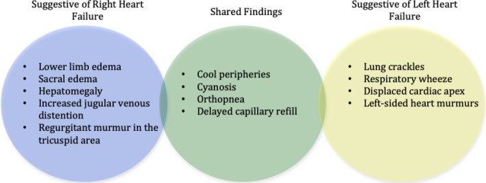
In the setting of CS, classic ACS symptoms and signs are combined with altered mental status, hypotension, arrhythmia, diminished pulses, dyspnea, peripheral edema, jugular venous distention, and orthopnea (Figure 1). These features reflect an infarction involving >40% of the left ventricle,17 and can occur in the setting of an acute infarct superimposed on an old MI or a new massive MI.
Figure 1.

Physical findings suggestive of the ventricle primarily involved in cardiogenic shock. Often pro‐inflammatory states induced by shock physiology causes a blunted performance of the less affected side. Both sides often contribute to the clinical presentation and physical exam findings.
Patients with CS most commonly present with cool extremities and signs of pulmonary congestion. This presentation is termed “cold and wet” and reflects a reduced cardiac index (CI), increased systemic vascular resistance, and increased PCWP. Patients may also present euvolemic or “dry and cold”, which indicates a reduced CI, increased systemic vascular resistance, and normal PCWP. Euvolemic presentations were more likely to have previous MI or chronic kidney disease in comparison with those with classic “cold and wet” features.18
An under‐recognized presentation of CS is the “wet and warm” subtype. This represents a systemic inflammatory response syndrome reaction in conjunction with an MI and is associated with a higher incidence of sepsis and mortality.13, 19, 20, 21 These patients have a reduced CI, low‐to‐normal systemic vascular resistance, and an elevated PCWP. Systemic inflammatory response syndrome should be suspected by the presence of fever, an elevated white cell count, and low systemic vascular resistance. Nineteen percent of patients had suspected sepsis in the SHOCK trial, with higher risk in younger patients and those with low systemic vascular resistance.21 ACS‐associated CS patients with culture‐positive sepsis have 2 times the risk of mortality.21 Systemic inflammatory response syndrome is prevalent on admission in 25% of patients with STEMI. Tachycardia, tachypnea, and leukocytosis are independent risk factors for mortality.20
Differential Diagnosis
Alternative diagnoses include other shock etiologies such as hypovolemic, distributive, and obstructive. Other types of shock may contribute to CS as either the main insult or in combination. Thorough medication reconciliation should be performed to discontinue agents that exacerbate hemodynamic dysfunction.
Initial Investigations
Cardiac catheterization is both the definitive diagnostic investigation and guides therapeutic intervention in CS complicating acute MI. Cardiac catheterization is typically preceded by several initial investigations and non‐interventional management strategies. However, CS is a clinical diagnosis and no investigation should delay emergent cardiac catheterization.
ECG
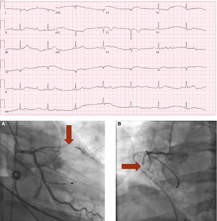
The ECG should be ordered within 10 minutes of presentation.22, 23 ECG findings in ACS are divided into 3 groups: ST‐segment elevation, ST‐segment depression, and non–ST‐segment deviation.24 Early ECG changes of early coronary occlusion and transmural infarction includes hyperacute T waves, which tend to be short‐lived and progress rapidly to ST‐segment elevation.25 The presence of ST‐segment elevation in ≥2 contiguous leads is an indication for urgent reperfusion.23 Transient ST‐segment elevation, ST‐segment depression, and/or T‐wave inversions should raise clinical suspicion of ACS. These patients should be treated with aggressive medical therapy and be evaluated immediately for early coronary angiography.23 New guidelines suggest that left bundle branch block (LBBB) is no longer an indication for urgent catheterization.26 In the appropriate clinical context and in the presence of suggestive diagnostic evidence, urgent catheterization should still be considered. Pathologic Q waves are a reflection of total size of MI, rather than transmural extent, and their presence predicts a lower ejection fraction (EF) and a larger MI.27 In the absence of previously described changes and high clinical suspicion of ACS, true posterior wall myocardial infarction is suggested by the following: ST‐segment depressions in the septal and anterior precordial leads (V1–V4), an R:S wave ratio >1 in V1‐V2, ST‐segment elevations in the posterior leads of a poster ECG (V7–V9).28 If there is co‐existing inferior wall myocardial infarction, there will be ST‐segment elevations in the inferior leads (II, III, and aVF). A normal ECG is not necessarily reassuring as posterior and lateral walls are not fully represented on ECG and thus may not exclude ischemia.23 STEs confer a higher mortality risk in ACS complicated by CS. It has been suggested that patients have a similar 90‐day prognosis if effective revascularization takes place, regardless of ST segment patterns.24 Other findings on (Figure 2) that are suggestive of ACS include sustained ventricular tachycardia, ventricular fibrillation, atrial fibrillation, new bundle branch block, or worsening of a symptomatic high‐degree atrioventricular block.29
Figure 2.

ECG and coronary angiogram of a 53‐year‐old male who presented following sudden onset of diaphoresis, nausea, and syncope. The patient was profoundly hypotensive on arrival and an ECG revealed complete atrioventricular dissociation with junctional bradycardia. Coronary angiography demonstrated (A) a high‐grade proximal LAD stenosis with Thrombolysis in Myocardial Infarction 2 flow and (B) a total thrombotic proximal right coronary artery (RCA) occlusion. An Impella 2.5 was inserted for left ventricular support and PCI to the LAD was performed with a drug eluting stent. He recovered, the Impella was discontinued, and he was discharged. LAD indicates left anterior descending; PCI, percutaneous intervention.
Routine Initial Investigations
Complete blood counts and metabolic panels should be obtained every 12 to 24 hours as they offer valuable information about oxygenation, electrolyte status, and end‐organ damage.
Type 1 MI is caused by an acute atherothrombosis as result of plaque rupture or erosion.30 Frequent monitoring of troponins may reflect extent of injury that is time‐dependent from the initial insult. In the setting of CS, as in STEMI, it is not recommended to wait for the presence of elevated cardiac enzymes before emergent catheterization. Troponins are typically trended every 6 hours starting from initial clinical suspicion.
N‐terminal pro‐B‐type natriuretic peptide (NT‐proBNP) will be elevated during an acute decompensation of heart failure. In CS resulting from ACS, raised levels of natriuretic peptides are associated with increased mortality.31, 32
CS causes reduced oxygenation to peripheral tissues that results in lower pO2 levels and elevated pCO2 levels. Higher levels of lactic acid can be associated with increased mortality.33, 34 Blood gas and lactic acid should be trended (eg, every 1–6 hours initially) to assess response to initial resuscitation.
Echocardiography may be beneficial, especially if there is clinical concern for an MI‐related mechanical complication precipitating CS; however, it should not delay cardiac catheterization. Ultimately, patients presenting with acute RVF or LVF of suspected ischemic etiology should undergo immediate cardiac catheterization for the assessment of coronary anatomy, intracardiac pressures, valvular dysfunction, and structural impairments that often complicate ACS and contribute to CS.
Stabilization and Resuscitation Strategy
Intravenous Fluids
Fluid resuscitation strategy is a clinical challenge in the early management of CS as it is often difficult to assess and can vary over time. In right‐sided heart failure, right atrial pressures and pulmonary artery wedge pressures are poor predictors of fluid response.15, 35 Echocardiography can assess right‐sided heart volume status and rule out pericardial fluid collection.15 The definitive method of volume status assessment and adequacy of resuscitation is right heart catheterization, which should be performed in conjunction with coronary angiography. If hypovolemia is present, conservative boluses of crystalloids (250–500 mL) are reasonable while the patient is being stabilized for cardiac catheterization.
Oxygenation and Ventilation
Continuous pulse oximetry should be used to monitor for respiratory compromise. Oxygen goals vary depending on patient comorbidities, but in the acute care setting blood oxygen saturations of >90% are acceptable.
When non‐invasive forms of oxygenation and ventilation are inadequate, invasive ventilation is required. Low tidal volumes (5–7 mL/kg of ideal body weight) used in the management of acute respiratory distress syndrome are considered lung protective and decrease the incidence of RVF from 60% to 25% in this cohort of patients.36 Low tidal volumes optimize blood flow between the pulmonary and parenchymal vasculature. The decreased resistance in the pulmonary circuit lowers stress on the RV, compared with higher tidal volumes. Therefore, a low tidal volume strategy is recommended when mechanically ventilating patients in CS.
Vasopressor Support
Vasopressors (Table 2) should be titrated to a mean arterial pressure with a typical goal of >65 mm Hg. Vasopressin has less pulmonary vasoconstriction than norepinephrine; and may be more beneficial as a first‐line vasopressor in patients with CS with acute RVF.37 Pulmonary vasoactivity can be modified by inodilators, phosphodiesterase III inhibitors, or nitric oxide (Table 3). When using these agents invasive blood pressure monitoring is required as they can rapidly induce hypotension.
Table 2.
Summary of Systemic Vasopressors
| Agents | Mechanism | Effect | Indications | Considerations |
|---|---|---|---|---|
| Phenylephrine | A1 agonist | Vasoconstriction | Various forms of shock | Caution in cardiac dysfunction as it increases afterload |
| Norepinephrine | A<B agonist | Inotropy, chronotropy, dromotropy, and vasoconstriction | Most common first line agent in shock | Most benefits demonstrated in septic shock |
| Epinephrine | A≪B agonist | Inotropy, chronotropy, dromotropy, and vasoconstriction | Commonly used as second line agent or first line in anaphylactic shock | Surviving Sepsis Guidelines has most data for epinephrine as second line agent |
| Dopamine | Dose dependent A, B, and D agonism | Inotropy, dromotropy, chronotropy, and vasoconstriction (at highest doses) | Second line agent in most forms of shock | SOAP II trial demonstrated more incidence of tachy‐arrythmias and increased mortality in CS patients when dopamine was used as first line |
| Vasopressin | V1 agonist | Vasoconstriction | Second line agent in most forms of shock | On or Off dosing, can cause hyponatremia |
| Dobutamine | B agonist | Inotropy and mild vasodilation | Commonly used in cardiogenic shock | May contribute to hypotension |
| Levosimendan | Myofilament Ca2+ sensitizer and K+ channel modifier | Ionotropy and inodilator | Used in acutely decompensated chronic heart failure | Minimal effect on myocardial oxygen consumption |
CS indicates cardiogenic shock; SOAP, Sepsis Occurrence in Acutely Ill Patients.
Table 3.
Summary of Vasoactive Agents Within the Pulmonary Circuit
| Agent | Mechanism | Route | Side Effects |
|---|---|---|---|
| Nitric Oxide | ↑ cGMP | Inhaled | Blurred vision, confusion, sweating, malaise, headache, bleeding |
| Milrinone | Phosphodiesterase 3 inhibitor | Intravenous | Bleeding, hypotension, chest pain, tremors, bronchospasm, hypokalemia |
| Prostacyclin | ↑ cAMP, ↑ K, ↓ ET‐1, and ↑ K+ | Inhaled or Intravenous | Bleeding, arrhythmias, diarrhea, edema, fevers, chills |
| Dobutamine | B agonist | Intravenous | Hypotension, tachyarrhythmia, headache, thrombocytopenia |
Continuous Renal Replacement Therapy
Acute kidney injury occurs in 13% to 28% in patients with CS, and 20% will require continuous renal replacement therapy.1, 38, 39 Continuous renal replacement therapy should be considered with stage 2 kidney injury as defined by elevated serum creatinine (≥2× baseline) and urine output <0.5 mL/kg per hour for ≥12 hours; or when life‐threatening changes in fluid, electrolyte, and acid‐base balance precipitates the need for dialysis.40
Hemodynamic Monitoring
Goals of hemodynamic monitoring should be focused on hemodynamic modification to produce stable vital signs and adequate tissue perfusion. Continuous blood pressure monitoring with an arterial line, telemetry, continuous pulse oximetry, temperature, respiratory rate, and urinary output are rudimentary parameters to monitor.
Mixed venous oxygen saturation (SvO2) is measured from a sample of blood drawn from the central venous system, ideally from the distal port of a pulmonary artery catheter. A low SvO2 may indicate reduced CO, anemia, hypoxemia, or increased oxygen consumption.41 A reduced SvO2 saturation is typically present in CS; however, this is also often the case in hypovolemic and obstructive shock. SvO2 measurement can help assess response to therapy when measured frequently. During the early stages of hemodynamic monitoring, SvO2 measurements should be drawn every 4 hours after central line placement.
Common structural complications of MI should be suspected by appearance of a new systolic murmur on clinical examination. Echocardiography can confirm early mechanical complications such as papillary muscle rupture, ventricular septal defect, and free wall rupture, which present most frequently within 24 hours of hospitalization.42, 43 Right ventricular free wall hypertrophy indicates long term right‐sided pressure elevation, while right ventricular dilation offers prognostic values.15, 44 During the treatment phase, echocardiography and catheterization are used together to assess the hemodynamic response to intervention.
A pulmonary artery catheter (PAC) is typically placed during cardiac catheterization and can assist with identification of patients requiring mechanical circulatory support. It often remains in place thereafter for continuous hemodynamic monitoring—including precise measurements of fluid states, central venous oxygen saturation, response to therapy, and indicates the effectiveness of ventricular support. PACs offer therapeutic advantages via continuous monitoring of cardiac output during inotrope and pulmonary artery vasodilator titration.15, 45 This intervention is helpful because patient response to mechanical circulatory support is dependent on several factors including volume status, intrinsic RV contractility, properties of the systemic and pulmonary vasculature, and the presence of valvular lesions.46 PACs can also aid the diagnosis of mechanical circulatory support device complications such as pump thrombosis.46 Pump thrombosis should be suspected in patients who exhibit clinical features of recurrent cardiogenic shock accompanied by sudden elevation of pulmonary artery or PCWP.
Despite its more precise measurements, PAC use does not confer a mortality benefit or reduce the length of intensive care unit or hospital stays. In fact its use in the critically ill has been associated with increased mortality.47 Complications of PACs include pulmonary infarcts, cardiac arrhythmias such as heart block, infection, and balloon rupture. An LBBB, commonly seen in ACS, is a contraindication to a PAC without backup ventricular pacing because of the risk of precipitating a right bundle branch block (RBBB). In critically ill patients, a PAC is not always time effective and clinical decisions are frequently made in the absence of this investigation.
MCS Devices
While inotropic agents are used widely, mortality is higher with an increased number of prescribed inotropes/vasopressors.48, 49 Furthermore, catecholamine therapy is associated with significant limitations including arrhythmias, increased myocardial oxygen consumption, and inadequate circulatory support.50 MCS devices (Table 4)11, 51, 52, 53 offer significant advantages over vasopressor therapy including substantial cardiovascular support without increased risk of myocardial ischemia and possible decreased myocardial oxygen demand.54 Most importantly, there are registry data indicating that early MCS device use is associated with improved survival rates.49 Thus, early use of support devices is an important therapeutic intervention. Options for acute percutaneous MCS include the intra‐aortic balloon pump (IABP), axial flow pumps (Impella LP 2.5, Impella CP), left atrial‐to‐femoral arterial ventricular assist devices (Tandem Heart) and venous‐arterial extracorporeal membrane oxygenation (ECMO).
Table 4.
Mechanical Circulatory Support Device Evidence
| Trial/Registry | Findings |
|---|---|
|
IABP Shock II (2012)4
Randomized Control Trial n=600 |
|
|
Shock Trial Registry analysis (2004)51
Prospective Analysis n=541 |
|
|
Protect II Trial (2012)52
Randomized Clinical Trial n=448 |
|
|
IMPRESS in Severe Shock (2017) Randomized Clinical Trial n=48 |
|
|
Catheter‐based Ventricular Assist Device Registry analysis (2017)49
Prospective Analysis n=287 |
|
|
Detroit Cardiogenic Shock Initiative (2018—ongoing)53
Randomized Control Trial n=500 (target enrollment) |
|
CPO indicates cardiac power output; CS, cariogenic shock; IABP, intra‐aortic balloon pump; MAE, major adverse events; MCS, mechanical circulatory support; OMT, optimal medical therapy; PCI, percutaneous coronary intervention; IMPRESS, IMPella versus IABP Reduces mortality in STEMI patients treated with primary PCI in Severe cardiogenic Shock.
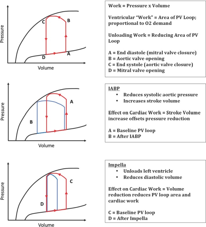
The left ventricular pressure‐volume loop (PVL) illustrates the 4 phases of the cardiac cycle—(1) isovolumetric contraction, (2) ejection, (3) isovolumetric relaxation and (4) filling. In the absence of pathology the loop is trapezoidal with a rounded top, but the position and morphology of the loop depend on ventricular preload and afterload. Preload is the cardiac “wall stress”; it is the end‐diastolic volume that results in the greatest average sarcomere stretch in the myocardium. Afterload is the pressure that the left ventricle contracts against and is determined by the hemodynamic characteristics of the vascular system. The PVL and normal LV mechanics provide a basis for understanding ventricular mechanical support devices (Figure 3). They also offer insight into myocardial oxygen consumption, which is related to the ventricular pressure‐volume area.46
Figure 3.

MCS devices effect on pressure‐volume loops. (1) The normal left ventricular pressure‐volume loop, (2) with effect of IABP and (3) with effect of Impella.55 IABP indicates intra‐aortic balloon pump, PV, pressure volume.
There are 3 circuit configurations for MCS devices—pumping from the (1) RA/central vein to a systemic artery, (2) LA to a systemic artery or (3) LV to a systemic artery. Peak flow rates of available devices range from 2.5 to 7 L/min.46
The IABP was introduced almost 5 decades ago and remains the most common support device used in CS. IABP is believed to decrease myocardial oxygen consumption, increase coronary artery perfusion, decrease afterload and modestly increase cardiac output (0.8–1 L/min).16 It is inserted via an 8Fr sheath in either the femoral or axillary artery.56
The IABP Shock II Trial included 600 patients with CS from acute MI receiving early revascularization and randomized them to IABP support or optimal medical therapy. The study showed no mortality benefit at 30 days.4 Follow‐up at 6 and 12 months showed no reduction in all‐cause mortality or improvement in quality of life assessments.57 These findings may be because of the fact that the IABP plays no role in myocardium salvage.57, 58 This study has a number of limitations. IABP insertion occurred within 24 hours, both before and after PCI. This does not align with contemporary thinking that emphasizes early MCS. However, pre‐ or post‐PCI IABP insertion showed no mortality difference. Additionally, the pursuit of culprit versus multivessel PCI was determined by the operator. Furthermore, 30 crossovers occurred (26 of these non‐protocol) to the IABP group and these may represent sicker patients.
There are a number of LV‐to‐aorta devices, however those most commonly used in the setting of CS are the Impella devices. The Impella devices are axial flow pumps that are advanced from the common femoral artery and passed retrograde across the aortic valve into the LV and eject blood into the ascending aorta. The Impella 2.5 and Impella CP devices are percutaneously inserted and can maintain a cardiac output of 2.5 to 4 L/min. Impella RP is a right‐sided device introduced via an 11Fr catheter that pumps blood from the inferior vena cava to the pulmonary artery and delivers a flow rate >4 L/min. The Impella 5.0 is a larger device that can achieve a cardiac output of 5 L/min, however, it requires a 22‐Fr sheath, necessitating a surgical cutdown of the femoral artery. Continuous pumping of blood from the LV, independent of the cardiac cycle, results in the loss of the normal isovolumic periods, transforming the PVL from its trapezoidal morphology to a triangular shape. In contrast to the IABP, the Impella acts independent of heart function and rhythm and as the pump flow rate increases it progressively unloads the LV (resulting in a leftward PVL shift), peak LV pressure decreases and there are decreases in pressure‐volume area and myocardial oxygen consumption. Also, aortic pressure increases with escalating flow rate causing a widening dissociation between aortic pressure and peak LV pressure (“LV‐Ao uncoupling”). This unloading also results in decreased LA and wedge pressures.46 Impella use is contraindicated in moderate‐to‐severe aortic valve disease, mechanical aortic valve and severe peripheral arterial disease.54
Analysis of the Shock Trial Registry showed that cardiac power output (CPO) is the strongest independent hemodynamic correlate of mortality in CS.51 CPO couples both pressure (mean arterial pressure) and flow (cardiac output) variables to derive a numerical value of cardiac pumping (CPO=mean arterial pressure×cardiac output/451). Impella has demonstrated greater intraprocedural hemodynamic stability (smaller decrease in mean arterial pressure and CPO).52 Thus, given the importance of CPO in CS and the improved hemodynamics offered by Impella, it appears to be the most optimal therapy.
The Protect II Trial showed that, in patients with complex triple‐vessel or left main stem disease and severely reduced LV function undergoing non‐emergent PCI, Impella provided superior hemodynamic support compared with IABP as measured by CPO. Notably, the incidence of major adverse events at 30 days was not statistically different between these 2 groups. However, at 90 days, Impella was associated with decreased major adverse events.52 It should be noted that Protect II did not, however, include patients with CS.
Other advantages of Impella over IABP includes that it acts independent of heart function, simultaneously unloads the left ventricle and supports arterial pressure, permits prolonged balloon inflations, multiple passes with atherectomy devices, and supports circulation during complex coronary interventional procedures.
Although there is some evidence that Impella use results in reduced peri‐ and post‐procedural major adverse events in high‐risk PCI,59, 60 the theoretical benefit of Impella over IABP is not borne out in larger trials of mechanical circulatory support in CS that are focused on major outcomes.
The IMPRESS in Severe Shock (IMPella versus IABP Reduces mortality in STEMI patients treated with primary PCI in Severe cardiogenic Shock) trial was a randomized comparison of Impella CP versus IABP in patients suffering acute MI with CS. The primary end point was 30‐day mortality and the study found no significant difference in 30‐day mortality (≈50% for both groups).11 A limitation of the study was the small sample size (n=48). Notably, it supported prior findings of increased bleeding risk with Impella.61 Overall, the study suggests that the clinical benefits of Impella may be more similar to IABP than expected.
The SHOCK,3 IABP‐SHOCK II,4 and IMPRESS in Severe Shock11 trials all showed ≈50% mortality over 6 to 12 months, illustrating the constant mortality outcomes in CS over the past 2 decades despite the widespread use of MCS devices. Recent analysis of the cVAD (Catheter‐based Ventricular Assist Device) Registry indicates that early MCS implantation in CS, before starting inotrope/vasopressor support and before PCI, is independently associated with improved survival rates in patients with CS because of acute MI.49 With this in mind, the Detroit Cardiogenic Shock Initiative proposed the use of standardized protocols with emphasis on early Impella insertion before PCI. The Detroit Cardiogenic Shock Initiative Pilot Study reported 76% survival to discharge with this approach and is expanding into a National Cardiogenic Shock Initiative.53
The Tandem Heart is an LA‐to‐arterial MCS device. A cannula is passed into the femoral vein and an atrial septal puncture is performed to access oxygenated LA blood, which is aspirated and pumped into one or both femoral arteries.54 Since blood is withdrawn directly from the LA this unloads the LV, resulting in decreased PCWP and LVEDP.46 It improves peripheral tissue perfusion in spite of the mild increase in afterload caused by the pumping of blood back into the femoral arteries.54 Use of Tandem Heart is limited by its requirement of a specialized skillset that includes transseptal puncture and time from door‐to‐LV unloading.
Venous arterial‐ECMO involves drainage of venous blood, passing it through an oxygenator and returning the oxygenated blood to systemic circulation using a centrifugal pump. It can be performed centrally by cannulation of the right atrium and aorta or peripherally with cannulation of the femoral artery and vein. Peripheral ECMO can reduce LV preload; however, this can cause increased ventricular wall tension due to retrograde flow from femoral artery cannulation and therefore requires closer monitoring than central ECMO.54 ECMO has a complex and variable hemodynamic response, which may be partially explained by the variability of secondary effects of ECMO on total peripheral resistance and left ventricular contractility.46 ECMO has been used in ≈13 000 patients and its rate of survival‐to‐discharge is 39% when used in cardiac support.62 The absence of large randomized controlled trials of ECMO in patients with CS consigns its use to refractory cases as a bridging therapy to LVAD or emergent heart transplantation.54
Coronary Angiography
The most important investigation in patients diagnosed with CS is coronary angiography (Figure 2). It enables physicians to identify the precise location of the lesion that precipitated CS. On coronary angiography ≈15% patients are found to have significant left main lesions and >50% have triple‐vessel disease. Mortality is associated with the culprit vessel—left main coronary artery (78.6%), saphenous vein graft (69.7%), circumflex coronary artery (42.4%), left anterior descending coronary artery (42.3%) and right coronary artery (37.4%). Additionally, mortality is inversely related to the Thrombolysis in Myocardial Infarction flow grade.63 After evaluation of coronary anatomy patients typically undergo primary percutaneous coronary intervention (PCI). In rare instances, and dependent on institutional resources, patients may proceed to coronary artery bypass graft surgery, hybrid coronary artery bypass graft/PCI or emergent cardiac transplantation.
PCI Strategy
Coronary reperfusion is an essential therapeutic intervention for patients with ACS complicated by CS. The SHOCK trial provided strong evidence supporting the use of PCI in cardiogenic shock. There were 302 patients diagnosed with acute MI complicated by CS who were randomized to emergency revascularization or medical stabilization. Overall mortality at 30 days was similar between the revascularization and medical therapy groups. However, at 6 months mortality rates were significantly lower in the revascularization cohort (50.3%) in comparison with the medical therapy group (63.1%).3 The marked mortality benefit in successful versus unsuccessful PCI was also clearly demonstrated, 35% versus 80% respectfully.3 Subgroup analysis of the SHOCK trial demonstrated a non‐significant trend towards increased 30‐day mortality in elderly patients receiving early revascularization versus initial medical stabilization.3 However, an early revascularization approach has subsequently been associated with lower short‐ (54.5% versus 72.1%) and medium‐term (60.4% versus 80.1%) mortality when compared with initial medical stabilization in this patient population.64 Of note, the SHOCK trial is now dated as only one‐third of the revascularization cohort received intracoronary stents.
Complete revascularization, addressing both culprit and hemodynamically significant non‐culprit lesions, has historically been the preferred strategy in patients with acute MI and CS and was recommended in recent guidelines;1 however, this paradigm has recently been challenged. The CULPRIT‐SHOCK (Culprit Lesion Only PCI versus Multivessel PCI in Cardiogenic Shock) Trial randomized 706 patients with STEMI/NSTEMI and an identifiable culprit lesion to multivessel or culprit lesion‐only PCI. The composite primary end point was death or renal failure requiring dialysis at 30 days. The trial demonstrated a 9.5% absolute risk reduction of the composite primary end point in the culprit lesion‐only group (7.3% of which was attributable to an absolute risk reduction in all‐cause mortality). Of note, the culprit‐lesion only cohort had the option for staged revascularization of non‐culprit lesions and almost 20% of patients underwent further staged or urgent PCI. Additionally, 75 patients crossed over from culprit lesion‐only to multivessel PCI raising the possibility of including more complex and comorbid patients in the multivessel PCI group, thus overestimating the benefit of culprit lesion‐only PCI. Also, greater dye loads in multivessel PCI may partially account for observed differences observed.65 Another limitation of the study was that low rates of MCS device use in the multivessel PCI group. One‐year follow‐up showed no mortality difference between the culprit lesion‐only and multivessel PCI groups (50% versus 56.9%, respectively). The CULPRIT‐SHOCK Trial contradicts widespread current practice and prior studies in non‐shock patients (DANAMI‐3‐PRIMULTI,66 PRAMI,67 CvLPRIT68) that suggested that there may be a benefit from complete revascularization.
Data from the KAMIR‐NIH (Korea Acute Myocardial Infarction‐National Institutes of Health) Registry are at odds with the findings from the CULPRIT‐SHOCK Trial. In this national multicenter prospective registry 659 patients with STEMI and CS who underwent PCI were studied. The risk of all‐cause death at 1 year was significantly lower in the multivessel PCI group versus the culprit lesion‐only group (21.3% versus 31.7%; P=0.001). Furthermore, multivessel PCI was associated with reduced rates in the composite outcome of all‐cause death, MI, and repeat revascularization (28.4% versus 42.6%; P<0.001).7 Larger trials that stratify patients according to door‐to‐LV unloading time in tandem with randomization to culprit‐lesion versus multivessel PCI are needed to resolve the discrepancy between the CULPRIT‐SHOCK and KAMIR‐NIH Registry findings.
Given the excellent long‐term patency rates of left internal mammary grafts coupled with the advances in minimally invasive techniques and stent technology, hybrid coronary revascularization procedures are a promising treatment modality for CS patients with multivessel disease. Hybrid coronary revascularization refers to combined surgical bypass with PCI during the same procedure or within 60 days.69
Despite significant advances in infarct management, persistently high mortality rates have been observed in CS over the past 2 decades. However, available and emerging evidence indicates promising avenues for contemporary management. A new approach that emphasizes rapid LV unloading and prompt coronary revascularization may reduce mortality of this devastating complication of AMI.
Disclosures
None.
(J Am Heart Assoc. 2019;8:e011991 DOI: 10.1161/JAHA.119.011991.)
References
- 1. van Diepen S, Katz JN, Albert NM, Henry TD, Jacobs AK, Kapur NK, Kilic A, Menon V, Ohman EM, Sweitzer NK, Thiele H, Washam JB, Cohen MG. Contemporary management of cardiogenic shock: a scientific statement from the American Heart Association. Circulation. 2017;136:e232–e268. [DOI] [PubMed] [Google Scholar]
- 2. Harjola V‐P, Lassus J, Sionis A, Køber L, Tarvasmäki T, Spinar J, Parissis J, Banaszewski M, Silva‐Cardoso J, Carubelli V, Di Somma S, Tolppanen H, Zeymer U, Thiele H, Nieminen MS, Mebazaa A; for the CardShock study investigators and the GREAT network . Clinical picture and risk prediction of short‐term mortality in cardiogenic shock: clinical picture and outcome of cardiogenic shock. Eur J Heart Fail. 2015;17:501–509. [DOI] [PubMed] [Google Scholar]
- 3. Hochman JS, Sleeper LA, Webb JG, Sanborn TA, White HD, Talley JD, Buller CE, Jacobs AK, Slater JN, Col J, McKinlay SM, LeJemtel TH. Early revascularization in acute myocardial infarction complicated by cardiogenic shock. SHOCK Investigators. Should We Emergently Revascularize Occluded Coronaries for Cardiogenic Shock. N Engl J Med. 1999;341:625–634. [DOI] [PubMed] [Google Scholar]
- 4. Thiele H, Zeymer U, Neumann F‐J, Ferenc M, Olbrich H‐G, Hausleiter J, Richardt G, Hennersdorf M, Empen K, Fuernau G, Desch S, Eitel I, Hambrecht R, Fuhrmann J, Böhm M, Ebelt H, Schneider S, Schuler G, Werdan K. Intraaortic balloon support for myocardial infarction with cardiogenic shock. N Engl J Med. 2012;367:1287–1296. [DOI] [PubMed] [Google Scholar]
- 5. Bauer T, Zeymer U, Hochadel M, Möllmann H, Weidinger F, Zahn R, Nef HM, Hamm CW, Marco J, Gitt AK. Use and outcomes of multivessel percutaneous coronary intervention in patients with acute myocardial infarction complicated by cardiogenic shock (from the EHS‐PCI Registry). Am J Cardiol. 2012;109:941–946. [DOI] [PubMed] [Google Scholar]
- 6. Ponikowski P, Voors AA, Anker SD, Bueno H, Cleland JGF, Coats AJS, Falk V, González‐Juanatey JR, Harjola V‐P, Jankowska EA, Jessup M, Linde C, Nihoyannopoulos P, Parissis JT, Pieske B, Riley JP, Rosano GMC, Ruilope LM, Ruschitzka F, Rutten FH, van der Meer P. 2016 ESC guidelines for the diagnosis and treatment of acute and chronic heart failure: the Task Force for the diagnosis and treatment of acute and chronic heart failure of the European Society of Cardiology (ESC). Developed with the special contribution of the Heart Failure Association (HFA) of the ESC. Eur Heart J. 2016;37:2129–2200. [DOI] [PubMed] [Google Scholar]
- 7. Lee JM, Rhee T‐M, Hahn J‐Y, Kim HK, Park J, Hwang D, Choi KH, Kim J, Park TK, Yang JH, Song YB, Choi J‐H, Choi S‐H, Koo B‐K, Kim YJ, Chae SC, Cho MC, Kim CJ, Gwon H‐C, Kim JH, Kim H‐S, Jeong MH; KAMIR Investigators . Multivessel percutaneous coronary intervention in patients with ST‐segment elevation myocardial infarction with cardiogenic shock. J Am Coll Cardiol. 2018;71:844–856. [DOI] [PubMed] [Google Scholar]
- 8. Bellumkonda L, Gul B, Masri SC. Evolving concepts in diagnosis and management of cardiogenic shock. Am J Cardiol. 2018;122:1104–1110. [DOI] [PubMed] [Google Scholar]
- 9. Kolte D, Khera S, Aronow WS, Mujib M, Palaniswamy C, Sule S, Jain D, Gotsis W, Ahmed A, Frishman WH, Fonarow GC. Trends in incidence, management, and outcomes of cardiogenic shock complicating ST‐elevation myocardial infarction in the United States. J Am Heart Assoc. 2014;3:e000590 DOI: 10.1161/JAHA.113.000590. [DOI] [PMC free article] [PubMed] [Google Scholar]
- 10. De Luca L, Olivari Z, Farina A, Gonzini L, Lucci D, Di Chiara A, Casella G, Chiarella F, Boccanelli A, Di Pasquale G, De Servi S, Bovenzi FM, Gulizia MM, Savonitto S. Temporal trends in the epidemiology, management, and outcome of patients with cardiogenic shock complicating acute coronary syndromes: management changes in cardiogenic shock. Eur J Heart Fail. 2015;17:1124–1132. [DOI] [PubMed] [Google Scholar]
- 11. Ouweneel DM, Eriksen E, Sjauw KD, van Dongen IM, Hirsch A, Packer EJS, Vis MM, Wykrzykowska JJ, Koch KT, Baan J, de Winter RJ, Piek JJ, Lagrand WK, de Mol BAJM, Tijssen JGP, Henriques JPS. Percutaneous mechanical circulatory support versus intra‐aortic balloon pump in cardiogenic shock after acute myocardial infarction. J Am Coll Cardiol. 2017;69:278–287. [DOI] [PubMed] [Google Scholar]
- 12. Mahmoud AN, Elgendy IY, Mojadidi MK, Wayangankar SA, Bavry AA, Anderson RD, Jneid H, Pepine CJ. Prevalence, causes, and predictors of 30‐day readmissions following hospitalization with acute myocardial infarction complicated by cardiogenic shock: findings from the 2013–2014 National Readmissions Database. J Am Heart Assoc. 2018;7:e008235 DOI: 10.1161/JAHA.117.008235. [DOI] [PMC free article] [PubMed] [Google Scholar]
- 13. Hochman JS. Cardiogenic shock complicating acute myocardial infarction: expanding the paradigm. Circulation. 2003;107:2998–3002. [DOI] [PubMed] [Google Scholar]
- 14. Prondzinsky R, Unverzagt S, Lemm H, Wegener N‐A, Schlitt A, Heinroth KM, Dietz S, Buerke U, Kellner P, Loppnow H, Fiedler MG, Thiery J, Werdan K, Buerke M. Interleukin‐6, ‐7, ‐8 and ‐10 predict outcome in acute myocardial infarction complicated by cardiogenic shock. Clin Res Cardiol. 2012;101:375–384. [DOI] [PubMed] [Google Scholar]
- 15. de Asua I, Rosenberg A. On the right side of the heart: medical and mechanical support of the failing right ventricle. J Intensive Care Soc. 2017;18:113–120. [DOI] [PMC free article] [PubMed] [Google Scholar]
- 16. Rihal CS, Naidu SS, Givertz MM, Szeto WY, Burke JA, Kapur NK, Kern M, Garratt KN, Goldstein JA, Dimas V, Tu T; Society for Cardiovascular Angiography and Interventions (SCAI), Heart Failure Society of America (HFSA), Society of Thoracic Surgeons (STS), American Heart Association (AHA), and American College of Cardiology (ACC) . 2015 SCAI/ACC/HFSA/STS clinical expert consensus statement on the use of percutaneous mechanical circulatory support devices in cardiovascular care: endorsed by the American Heart Assocation, the Cardiological Society of India, and Sociedad Latino Americana de Cardiologia Intervencion; Affirmation of Value by the Canadian Association of Interventional Cardiology‐Association Canadienne de Cardiologie d'intervention. J Am Coll Cardiol. 2015;65:e7–e26. [DOI] [PubMed] [Google Scholar]
- 17. McCallister BD, Christian TF, Gersh BJ, Gibbons RJ. Prognosis of myocardial infarctions involving more than 40% of the left ventricle after acute reperfusion therapy. Circulation. 1993;88:1470–1475. [DOI] [PubMed] [Google Scholar]
- 18. Menon V, White H, LeJemtel T, Webb JG, Sleeper LA, Hochman JS. The clinical profile of patients with suspected cardiogenic shock due to predominant left ventricular failure: a report from the SHOCK Trial Registry. SHould we emergently revascularize Occluded Coronaries in cardiogenic shocK? J Am Coll Cardiol. 2000;36:1071–1076. [DOI] [PubMed] [Google Scholar]
- 19. Lim N, Dubois M‐J, De Backer D, Vincent J‐L. Do all nonsurvivors of cardiogenic shock die with a low cardiac index? Chest. 2003;124:1885–1891. [DOI] [PubMed] [Google Scholar]
- 20. van Diepen S, Vavalle JP, Newby LK, Clare R, Pieper KS, Ezekowitz JA, Hochman JS, Mahaffey KW, Armstrong PW, Granger CB. The systemic inflammatory response syndrome in patients with ST‐segment elevation myocardial infarction. Crit Care Med. 2013;41:2080–2087. [DOI] [PubMed] [Google Scholar]
- 21. Kohsaka S, Menon V, Lowe AM, Lange M, Dzavik V, Sleeper LA, Hochman JS; SHOCK Investigators . Systemic inflammatory response syndrome after acute myocardial infarction complicated by cardiogenic shock. Arch Intern Med. 2005;165:1643–1650. [DOI] [PubMed] [Google Scholar]
- 22. Amsterdam EA, Wenger NK, Brindis RG, Casey DE, Ganiats TG, Holmes DR, Jaffe AS, Jneid H, Kelly RF, Kontos MC, Levine GN, Liebson PR, Mukherjee D, Peterson ED, Sabatine MS, Smalling RW, Zieman SJ. 2014 AHA/ACC guideline for the management of patients with non–ST‐elevation acute coronary syndromes. J Am Coll Cardiol. 2014;64:e139–e228. [DOI] [PubMed] [Google Scholar]
- 23. Makki N, Brennan TM, Girotra S. Acute coronary syndrome. J Intensive Care Med. 2015;30:186–200. [DOI] [PubMed] [Google Scholar]
- 24. Javanainen T, Tolppanen H, Lassus J, Nieminen MS, Sionis A, Spinar J, Silva‐Cardoso J, Greve Lindholm M, Banaszewski M, Harjola V‐P, Jurkko R. Predictive value of the baseline electrocardiogram ST‐segment pattern in cardiogenic shock: results from the CardShock Study. Ann Noninvasive Electrocardiol. 2018;23:e12561. [DOI] [PMC free article] [PubMed] [Google Scholar]
- 25. Levis JT. ECG diagnosis: hyperacute T waves. Perm J. 2015;19:79. [DOI] [PMC free article] [PubMed] [Google Scholar]
- 26. Behuria S, Yu JC, Sidhu R, Fisher P, Misra D, Schweitzer P (Pavol), Kanei Y. Emergency room evaluation of patients with cardiac complaints and new left bundle branch block: the utility of the Sgarbossa and modified Sgarbossa criteria. J Am Coll Cardiol. 2017;69:1270.28279293 [Google Scholar]
- 27. Moon JCC, Perez De Arenaza D, Elkington AG, Taneja AK, John AS, Wang D, Janardhanan R, Senior R, Lahiri A, Poole‐Wilson PA, Pennell DJ. The pathologic basis of Q‐wave and non‐Q‐wave myocardial infarction. J Am Coll Cardiol. 2004;44:554–560. [DOI] [PubMed] [Google Scholar]
- 28. Levis JT. ECG diagnosis: isolated posterior wall myocardial infarction. Perm J. 2015;19:e143–e144. [DOI] [PMC free article] [PubMed] [Google Scholar]
- 29. Gorenek B, Blomström Lundqvist C, Brugada Terradellas J, Camm AJ, Hindricks G, Huber K, Kirchhof P, Kuck K‐H, Kudaiberdieva G, Lin T, Raviele A, Santini M, Tilz RR, Valgimigli M, Vos MA, Vrints C, Zeymer U; Document Reviewers , Lip GYH, Potpara T, Fauchier L, Sticherling C, Roffi M, Widimsky P, Mehilli J, Lettino M, Schiele F, Sinnaeve P, Boriani G, Lane D, Savelieva I. Cardiac arrhythmias in acute coronary syndromes: position paper from the joint EHRA, ACCA, and EAPCI task force. Europace. 2014;16:1655–1673. [DOI] [PubMed] [Google Scholar]
- 30. Gupta S, Vaidya SR, Arora S, Bahekar A, Devarapally SR. Type 2 versus type 1 myocardial infarction: a comparison of clinical characteristics and outcomes with a meta‐analysis of observational studies. Cardiovasc Diagn Ther. 2017;7:348–358. [DOI] [PMC free article] [PubMed] [Google Scholar]
- 31. Shah NR, Bieniarz MC, Basra SS, Paisley RD, Loyalka P, Gregoric ID, Mann DL, Kar B. Serum biomarkers in severe refractory cardiogenic shock. JACC Heart Fail. 2013;1:200–206. [DOI] [PubMed] [Google Scholar]
- 32. Lemm H, Prondzinsky R, Geppert A, Russ M, Huber K, Werdan K, Buerke M. BNP and NT‐proBNP in patients with acute myocardial infarction complicated by cardiogenic shock: results from the IABP Shock trial. Crit Care. 2010;14:P146. [Google Scholar]
- 33. Attanà P, Lazzeri C, Chiostri M, Picariello C, Gensini GF, Valente S. Strong‐ion gap approach in patients with cardiogenic shock following ST‐elevation myocardial infarction. Acute Card Care. 2013;15:58–62. [DOI] [PubMed] [Google Scholar]
- 34. Valente S, Lazzeri C, Vecchio S, Giglioli C, Margheri M, Bernardo P, Comeglio M, Chiocchini S, Gensini GF. Predictors of in‐hospital mortality after percutaneous coronary intervention for cardiogenic shock. Int J Cardiol. 2007;114:176–182. [DOI] [PubMed] [Google Scholar]
- 35. Jung C, Lauten A, Ferrari M. Microcirculation in cardiogenic shock: from scientific bystander to therapy target. Crit Care. 2010;14:193. [DOI] [PMC free article] [PubMed] [Google Scholar]
- 36. Price LC, Wort SJ, Finney SJ, Marino PS, Brett SJ. Pulmonary vascular and right ventricular dysfunction in adult critical care: current and emerging options for management: a systematic literature review. Crit Care. 2010;14:R169. [DOI] [PMC free article] [PubMed] [Google Scholar]
- 37. Gordon AC, Wang N, Walley KR, Ashby D, Russell JA. The cardiopulmonary effects of vasopressin compared with norepinephrine in septic shock. Chest. 2012;142:593–605. [DOI] [PubMed] [Google Scholar]
- 38. Lauridsen MD, Gammelager H, Schmidt M, Rasmussen TB, Shaw RE, Bøtker HE, Sørensen HT, Christiansen CF. Acute kidney injury treated with renal replacement therapy and 5‐year mortality after myocardial infarction‐related cardiogenic shock: a nationwide population‐based cohort study. Crit Care. 2015;19:452. [DOI] [PMC free article] [PubMed] [Google Scholar]
- 39. Adler C, Reuter H, Seck C, Hellmich M, Zobel C. Fluid therapy and acute kidney injury in cardiogenic shock after cardiac arrest. Resuscitation. 2013;84:194–199. [DOI] [PubMed] [Google Scholar]
- 40. Section 2: AKI definition. Available at: https://www.ncbi.nlm.nih.gov/pmc/articles/PMC4089595/. Accessed May 29, 2018.
- 41. Laher AE, Watermeyer MJ, Buchanan SK, Dippenaar N, Simo NCT, Motara F, Moolla M. A review of hemodynamic monitoring techniques, methods and devices for the emergency physician. Am J Emerg Med. 2017;35:1335–1347. [DOI] [PubMed] [Google Scholar]
- 42. Kutty RS, Jones N, Moorjani N. Mechanical complications of acute myocardial infarction. Cardiol Clin. 2013;31:519–531. [DOI] [PubMed] [Google Scholar]
- 43. Menon V, Webb JG, Hillis LD, Sleeper LA, Abboud R, Dzavik V, Slater JN, Forman R, Monrad ES, Talley JD, Hochman JS. Outcome and profile of ventricular septal rupture with cardiogenic shock after myocardial infarction: a report from the SHOCK Trial Registry. J Am Coll Cardiol. 2000;36:1110–1116. [DOI] [PubMed] [Google Scholar]
- 44. Stretch R, Sauer CM, Yuh DD, Bonde P. National trends in the utilization of short‐term mechanical circulatory support. J Am Coll Cardiol. 2014;64:1407–1415. [DOI] [PubMed] [Google Scholar]
- 45. Jung C, Lauten A, Roediger C, Fritzenwanger M, Schumm J, Figulla HR, Ferrari M. In vivo evaluation of tissue microflow under combined therapy with extracorporeal life support and intra‐aortic balloon counterpulsation. Anaesth Intensive Care. 2009;37:833–835. [DOI] [PubMed] [Google Scholar]
- 46. Burkhoff D, Sayer G, Doshi D, Uriel N. Hemodynamics of mechanical circulatory support. J Am Coll Cardiol. 2015;66:2663–2674. [DOI] [PubMed] [Google Scholar]
- 47. Connors AF, Speroff T, Dawson NV, Thomas C, Harrell FE, Wagner D, Desbiens N, Goldman L, Wu AW, Califf RM, Fulkerson WJ, Vidaillet H, Broste S, Bellamy P, Lynn J, Knaus WA. The effectiveness of right heart catheterization in the initial care of critically ill patients. SUPPORT Investigators. JAMA. 1996;276:889–897. [DOI] [PubMed] [Google Scholar]
- 48. Samuels LE, Kaufman MS, Thomas MP, Holmes EC, Brockman SK, Wechsler AS. Pharmacological criteria for ventricular assist device insertion following postcardiotomy shock: experience with the Abiomed BVS system. J Card Surg. 1999;14:288–293. [DOI] [PubMed] [Google Scholar]
- 49. Basir MB, Schreiber TL, Grines CL, Dixon SR, Moses JW, Maini BS, Khandelwal AK, Ohman EM, O'Neill WW. Effect of early initiation of mechanical circulatory support on survival in cardiogenic shock. Am J Cardiol. 2017;119:845–851. [DOI] [PubMed] [Google Scholar]
- 50. Werdan K, Gielen S, Ebelt H, Hochman JS. Mechanical circulatory support in cardiogenic shock. Eur Heart J. 2014;35:156–167. [DOI] [PubMed] [Google Scholar]
- 51. Fincke R, Hochman JS, Lowe AM, Menon V, Slater JN, Webb JG, LeJemtel TH, Cotter G; SHOCK Investigators . Cardiac power is the strongest hemodynamic correlate of mortality in cardiogenic shock: a report from the SHOCK trial registry. J Am Coll Cardiol. 2004;44:340–348. [DOI] [PubMed] [Google Scholar]
- 52. O'Neill WW, Kleiman NS, Moses J, Henriques JPS, Dixon S, Massaro J, Palacios I, Maini B, Mulukutla S, Dzavík V, Popma J, Douglas PS, Ohman M. A prospective, randomized clinical trial of hemodynamic support with Impella 2.5 versus intra‐aortic balloon pump in patients undergoing high‐risk percutaneous coronary intervention: the PROTECT II study. Circulation. 2012;126:1717–1727. [DOI] [PubMed] [Google Scholar]
- 53. Basir MB, Schreiber T, Dixon S, Alaswad K, Patel K, Almany S, Khandelwal A, Hanson I, George A, Ashbrook M, Blank N, Abdelsalam M, Sareen N, Timmis SBH, O'Neill Md WW. Feasibility of early mechanical circulatory support in acute myocardial infarction complicated by cardiogenic shock: the Detroit cardiogenic shock initiative. Catheter Cardiovasc Interv. 2018;91:454–461. [DOI] [PubMed] [Google Scholar]
- 54. Khan MH, Corbett BJ, Hollenberg SM. Mechanical circulatory support in acute cardiogenic shock. F1000Prime Rep. 2014;6:91. [DOI] [PMC free article] [PubMed] [Google Scholar]
- 55. Jones HA, Kalisetti DR, Gaba M, McCormick DJ, Goldberg S. Left ventricular assist for high‐risk percutaneous coronary intervention. J Invasive Cardiol. 2012;24:544–550. [PubMed] [Google Scholar]
- 56. Ouweneel DM, Henriques JPS. Percutaneous cardiac support devices for cardiogenic shock: current indications and recommendations. Heart. 2012;98:1246–1254. [DOI] [PubMed] [Google Scholar]
- 57. Thiele H, Zeymer U, Neumann F‐J, Ferenc M, Olbrich H‐G, Hausleiter J, de Waha A, Richardt G, Hennersdorf M, Empen K, Fuernau G, Desch S, Eitel I, Hambrecht R, Lauer B, Böhm M, Ebelt H, Schneider S, Werdan K, Schuler G. Intra‐aortic balloon counterpulsation in acute myocardial infarction complicated by cardiogenic shock (IABP‐SHOCK II): final 12 month results of a randomised, open‐label trial. Lancet. 2013;382:1638–1645. [DOI] [PubMed] [Google Scholar]
- 58. Patel MR, Smalling RW, Thiele H, Barnhart HX, Zhou Y, Chandra P, Chew D, Cohen M, French J, Perera D, Ohman EM. Intra‐aortic balloon counterpulsation and infarct size in patients with acute anterior myocardial infarction without shock: the CRISP AMI randomized trial. JAMA. 2011;306:1329. [DOI] [PubMed] [Google Scholar]
- 59. Kovacic JC, Kini A, Banerjee S, Dangas G, Massaro J, Mehran R, Popma J, O'Neill WW, Sharma SK. Patients with 3‐vessel coronary artery disease and impaired ventricular function undergoing PCI with Impella 2.5 hemodynamic support have improved 90‐day outcomes compared to intra‐aortic balloon pump: a sub‐study of the PROTECT II trial. J Interv Cardiol. 2015;28:32–40. [DOI] [PubMed] [Google Scholar]
- 60. Dangas GD, Kini AS, Sharma SK, Henriques JPS, Claessen BE, Dixon SR, Massaro JM, Palacios I, Popma JJ, Ohman M, Stone GW, O'Neill WW. Impact of hemodynamic support with Impella 2.5 versus intra‐aortic balloon pump on prognostically important clinical outcomes in patients undergoing high‐risk percutaneous coronary intervention (from the PROTECT II randomized trial). Am J Cardiol. 2014;113:222–228. [DOI] [PubMed] [Google Scholar]
- 61. Manzo‐Silberman S, Fichet J, Mathonnet A, Varenne O, Ricome S, Chaib A, Zuber B, Spaulding C, Cariou A. Percutaneous left ventricular assistance in post cardiac arrest shock: comparison of intra aortic blood pump and IMPELLA Recover LP2.5. Resuscitation. 2013;84:609–615. [DOI] [PubMed] [Google Scholar]
- 62. Paden ML, Conrad SA, Rycus PT, Thiagarajan RR; ELSO Registry . Extracorporeal life support organization registry report 2012. ASAIO J. 2013;59:202–210. [DOI] [PubMed] [Google Scholar]
- 63. Wong SC, Sanborn T, Sleeper LA, Webb JG, Pilchik R, Hart D, Mejnartowicz S, Antonelli TA, Lange R, French JK, Bergman G, LeJemtel T, Hochman JS. Angiographic findings and clinical correlates in patients with cardiogenic shock complicating acute myocardial infarction: a report from the SHOCK Trial Registry. SHould we emergently revascularize Occluded Coronaries for cardiogenic shocK? J Am Coll Cardiol. 2000;36:1077–1083. [DOI] [PubMed] [Google Scholar]
- 64. Rogers PA, Daye J, Huang H, Blaustein A, Virani S, Alam M, Kumar A, Paniagua D, Kar B, Bozkurt B, Ballantyne CM, Deswal A, Jneid H. Revascularization improves mortality in elderly patients with acute myocardial infarction complicated by cardiogenic shock. Int J Cardiol. 2014;172:239–241. [DOI] [PubMed] [Google Scholar]
- 65. Thiele H, Akin I, Sandri M, Fuernau G, de Waha S, Meyer‐Saraei R, Nordbeck P, Geisler T, Landmesser U, Skurk C, Fach A, Lapp H, Piek JJ, Noc M, Goslar T, Felix SB, Maier LS, Stepinska J, Oldroyd K, Serpytis P, Montalescot G, Barthelemy O, Huber K, Windecker S, Savonitto S, Torremante P, Vrints C, Schneider S, Desch S, Zeymer U; CULPRIT‐SHOCK Investigators . PCI strategies in patients with acute myocardial infarction and cardiogenic shock. N Engl J Med. 2017;377:2419–2432. [DOI] [PubMed] [Google Scholar]
- 66. Engstrøm T, Kelbæk H, Helqvist S, Høfsten DE, Kløvgaard L, Holmvang L, Jørgensen E, Pedersen F, Saunamäki K, Clemmensen P, De Backer O, Ravkilde J, Tilsted H‐H, Villadsen AB, Aarøe J, Jensen SE, Raungaard B, Køber L; DANAMI‐3—PRIMULTI Investigators . Complete revascularisation versus treatment of the culprit lesion only in patients with ST‐segment elevation myocardial infarction and multivessel disease (DANAMI‐3—PRIMULTI): an open‐label, randomised controlled trial. Lancet. 2015;386:665–671. [DOI] [PubMed] [Google Scholar]
- 67. Wald DS, Morris JK, Wald NJ, Chase AJ, Edwards RJ, Hughes LO, Berry C, Oldroyd KG; PRAMI Investigators . Randomized trial of preventive angioplasty in myocardial infarction. N Engl J Med. 2013;369:1115–1123. [DOI] [PubMed] [Google Scholar]
- 68. Gershlick AH, Khan JN, Kelly DJ, Greenwood JP, Sasikaran T, Curzen N, Blackman DJ, Dalby M, Fairbrother KL, Banya W, Wang D, Flather M, Hetherington SL, Kelion AD, Talwar S, Gunning M, Hall R, Swanton H, McCann GP. Randomized trial of complete versus lesion‐only revascularization in patients undergoing primary percutaneous coronary intervention for STEMI and multivessel disease: the CvLPRIT trial. J Am Coll Cardiol. 2015;65:963–972. [DOI] [PMC free article] [PubMed] [Google Scholar]
- 69. Saha T, Naqvi SY, Goldberg S. Hybrid revascularization: a review. Cardiology. 2018;140:35–44. [DOI] [PubMed] [Google Scholar]


