Abstract
Whole-genome sequencing is increasingly adopted in clinical settings to identify pathogen transmissions, though largely as a retrospective tool. Prospective monitoring, in which samples are continuously added and compared to previous samples, can generate more actionable information. To enable prospective pathogen comparison, genomic relatedness metrics based on single-nucleotide differences must be consistent across time, efficient to compute and reliable for a large variety of samples. The choice of genomic regions to compare, i.e., the core genome, is critical to obtain a good metric. We propose a novel core genome method that selects conserved sequences in the reference genome by comparing its k-mer content to that of publicly available genome assemblies. The conserved-sequence genome is sample set-independent, which enables prospective pathogen monitoring. Based on clinical data sets of 3436 S. aureus, 1362 K. pneumoniae and 348 E. faecium samples, ROC curves demonstrate that the conserved-sequence genome disambiguates same-patient samples better than a core genome consisting of conserved genes. The conserved-sequence genome confirms outbreak samples with high sensitivity: in a set of 2335 S. aureus samples, it correctly identifies 44 out of 44 known outbreak samples, whereas the conserved-gene method confirms 38 known outbreak samples.
Subject terms: Microbial genetics, Genome informatics, Infectious diseases
Introduction
Genomic analysis has become an indispensable tool for identification, disambiguation, and classification of infectious agents. As the cost of whole-genome sequencing has dropped to around two hundred dollars per bacterial sample1, infectious disease practice is increasingly turning to genomic sequencing as a routine method to monitor infections and inform antibiotic stewardship programs2–6. Healthcare-acquired infections in particular present a significant threat to patient safety, as well as a financial burden to the healthcare system7,8. With whole-genome sequencing analysis, it is possible to identify potential transmissions, suggest sources of transmission and provide insight into the nature and phenotypes of pathogens. The adoption of this relatively new technology calls for best practices and standards to compare microbial genomes in a manner that is consistent across time and location.
While whole-genome comparison provides better resolution for pathogen disambiguation than FPGE9 or gene-based or multi-locus sequence typing10–14, it also comes with bigger challenges4. The genomic ‘distance’ between pathogens can be defined in many ways, with or without a reference genome to guide the comparison15. Notable reference-free approaches include k-mer composition-based metrics and an assembly-based core genome MLST16,17. In this work, we will focus on alignment-based pathogen comparison, since the use of a reference genome can aid in interpretation and provide information on the location of mutations. In this paradigm, samples are aligned against a common reference genome and genomic distances are computed as the number of nucleotide differences between the consensus genomes, typically including only single-nucleotide variations (SNVs).
There are several challenges in quantifying SNV distances reliably in a clinical setting. First, clinical sites typically experience a wide variety of samples and the reference genome used for the analysis – usually one per species – may be quite distant from some of the samples observed, which leads to a situation in which small differences between large numbers of SNVs need to be detected reliably. Moreover, the observed pathogens may lack some of the regions in the reference genome, in which case the comparison becomes problematic. Second, mutation rates can vary greatly across the genome18,19 and bias a distance metric towards hypervariable regions. Third, the distance metric needs to be sample-independent in prospective studies such that the distances between samples a and b do not change when samples c, d, … are added. This requirement ensures that the interpretation of sample relationships does not change over time.
The choice of core genome, i.e., the regions included in the genome comparison, is crucial to obtain a reliable and consistent genomic distance metric. We have developed a method for defining a conserved-sequence genome, and we demonstrate this with large clinical data sets for gram-positive and gram-negative pathogens: S. aureus, E. faecium and K. pneumoniae. The conserved sequences that make up the core genome can be computed efficiently (without the need for multi-sequence alignment) from publicly available genome assemblies based on k-mer frequency analysis.
Our goal is to compare and evaluate this conserved-sequence method against other core genome approaches typically used for whole-genome SNV comparison. Few prior work exists on core genome approaches for applications in clinical epidemiology13, so we aim to provide insight into the advantages and drawbacks of commonly used methods, including conserved-gene approaches20 and approaches that select genome regions with sufficient coverage in a set of samples21–23. We illustrate that sample-dependent core genome definitions are not suitable for prospective studies because they lead to variable SNV distances as samples are added, which complicates clinical decision-making. Conserved-gene methods, on the other hand, are suitable for prospective whole-genome comparison, as is the conserved-sequence approach we developed. We compare these approaches on several aspects: (i) their ability to separate same-patient samples from different-patient samples, (ii) their use in comparing samples across time and location, and (iii) their ability to confirm previously published outbreak samples of S. aureus in a neonatal ICU unit21. We also detail a methodology to determine thresholds for pathogen relatedness for these core genome methods.
Results
Sample-dependent core genomes yield variable genomic distances in a prospective setting
The published body of genomic epidemiology work overwhelmingly deals with retrospective studies21,24–27, in which a defined set of samples is analyzed simultaneously and independently of other sample sets. As genomics is moving into the clinic as a routine methodology for infection control, a prospective mode of operation is needed to provide actionable results at any point in time that are consistent with future sample analysis. Such consistency constraints are typically not accounted for by popular methods for genomic distance calculation.
Retrospective clinical studies often compute SNV distances across an intersection of nucleotides that are unambiguously determined in all samples, an approach we will refer to as the “intersection” core genome (see Method section 3). This approach is practically infeasible to apply in a prospective setting, because the shared regions with sufficient coverage and unambiguously determined bases decrease as the sample set grows. This implies that all distances between samples, including previously compared samples, have to be recomputed and that the resulting genomic distances decrease as samples accumulate.
To illustrate the variability of genomic distances with the intersection core genome, we simulate a prospective analysis of a K. pneumoniae data set28 by analyzing the samples incrementally (Fig. 1). Each time samples are added, all sample distances have to be recomputed, which can lead to a significant computational overhead: comparing around 2,000 samples in a pairwise fashion amounts to nearly 2 million core genome distances to recompute. More importantly, the SNV threshold that separates same-patient samples from different-patient samples keeps decreasing as samples are added. This highlights the problem of interpretation: the thresholds would have to be re-evaluated continuously and, even with such variable thresholds, groupings of genetically similar samples would not be guaranteed to be stable over time.
Figure 1.
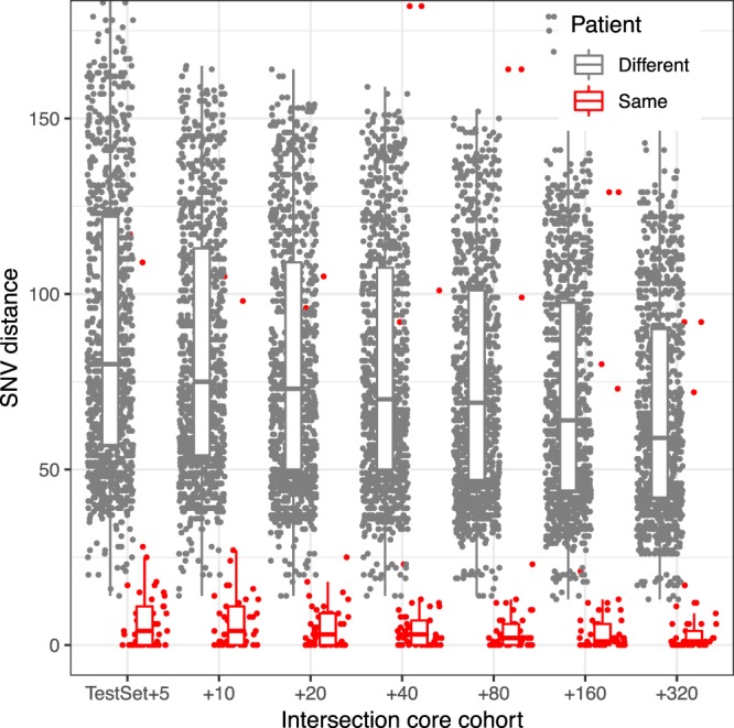
Changes in the intersection core genome SNV distances with increasing cohort size for a set of K. pneumoniae samples. Within-patient and between-patient SNV distances are shown for the same set of 49 samples as the core genome evolves when samples are added. The x axis indicates the number of samples added to the original set.
Sample-independent core genome methods rely on conserved genome regions
To deliver consistent pathogen similarities and facilitate their computation in a clinical setting, the core genome must be defined from the start of the prospective study and kept fixed throughout the study. The core genome therefore needs to rely on genomic regions that are shared by the majority of strains to be compared. However, clinical settings typically see a wide variety of strains and should be able to deal with unexpected or novel strains.
A common solution is to use “housekeeping” genes, that is, genes that are highly conserved within the species, as a core genome29. These genes can be identified in a sample set-independent manner by comparing gene content in publicly available finished genomes and assemblies. In this work we refer this method as “conserved-gene” core genome. A drawback of this approach is that mutation rates can vary locally, even within genes, and this can lead to an over-representation of highly variable sites within the SNV distance.
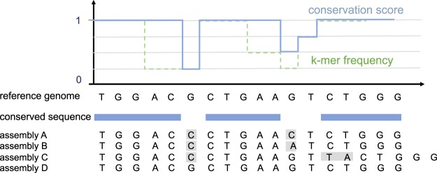
To create an SNV distance metric built on genomic sites with similar mutation rates, we developed a core genome that consists of highly conserved sequences, regardless of their gene content. We will refer to this method as the “conserved-sequence” core genome. Nucleotide conservation scores can be difficult to compute since multi-sequence alignment is computationally challenging for a large number of sequences30,31. Instead of using multi-sequence alignment, we estimate the conservation of each nucleotide in the reference genome through the relative frequency of k-mers spanning the nucleotide in a set of genome assemblies (Method section 5). The conservation scores are computed in three steps (Fig. 2). First, the reference genome and all genome assemblies are k-merized. Then, for each k-mer in the reference genome, the relative number of assemblies that contain the same canonical k-mer is computed and associated with the k-mer’s start position p in the reference genome (the function ‘k-mer frequency’ in Fig. 2). Finally, a conservation score for each position p is obtained from this function by taking a running maximum in a window [p − k + 1, p] with k the k-mer length (the function ‘conservation score’ in Fig. 2). Although smaller k-mers might be able to resolve shorter conserved sequences between SNVs, longer k-mers are more likely to ensure unique associations between the genome position and the k-mer frequency. For this reason, we choose to work with 31-mers. Like the conserved-gene approach, the conserved-sequence genome can be composed from genome assemblies, which enables it to capture a large variety of strains while remaining sample-set independent.
Figure 2.
Illustration of the k-mer frequency method used to select conserved sequences in the reference genome for an example using 3-mers, a hypothetical set of 4 short genome assemblies and a frequency cutoff of 95% (in practice, canonical but longer k-mers are used to favor unique mappings to the genome). The function ‘k-mer frequency’ represents the relative frequency of the reference genome k-mers in the genome assembly set, and the function ‘conservation score’ is generated from this by taking a running maximum in an interval of 3 nucleotides.
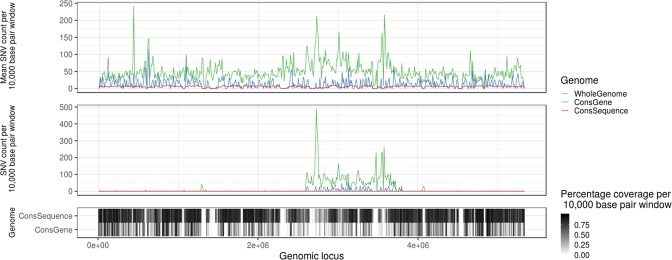
We built conserved-gene and conserved-sequence genomes from all available RefSeq assemblies for S. aureus, E. faecium and K. pneumoniae (Method section 1) with a 95% conservation threshold for both methods. Since both of these core genome approaches rely on conserved genome regions, they largely overlap in core gene regions, but differ on a nucleotide level. While the conserved-gene core genome targets housekeeping genes that are expected to be shared by nearly all strains, the conserved-sequence core genome is not limited to gene sequences, resulting in a larger core genome (Table S1; Fig. 3). The number of SNVs within the core regions is considerably smaller for the conserved-sequence than the conserved-gene cores (Fig. 3), even though the latter covers a smaller part of the genome. This reflects the success of our strategy to remove regions commonly displaying variation between strains, resulting in a focus on regions with similar mutation rates, where only de novo mutations are expected.
Figure 3.
Histogram of the number of SNV counts over 10,000 base pair windows across K. pneumoniae genome, averaged across all samples (top panel) and in one random sample (middle panel). The percentage of bases covered by a core genome in the corresponding windows is shown in the bottom.
Comparison of core genome approaches on distinction of same-patient samples
To compare the conserved-gene and conserved-sequence core genome, we evaluate their ability to separate same-patient (and same strain type) samples from different-patient samples within large clinical data sets. The data sets are not enriched for outbreak samples, so we assume that the same-patient samples largely represent same-pathogen samples. This assumption is not perfect – patients can have multiple infections of the same strain type or have a transmitted infection – but it is a useful heuristic to compare different core genome approaches on actual clinical data.
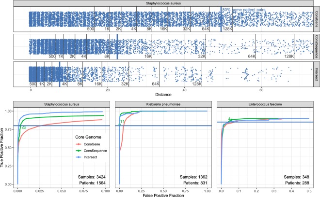
We find that the conserved-sequence core genome obtains a better separation of same-patient samples than the conserved-gene core genome, as measured by their ROC curves for large data sets for S. aureus, K. pneumoniae and E. faecium (Fig. 4). For the large set of over three thousand S. aureus samples, 80% of same-patient pairs can be captured with SNV thresholds of 49, 22 and 7 for the conserved-gene, conserved-sequence and intersection method, respectively For this threshold, the conserved-gene method has the highest rate of false positives as it captures 95,464 different-patient samples, while the conserved-sequence method captures 9,605 different-patient pairs and the intersection method captures 3,292 different-patient pairs. While some of these different-patient pairs may represent actual transmissions, the disparity between these numbers suggest that the majority of the 95,464 pairs are false positives. This also illustrates how a well-chosen core genome can reduce the number of cases that need to be investigated by infection control teams.
Figure 4.
A comparison of different core genome methods to distinguish same-patient samples from different-patient samples. Top figure shows the SNV distances for same-patient pairs with blue dots. The black lines indicate the cumulative number of different-patient pairs that have SNV distances below the distance indicated on the x axis. The SNV thresholds that capture 80% of same-patient SNV distances correspond to 49 for the conserved-gene, 22 for conserved-sequence and 7 for intersection core genome. Bottom figures show ROC curves for same-patient versus different-patient sample distinction for S. aureus (same data as top panel), K.pneumoniae and E. faecium (selected SNV threshold in green). Note that the x-axis may be truncated for clarity.
The 80% same-patient threshold does not necessarily represent the best true/false positive trade-off for each method, but it establishes a point to compare. The ability to discriminate between same-patient and different-patient samples for different true/false positive trade-offs is summarized by the ROC curve (Fig. 4). For all species, the conserved-gene method demonstrates the poorest performance. The conserved-sequence method is outperformed by the intersection core genome in S. aureus, as illustrated for the 80% same-patient threshold, but performs better than the intersection core genome for K. pneumoniae. For E. faecium, it is difficult to detect meaningful differences since the data set is small and contains a limited number of same-patient replicates.
The ROC curves that display same-patient versus different-patient sample distinction can be used to set SNV thresholds for the detection of potential transmission clusters. In this work, we use the SNV thresholds corresponding to the 80% or 85% true positive rates, rounded up to the nearest integer (Table 1). In general, the desired true/false positive rate will depend on the application. It should be noted that the ideal data set would be free of transmissions, since transmissions between patients would be counted as false positives and could lead to an underestimation of the SNV threshold. It is also important to note that these thresholds are not universal – they depend on the data used, the choice of core genome and the bioinformatic analysis pipeline used to process the samples. Given these parameters, the ROC curves can be used to set thresholds for a particular application.
Table 1.
SNV thresholds corresponding to given true positive rate (TPR) in ROC curves.
| TPR | ConsGene | ConsSequence | Intersection | |
|---|---|---|---|---|
| S. aureus | 80% | 49 | 22 | 7 |
| K. pneumoniae | 80% | 37 | 11 | 4 |
| E. faecium | 85% | 5 | 4 | 9 |
For E. faecium, we choose a slightly higher TPR to obtain a threshold that leaves some room for error.
Benchmarking the conserved-sequence genome through verification of replicates
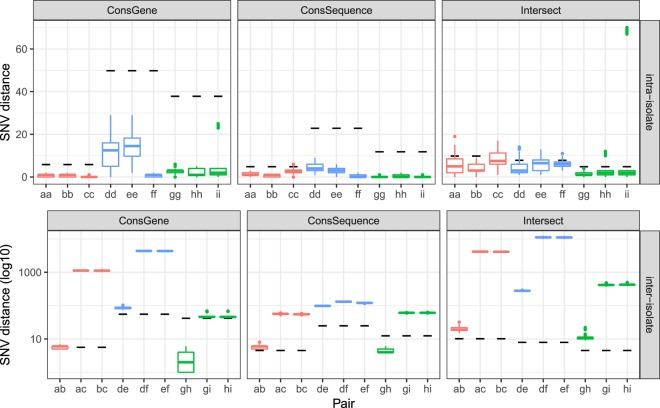
To evaluate whether the conserved-sequence core genome approach correctly identifies same-pathogen samples – a measure of the true positive rate for transmission detection – we applied it to a set of 81 technical and biological replicates generated from three isolates for each species considered (see Method section 1). Since these are small sample sets, we use the intersection core genome as defined by the large data sets in previous section. Using the SNV thresholds described in previous section, nearly all intra-isolate distances for E. faecium, S. aureus, and K. pneumoniae fall below the thresholds, while the inter-isolate distances lie above the thresholds (Fig. 5), except for isolates g-h, which are same-patient samples. While the conserved-gene and conserved-sequence method provide similar results, the intersection core genome leads to some outlying distances and larger distances for the same-patient isolates g-h. These differences could result from our use of the intersection core genome defined by the ROC sample set, perhaps reflecting that the intersection core genome does not generalize as well as the conserved genome approaches. The conserved genome approaches provide consistent results across a variety of strains and variation in experimental setup and sample processing, resulting in genome coverages ranging from 50× to 200×.
Figure 5.
Distribution of intra-isolate (upper panel) and inter-isolate (lower panel) SNV distances between replicates for E. faecium (red), S. aureus (blue), and K. pneumoniae (green), represented by boxplots. The SNV thresholds for genomic relatedness for each species are indicated with black dashes.
Comparison of genomic samples across geographical locations
The conserved-sequence and conserved-gene approaches produce SNV distances that are independent of the sample set. This is not only useful to compare samples across time frames – it also makes it possible to compare samples across locations without having to anticipate the samples for all locations. Here, we test how samples compare across clinical sites with different core genome methods.
We consider S. aureus cohorts from multiple locations in the U.K.32,33. and E. faecium from Australia34 and the U.K.35. Since we expect to see few direct transmissions between these geographically distinct sites, the SNV distances should be clearly distinguishable from the same-patient distances for these data sets. We tested this for the three different core genome methods considered (Fig. 6). Comparing the inter-site sample SNV distances to the quantiles obtained for the same-patient distances, we see that the E. faecium inter-site distances fall well above the 80% same-patient threshold for all core genome methods. For S. aureus, inter-site distances are closer to the 80% same-patient threshold, perhaps reflecting S. aureus’ presence as a community pathogen. The conserved-gene method finds many inter-site sample distances below the 80% same-patient threshold, most of which are likely false positives. The conserved-sequence and intersection core genome find hardly any inter-site sample distances below the 80% same-patient threshold. The intersection core genome method leads to a better separation of inter-site sample distances than the conserved-sequence method but is difficult to apply in a prospective manner across locations because it requires prior knowledge of all samples to define the core genome. Using a conserved-sequence core genome and similar data processing pipelines across medical centers, sample comparison across sites becomes trivial. The ability to compare genomic results efficiently across medical centers and time frames enables real-time tracking of outbreaks and emerging threats.
Figure 6.
Comparison of SNV distances (in red) between samples from geographically distinct cohorts – between S. aureus samples collected in East England and Oxford, U.K., respectively, and between E. faecium samples collected in Victoria, Australia and Cambridge, U.K. – to the quantiles obtained for same-patient distances (in blue). Most of the inter-site distances fall above the 80% same-patient threshold (see Fig. 4), except for the conserved-gene method.
Comparison of core genome approaches on a confirmed S. aureus outbreak
We compare the different core genome approaches’ ability to confirm and distinguish previously determined outbreak samples from an S. aureus outbreak in a neonatal intensive care in Cambridge, U.K.21. Of the 56 samples from this study, 44 are labeled as part of the outbreak (1 of the original samples was dropped due to insufficient coverage) and 12 as different from the outbreak. To obtain a more realistic – and more challenging – test scenario, we add the set of 2278 East England S. aureus samples such that the outbreak samples present a small fraction of samples examined. As we showed in the Results, consistency requirements and computational considerations essentially rule out the intersection core genome for prospective application, but it nonetheless represents an important benchmark. We defined the SNV thresholds for pathogen relatedness as the 80% same-patient threshold in the ROC curves (Fig. 4), corresponding to a similar sensitivity on the large S. aureus data set for all methods.
The intersection core genome and conserved-sequence core genome find highly similar transmission clusters for the S. aureus outbreak samples (Fig. 7). The conserved-sequence method confirms 44 out of 44 outbreak samples and finds 10 of the East England samples similar to the outbreak; the intersection method confirms 43 and adds 9 East England samples to the outbreak cluster, all 9 of which agree with those found by the conserved-sequence method. These 9 samples are of the same sequence type as the outbreak and may be genomically highly similar, since the East England samples were collected in the same geographical area as the outbreak and S. aureus is known to be an important community pathogen. The conserved-gene method, on the other hand, fails to confirm 9 of the outbreak samples and does not add any East England samples. A narrow focus on the outbreak cluster may suggest that its performance can simply be improved by increasing the SNV threshold, but this would lead to a very large number of potential transmissions in the overall data set. In order to confirm the 44 outbreak samples, the conserved-gene threshold would need to be raised to 89 SNV’s (Fig. S5), at which point 1,110,677 out of 3,235,210 different-patient sample pairs in the ROC curve would be captured below the threshold (Fig. 4). Another observation we make is that the conserved-gene distances cluster the outbreak samples together by virtue of single linkage but are much less consistent than the conserved-sequence and the intersection distances (Figs S2–S4).
Figure 7.
Network plot displaying single-linkage clusters of confirmed S. aureus outbreak samples (blue dots) and the 45 closest other samples (red dots) based on genomic distances computed with different core genome methods. Connected dots represent clusters of samples with SNV distances below the threshold for relatedness (i.e., the 80% same-patient pair threshold in Fig. 4).
This example illustrates how the SNV thresholds and conserved-gene and conserved-sequence genomes, defined independently of the outbreak data set, might apply to new data sets. The conserved-sequence method confirms the known outbreak samples with similar sensitivity and precision to the intersection core genome method, even though its definition did not rely on any of the outbreak sample data.
Discussion
Each core genome method has advantages and disadvantages and the most suitable method depends on the application. In a clinical environment, in which samples are continuously added and analyzed, stable SNV distances are needed to provide consistent insights over time and to avoid recomputing SNV distances. The commonly used intersection core genome, consisting of genome regions with high-confidence reference/variant calls in all samples, does not provide such consistency (Fig. 1). Conserved-gene and conserved-sequence approaches, such as the one we introduced here, consider regions of the genome that are conserved among strains and can be applied invariably to all samples. The resulting SNV distances do not depend on the sample set and thus provide consistent genomic relationships across time and location. While defining the core genomes a priori may trade in some resolution for consistency across sample sets, the ability to compare samples efficiently and dynamically within and across hospitals is crucial to identify outbreaks and emerging threats.
The conserved-sequence method we developed is suitable for dynamic clinical studies and provides a better sensitivity/specificity tradeoff for pathogen relatedness over the conserved-gene method, as measured by ROC curves for large clinical data sets. This is true for all species examined (Fig. 4). Comparing S. aureus and E. faecium samples from different locations (Fig. 6), the conserved-sequence method finds few small SNV distances, consistent with a low false-positive rate. The difference in accuracy between the conserved genome methods is very clear for the S. aureus outbreak, for which the conserved-sequence method confirms 44 out of 44 known outbreak samples (Fig. 7), while the conserved-gene method confirms only 36. The conserved-sequence method performs as well as the intersection core genome on this outbreak study but has the advantage that it can be applied towards prospective infectious disease epidemiology.
While the intersection core genome approach is not suitable for dynamic clinical analysis, the conserved genome approaches have the drawback that they put a stronger requirement on the consistency of variant calls across samples: inconsistent calls add to the error on the genomic distance metric in fixed core genome approaches, while they are often excluded from consideration in an intersection core genome approach because they correspond to low-confidence calls. The variability introduced by variant calling and experimental errors can be benchmarked via sample replicates (Fig. 5). The ability of the core-genome SNV distances to separate same-pathogen samples from different-pathogen samples is therefore a function of the core genome method, the choice of reference genome and the bioinformatics analysis. These factors should be considered when formulating SNV thresholds for pathogen relatedness. In the absence of large same-pathogen labeled data sets, SNV thresholds can be determined via ROC curves for same-patient (same-strain-type) samples versus different-patient samples (Fig. 4).
The conserved-sequence approach can be further optimized. To capture conserved sequences, diversity of the genome assemblies (in terms of sequence type or subspecies) is more important than the absolute number of assemblies. For instance, using a random subsample of 2,000 of the 8,274 S. aureus assemblies generates a highly similar conserved-sequence genome to the full set. Instead of using all available assemblies for the species, stratifying them by their clinical importance or stratifying them equally among different strain types can potentially improve results.
Methods
Data sources
The fixed core genome approaches we evaluate build a core genome based on reference genome data, typically genome assemblies. We use all available RefSeq genome assemblies for the species considered, including 782 assemblies for E. faecium, 2,876 for K. pneumoniae and 8,274 for S. aureus.
The replicate study consists of 3 isolates for each species, obtained from Westchester Medical Center. For each isolate that is sequenced, two additional sequencing replicates, two additional library replicates, and two additional colonies from the same plate are analyzed. Two additional culture replicates (from the same colony) are sequenced one and two days after the original isolate, yielding a total of 9 replicates for each isolate. The libraries are prepared using Nextera XT DNA library preparation kits and sequenced using Illumina 2 × 150 bp reads.
All other data used are public data sets from the NCBI Sequencing Read Archive (SRA): a 57 sample set of S. aureus from Cambridge, U.K. (PRJEB2737)21, a 1158 sample set of S. aureus from Oxford, U.K. (PRJNA369475)33, a 2278 sample set of methicillin-resistant S. aureus (MRSA) from East England (PRJEB3174)32, a 1362 sample set of predominantly extended-spectrum beta-lactamase producing K. pneumoniae from Houston, TX (PRJNA376414)28, a 310 sample set of vancomycin-resistant Enterococcus (VRE) from Victoria, Australia (PRJNA433676)34 and a 175 sample set of VRE from Cambridge, U.K. (PRJEB12937)35. The VRE set from Cambridge contains longitudinal sampling and same-day replicates from a small number of patients. We remove the same-day replicates by selecting only the sample with the greatest sequencing depth for the same combination of patient ID, sequence type, sample source and sampling date. We also redefine patients as unique combinations of patient and sequence type of the pathogen in this study, since several patients had multiple distinct infections with different sequence types.
Genome alignment and variant calling
All sequencing data consist of Illumina paired-end reads, which are trimmed using Trimmomatic36 and aligned with BWA-mem37 against the following reference genomes: Staphylococcus aureus NCTC 8325 (GenBank CP000253.1), Enterococcus faecium DO (GenBank CP003583.1), Klebsiella pneumoniae HS11286 (GenBank CP003200.1). Single-nucleotide variants (SNVs) are then called with FreeBayes38 with a minimum base quality of 20 and subsequently filtered with vcffilter to keep SNV’s with allele frequency greater than 0.9 and a quality score greater than 5. The core genome definitions are stored in BED format, and this format is used to filter the SNV calls for the core genome regions using BCFtools’39 intersection operation. SNV distances are then computed based on the consensus sequences filtered for the core genome regions. Only samples with coverage breadth >80% and depth >30x and fewer than 5000 ambiguous base calls (including low coverage and variant calls with <90% allele frequencies) are included in our analysis (Supplementary Fig. S1). In the Klebsiella dataset, we additionally remove samples for which were unable to call a multi-locus sequence type based on the variant calls.
Intersection core genome
The intersection core genome approach selects the reference genome nucleotides for which all samples have sufficient read coverage and unambiguous variant/reference calls. More specifically, a position p in the reference genome is included in the intersection core genome if all samples considered have read coverage depth greater than 20x at this position after alignment to the reference and no sample has an ambiguous variant call in this position (i.e., a variant call with <90% allele frequency).
Conserved-gene core genome
The conserved-gene core genome consists of genes found in greater than 95% of all RefSeq reference assemblies considered. The genes in these assemblies are identified with Prokka40. The genes are then clustered by identity and for each gene cluster, the number of assemblies containing a gene in the cluster is counted using Roary41. The genes that occur in greater than 95% of assemblies are then selected as the core genome.
Conserved-sequence core genome
The conserved-sequence core genome consists of conserved sequences within the reference genome. Nucleotide-based conservation scores are computed from all RefSeq assemblies for the same species via a k-mer frequency heuristic (see Results). The conservation scores reflect the fraction of assemblies that contain the same nucleotide as the reference genome in a given position. To k-merize the genome assemblies, we use Jellyfish42, with 31 as the k-mer length. We use a 95% cutoff on the conservation score to select the core genome.
Intersection core genome evolution with increasing cohort size
To visualize the intersection core genome SNV distances with increasing cohort size (Fig. 1), we select all sequence type 307 samples from the Houston K. pneumoniae set for which the same patient was sampled at least three times (49 samples). We use the procedure described above to define the intersection core genomes, while adding increasing numbers of arbitrarily chosen samples (of any sequence type) from the same Houston cohort to the original set.
ROC curves for same-patient sample distinction
To compose ROC curves for disambiguation of same-patient (positive) sample pairs, we use all datasets combined per species, except for the S. aureus outbreak data. Each point on the ROC curve represents a true-positive/false-positive tradeoff with a particular SNV threshold. For combined data sets, only distances within datasets could be labeled as same-patient or different-patient, so distances between datasets were excluded.
S. aureus outbreak case study
For this study, we combine samples from the Cambridge outbreak21 with the East England MRSA study32, which contains samples from Cambridge and surrounding locations. For each core genome method, the plot displays the 44 outbreak samples and the 45 other samples with the smallest distances to the outbreak samples.
Conclusion
The adoption of rapid whole-genome-based pathogen profiling for clinical epidemiology comes with a need for standards and methods that provide consistent genomic insights. Perhaps most importantly, we need methods that quantify pathogen similarity in a manner that is robust, universally applicable and suitable for dynamic analysis, in which samples are added and analyzed incrementally. This is different from a typical retrospective research setup, in which the entire sample set and the target pathogen are known and reference genomes can be chosen to be similar to the target pathogen. In a prospective clinical application, typically one reference genome is used per species and the pathogen comparison has to be robust to cover a large variety of strains within the species. As we have illustrated, prospective studies require a core genome that remains fixed throughout the duration of the study to quantify pathogen similarities consistently across time.
In this work, we introduced a conserved-sequence genome, composed by analyzing k-mer frequencies in publicly available genome assemblies, that can be applied universally to obtain consistent SNV distances across time and location. The k-mer frequency analysis used to compose the conserved-sequence genome can be applied to any set of assemblies, or even to sample data, allowing the core genome to capture the conserved sequences across a large variety of strains. Several optimizations are possible, such as using a set of assemblies stratified by strain type according to their clinical incidence. The SNV distances could also be improved by taking SNV read signals into account rather than a binary present/absent comparison.
While a priori-defined core genomes may trade in some resolution to achieve consistency, we demonstrated with large clinical datasets that the conserved-sequence method detects potential transmissions with better sensitivity/specificity than the conserved-gene method. On a small set of labeled outbreak data, the conserved-sequence method identifies known outbreak samples with similar sensitivity and specificity to the intersection core genome method, but it has the advantage that it enables prospective genomic studies. This approach paves the way towards accurate, dynamic whole-genome pathogen comparison, making it possible to identify pathogen transmissions as they happen.
Supplementary information
Author Contributions
H.vA., R.K., Y.F., M.F.H., J.J.C. and B.D.G. contributed to the design of the study and editing of the manuscript. H.v.A. conceived conserved-sequence methodology, performed sample replicate analysis, created Figures 2, 5, and wrote the manuscript. R.K. performed statistical analyses, conceived testing methodology and created data visualizations 1,4,6,7. H.C. processed genomic data for public data sets. J.L. performed data analysis and plotting for Figure 3. Y.F. created the conserved-gene methodology pipeline. J.T.F., W.H. and G.W. executed experiments on sample replicates. All authors have read and approved the final manuscript.
Data Availability
The sequences from the replicate study have been submitted to the NCBI SRA database with the Accession Number PRJNA512284.
Competing Interests
H.vA., R.K., J.L., H.C., Y.F., M.F.H., J.J.C. and B.D.G. are employees of Philips Research. H.vA. and B.D.G. have submitted a patent application on the core genome methodology (62/684323, 06/13/2018). J.T.F., W.H. and G.W. have received funding from Philips Research towards the work described herein and other work related to infectious disease research.
Footnotes
Publisher’s note: Springer Nature remains neutral with regard to jurisdictional claims in published maps and institutional affiliations.
Supplementary information
Supplementary information accompanies this paper at 10.1038/s41598-019-44189-0.
References
- 1.Rossen JWA, Friedrich AW, Moran-Gilad J. Practical issues in implementing whole-genome-sequencing in routine diagnostic microbiology. Clin. Microbiol. Infect. 2018;24:355–360. doi: 10.1016/j.cmi.2017.11.001. [DOI] [PubMed] [Google Scholar]
- 2.Tagini F, Greub G. Bacterial genome sequencing in clinical microbiology: a pathogen-oriented review. Eur. J. Clin. Microbiol. Infect. Dis. 2017;36:2007–2020. doi: 10.1007/s10096-017-3024-6. [DOI] [PMC free article] [PubMed] [Google Scholar]
- 3.Gardy JL, Loman NJ. Towards a genomics-informed, real-time, global pathogen surveillance system. Nat. Rev. Genet. 2017;19:9. doi: 10.1038/nrg.2017.88. [DOI] [PMC free article] [PubMed] [Google Scholar]
- 4.Tang P, Croxen MA, Hasan MR, Hsiao WWL, Hoang LM. Infection control in the new age of genomic epidemiology. Am. J. Infect. Control. 2017;45:170–179. doi: 10.1016/j.ajic.2016.05.015. [DOI] [PubMed] [Google Scholar]
- 5.Aarestrup FM, et al. Integrating Genome-based Informatics to Modernize Global Disease Monitoring, Information Sharing, and Response. Emerg. Infect. Dis. 2012;18:e1–e1. doi: 10.3201/eid1811.120453. [DOI] [PMC free article] [PubMed] [Google Scholar]
- 6.Besser J, Carleton HA, Gerner-Smidt P, Lindsey RL, Trees E. Next-generation sequencing technologies and their application to the study and control of bacterial infections. Clin. Microbiol. Infect. 2018;24:335–341. doi: 10.1016/j.cmi.2017.10.013. [DOI] [PMC free article] [PubMed] [Google Scholar]
- 7.Zimlichman E, et al. Health Care–Associated Infections: A Meta-analysis of Costs and Financial Impact on the US Health Care System. JAMA Intern. Med. 2013;173:2039. doi: 10.1001/jamainternmed.2013.9763. [DOI] [PubMed] [Google Scholar]
- 8.Stone PW. Economic burden of healthcare-associated infections: an American perspective. Expert Rev. Pharmacoecon. Outcomes Res. 2009;9:417–422. doi: 10.1586/erp.09.53. [DOI] [PMC free article] [PubMed] [Google Scholar]
- 9.Salipante SJ, et al. Application of Whole-Genome Sequencing for Bacterial Strain Typing in Molecular Epidemiology. J. Clin. Microbiol. 2015;53:1072–1079. doi: 10.1128/JCM.03385-14. [DOI] [PMC free article] [PubMed] [Google Scholar]
- 10.Maiden MCJ, et al. Multilocus sequence typing: A portable approach to the identification of clones within populations of pathogenic microorganisms. Proc. Natl. Acad. Sci. 1998;95:3140–3145. doi: 10.1073/pnas.95.6.3140. [DOI] [PMC free article] [PubMed] [Google Scholar]
- 11.Bekal S, et al. Usefulness of High-Quality Core Genome Single-Nucleotide Variant Analysis for Subtyping the Highly Clonal and the Most Prevalent Salmonella enterica Serovar Heidelberg Clone in the Context of Outbreak Investigations. J. Clin. Microbiol. 2016;54:289–295. doi: 10.1128/JCM.02200-15. [DOI] [PMC free article] [PubMed] [Google Scholar]
- 12.Kwong JC, Mccallum N, Sintchenko V, Howden BP. Whole genome sequencing in clinical and public health microbiology. Pathology (Phila.) 2015;47:199–210. doi: 10.1097/PAT.0000000000000235. [DOI] [PMC free article] [PubMed] [Google Scholar]
- 13.Pearce ME, et al. Comparative analysis of core genome MLST and SNP typing within a European Salmonella serovar Enteritidis outbreak. Int. J. Food Microbiol. 2018;274:1–11. doi: 10.1016/j.ijfoodmicro.2018.02.023. [DOI] [PMC free article] [PubMed] [Google Scholar]
- 14.Harris SR, et al. Whole-genome analysis of diverse Chlamydia trachomatis strains identifies phylogenetic relationships masked by current clinical typing. Nat. Genet. 2012;44:413. doi: 10.1038/ng.2214. [DOI] [PMC free article] [PubMed] [Google Scholar]
- 15.Schürch AC, Arredondo-Alonso S, Willems RJL, Goering RV. Whole genome sequencing options for bacterial strain typing and epidemiologic analysis based on single nucleotide polymorphism versus gene-by-gene–based approaches. Clin. Microbiol. Infect. 2018;24:350–354. doi: 10.1016/j.cmi.2017.12.016. [DOI] [PubMed] [Google Scholar]
- 16.Maiden MCJ, et al. MLST revisited: the gene-by-gene approach to bacterial genomics. Nat. Rev. Microbiol. 2013;11:728. doi: 10.1038/nrmicro3093. [DOI] [PMC free article] [PubMed] [Google Scholar]
- 17.Mellmann A, et al. Prospective Genomic Characterization of the German Enterohemorrhagic Escherichia coli O104:H4 Outbreak by Rapid Next Generation Sequencing Technology. PLoS ONE. 2011;6:e22751. doi: 10.1371/journal.pone.0022751. [DOI] [PMC free article] [PubMed] [Google Scholar]
- 18.Denamur E, Matic I. Evolution of mutation rates in bacteria. Mol. Microbiol. 2006;60:820–827. doi: 10.1111/j.1365-2958.2006.05150.x. [DOI] [PubMed] [Google Scholar]
- 19.Martinez JL, Baquero F. Mutation Frequencies and Antibiotic Resistance. Antimicrob. Agents Chemother. 2000;44:1771–1777. doi: 10.1128/AAC.44.7.1771-1777.2000. [DOI] [PMC free article] [PubMed] [Google Scholar]
- 20.Chen C, et al. Minimum Core Genome Sequence Typing of Bacterial Pathogens: a Unified Approach for Clinical and Public Health Microbiology. J. Clin. Microbiol. 2013;51:2582–2591. doi: 10.1128/JCM.00535-13. [DOI] [PMC free article] [PubMed] [Google Scholar]
- 21.Harris SR, et al. Whole-genome sequencing for analysis of an outbreak of meticillin-resistant Staphylococcus aureus: a descriptive study. Lancet Infect. Dis. 2013;13:130–136. doi: 10.1016/S1473-3099(12)70268-2. [DOI] [PMC free article] [PubMed] [Google Scholar]
- 22.Bowers JR, et al. Genomic Analysis of the Emergence and Rapid Global Dissemination of the Clonal Group 258 Klebsiella pneumoniae Pandemic. PLOS ONE. 2015;10:e0133727. doi: 10.1371/journal.pone.0133727. [DOI] [PMC free article] [PubMed] [Google Scholar]
- 23.Grad YH, et al. Genomic epidemiology of the Escherichia coli O104:H4 outbreaks in Europe, 2011. Proc. Natl. Acad. Sci. 2012;109:3065–3070. doi: 10.1073/pnas.1121491109. [DOI] [PMC free article] [PubMed] [Google Scholar]
- 24.Parcell BJ, et al. Pseudomonas aeruginosa intensive care unit outbreak: winnowing of transmissions with molecular and genomic typing. J. Hosp. Infect. 2018;98:282–288. doi: 10.1016/j.jhin.2017.12.005. [DOI] [PMC free article] [PubMed] [Google Scholar]
- 25.Leong KWC, et al. Emergence of Vancomycin-Resistant Enterococcus faecium at an Australian Hospital: A Whole Genome Sequencing Analysis. Sci. Rep. 2018;8:6274. doi: 10.1038/s41598-018-24614-6. [DOI] [PMC free article] [PubMed] [Google Scholar]
- 26.Kumar N, et al. Genome-Based Infection Tracking Reveals Dynamics of Transmission and Disease Recurrence. Clin. Infect. Dis. 2016;62:746–752. doi: 10.1093/cid/civ1031. [DOI] [PMC free article] [PubMed] [Google Scholar]
- 27.Sui, W. et al. Whole genome sequence revealed the fine transmission map of carbapenem-resistant Klebsiella pneumonia isolates within a nosocomial outbreak. Antimicrob. Resist. Infect. Control7 (2018). [DOI] [PMC free article] [PubMed]
- 28.Long, S. W. et al. Population Genomic Analysis of 1,777 Extended-Spectrum Beta-Lactamase-Producing Klebsiella pneumoniae Isolates, Houston, Texas: Unexpected Abundance of Clonal Group 307. mBio8 (2017). [DOI] [PMC free article] [PubMed]
- 29.Segerman, B. The genetic integrity of bacterial species: the core genome and the accessory genome, two different stories. Front. Cell. Infect. Microbiol. 2 (2012). [DOI] [PMC free article] [PubMed]
- 30.Wang L, Jiang T. On the Complexity of Multiple Sequence Alignment. J. Comput. Biol. 1994;1:337–348. doi: 10.1089/cmb.1994.1.337. [DOI] [PubMed] [Google Scholar]
- 31.Chatzou M, et al. Multiple sequence alignment modeling: methods and applications. Brief. Bioinform. 2016;17:1009–1023. doi: 10.1093/bib/bbv099. [DOI] [PubMed] [Google Scholar]
- 32.Coll Francesc, Harrison Ewan M., Toleman Michelle S., Reuter Sandra, Raven Kathy E., Blane Beth, Palmer Beverley, Kappeler A. Ruth M., Brown Nicholas M., Török M. Estée, Parkhill Julian, Peacock Sharon J. Longitudinal genomic surveillance of MRSA in the UK reveals transmission patterns in hospitals and the community. Science Translational Medicine. 2017;9(413):eaak9745. doi: 10.1126/scitranslmed.aak9745. [DOI] [PMC free article] [PubMed] [Google Scholar]
- 33.Young, B. C. et al. Severe infections emerge from commensal bacteria by adaptive evolution. eLife6 (2017). [DOI] [PMC free article] [PubMed]
- 34.Lee, R. S. et al. The changing landscape of VREfm in Victoria, Australia: a state-wide genomic snapshot, 10.1101/289975 (2018).
- 35.Moradigaravand, D. et al. Within-host evolution of Enterococcus faecium during longitudinal carriage and transition to bloodstream infection in immunocompromised patients. Genome Med. 9 (2017). [DOI] [PMC free article] [PubMed]
- 36.Bolger AM, Lohse M, Usadel B. Trimmomatic: a flexible trimmer for Illumina sequence data. Bioinformatics. 2014;30:2114–2120. doi: 10.1093/bioinformatics/btu170. [DOI] [PMC free article] [PubMed] [Google Scholar]
- 37.Li, H. Aligning sequence reads, clone sequences and assembly contigs with BWA-MEM. ArXiv13033997 Q-Bio (2013).
- 38.Garrison, E. & Marth, G. Haplotype-based variant detection from short-read sequencing. arXiv:1207.3907 (2013).
- 39.Li H. A statistical framework for SNP calling, mutation discovery, association mapping and population genetical parameter estimation from sequencing data. Bioinformatics. 2011;27:2987–2993. doi: 10.1093/bioinformatics/btr509. [DOI] [PMC free article] [PubMed] [Google Scholar]
- 40.Seemann T. Prokka: rapid prokaryotic genome annotation. Bioinformatics. 2014;30:2068–2069. doi: 10.1093/bioinformatics/btu153. [DOI] [PubMed] [Google Scholar]
- 41.Page AJ, et al. Roary: rapid large-scale prokaryote pan genome analysis. Bioinformatics. 2015;31:3691–3693. doi: 10.1093/bioinformatics/btv421. [DOI] [PMC free article] [PubMed] [Google Scholar]
- 42.Marçais G, Kingsford C. A fast, lock-free approach for efficient parallel counting of occurrences of k-mers. Bioinformatics. 2011;27:764–770. doi: 10.1093/bioinformatics/btr011. [DOI] [PMC free article] [PubMed] [Google Scholar]
Associated Data
This section collects any data citations, data availability statements, or supplementary materials included in this article.
Supplementary Materials
Data Availability Statement
The sequences from the replicate study have been submitted to the NCBI SRA database with the Accession Number PRJNA512284.


