Abstract
Shotgun lipid analysis based on electrospray ionization-tandem mass spectrometry (ESI-MS/MS) is increasingly used in lipidomic studies. One challenge for the shotgun approach is the discrimination of lipid isomers and isobars. Gas-phase charge inversion via ion/ion reactions has been used as an effective method to identify multiple isomeric/isobaric components in a single MS peak by exploiting the distinctive functionality of different lipid classes. In doing so, fatty acyl chain information can be obtained without recourse to condensed-phase separations or derivatization. This method alone, however, cannot provide carbon-carbon double bond (C=C) location information from fatty acyl chains. Herein, we provide an enhanced method pairing photochemical derivatization of C=C via the Paterno-Buchi reaction with charge inversion ion/ion tandem mass spectrometry. This method was able to provide gas-phase separation of phosphatidylcholines and phosphatidylethanolamines, the fatty acyl compositions, and the C=C location within each fatty acyl chain. We have successfully applied this method to bovine liver lipid extracts and identified 40 molecular species of glycerophospholipids with detailed structural information including head group, fatty acyl composition, and C=C location.
Keywords: lipidomics, the Paternò-Büchi Reaction, unsaturated lipid, charge inversion, mass spectrometry
Graphical Abstract
INTRODUCTION
Amongst all the classes of lipids in mammalian cells, glycerophospholipids (GPs) are the most abundant, constituting approximately 60 mol% of lipid mass within the cell membrane.[1–2] GPs are the key component in cell membrane structure3; they function as signaling molecules [1,2] and secondary messengers in cell metabolism.[1–3] GP profiles have been used as phenotypical signals [3–5] for imaging of diseased tissues [6] and systems biology studies. [7–9] GPs are assembled from three building blocks, a glycerol backbone, two fatty acyl or alkyl chains linked at the sn1 and sn2 positions of the glycerol, and a phosphate-containing head group. [10–13] The identity of the head group defines the GP subclass; for example, phosphatidylethanolamine (PE) and phosphatidylcholine (PC) head groups consist of ethanolamine and choline esterified to the phosphate, respectively.
Lipid analysis via mass spectrometry (MS) can be performed using two main approaches: liquid chromatography-mass spectrometry (LC/MS) and direct-infusion electrospray mass spectrometry. The use of LC/MS allows for complex lipid components in a sample to undergo chromatographic separation prior to being analyzed by the MS, thus providing increased sensitivity and selectivity. [14–17] Direct-infusion electrospray mass spectrometry, or shotgun lipid analysis, is a fast and sensitive method that analyzes the lipids directly from the crude extract without chromatographic separation. [18–24] Shotgun analysis benefits from the use of high-resolution MS instruments, which helps distinguish isobaric peaks and allows for more accurate identification due to better peak separation. However, high mass resolving power alone cannot provide isomeric separation. Separation in shotgun lipidomic approaches can also be done via in-solution modifications. Functional group selective modification, such as targeting the vinyl ether bond, [4] on the GP head groups [25,26] or a one-step methylation of the phosphate, [27] has been demonstrated to improve identification and quantitation for isomeric/isobaric GPs and boosting the detection of low abundance GPs.
Gas-phase ion/ion reactions have been used to separate isomeric and isobaric species post-ionization thereby obviating the need for in-solution modification. Spraying PCs and PEs in the positive ion mode and subjecting the ions to gas-phase ion/ion interaction with a dicarboxylate reagent effectively charge inverts the cationic lipids and separates PC and PE isomers based on distinct reaction chemistry between their head groups and the reagent. [28,29]. The negatively charged ions resulting from charge inversion ion/ion reactions provide abundant fatty acyl fragment ions upon collision-induced dissociation (CID), leading to confident identification of fatty acyl composition including the number of carbons and degree of unsaturation. [30–31] However, the carbon-carbon double bond (C=C) location cannot be determined from such a process.
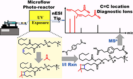
Notable gas-phase activation methods of determining C=C location of lipids include charge-remote fragmentation induced by high energy CID,[32] ozone-induced dissociation (OzID),[33–35] 193 nm ultraviolet photodissociation (UVPD),[36] radical directed dissociation (RDD),37,38 helium metastable atom-activated dissociation (He-MAD),[39,40] and electron impact excitation of ions from organics (EIEIO).[41,42] All these methods either utilize high-energy collisions or require significant instrument alterations. The Xia group introduced using UV-initiated reactions without instrument modification for localization of C=Cs, including ozonolysis [43,44] and the in-solution Paternò-Büchi (PB). [45–48] The PB reaction has been employed with subsequent ESI-MS/MS via low energy CID on multiple classes of lipids. [49] In such a method, acetone serves both as the PB reagent and a co-solvent for ESI-MS of lipids. Upon 254 nm UV irradiation of the solution, acetone selectively adds onto a C=C, forming a four-membered oxetane ring structure. Low energy CID of the PB reaction products ruptures the oxetane ring yielding fragment ions that are specific to the C=C location. The PB-MS/MS method is versatile as it can be performed on various MS instruments that have CID and ESI capabilities. Confident structural identification of unsaturated GPs at C=C location level builds upon the capability of determining fatty acyl/alkyl composition. For situations where lipids experience competitive ion suppression, such as detecting lower abundance PC in negative ion mode, or when lipid isobaric and isomeric isomers co-exist, fatty acyl chain assignment from MS2 CID in negative ion mode is often complicated by chemical interferences. [47] Combining the in-solution PB reaction and the gas-phase charge inversion reaction is promising for high level characterization of GP structures for shotgun lipidomics. In this study, synthetic standards of unsaturated PC and PE were used for method development. The performance of pairing the PB reaction with ion/ion charge inversion for complex mixture analysis was evaluated with a polar lipid extract of bovine liver, which led to confident identification of 40 PC and PE molecular species with detailed structural information including head group, fatty acyl composition, and C=C location.
MATERIALS & METHODS
Nomenclature.
We follow the lipid annotation recommended by LIPID MAPS. [50] In short, PC 16:0_18:2(Δ9,12) identifies the phosphocholine (PC) head group, two fatty acyl chains with the carbon number (the value before the colon, i.e. 16 and 18), the degree of unsaturation (the value after the colon, i.e. 0 and 2), and the location of C=C counting from the carboxylic carbon towards the fatty chain end (i.e. Δ9 and Δ12 in C18:2). The underscore (_) suggests that the sn-positions of fatty acyl chains are not specified, while a forward slash (/), identifies the fatty acyls occupy the sn1 and sn2 location, respectively, as the order indicated in the annotation. Only when the conformation of a C=C is known, a letter E or Z is used to indicate the trans- or cis-conformation, respectively.
Materials.
1-palmitoyl-2-oleoyl-glycero-3-phosphocholine (PC 16:0/18:1(9Z)), 1-palmitoyl-2-oleoyl-sn-glycero-3-phosphoethanolamine (PE 16:0/18:1(9Z)), and polar lipid extract of bovine liver (in chloroform) were purchased from Avanti Polar Lipid, Inc. (Alabaster, AL). 1,4-phenylenedipropionic acid (PDPA) and ammonium acetate were purchased from Sigma Aldrich (St. Louis, MO). Formic acid was purchased from Fisher Chemical. Organic solvents were LC-grade, and ultrapure water was obtained from a purification system at 0.03 μS cm.
The PB reactions in an offline flow microreactor.
A flow microreactor was constructed using UV transparent fused silica capillary as the flow cell (fluoropolymer-coated, 100 μm i.d. 363 μm o.d.; Polymicro Technologies/Molex; Phoenix, AZ). The fused silica capillary and a low-pressure mercury lamp with emission centered around 254 nm wavelength (BHK, Inc. Ontario, CA) were placed in parallel with a 1 cm distance, providing an effective UV exposure length of 4 cm. For safety considerations, the reactor was enclosed in an aluminum-lined cardboard box to prevent stray UV light. When conducting the PB reaction, the UV lamp was turned on and the lipid solution was pumped through the reactor via a syringe pump at a flowrate of 4–6 μL/min, enabling 4–5 second reaction times. All lipids were dissolved in 69/29/1/1 (v/v/v/v) of acetone/water/formic acid/isopropyl alcohol solution for the PB reaction. Ammonium acetate (10 mM) was added to the solvent system for conducting experiments when direct-injection negative ion mode was used. About 5–10 μL of the reaction solution was collected in a pre-pulled nanoESI tip and immediately subjected to subsequent MS analysis.
Mass Spectrometry
For prior analysis of the lipids in the bovine liver polar lipid extract, samples were analyzed using a QTRAP 4000 hybrid triple quadrupole/linear ion-trap mass spectrometer (Sciex, Toronto, ON, Canada) equipped with a home-built nanoESI source. The lipid extract was diluted to 100 μM in a 69/29/1/1 (v/v/v/v) of acetone/water/formic acid/isopropyl alcohol solvent system. Mass spectrometer parameters were as follows: spray voltage 1450–1600 V; curtain gas, 3 psi; declustering potential, 80 V; collision gas, high. Precursor ion scan (PIS) and neutral loss (NL) scan (NLS) were employed to identify the head group of the GPs.
Gas-phase charge inversion ion/ion reactions.
Structural analyses were performed using a TripleTOF 5600 triple quadruple/time-of-flight mass spectrometer (Sciex, Concord, ON, Canada) that was previously modified to perform mutual storage of cation and anions. [51] The dual nanoESI emitter setup allowed sequential formation of the PB modified lipid cations and PDPA dianions ([PDPA - 2H]2−), which were each mass selected in Q1 and transferred into the q2 linear ion trap for ion/ion reactions. The ions in q2 were mutually stored for 30 ms and the products of interest were isolated via a notched broadband waveform. CID was performed using a single frequency resonance excitation at q-value of 0.18. Third generation ion of interest was fragmented by placing it at a q-value of 0.14 and implementing a third resonance excitation event.
RESULTS AND DISCUSSION
Conducting the PB reactions in an off-line microreactor.
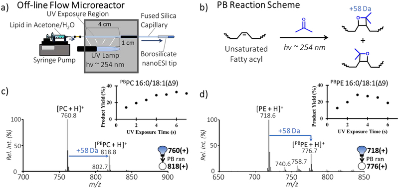
A graphic representation of the off-line flow microreactor for conducting the PB reaction is shown in Figure 1a and the reaction scheme is shown in Figure 1b. We have previously shown that using a flow microreactor to initiate the PB reaction allows for high photon efficiency leading to higher yield and the ability to control side reactions. [46] This setup is especially beneficial for the dual-emitter apparatus for alternatively injecting cations and anions for ion/ion reactions. The PB reaction conditions for unsaturated PC and PE were optimized by monitoring the PB product percent yield (the relative ion intensity of the PB products normalized to that of the intact lipid before UV exposure), using PC 16:0/18:1(9Z) and PE 16:0/18:1(9Z) as model compounds, respectively. Figure 1c and 1d represent typical nanoESI-MS spectra of the unsaturated PC and PE after the PB reactions. The PB products are clearly detected at m/z 818.7 for protonated PC 16:0/18:1(9Z), of the form [PBPC + H]+ in Figure 1c and at m/z 776.7 for protonated PE 16:0/18:1(9Z), of the form [pbPE + H]+ in Figure 1d, with limited evidence for side reactions resulting from Norrish Type I cleavage of acetone. [46] These PB products have a characteristic 58 Da increase in mass relative to the intact lipids, consistent with one acetone addition to the C=C in lipids. By monitoring the reaction kinetics, we found the PB reaction of PC 16:0/18:1(9Z) plateaued at approximately 6 seconds with 35% yield (inset of Figure 1c). For PE 16:0/18:1(9Z), the PB reaction yield was maximized around 3 seconds at 30% (kinetic curve shown in the inset of Figure 1d), while longer reaction times slightly decreased the yield due to increased contributions from side reactions. Based on these observations, the reaction time used for later studies was generally between 4–5 seconds to ensure adequate PB-product yield and minimize the extent of side reactions.
Fig. 1:
a) Schematic of an off-line flow microreactor for conducting the PB reaction; b) The PB reaction scheme, involving acetone addition to a C=C in a fatty acyl chain; the positive ion mode nanoESI-MS spectra of c) 5 μM PC 16:0/18:1(9Z) after 5 s UV exposure d) 5 μM PE 16:0/18:1(9Z) after 4 s UV exposure. Insets in c) and d) represent the PB reaction kinetic curve of PC and PE standard, respectively, with respect to UV exposure time. Positively charged ions are represented by (+).
Charge inversion of the PB reaction products.
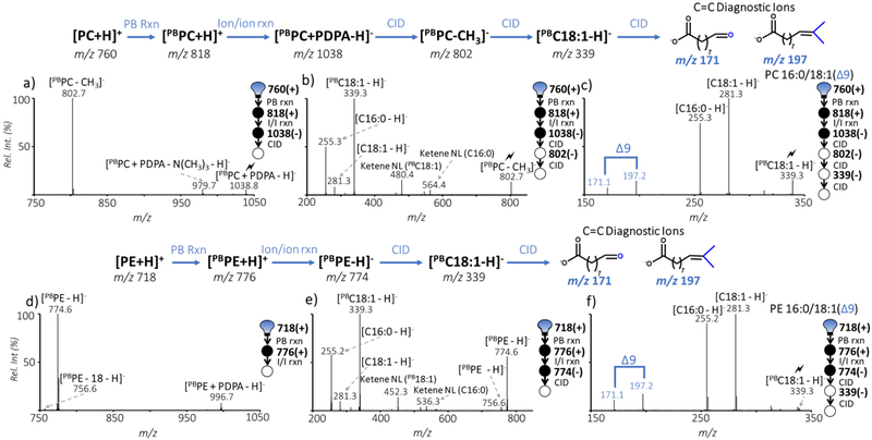
Using the optimized PB conditions for unsaturated PC and PE, we further explored pairing the PB reaction with charge inversion ion/ion reactions and subsequent CID experiments for structural characterization to the C=C location level. The typical workflow for unsaturated PC analysis is illustrated in Figure 2a–c, using the model compound PC 16:0/18:1(9Z) as an example. First, the PB product of PC 16:0/18:1(9Z) ([PBPC + H]+) was mass-isolated and subjected to ion/ion reaction with [PDPA - 2H]2-. The ion/ion reaction products included dominant complex formation of the two reactant ions, [PBPC + PDPA - H]− (m/z 1038), and a minor peak at m/z 802 resulting from demethylation of the PBPC anion (see Electronic Supplementary Material (ESM) Fig. S1). This ion/ion reaction phenomenon is identical to that of the intact PC cation, suggesting that the modification at the C=C from the PB reaction does not interfere with the charge inversion chemistry (ESM Fig. S2). Collisional activation of the complex ion at m/z 1038 mainly led to the formation of [PBPC - CH3]− ion (m/z 802.7, Figure 2a), formed from NL of methylated PDPA (236 Da). In previous work, the operational efficiency of protonated PC species to demethylated species was calculated as about 50%, although absolute efficiency could not be calculated due to difference in detector response to positive and negative ions. [28] Further CID of this fragment produced abundant fatty acyl anions (Figure 2b), including [C16:0 - H]− (m/z 255.3) and the PB modified C18:1 ([pbC18:1 - H]−, m/z 339.2), and fragments (m/z 480.4 and m/z 536.3) due to ketene NL of PBC18:1 and C16:0, respectively. A small intact C18:1 anion peak at m/z 281.3 was also observed, likely formed from loss of acetone from [PBC18:1 - H]− due to sequential fragmentation. The relatively higher abundances of ketene NL ions and carboxylate anions from the sn2 chain relative to those formed from sn1 chain, corroborate that the sn1 and sn2 chains are using PE 16:0/18:1(9Z) as an example. The workflow is slightly different from that of the PC, due to the difference in charge inversion ion/ion chemistry between a primary amine (in PE) and a quaternary amine (in PC). As shown in Figure 2d, the dominant charge inversion product of [pbPE + H]+ (m/z 776.6) is the corresponding deprotonated ion ([pbPE - H]−, m/z 774.6), accompanied only by a very small extent of complex formation (the peak at m/z 996.7). Collisional activation of [pbPE - H]− (Figure 2e) resulted in the formation of abundant PB reaction modified C18:1 ([PBC18:1 - H]−, m/z 339.3) and C16:0 ([C16:0 - H]−, m/z 255.2) fatty acyl anions as well as the ketene NL of the sn2 chain (m/z 452.3) and sn1 chain (m/z 536.3). Subsequent CID of [PBC18:1 - H]− produced characteristic C=C diagnostic ions that are 26 Da apart at m/z 171.1 and 197.2, confirming the C=C location at Δ9 in C18:1.
Fig. 2:
Sequence of events post-ion/ion reaction between GP standard cations and PDPA dianion; a) Ion trap CID of [PBPC + PDPA - H]−; b) Ion trap CID of [PBPC - CH3]−; c) Subsequent ion trap CID of [PBC18:1 - H]− d) Result of ion/ion reaction between [PDPA-2H]2− and [PBPE + H]+; e) Ion trap CID of [PBPE - H]−; f) Subsequent ion trap CID of [PBC18:1 - H]−; Schematics in a-c and d-f are layouts of the process for determining the GP structure. CID of a target ion is depicted by a lightning bolt ( ). Cations and anions are represented by (+) or (−), respectively.
The above analysis using PC and PE synthetic standards demonstrates that the PB reaction and gas-phase charge inversion ion/ion reactions can be efficiently paired and applied for confident structural identification of GPs at the C=C location level. Charge inversion of PB products exhibits equivalent information to unmodified GPs with the addition of the C=C localization and was applied to a biological extraction sample.
Shotgun analysis of the polar lipid extract from bovine liver.
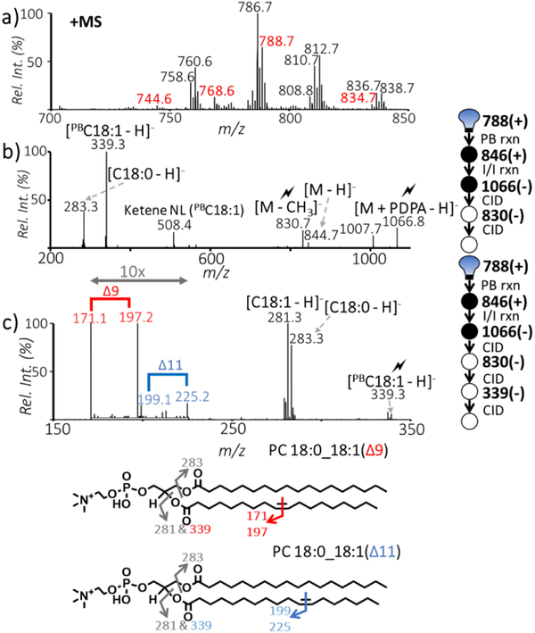
Polar lipid extract from bovine liver was employed as the benchmark test to evaluate the performance of coupling the PB reaction with ion/ion reaction for shotgun lipid analysis. The positive ion mode nanoESI mass spectrum of the lipid extract is shown in Figure 3a. By employing linked scans in positive ion mode, i.e. PE via 141 Da NLS and PC via m/z 184 PIS, these two lipid subclasses were identified (ESM Fig. S3); however, the fatty acyl composition, could not be obtained. Herein, we exemplify the power of the detailed structural analysis for unsaturated PC and PE by pairing the PB reaction and charge inversion ion/ion reactions. For clarity, those peaks that we demonstrate in later discussions are labelled in red in Figure 3a (i.e. m/z 744.6, m/z 768.6, m/z 788.7, and m/z 834.7).
Fig. 3:
a) Positive ion mode nanoESI MS spectrum of polar lipid extract from bovine liver (100 μM) without PB reaction, b) Subjecting ions at m/z 788.7 in panel (a) to the PB reaction (m/z 846.8 isolated in positive ion mode), charge inversion ion/ion reaction, and CID of post-ion/ion reaction product ions at m/z 1066.8 and 830.7 (negative ion mode) allows confident identification of this lipid at fatty acyl level, c) CID spectrum of m/z 339.3 produced in panel (b). Detection of two pairs of C=C diagnostic ions at m/z 171.1/197.2 and m/z 199.1/225.2 from C18:1 chain identifies the lipid at m/z 788.7 in panel (a) as PC 18:0_18:1(Δ9) and PC18:0_18:1(Δ11).
Individual PB reacted peaks were isolated from the UV irradiated, diluted lipid extract. The PB reaction product (m/z 846.8) of a relatively abundant lipid peak (m/z 788.7) was isolated using a unit mass isolation window and subjected to ion/ion reaction with the PDPA dianion. The resulting spectrum was dominated by ions at m/z 1066.8 from complex formation ([PBM + PDPA - H]−), a very minor peak at m/z 844.7 due to double proton transfer, and a small fragment ion (m/z 830.7) due to NL of methylated PDPA (ESM Fig. S4). Based on the distinct ion/ion charge inversion chemistry of PC and PE, i.e. complex formation vs. double proton transfer, it is evident that the peak at m/z 788.7 in Figure 3a consists of PC 36:1 as a major component and PE 39:1 as a minor component. In order to identify the fatty acyl composition and C=C location in PC 36:1, the ion complex at m/z 1066.8 was subjected to sequential CID experiments as depicted in Figure 2a. The CID spectrum of m/z 830.7 (([PBPC - CH3]−, Figure 3b) indicated that PC 36:1 consists primarily of C18:0 and C18:1. The glycerol backbone location of C18:1 is likely at sn2 given the high abundance of associated ketene NL, however, the method currently is limited in its ability to determine sn position due to possible coexisting isomers. This is consistent with the fatty acyl composition inferred from charge inversion and CID of the intact ions at m/z 788 (ESM Fig. S5). The PB modified C18:1 ion population was detected at m/z 339.3; further CID of these ions produced two C=C diagnostic ion pairs at m/z 171.1/197.2 and m/z 199.1/225.2, providing definitive evidence for locations of C=Cs at Δ9 and Δ11 in C18:1, respectively. Combining all above information, PC 36:1 could be confidently identified as a mixture of PC 18:0_18:1(Δ9) and PC 18:0_18:1(Δ11). The PC 18:0_18:1(Δ9) specie is the major isomer based on the relative abundances of its C=C diagnostic ions relative to those of the Δ11 isomer (Figure 3c). Due to the low ion signal of PE 39:1, C=C location determination could not be obtained after ion/ion reaction. The above data highlight that in a single spectrum, it is possible to differentiate GP class isomers, fatty acyl isomers, and C=C isomers (ESM Fig. S6).
Although charge inversion of the PB products was powerful in pinpointing detailed structural information of unsaturated PC and PE, the need of performing multi-step CID made it difficult to analyze lower abundance lipids. Furthermore, CID of the PB modified polyunsaturated fatty acid anions would not provide as abundant C=C diagnostic ions as performed in the positive ion mode. This limitation can be overcome by conducting ion/ion charge inversion and the PB reaction in two separate experiments. That is, first to charge invert the peak of interest to verify headgroup and fatty acyl identification, then use positive ion mode PB-MS/MS for determination of C=C location. Separately using PB-MS/MS and ion/ion reaction allows for increased sensitivity of C=C diagnostic information and a rapid structural analysis compared to combination of the methods.
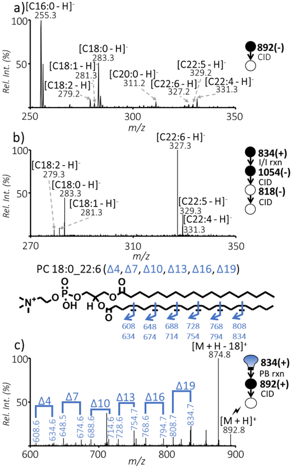
Structural analysis of polyunsaturated GPs is demonstrated by probing the ions at m/z 834.7 ([M + H]+) in Figure 3a, because of the structural complexity and minor relative abundance. Based on the PIS for m/z 184 combined with the LipidMaps structural database, the lipid peak was expected to contain PC 40:6. Following the conventional method for fatty acyl identification, the possible PC species (acetate adduct at m/z 892.8, [M+Ac]−) was probed in the negative ion mode from nanoESI of lipid solution added with 10 mM of ammonium acetate. Figure 4a shows the MS2 CID spectrum of [M+Ac]−, the same nominal m/z as the acetate adduct ion of PC 40:6. The major fragment peaks include ions at m/z 255.3 and 281.3, corresponding to C16:0 and C18:0, respectively, with a few smaller peaks corresponding to other fatty acyl chains. The combination of fatty acyl chains C16:0 and C18:0 would not provide the correct identification of PC 40:6 achieved from positive ion mode, largely due to poor ionization efficiency of the acetate adduct and ion suppression of PC in negative ion mode. Thus, it is evident that in negative ion mode the ions presented at m/z 892.8 contained isobaric GP species that were not the analyte of interest. Charge inversion ion/ion reaction of m/z 834.7 from positive ion mode produced abundant ions at m/z 1054.8 and 818.8, which are characteristic for the presence of PC 40:6. The appearance of ions at m/z 832.7 suggested the presence of PE 43:6 as a minor component. The CID spectrum of the demethylated PC 40:6 (m/z 818.8) is shown in Figure 4b. The negative ion mode spectrum contains abundant peaks of C18:0 and C22:6 at m/z 283.3 and 327.3, respectively. This set of data demonstrates the utility of charge inversion ion/ion chemistry in confidently providing fatty acyl chain information for less abundant PCs, which otherwise would be difficult to obtain using conventional shotgun lipid analysis methods.
Fig. 4:
a) Direct negative ionization of bovine liver polar lipid extract. Isolation followed by beam-type CID of ions at m/z 892.8 (positive m/z 834.7 from Figure 3a complexed with acetate anion, [M+Ac]−, 100 with 10 mM ammonium acetate); b) Charge inversion and subsequent ion trap CID spectrum of the peak found at m/z 834.7 in the positive ion mode spectrum; c) Positive ion mode ion-trap CID of the PB product of m/z 834.7 peak, [PBM + H]+, at m/z 892.8.
In order to assign the locations of unsaturation in C22:6, the PB-MS/MS experiment was performed separately in positive ion mode. Figure 4c shows ion-trap CID of m/z 892.8 ([PBM + H]+), which contains six distinct pairs of diagnostic ions (m/z 608.6/634.6, m/z 648.5/674.6, m/z 688.6/714.6, m/z 728.6/754.7, m/z 768.6/794.7, and m/z 808.7/834.7) that align with C=Cs at positions Δ4, Δ7, Δ10, Δ13, Δ16, and Δ19. The diagnostic ions thus allowed for confident identification of PC 18:0_22:6(Δ4, Δ7, Δ10, Δ13, Δ16, Δ19), which contains polyunsaturated omega-3 fatty acyl.
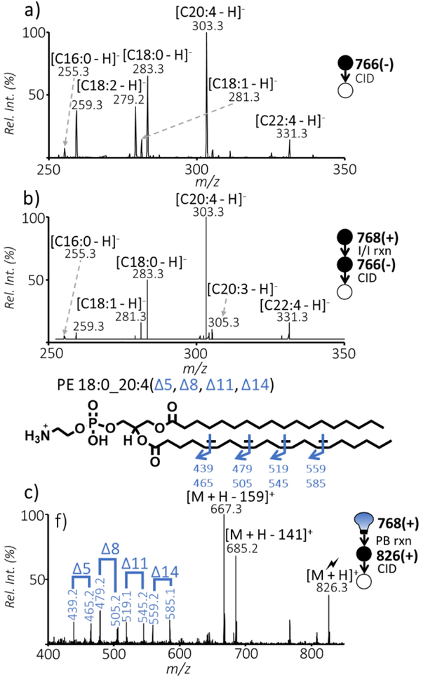
Figure 5a shows MS2 CID of ions at m/z 766.5, the same nominal mass as deprotonated PE 38:4 in negative ion mode. The fatty acyl chains that correspond to the PE species can be determined as mainly C20:4 and C18:0 classifying the peak as PE 18:0_20:4. Based on Figure 5a, however, there were still peaks present in high abundance that do not correspond to the lipid class being targeted. For example, a peak that corresponds to C18:2 at m/z 279.2 is prominent, but there is no parallel ion for C20:2 (m/z 307), the fatty acyl chain that would complement C18:2, suggesting that the C18:2 ion may arise from an isobaric species not related to PE, despite its high abundance. Figure 5b shows the charge inversion of the m/z 768.6 [M + H]+ ion and subsequent ion trap CID of 766.5 ([M - H]−) to determine that the fatty acyl chains present for PE 38:4 to be C18:0 and C20:4 at m/z 283.3 and 303.3, respectively, identifying the major species as PE 18:0_20:4, while several other minor components, i.e., PE 18:1_20:3 and PE 16:0_22:4, coexist. Note that the prominent m/z 279.2 ion observed in Figure 5a is largely absent in Figure 5b, which further suggests that the charge inversion process provides a degree of discrimination against possible isobaric interferences presence in direct negative ion mode ionization. Positive ion mode CID of the m/z 768.6 PB product at m/z 826.3, shown in Figure 5c, displays four C=C diagnostic ion pairs (m/z 439.2/465.2, m/z 479.2/505.2, m/z 519.1/545.2, and m/z 559.2/585.1), corresponding to the Δ5, Δ8, Δ11, and Δ14 C=C locations on the 20:4 fatty acyl chain (an omega-6 fatty acyl). This set of data identified that PE 18:0_20:4 (Δ5, Δ8, Δ11, Δ14) was the major component for the peak m/z 786.6 from shotgun analysis in the positive ion mode.
Fig. 5:
a) Direct ionization of bovine liver polar lipid extract in negative ion mode. Isolation and beam-type CID of ions at m/z 766.5 (positive m/z 768.6 from Figure 3a); b) Charge inversion and subsequent ion trap CID spectrum originating from positive ions present at m/z 768.6; c) Positive ion mode beam-type CID of the peak at m/z 826.3 (PB product of m/z 768.6).
When compared to using direct negative ionization for PC and PE analysis, charge inversion is shown to be efficient at determining fatty acyl chain information that is relevant to the peak of interest. The added ability to pair the fatty acyl chains determined to C=C location information allows novel information where ambiguity is minimized. Polyunsaturated PCs and PEs with greater than two double bonds, along with those with relative abundances less than 4% were analyzed using this method for confident double bond position siting.
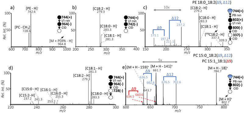
It is desirable for a method to be able to distinguish PC and PE isomers with the added ability to determine the C=C location. Investigation of the m/z 744.6 ions in the bovine liver extract via charge inversion is provided in Figure 6 to illustrate such a capability. Charge inversion followed by CID of the m/z 964.8 ion showed a dominant peak at m/z 742.6, a deprotonated PE 36:2 as well as a lower abundance peak at m/z 728.6 (demethylated anions of PC 33:2), indicating the co-existence of isomeric PE and PC in the lipid sample (Figure 6a). Ion trap CID of the m/z 742.6 ion resulted in ions at m/z 279.3, 283.3, and 281.3 representing fatty acyls C18:2, C18:0 and C18:1, respectively, as shown in Figure 6b. Based on the relative ion intensities of these fatty acyl anions, the PE 36:2 species in the sample was determined to contain PE 18:0_18:2 as a major component with PE 18:1_18:1 as a minor component. The minor PE species was identified as PE 18:1(Δ9)_18:1(Δ9) using beam-type CID of the PB product (m/z 802.7) in positive ion mode (ESM Fig. S7). The PB product (m/z 802.7) of the peak at m/z 744.6 was subjected to charge inversion followed by sequential steps of CID of the ions of m/z 1022.7 and m/z 800.6. Fatty acyl related products are detected at m/z 337.3, 283.3, 339.3, and 281.3 further confirming that PE 18:0_18:2 and PE 18:1_18:1 are present. The relative abundance of the m/z 337.3 ion was sufficiently high to allow for a subsequent CID for C=C localization. Diagnostic peaks with 26 Da differences are present at m/z 171/197 and m/z 211/237 to determine the double bond locations to be at Δ9 and Δ12 identifying the major PE species content to PE 18:0_18:2(Δ9, Δ12) (Figure 5c).
Fig. 6:
a) Charge inversion of cations at m/z 744.6 followed by ion trap CID of ions at m/z 964.8; b) Sequential CID of ions at m/z 742.8; c) CID of ions at m/z 337.3 resulting from sequential CID of charge inverted PB products; e) Positive ion trap CID of PB reaction product at m/z 802.7.
The isomeric PC 33:2 was analyzed by subjecting the demethylated anions to CID (Figure 5d). The detection of fatty acyl anions at m/z 281.3, 279.3, 283, 241.3, and 237.2 are C18:1, C18:2, C18:0, C15:0 and C15:2, respectively, meaning isomers PC 15:0_18:2, PC 15:1_18:1, and PC 15:2_18:0 are present. Positive PB product analysis (Figure 5e) was used to analyze the C=C location. Ion-trap CID in positive ion mode resulted in diagnostic ions that are 26 Da apart determining the major components to be PC 15:0_18:2(Δ9, Δ12) and PC 15:1_18:1(Δ9).
It is worth noting that the diagnostic ions for C15:1 with a Δ9 C=C location would be at m/z 676.5 and 702.6, meaning that they overlap with the diagnostic ions of Δ12 C=C in PC 15:0_18:2(Δ9, Δ12). This is a limitation of the direct positive ion mode approach that can be overcome via the charge inversion route, but only when the fatty acyl anion abundances are sufficiently high. The commonly present C18:2(Δ9, Δ12) motif inclined us to ascribe the m/z 676.5 and 702.6 ions as arising from the Δ12 position and do not report the presence of C15:1(Δ9). Further improvement of signal for low abundant monounsaturated fatty acyls will allow for the confident determination of C=C. Nonetheless, by pairing ion/ion charge inversion with the PB reaction, 40 distinct unsaturated PC and PE structures were determined with fatty acyl composition and C=C locations from bovine polar lipid extract as shown in Table 1 (detailed list in ESM Table S1j. This level of identification compares favorably to those reported from UVPD [36] and epoxidation.52
Table 1:
The identified PCs and PEs in bovine liver polar lipid extract via pairing the PB reaction with ion/ion charge inversion.
| [M + H]+ (m/z) |
Identity of GPs |
|---|---|
| 718.6 | PE 16:0_18:1(Δ9) |
| PE 17:0_18:1(Δ9) | |
| PC 14:0_18:1(Δ9) | |
| 732.6 | PC 14:0_18:1(Δ11) |
| PC 16:0_16:1(Δ9) | |
| 740.6 | PE 18:2(Δ9, Δ12)_18:2(Δ9, Δ12) |
| PE 18:0_18:2(Δ9, Δ12) | |
| 744.6 | PE 18:1(Δ9)_18:1(Δ9) |
| PC 15:1_18:1(Δ9) | |
| PC 15:1_18:1(Δ11) | |
| PC 15:0_18:2(Δ9, Δ12) | |
| PE 18:0_18:1(Δ9) | |
| PE 18:0_18:1(Δ11) | |
| 746.6 | PC 15:0_18:1(Δ9) |
| PC 15:0_18:1(Δ11) | |
| PC 17:1(Δ9)_16:0 | |
| PC 15:1(Δ9)_18:0 | |
| 758.6 | PC 16:0_18:2(Δ9, Δ12) |
| 760.7 | PC 16:0_18:1(Δ9) |
| PC 16:0_18:1(Δ11) | |
| 766.6 | PE 20:5(Δ5, Δ8, Δ11, Δ14, Δ17)_18:0 |
| 768.6 | PE 18:0_20:4(Δ5, Δ8, Δ11, Δ14) |
| 772.6 | PC 17:0_18:2(Δ9, Δ12) |
| 774.7 | PC 17:0_18:1(Δ9) |
| PC 18:0_17:1(Δ9) | |
| 782.6 | PC 16:0_20:4(Δ5, Δ8, Δ11, Δ14) |
| 784.6 | PC 16:0_20:3(Δ8, Δ11,Δ14) |
| PC 18:1(Δ9)_18:2(Δ9, Δ12) | |
| 786.7 | PC 18:0_18:2(Δ9, Δ12) |
| PC 18:1(Δ9)_18:1(Δ9) | |
| 788.7 | PC 18:0_18:1(Δ9) |
| PC 18:0_18:1(Δ11) | |
| 808.7 | PC 18:1_20:4(Δ5, Δ8, Δ11, Δ14) |
| PC 18:0_20:5(Δ5, Δ8, Δ11, Δ14, Δ17) | |
| 810.7 | PC 18:0_20:4(Δ5, Δ8, Δ11, Δ14) |
| 812.7 | PC 18:0_20:3(Δ8, Δ11, Δ14) |
| 834.7 | PC 18:0_22:6(Δ4, Δ7, Δ10, Δ13, Δ16, Δ19) |
| 836.7 | PC 18:0_22:5(Δ7, Δ10, Δ13, Δ16, Δ19) |
| PC 18:1_22:4(Δ7, Δ10, Δ13, Δ16) | |
| 838.7 | PC 18:0_22:4(Δ7, Δ10, Δ13, Δ16) |
CONCLUSION
In this work, we have paired the Paternò–Büchi reaction with charge inversion by ion/ion reaction aiming to achieve high level structural information for unsaturated PEs and PCs using the shotgun approach. The method allows for confident identification of the double bond location in a specific fatty acyl chain containing one or two degrees of unsaturation. Double bond location isomers were distinguished along with the ability to characterize PE and PC isomers. A limitation to conducting charge inversion and PB-MS/MS in one experiment is that C=C diagnostic fragment ions of polyunsaturated fatty acyl chain is difficult to obtain in negative ion mode.49 This limitation can be overcome by merging accurate fatty acyl chain information from charge inversion in negative ion mode with the information provided by positive ion mode PB-MS/MS for C=C location in two separate experiments.
For relatively abundant PCs and PEs or situations where chemical interference is minimal, charge inversion is not a necessity. However, for PCs and PEs at lower abundances, i.e. 2–10% of the most abundance species, charge inversion significantly reduces chemical interference, leading to both improved sensitivity and specificity for structural identification. As a simple comparison, without charge inversion we identified 24 GP species (18 PCs and 6 PEs) at C=C location level; while the combined methods allowed for identification of 40 GP species (31 PCs and 9 PEs). Dynamic range of such method is estimated to be 50-times. For GPs of lower than 2% relative abundance, a possible path to improve their identification will be application of this method on GPs that have been modified to improve ionization efficiency, suchas 13C-Trimethylation Enhancement Using Diazomethane.
Although not demonstrated here, quantitation for lipid C=C location isomers can be achieved in positive ion mode using methods already established from PB-MS/MS, while relative quantitation for fatty acyl composition isomers can be obtained from charge inversion followed by CID. A home-modified Q-TOF instrument has been employed in this study for charge inversion ion/ion reactions and PB-MS/MS; in principle, commercial MS instruments capable of ETD, such as an LTQ-Orbitrap, should support charge inversion as long as the doubly charged reagent anions can be introduced into linear ion trap for ion/ion reactions. Overall, the present work shows that gas-phase charge inversion paired with the PB reaction is a useful method for structural elucidation of unsaturated phospholipids.
Supplementary Material
ACKNOWLEDGEMENTS
This research was supported by National Institutes of Health (R01GM118484 to Y. X. and GM R37–45372 to S. A. M.) and Sciex.
Footnotes
Published in the topical collection Young Investigators in (Bio-)Analytical Chemistry with guest editors Erin Baker, Kerstin Leopold, Francesco Ricci, and Wei Wang.
CONFLICT OF INTEREST
The authors declare that they have no conflict of interest.
REFERENCES
- 1.Han X, Gross RW. Shotgun Lipidomics: Electrospray Ionization Mass Spectrometric Analysis and Quantitation of Cellular Lipidomes Directly from Crude Extracts of Biological Samples. Mass Spectrom Rev. 2005;24(3):367–412. [DOI] [PubMed] [Google Scholar]
- 2.Armstrong D, editor. Lipidomics [Internet]. Totowa, NJ: Humana Press; 2009. [cited 2017 Oct 8]. (Methods in Molecular Biology; vol. 579). Available from: http://link.springer.com/10.1007/978-1−60761−322-0 [Google Scholar]
- 3.Han X, Gross RW. Shotgun Lipidomics: Multidimensional MS Analysis of Cellular Lipidomes. Expert Rev Proteomics. 2005;2(2):253–64. [DOI] [PubMed] [Google Scholar]
- 4.Fhaner CJ, Liu S, Ji H, Simpson RJ, Reid GE. Comprehensive Lipidome Profiling of Isogenic Primary and Metastatic Colon Adenocarcinoma Cell Lines. Anal Chem. 2012;84(21):8917–26. [DOI] [PMC free article] [PubMed] [Google Scholar]
- 5.van Meer G, Voelker DR, Feigenson GW. Membrane lipids: Where They Are and How They Behave. Nat Rev Mol Cell Bio. 2008;9(2): 112–24. [DOI] [PMC free article] [PubMed] [Google Scholar]
- 6.Eberlin LS, Dill AL, Golby AJ, Ligon KL, Wiseman JM, Cooks RG, Agar NYR. Discrimination of Human Astrocytoma Subtypes by Lipid Analysis Using Desorption Electrospray Ionization Imaging Mass Spectrometry. Angew Chem Int Ed. 2010;49(34):5953–6. [DOI] [PMC free article] [PubMed] [Google Scholar]
- 7.Gross RW, Han X. Lipidomics at the Interface of Structure and Function in Systems Biology. Chem Biol. 2011;18(3):284–91. [DOI] [PMC free article] [PubMed] [Google Scholar]
- 8.Ejsing CS, Sampaio JL, Surendranath V, Duchoslav E, Ekroos K, Klemm RW, Simons K, Shevchenko A. Global Analysis of the Yeast Lipidome by Quantitative Shotgun Mass Spectrometry. Proc Natl Acad Sci U S A. 2009;106(7):2136–41. [DOI] [PMC free article] [PubMed] [Google Scholar]
- 9.Loizides-Mangold U On the Future of Mass Spectrometry-based Lipidomics. FEBS J. 2013;280(12):2817–29. [DOI] [PubMed] [Google Scholar]
- 10.Fahy E, Subramaniam S, Murphy RC, Nishijima M, Raetz CRH, Shimizu T, Spener F, van Meer G, Wakelam MJO, Dennis EA. Update of the LIPID MAPS Comprehensive Classification System for Lipids. J Lipid Res. 2009;50(Supplement):S9–14. [DOI] [PMC free article] [PubMed] [Google Scholar]
- 11.Whiley L, Sen A, Heaton J, Proitsi P, García-Gómez D, Leung R, Smith N, Thambisetty M, Kloszewska I, Mecocci P, Soininen H, Tsolaki M, Vellas B, Lovestone S, Legido-Quigley C. Evidence of Altered Phosphatidylcholine Metabolism in Alzheimer’s Disease. Neurobiol Aging. 2014;35(2):271–8. [DOI] [PMC free article] [PubMed] [Google Scholar]
- 12.Phoenix DA, Harris F, Mura M, Dennison SR. The Increasing Role of Phosphatidylethanolamine as a Lipid Receptor in the Action of Host Defense Peptides. Prog Lipid Res. 2015;59:26–37. [DOI] [PubMed] [Google Scholar]
- 13.Guan Z-Z, Wang Y-N, Xiao K-Q, Hu P-S, Liu J-L. Activity of Phosphatidylethanolamine-N-Methyltransferase in Brain affected by Alzheimers disease. Neurochem Int. 1999;34(1):41–7. [DOI] [PubMed] [Google Scholar]
- 14.Brouwers JF. Liquid Chromatographic-Mass Spectrometric Analysis of Phospholipids. Chromatography, Ionization and Quantification. BBA-Mol Cell Bio Lipids 2011;1811(11):763–75. [DOI] [PubMed] [Google Scholar]
- 15.Wang C, Kong H, Guan Y, Yang J, Gu J, Yang S, Xu G. Plasma Phospholipid Metabolic Profiling and Biomarkers of Type 2 Diabetes Mellitus Based on High-Performance Liquid Chromatography/Electrospray Mass Spectrometry and Multivariate Statistical Analysis. Anal Chem. 2005;77(13):4108–16. [DOI] [PubMed] [Google Scholar]
- 16.Bird SS, Marur VR, Sniatynski MJ, Greenberg HK, Kristal BS. Lipidomics Profiling by High-Resolution LC-MS and High-Energy Collisional Dissociation Fragmentation: Focus on Characterization of Mitochondrial Cardiolipins and Monolysocardiolipins. Anal Chem. 2011;83(3):940–9. [DOI] [PMC free article] [PubMed] [Google Scholar]
- 17.Nygren H, Seppänen-Laakso T, Castillo S, Hyötyläinen T, Orešič M. Liquid Chromatography-Mass Spectrometry (LC-MS)-Based Lipidomics for Studies of Body Fluids and Tissues In: Metz TO, editor. Metabolic Profiling [Internet]. Totowa, NJ: Humana Press; 2011. [cited 2017 Oct 17]. p. 247–57. Available from: http://link.springer.com/10.1007/978-1-61737-985-715 [DOI] [PubMed] [Google Scholar]
- 18.Brügger B, Erben G, Sandhoff R, Wieland FT, Lehmann WD. Quantitative Analysis of Biological Membrane Lipids at the Low Picomole Level by Nano-Electrospray Ionization Tandem Mass Spectrometry. Proc Natl Acad Sci U S A. 1997;94(6):2339. [DOI] [PMC free article] [PubMed] [Google Scholar]
- 19.Han X, Gross RW. Electrospray Ionization Mass Spectroscopic Analysis of Human Erythrocyte Plasma Membrane Phospholipids. Proc Natl Acad Sci USA. 1994;91(22):10635–9. [DOI] [PMC free article] [PubMed] [Google Scholar]
- 20.Han X, Gross RW. Global Analyses of Cellular Lipidomes Directly from Crude Extracts of Biological Samples by ESI Mass Spectrometry: A Bridge to Lipidomics. J Lipid Res. 2003;44(6):1071–9. [DOI] [PubMed] [Google Scholar]
- 21.Han X, Yang K, Gross RW. Multi-Dimensional Mass Spectrometry-based Shotgun Lipidomics and Novel Strategies for Lipidomic Analyses. Mass Spectrom Rev. 2012;31(1):134–78. [DOI] [PMC free article] [PubMed] [Google Scholar]
- 22.Lintonen TPI, Baker PRS, Suoniemi M, Ubhi BK, Koistinen KM, Duchoslav E, Campbell JL, Ekroos K. Differential Mobility Spectrometry-Driven Shotgun Lipidomics. Anal Chem. 2014;86(19):9662–9. [DOI] [PubMed] [Google Scholar]
- 23.Buré C, Ayciriex S, Testet E, Schmitter J-M. A Single Run LC-MS/MS Method for Phospholipidomics. Anal Bioanal Chem. 2013;405(1):203–13. [DOI] [PubMed] [Google Scholar]
- 24.Hsu F-F, Turk J. Electrospray Ionization with Low-energy Collisionally Activated Dissociation Tandem Mass Spectrometry of Glycerophospholipids: Mechanisms of Fragmentation and Structural Characterization. J Chromatogr B. 2009;877(26):2673–95. [DOI] [PMC free article] [PubMed] [Google Scholar]
- 25.Wasslen KV, Canez CR, Lee H, Manthorpe JM, Smith JC. Trimethylation Enhancement Using Diazomethane (TrEnDi) II: Rapid In-Solution Concomitant Quaternization of Glycerophospholipid Amino Groups and Methylation of Phosphate Groups via Reaction with Diazomethane Significantly Enhances Sensitivity in Mass Spectrometry Analyses via a Fixed, Permanent Positive Charge. Anal Chem. 2014;86(19):9523–32. [DOI] [PubMed] [Google Scholar]
- 26.Canez CR, Shields SWJ, Bugno M, Wasslen KV, Weinert HP, Willmore WG, Manthorpe JM, Smith JC. Trimethylation Enhancement Using13 C-Diazomethane (13C-TrEnDi): Increased Sensitivity and Selectivity of Phosphatidylethanolamine, Phosphatidylcholine, and Phosphatidylserine Lipids Derived from Complex Biological Samples. Anal Chem. 2016;88(14):6996–7004. [DOI] [PubMed] [Google Scholar]
- 27.Wang M, Palavicini JP, Cseresznye A, Han X. Strategy for Quantitative Analysis of Isomeric Bis(monoacylglycero)phosphate and Phosphatidylglycerol Species by Shotgun Lipidomics after One-Step Methylation. Anal Chem. 2017;89(16):8490–5. [DOI] [PubMed] [Google Scholar]
- 28.Stutzman JR, Blanksby SJ, McLuckey SA. Gas-Phase Transformation of Phosphatidylcholine Cations to Structurally Informative Anions via Ion/Ion Chemistry. Anal Chem. 2013;85(7):3752–7. [DOI] [PMC free article] [PubMed] [Google Scholar]
- 29.Rojas-Betancourt S, Stutzman JR, Londry FA, Blanksby SJ, McLuckey SA. Gas-Phase Chemical Separation of Phosphatidylcholine and Phosphatidylethanolamine Cations via Charge Inversion Ion/Ion Chemistry. Anal Chem. 2015;87(22):11255–62. [DOI] [PMC free article] [PubMed] [Google Scholar]
- 30.Pulfer M, Murphy RC. Electrospray Mass Spectrometry of Phospholipids. Mass Spectrom Rev. 2003;22(5):332–64. [DOI] [PubMed] [Google Scholar]
- 31.Hsu F-F, Turk J. Electrospray Ionization with Low-Energy Collisionally Activated Dissociation Tandem Mass Spectrometry of Glycerophospholipids: Mechanisms of Fragmentation and Structural Characterization. J Chromatogra B. 2009;877(26):2673–95. [DOI] [PMC free article] [PubMed] [Google Scholar]
- 32.Tomer KB, Crow FW, Gross ML. Location of Double-Bond Position in Unsaturated Fatty Acids by Negative Ion MS/MS. J Am Chem Soc. 1983;105(16):5487–8. [Google Scholar]
- 33.Thomas MC, Mitchell TW, Harman DG, Deeley JM, Nealon JR, Blanksby SJ. Ozone-Induced Dissociation: Elucidation of Double Bond Position within Mass-Selected Lipid Ions. Anal Chem. 2008;80(1):303–11. [DOI] [PubMed] [Google Scholar]
- 34.Brown SHJ, Mitchell TW, Blanksby SJ. Analysis of Unsaturated Lipids by Ozone-Induced Dissociation. BBA-Mol Cell Bio Lipids. 2011;1811(11):807–17. [DOI] [PubMed] [Google Scholar]
- 35.Pham HT, Maccarone AT, Thomas MC, Campbell JL, Mitchell TW, Blanksby SJ. Structural Characterization of Glycerophospholipids by Combinations of Ozone- and Collision-Induced Dissociation Mass Spectrometry: The Next Step towards “Top-Down” Lipidomics. Analyst. 2014;139(1):204–14. [DOI] [PubMed] [Google Scholar]
- 36.Klein DR, Brodbelt JS. Structural Characterization of Phosphatidylcholines Using 193 nm Ultraviolet Photodissociation Mass Spectrometry. Anal Chem. 2017;89(3):1516–22. [DOI] [PMC free article] [PubMed] [Google Scholar]
- 37.Pham HT, Trevitt AJ, Mitchell TW, Blanksby SJ. Rapid Differentiation of Isomeric Lipids by Photodissociation Mass Spectrometry of Fatty Acid Derivatives: Photodissociation of Derivatized Fatty Acids. Rapid Commun Mass Spectrom. 2013;27(7):805–15. [DOI] [PubMed] [Google Scholar]
- 38.Pham HT, Julian RR. Radical Delivery and Fragmentation for Structural Analysis of Glycerophospholipids. Int J Mass Spectrom. 2014;370:58–65. [Google Scholar]
- 39.Deimler RE, Sander M, Jackson GP. Radical-Induced Fragmentation of Phospholipid Cations using Metastable Atom-Activated Dissociation Mass Spectrometry (MAD-MS). Int J Mass Spectrom. 2015;390:178–86. [DOI] [PMC free article] [PubMed] [Google Scholar]
- 40.Li P, Hoffmann WD, Jackson GP. Multistage Mass Spectrometry of Phospholipids using Collision-Induced Dissociation (CID) and Metastable Atom-Activated Dissociation (MAD). Int J Mass Spectrom. 2016;403:1–7. [DOI] [PMC free article] [PubMed] [Google Scholar]
- 41.Campbell JL, Baba T. Near-Complete Structural Characterization of Phosphatidylcholines Using Electron Impact Excitation of Ions from Organics. Anal Chem. 2015;87(11):5837–45. [DOI] [PubMed] [Google Scholar]
- 42.Baba T, Campbell JL, Le Blanc JCY, Baker PRS. Structural Identification of Triacylglycerol Isomers using Electron Impact Excitation of Ions from Organics (EIEIO). J Lipid Res. 2016;57(11):2015–27. [DOI] [PMC free article] [PubMed] [Google Scholar]
- 43.Stinson CA, Zhang W, Xia Y. UV Lamp as a Facile Ozone Source for Structural Analysis of Unsaturated Lipids Via Electrospray Ionization-Mass Spectrometry. J Am Soc Mass Spectr. 2018;29(3):481–9. [DOI] [PMC free article] [PubMed] [Google Scholar]
- 44.Harris RA, May JC, Stinson CA, Xia Y, McLean JA. Determining Double Bond Position in Lipids Using Online Ozonolysis Coupled to Liquid Chromatography and Ion Mobility-Mass Spectrometry. Anal Chem. 2018;90(3):1915–24. [DOI] [PMC free article] [PubMed] [Google Scholar]
- 45.Ma X, Xia Y. Pinpointing Double Bonds in Lipids by Paternò-Büchi Reactions and Mass Spectrometry. Angew Chem Int Ed. 2014;53(10):2592–6. [DOI] [PubMed] [Google Scholar]
- 46.Stinson CA, Xia Y. A Method of Coupling the Paternò-Büchi Reaction with Direct Infusion ESI-MS/MS for Locating the C=C Bond in Glycerophospholipids. Analyst. 2016;141(12):3696–704. [DOI] [PubMed] [Google Scholar]
- 47.Ma X, Chong L, Tian R, Shi R, Hu TY, Ouyang Z, Xia Y. Identification and Quantitation of Lipid C=C Location Isomers: A Shotgun Lipidomics Approach Enabled by Photochemical Reaction. Proc Natl Acad Sci U S A. 2016;113(10):2573–8. [DOI] [PMC free article] [PubMed] [Google Scholar]
- 48.Ren J, Franklin ET, Xia Y. Uncovering Structural Diversity of Unsaturated Fatty Acyls in Cholesteryl Esters via Photochemical Reaction and Tandem Mass Spectrometry. J Am Soc Mass Spectrom. 2017;28(7):1432–41. [DOI] [PMC free article] [PubMed] [Google Scholar]
- 49.Murphy RC, Okuno T, Johnson CA, Barkley RM. Determination of Double Bond Positions in Polyunsaturated Fatty Acids Using the Photochemical Paterno-Buchi Reaction with Acetone and Tandem Mass Spectrometry. Anal Chem. 2017;89(16):8545–53. [DOI] [PubMed] [Google Scholar]
- 50.Liebisch G, Vizcaíno JA, Köfeler H, Trötzmuller M, Griffiths WJ, Schmitz G, Spener F, Wakelam MJO. Shorthand Notation for Lipid Structures Derived from Mass Spectrometry. J Lipid Res. 2013;54(6):1523–30. [DOI] [PMC free article] [PubMed] [Google Scholar]
- 51.Xia Y, Chrisman PA, Erickson DE, Liu J, Liang X, Londry FA, Yang MJ, McLuckey SA. Implementation of Ion/Ion Reactions in a Quadrupole/Time-of-Flight Tandem Mass Spectrometer. Anal Chem. 2006;78(12):4146–54. [DOI] [PMC free article] [PubMed] [Google Scholar]
- 52.Cao W, Ma X, Li Z, Zhou X, Ouyang Z. Locating Carbon-Carbon Double Bonds in Unsaturated Phospholipids by Epoxidation Reaction and Tandem Mass Spectrometry. Anal Chem. 2018;90(17):10286–92. [DOI] [PubMed] [Google Scholar]
Associated Data
This section collects any data citations, data availability statements, or supplementary materials included in this article.


